table of contents - bloodstain pattern analysisiabpa.org/uploads/files/iabpa publications/march 2009...
TRANSCRIPT
I.A.B.P.A. News 1 March 2009
Table of Contents
Table of Contents ……………….………………………………………………….…. 1
IABPA 2009 Officers …………………………………………….……...………...…. 2
President’s Message ……………………………………………………………....…... 3
Newly Elected Officers for 2009 ……………………………………………………... 4
The Identification and Significance of Hemospheresin Crime Scene InvestigationJames O. Pex, M.S., D-ABC …………………………………………………………. 8
Guide to Capturing Photographs of Bloodstainsfor 3D MeasurementEugene Liscio, P.Eng. …………………………………………………………………. 19
An Expiratory Bloodstain Pattern Due to Medical InterventionPeter Lamb and Gillian Leak …………………………………………………………. 27
Abstracts of Recent BPA Related Articles Publishedin the Scientific Literature …………………………………………………………… 29
2009 IABPA Training Conference …………………………………………………… 30
Organizational Notices ……………………………………………………………….. 31
Training Opportunities ……………………………………………………………...... 32
Editor’s Corner ……………………………………………………………………...... 34
Past Presidents of the IABPA ……………………………………………………..…. 35
Associate Editors of the IABPA NEWS ……………………………………….….…. 35
I.A.B.P.A. News 2 March 2009
2009 I.A.B.P.A. Officers
PRESIDENT
Iris [email protected]
Vice President, Region I Vice President, Region IICarolyn Gannett John [email protected] [email protected]
Vice President, Region III Vice President, Region IVTodd A. Thorne Craig [email protected] [email protected]
Vice President, Region V Vice President, Region VIAndre Hendrix Mark [email protected] [email protected]
Secretary / Treasurer Sergeant at ArmsNorman Reeves Jeff [email protected] [email protected]
Immediate Past President HistorianLeeAnn Singley Herbert Leon [email protected] [email protected]
I.A.B.P.A. News 3 March 2009
President’s Message
As I write my first message to you as President, I first thank you all for allowing me toserve in this office. It is my sincere hope that as we go forward that I will not disappoint thetrust that you have given me. President LeeAnn Singley did a superb job and continues toserve and support the IABPA as our Immediate Past President. I greatly appreciate herexample and assistance.
We welcome several new Board Members. Region I Vice-President Carolyn Gannett isfrom San Diego, California. Carolyn did most of the work on the bylaws revisions last year.Region III Vice-President Todd Thorne is from Kenosha, Wisconsin. Todd is an associateeditor of the IABPA NEWS. Sergeant-at-Arms Jeff Scozzafava is from New Jersey. Jeff hasoffered to host the 2010 Conference in Atlantic City, New Jersey. Each of these individualshas shown dedication and support to this Association by their active participation in theconferences and their willingness to assist others and promote the mission of the IABPA.
Pam Bordner is working on preparations for the 2009 Training Conference in Portland,Oregon. Information is posted on the website and this issue of the NEWS. I encourageeveryone to be making plans to attend that conference. It’s not too early to get thosereservations in! If you wish to present a paper or workshop or the help in some other way,please contact Pam. Her information is on our website and in the NEWS.
In 1992, I attended my first IABPA Training Conference in Colorado Springs, Colorado. Iwas in awe of the people making the presentations, in awe of the depth of research and thedetail of case investigations. I wished that I had the fortitude to someday stand up there infront of that auspicious group. I knew, of course, that I could never do that! Sitting in theback of that conference room, I was quietly shaking in my boots! Will wonders never cease?With constant training and encouragement I received from other IABPA members, I wasconvinced to make my first presentation. I was still shaking, but I got through it. The rest ishistory, and now I am writing as your President, hoping that someday I can be as worthy asthose who have served before me and with me. I sit in this office today because of thosemembers who were fulfilling the mission of the IABPA.
Gratefully,
Iris Dalley
I.A.B.P.A. News 4 March 2009
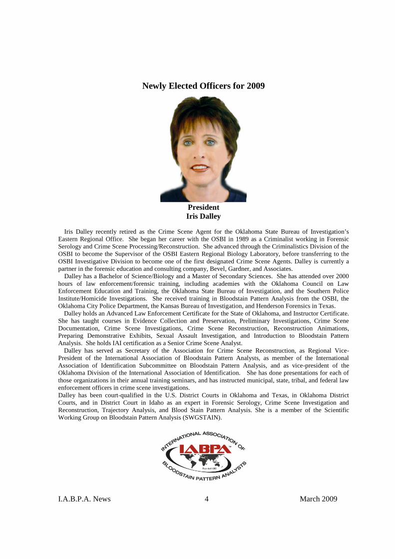
Newly Elected Officers for 2009
PresidentIris Dalley
Iris Dalley recently retired as the Crime Scene Agent for the Oklahoma State Bureau of Investigation’sEastern Regional Office. She began her career with the OSBI in 1989 as a Criminalist working in ForensicSerology and Crime Scene Processing/Reconstruction. She advanced through the Criminalistics Division of theOSBI to become the Supervisor of the OSBI Eastern Regional Biology Laboratory, before transferring to theOSBI Investigative Division to become one of the first designated Crime Scene Agents. Dalley is currently apartner in the forensic education and consulting company, Bevel, Gardner, and Associates.
Dalley has a Bachelor of Science/Biology and a Master of Secondary Sciences. She has attended over 2000hours of law enforcement/forensic training, including academies with the Oklahoma Council on LawEnforcement Education and Training, the Oklahoma State Bureau of Investigation, and the Southern PoliceInstitute/Homicide Investigations. She received training in Bloodstain Pattern Analysis from the OSBI, theOklahoma City Police Department, the Kansas Bureau of Investigation, and Henderson Forensics in Texas.
Dalley holds an Advanced Law Enforcement Certificate for the State of Oklahoma, and Instructor Certificate.She has taught courses in Evidence Collection and Preservation, Preliminary Investigations, Crime SceneDocumentation, Crime Scene Investigations, Crime Scene Reconstruction, Reconstruction Animations,Preparing Demonstrative Exhibits, Sexual Assault Investigation, and Introduction to Bloodstain PatternAnalysis. She holds IAI certification as a Senior Crime Scene Analyst.
Dalley has served as Secretary of the Association for Crime Scene Reconstruction, as Regional Vice-President of the International Association of Bloodstain Pattern Analysts, as member of the InternationalAssociation of Identification Subcommittee on Bloodstain Pattern Analysis, and as vice-president of theOklahoma Division of the International Association of Identification. She has done presentations for each ofthose organizations in their annual training seminars, and has instructed municipal, state, tribal, and federal lawenforcement officers in crime scene investigations.Dalley has been court-qualified in the U.S. District Courts in Oklahoma and Texas, in Oklahoma DistrictCourts, and in District Court in Idaho as an expert in Forensic Serology, Crime Scene Investigation andReconstruction, Trajectory Analysis, and Blood Stain Pattern Analysis. She is a member of the ScientificWorking Group on Bloodstain Pattern Analysis (SWGSTAIN).
I.A.B.P.A. News 5 March 2009
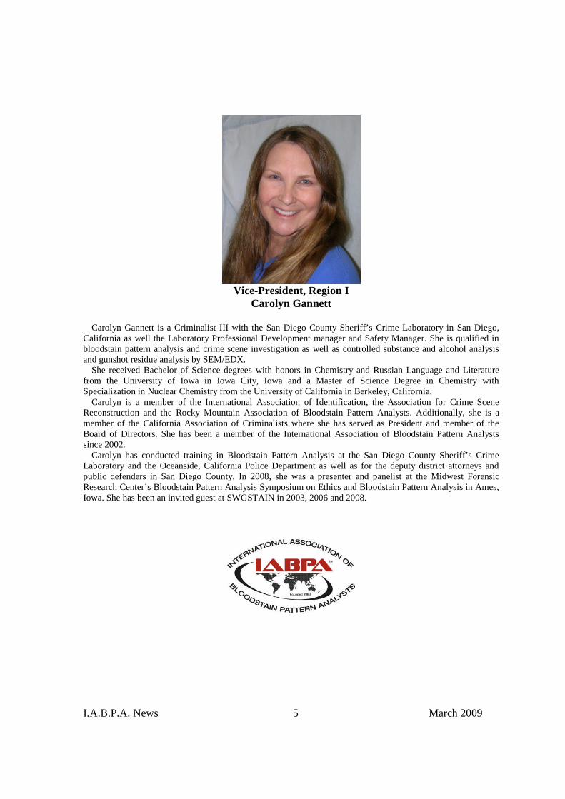
Vice-President, Region ICarolyn Gannett
Carolyn Gannett is a Criminalist III with the San Diego County Sheriff’s Crime Laboratory in San Diego,California as well the Laboratory Professional Development manager and Safety Manager. She is qualified inbloodstain pattern analysis and crime scene investigation as well as controlled substance and alcohol analysisand gunshot residue analysis by SEM/EDX.
She received Bachelor of Science degrees with honors in Chemistry and Russian Language and Literaturefrom the University of Iowa in Iowa City, Iowa and a Master of Science Degree in Chemistry withSpecialization in Nuclear Chemistry from the University of California in Berkeley, California.
Carolyn is a member of the International Association of Identification, the Association for Crime SceneReconstruction and the Rocky Mountain Association of Bloodstain Pattern Analysts. Additionally, she is amember of the California Association of Criminalists where she has served as President and member of theBoard of Directors. She has been a member of the International Association of Bloodstain Pattern Analystssince 2002.
Carolyn has conducted training in Bloodstain Pattern Analysis at the San Diego County Sheriff’s CrimeLaboratory and the Oceanside, California Police Department as well as for the deputy district attorneys andpublic defenders in San Diego County. In 2008, she was a presenter and panelist at the Midwest ForensicResearch Center’s Bloodstain Pattern Analysis Symposium on Ethics and Bloodstain Pattern Analysis in Ames,Iowa. She has been an invited guest at SWGSTAIN in 2003, 2006 and 2008.
I.A.B.P.A. News 6 March 2009
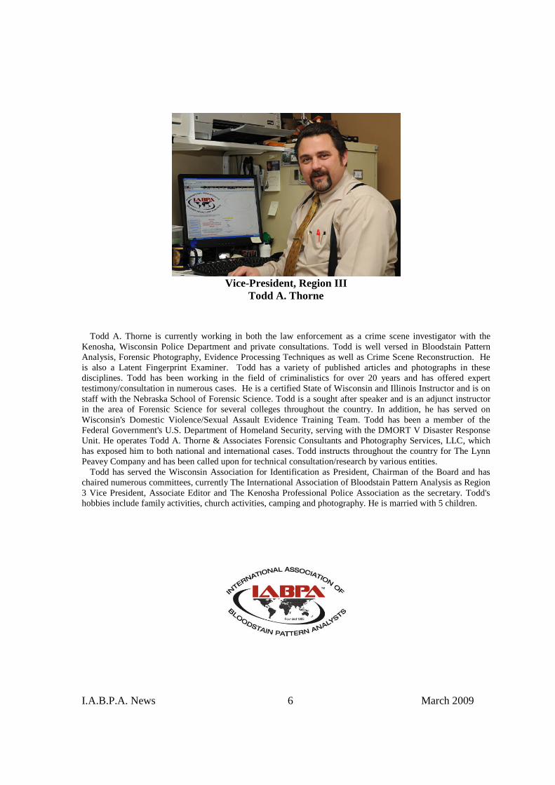
Vice-President, Region IIITodd A. Thorne
Todd A. Thorne is currently working in both the law enforcement as a crime scene investigator with theKenosha, Wisconsin Police Department and private consultations. Todd is well versed in Bloodstain PatternAnalysis, Forensic Photography, Evidence Processing Techniques as well as Crime Scene Reconstruction. Heis also a Latent Fingerprint Examiner. Todd has a variety of published articles and photographs in thesedisciplines. Todd has been working in the field of criminalistics for over 20 years and has offered experttestimony/consultation in numerous cases. He is a certified State of Wisconsin and Illinois Instructor and is onstaff with the Nebraska School of Forensic Science. Todd is a sought after speaker and is an adjunct instructorin the area of Forensic Science for several colleges throughout the country. In addition, he has served onWisconsin's Domestic Violence/Sexual Assault Evidence Training Team. Todd has been a member of theFederal Government's U.S. Department of Homeland Security, serving with the DMORT V Disaster ResponseUnit. He operates Todd A. Thorne & Associates Forensic Consultants and Photography Services, LLC, whichhas exposed him to both national and international cases. Todd instructs throughout the country for The LynnPeavey Company and has been called upon for technical consultation/research by various entities.
Todd has served the Wisconsin Association for Identification as President, Chairman of the Board and haschaired numerous committees, currently The International Association of Bloodstain Pattern Analysis as Region3 Vice President, Associate Editor and The Kenosha Professional Police Association as the secretary. Todd'shobbies include family activities, church activities, camping and photography. He is married with 5 children.
I.A.B.P.A. News 7 March 2009
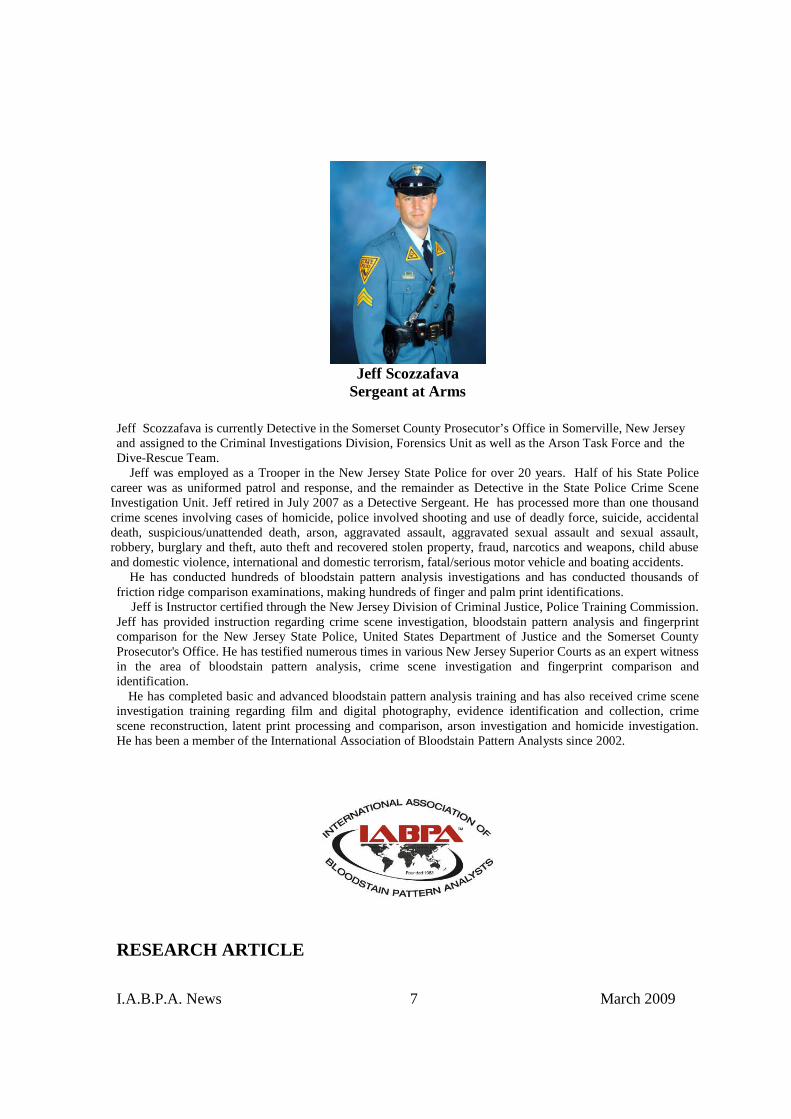
Jeff ScozzafavaSergeant at Arms
Jeff Scozzafava is currently Detective in the Somerset County Prosecutor’s Office in Somerville, New Jerseyand assigned to the Criminal Investigations Division, Forensics Unit as well as the Arson Task Force and theDive-Rescue Team.
Jeff was employed as a Trooper in the New Jersey State Police for over 20 years. Half of his State Policecareer was as uniformed patrol and response, and the remainder as Detective in the State Police Crime SceneInvestigation Unit. Jeff retired in July 2007 as a Detective Sergeant. He has processed more than one thousandcrime scenes involving cases of homicide, police involved shooting and use of deadly force, suicide, accidentaldeath, suspicious/unattended death, arson, aggravated assault, aggravated sexual assault and sexual assault,robbery, burglary and theft, auto theft and recovered stolen property, fraud, narcotics and weapons, child abuseand domestic violence, international and domestic terrorism, fatal/serious motor vehicle and boating accidents.
He has conducted hundreds of bloodstain pattern analysis investigations and has conducted thousands offriction ridge comparison examinations, making hundreds of finger and palm print identifications.
Jeff is Instructor certified through the New Jersey Division of Criminal Justice, Police Training Commission.Jeff has provided instruction regarding crime scene investigation, bloodstain pattern analysis and fingerprintcomparison for the New Jersey State Police, United States Department of Justice and the Somerset CountyProsecutor's Office. He has testified numerous times in various New Jersey Superior Courts as an expert witnessin the area of bloodstain pattern analysis, crime scene investigation and fingerprint comparison andidentification.
He has completed basic and advanced bloodstain pattern analysis training and has also received crime sceneinvestigation training regarding film and digital photography, evidence identification and collection, crimescene reconstruction, latent print processing and comparison, arson investigation and homicide investigation.He has been a member of the International Association of Bloodstain Pattern Analysts since 2002.
RESEARCH ARTICLE
I.A.B.P.A. News 8 March 2009
The Identification and Significanceof Hemospheres in Crime Scene
Investigation
James O. Pex, M.S., D-ABCPex Forensic Consulting, Inc.
North Bend, Oregon
Abstract
The collection of microscopic evidence at a crime scene has been common for many years.The collection techniques involve use of enhanced visual examination, adhesive lifts andvacuum sweeping with a filter capture system. Identification of hemospheres may addconfirmation of blood to non-specific patterns observed in Luminol testing. For purposes ofthis paper, hemospheres are defined as extremely small, sub-millimeter droplets of blood.The observation, collection, and relevance of hemospheres at a scene are reviewed.
Introduction
In 1769, a plumber named William Watts poured molten lead through a screen andcaptured the resulting lead pellets in water. Current techniques for shotshell pellets have notchanged much over the years (1). The usual height required for lead to form uniform roundpellets is 133 to 200 feet. The size of the pellets was controlled by the size of the holes in thescreen. The conversion of liquids to a solid by falling in air has been well established.
Several times over the last decade, microscopic examination of crime scene evidence hasdemonstrated bloodstain formation from spheres to flat rings. Experiments conducted duringtraining courses to demonstrate these shapes utilized an electric motor with a wood paddleattached. Liquid blood was dropped on the rotating paddle and small drops of blood werecreated. Larger drops traveled greater distances than smaller drops. These drops of onemillimeter or less in size dried almost immediately upon impact with a surface. If the surfacewas absorbent and close to the motor, the characteristic ring formation was noted. If Mylar®
was substituted for the non-absorbent impact surface, various shapes would be noted (2).Some drops dried before becoming a flat ring and demonstrated a volcano-like shape. Asnoted in other blood drying time tests, the center is always last to dehydrate.
In a homicide investigation in the late 1980s, nearly spheroid drops of blood less than onemillimeter in diameter were removed from a velour shirt utilizing the standard scrapingtechnique onto white paper. The sizes of these drops ranged from 0.1 to 0.5 mm in diameter.Scanning electron microscopy (SEM) along with micro Kastle-Meyer testing confirmed thesewere blood. In the examination of this shirt, impact spatter was re-created experimentallywith the discharge of a firearm and the results were evaluated. The bloodstains of interestwere extremely small and spherical.

I.A.B.P.A. News 9 March 2009
ExperimentationA bullet trap was placed on a work table with the center 42 inches above the floor. A 2’ x2’
swatch of new carpet was placed on the floor directly below the bullet trap. A control swatchof carpet was vacuumed and sprayed with Luminol prior to firearms testing.
Human blood, preserved in EDTA, was used to saturate a sponge. The sponge was placedinside a tight-fitting plastic bag and attached to the front of a bullet trap. A .38 Specialrevolver was fired with the muzzle in loose contact with the sponge. The testing took placeindoors at a temperature of 71 degrees Fahrenheit. Backspatter was noted on the weapon, thefiring hand and the shirt sleeve of the forearm. The carpet was visually examined, andvacuumed with a 3M brand model C-1000 Trace Evidence Vacuum. The vacuumed materialwas collected in C-1010 Microfilters.
A second test was performed on another segment of new carpet and retained for thirty days.The carpet was exposed to light foot traffic prior to vacuuming. The filters were examinedwith a 7X to 45X microscope. Luminol testing was performed after the vacuum collection(Figure 1). The carpet was re-examined for any residual bloodspatter.
A third test was performed to determine the solubility of hemispheres by placing several ina small quantity of water. Observations were made over a three hour period.
Figure 1. Positive Luminol reaction on carpet.
Method for the Presumptive Testing of Microscopic Impact Spatter
The detection and subsequent chemical testing on sub-millimeter amounts of blood hasbeen problematic in the past due to the limited sample available. The transfer of the bloodparticles to a stable media, then performance of a test that is sufficiently sensitive to providea visual response can be achieved by the following method:

I.A.B.P.A. News 10 March 2009
Reagent preparation of Leucomalachite Green (LMG)
Sodium Perborate 3.2 gLeucomalachite Green 0.1 gGlacial Acetic Acid 66 mLDistilled Water 33 mL
Procedure
1. Apply a thin film of clean paraffin to the surface of a glass slide.2. Prepare positive and negative control slides.3. Utilizing a fine tip probe, pick up minute stains with the aid of a microscope and
cluster them in the paraffin.4. After collection of several minute stains, gently heat the underside of the slide with a
match or heating block, just enough to melt the paraffin. Allow to cool and clean thebottom of the slide.
5. Recheck the location of the potential bloodstains with a microscope.6. Apply a drop of the LMG solution to the test slide and prepared positive and negative
control slides.7. The production of a green color indicates a positive test.
Although the Kastle-Meyer (KM) test is the most common presumptive test for blood,weak pink reactions were difficult to see at the microscopic level. If LMG is not readilyavailable, Hemastix® Reagent Strips used for urinalysis testing can be utilized. Hemastix® ismanufactured by Bayer and is available in most pharmacies as well as forensic productcompanies. Take two strips and dip the patches in 0.5 mL of distilled water for a minute.Apply a drop of the reagent water to the slide and a positive and negative control slide. Agreen/blue reaction offers a high contrast to the color of the blood and can be observedmicroscopically on the blood, if present. From a chemist’s perspective, the use of Hemastix®
is a bit crude, but efficient.Presumptive tests can produce false positives with certain substances such as metals,
oxidizing agents and peroxidases. If the test is positive, confirmatory testing should beperformed if possible.
Results
Examination of the control microfilter did not reveal any hemospheres or blood fragmentsas expected. Luminol testing did reveal a few nonspecific luminescent areas. Examination ofthe vacuum sweepings from the two other carpets used in test firings revealed a number ofinteresting shapes of bloodspatter, all less than one millimeter in diameter (Figure 2).Measurements were made with software associated with an MD 900 five megapixel cameraattached to an AmScope 900 Microscope. The measurements were standardized with aNational Institute of Standards and Technology (NIST) micro ruler.
Microscopic examination of the post-test carpet revealed the same characteristic shapes ofblood spatter often seen on a larger scale. The standard experimental model used to producevarious shapes of blood spatter is to use a paddle wheel attached to an electric motor. Bloodis dropped on the rotating paddle and spatter is directed to absorbent and non-absorbent
I.A.B.P.A. News 11 March 2009
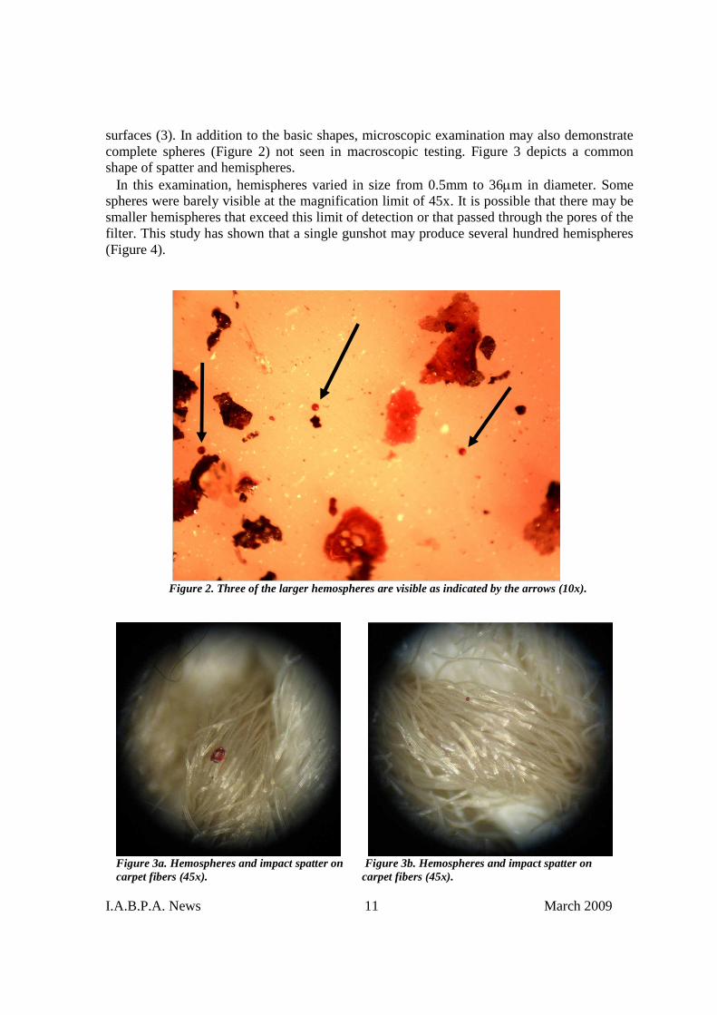
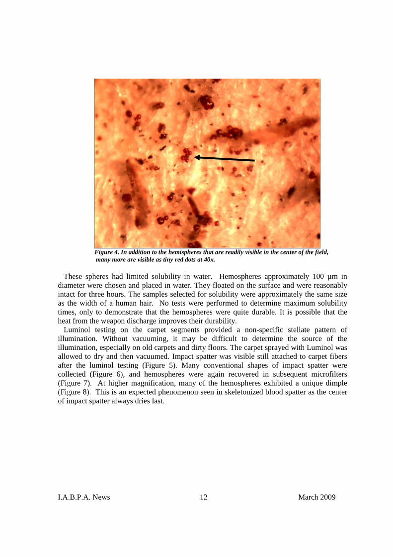
surfaces (3). In addition to the basic shapes, microscopic examination may also demonstratecomplete spheres (Figure 2) not seen in macroscopic testing. Figure 3 depicts a commonshape of spatter and hemispheres.
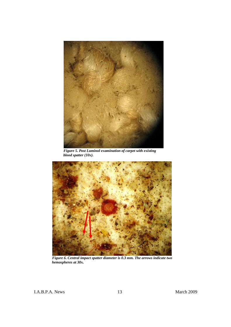
In this examination, hemispheres varied in size from 0.5mm to 36m in diameter. Somespheres were barely visible at the magnification limit of 45x. It is possible that there may besmaller hemispheres that exceed this limit of detection or that passed through the pores of thefilter. This study has shown that a single gunshot may produce several hundred hemispheres(Figure 4).
Figure 2. Three of the larger hemospheres are visible as indicated by the arrows (10x).
Figure 3a. Hemospheres and impact spatter on Figure 3b. Hemospheres and impact spatter oncarpet fibers (45x). carpet fibers (45x).
I.A.B.P.A. News 12 March 2009
Figure 4. In addition to the hemispheres that are readily visible in the center of the field,many more are visible as tiny red dots at 40x.
These spheres had limited solubility in water. Hemospheres approximately 100 µm indiameter were chosen and placed in water. They floated on the surface and were reasonablyintact for three hours. The samples selected for solubility were approximately the same sizeas the width of a human hair. No tests were performed to determine maximum solubilitytimes, only to demonstrate that the hemospheres were quite durable. It is possible that theheat from the weapon discharge improves their durability.
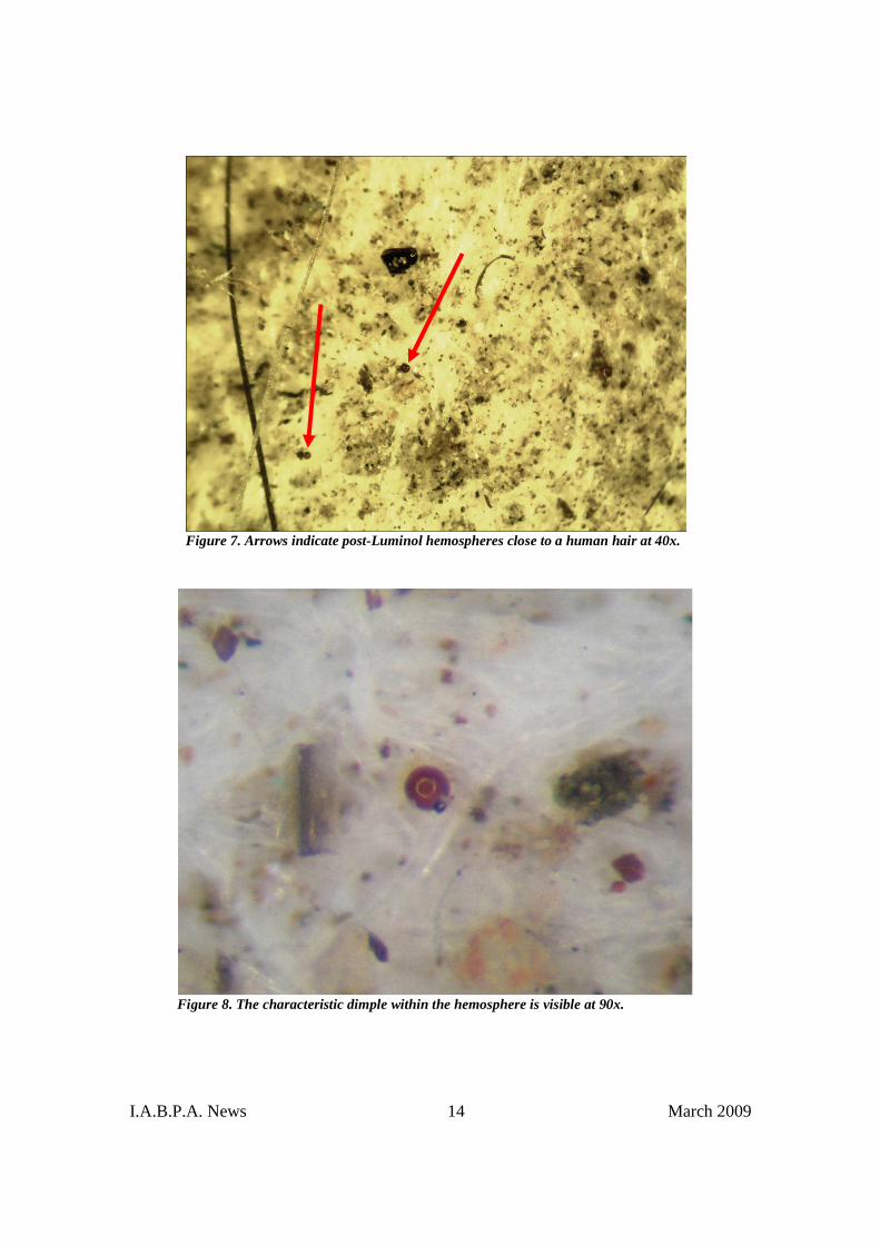
Luminol testing on the carpet segments provided a non-specific stellate pattern ofillumination. Without vacuuming, it may be difficult to determine the source of theillumination, especially on old carpets and dirty floors. The carpet sprayed with Luminol wasallowed to dry and then vacuumed. Impact spatter was visible still attached to carpet fibersafter the luminol testing (Figure 5). Many conventional shapes of impact spatter werecollected (Figure 6), and hemospheres were again recovered in subsequent microfilters(Figure 7). At higher magnification, many of the hemospheres exhibited a unique dimple(Figure 8). This is an expected phenomenon seen in skeletonized blood spatter as the centerof impact spatter always dries last.
I.A.B.P.A. News 13 March 2009
Figure 5. Post Luminol examination of carpet with existingblood spatter (10x).
Figure 6. Central impact spatter diameter is 0.3 mm. The arrows indicate twohemospheres at 30x.
I.A.B.P.A. News 14 March 2009
Figure 7. Arrows indicate post-Luminol hemospheres close to a human hair at 40x.
Figure 8. The characteristic dimple within the hemosphere is visible at 90x.
I.A.B.P.A. News 15 March 2009
Discussion
The reasonable explanation for the appearance of hemospheres is similar to a shot tower.As a spray of spattered blood comes back toward the shooter (4), some droplets travel uphigh enough to dry prior to reaching the carpet. Some travel down and forms various othershapes depending on the diameter and the time of travel to the impacted surface. Theminimum distance from the floor or the minimum caliber required was not determined.However, hemospheres were able to be created with a .22LR pistol contact shot into a blood-soaked sponge.
In this type of testing under controlled circumstances, it is easy to identify the observedspheres as blood. However, in the crime laboratory environment, the issue becomes morecomplex with a need to confirm these spheres as blood. Their lack of solubility in wateraffects the testing with most blood screening agents. The quantity of material may requirePCR or STR techniques to determine human origin. The size and dispersal of these spheresrenders Luminol inconclusive (5). It cannot be determined if a minute spot of light originatedfrom un-dissolved chemicals in the sprayer, unknown substances on a soiled surface, or theblood on the fabric. Metallic fragments were also noted in the carpet that may haveoriginated from the bore of the weapon at the time of discharge.
Dixon, et al. (7) described a technique for blood identification utilizing scanning electronmicroscopy (SEM). Elemental composition may be a way to support the physical appearanceof the spheres in conjunction with presumptive testing. The distribution of these hemospheresstill remains a question. EPA documents (6) indicate that particles greater than 50-100 µmusually remain in the air for a few minutes before settling near a surface. Considering that asingle intact red blood cell is 7 µm in diameter, a hemosphere is a minute volume of blood.Consequently, these blood particles may remain suspended in air for extended periods oftime. Since we do not know the minimum size of hemospheres created, a health issue comesto the forefront in a shooting environment. Studies of GSR have shown that these particlesare easily affected by air currents. An open window at a scene could relocate them to otherareas.
Hemospheres, the smallest form of impact blood spatter, were detectable on the clothing ofa person in the vicinity of a shooting. It is not known, but certainly also possible that thesehemospheres may occur with forward spatter or other forms of impact spatter mechanisms.Because these small particulates may remain suspended as in GSR testing, some people maybe contaminated but may not have committed the crime.
The decision to vacuum should be a consideration at the crime scene. Hemospheres werefound in higher concentrations before the application of any liquid blood detection solutions.However, their durability does not prevent the test from being conducted after the surfacehad dried. It is recommended that an area such as a room be divided into one yard squaresand each square vacuumed, the canister changed and the next area tested. The area with thehighest concentration of hemospheres should represent where the shooting took place.Figure 9 is a SEM photograph of a bisected hemosphere. This was removed from a shirt andthe placement of the original fiber can be seen at the bottom of the sphere halves. Thephotograph is also interesting in that the sphere was hollow. Observations of micro bloodspatter up to ninety magnifications revealed a variety of shapes and sizes common to thoseseen with the naked eye. Many of the shapes observed with the microscope would be difficult

I.A.B.P.A. News 16 March 2009
to defend as bloodspatter to the exclusion of all other possibilities. Hemospheres present aunique size and shape that is reproducible.
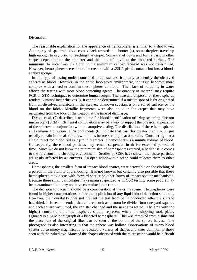
While examining for hemospheres, fragments of burned gunpowder were seen in themicrofilters (Figure 10). The combination of hemospheres and partially burned gunpowderparticles could be an important combination when investigating a crime scene anddetermining the location of a shooting; especially if the body was missing or suspected ofhaving been moved to another location.
It is recommended that the analyst repeat these tests and become familiar with the shapesseen under the microscope. Fragments of larger bloodstains are sometimes seen and easilyrecognized by their irregular edges.
Figure 9. Hemosphere split in two parts with concave red blood cells (RBCs) visible. TheU-shaped fragment below the RBCs is the fiber attachment (SEM).
I.A.B.P.A. News 17 March 2009
Figure 10. Bloodspatter and possible GSR at 45x.
Conclusion
The identification and documentation of hemospheres offers new evidence for shootingscene reconstruction. Difficulty with current screening tests and confirmation as humanblood has to be overcome to realize their full potential. The visual appearance under themicroscope may be seen as consistent with hemospheres until confirmation becomesavailable. Within the vacuum sweepings, these spheres compliment other evidence of firearmdischarge such as burned and unburned gun powder particles. This carries the importantconcept from just the discharge of a firearm to the reality that someone was actually struckby a bullet, if other bloodstains are absent. It is a reasonable expectation that barium,antimony and lead may be found within hemospheres.
Hemospheres have been observed on three occasions in the past. Unfortunately, vacuumsweepings from shooting homicides were normally done only to look for gun powderparticles. Not every shooting scene requires this type of examination. It would be of greatvalue for investigators to begin looking for these spheres and to determine their frequency inshooting incidents along with the circumstances in which they occurred.
I.A.B.P.A. News 18 March 2009
Unusual Case Circumstance
In the late 1980s, a homicide occurred at a local convenience store. The victim was shot inthe back of the head several times while lying on the floor. Suspects were later identified andtheir clothing submitted to the laboratory for examination. One suspect had a red velour shirt.No blood was visible. Standard scraping of the back of the shirt onto white paper for traceevidence produced small spherical drops of blood. These were approximately 0.1-0.5 mm indiameter. The Kastle-Meyer test was positive on the larger spheres. These were subjected toSEM microscopy and it could be seen that an area of the drop had previously been attachedto a fiber. At that time, enzyme phenotyping was not sufficiently sensitive to detect anyphenotypes. Inadequate research had been done at the time to appreciate the value of thisevidence. Approximately ten years later, the case was reopened. DNA testing was successfulon the remaining spheres and showed the blood originated from the victim. Due to theirlocation on the back of the shirt and other facts in the case, it was suspected that thisindividual may have held the victim down when the victim was shot by another person.
References
1. Mann, M., Espinoza, E., Ralston, R., Stroud, R., Scanlan, M., and Strauss, S.Shot Pellets: An Overview. AFTE, 1994 July; Volume 26, No. 3.
2. Pex, J. and Knowles, G. Bloodstain Pattern Analysis Training Manual, OSP,Salem, Oregon, 1991.
3. MacDonell, H. and Bialousz, L. Laboratory Manual on the GeometricInterpretation of Human Bloodstain Evidence, 1973 Jan., pp. 51-52.
4. Pex, J. and Vaughan, C. “Observations of High Velocity Bloodspatter onAdjacent Objects, ”J. Forensic Sci., Vol. 32, No. 6, Nov. 1987, pp. 1587-1594.
5. McGrath, J. The Chemical Luminescence Test for Blood. Forensic andChemical Applications. British Medical Journal. Vol. 28, Aug. 1942, pp. 156-157.
6. Airborne Particulates.http://www.epa.qld.gov.au/environmental_management/air/air quality_monitoring/air_pollutants
7. Dixon, T., Samudra, V., Stewart, W.D., and Johari, O. A Scanning ElectronMicroscope Study of Dried Blood, J. Forensic Sci., Vol. 21, Issue 4, Oct.1976.
I.A.B.P.A. News 19 March 2009
Guide to Capturing Photographs of Bloodstains for 3D Measurement
Eugene Liscio, P.Eng.AI2–3D Forensic AnimationsWoodbridge, Ontario, Canada
www.ai2-3d.com
Introduction
When one stops to think how many decades have passed since the camera first becamecommon place in law enforcement agencies, it is quite incredible to think that still today,taking photographs is one of the most common methods of documenting evidence. Theevolution of the computer and digital technologies has changed the way photographs arerecorded and stored, but they have not lessened the importance of documenting evidencethrough photography. Although more and more law enforcement agencies are becomingequipped with high tech 3D laser scanners and reflector-less total stations, most people find itsurprising to note that it is possible to record accurate 3D coordinate information by usingjust about any digital camera. The key is in how to take the photographs.
Often, a bloodstain expert is retained, weeks, months or years after a brutal crime has takenplace and the key pieces of evidence are provided in the form of police photographs, reportsand diagrams. Most bloodstain experts know that without placing a reference set of axes inthe camera’s view, it becomes next to impossible to get accurate measurements for keypieces of evidence. Even with a reference set of axes, trying to visualize a specificbloodstain pattern with a high degree of perspective is next to impossible and key details areeasily missed.
This guide has been written with the intent of assisting Crime Scene Investigators withcapturing photographs of bloodstains (or other evidence) that will preserve 3D geometricdata for future analysis by a photogrammetry specialist.
Photogrammetry
Photogrammetry is the science of taking geometric measurements from photographs.“Photogram” is a photograph and “metry” is the science of measuring. Since a photographtakes images from the 3D world and projects it on a flat 2D image plane, we lose the depthinformation. However, photogrammetry can be viewed as the process by which we do thereverse. By knowing some information about the camera which took the photographs and byhaving two or more photos of the same object from a different perspective, we can gain some3D information back.
This is normally accomplished by taking several photographs of a particular piece ofevidence from wide angles of separation and by recording a reference dimension. Once thisis done a subsequent analysis can be sent to a photogrammetry specialist to solve for:
1. The focal length of the camera being used to take the photographs.2. The position of the camera in 3D space.3. The camera’s orientation in 3D space.4. 3D Measurements for specific points that appear in at least 2 or more photographs.
I.A.B.P.A. News 20 March 2009
5. 2D measurements for specific points that appear on flat surfaces.
It is not necessary to record the camera’s position or focal length since these will be solvedthrough the photogrammetry analysis. However, in practice, it is beneficial to know thesevalues since they provide a “ballpark” verification of the solution.
Focal Length
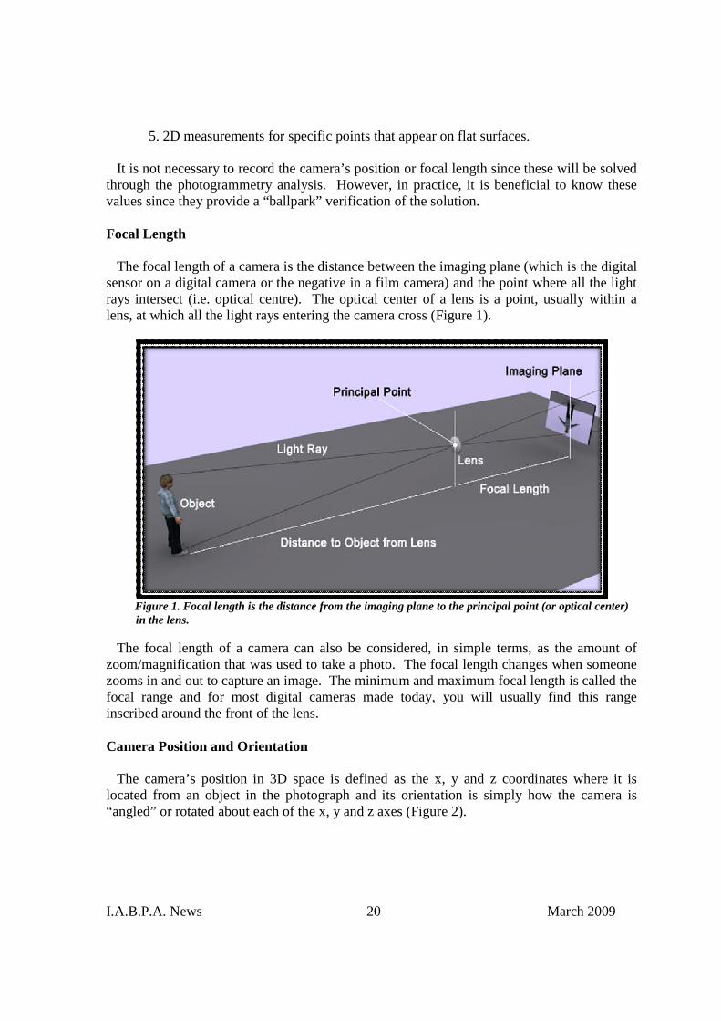
The focal length of a camera is the distance between the imaging plane (which is the digitalsensor on a digital camera or the negative in a film camera) and the point where all the lightrays intersect (i.e. optical centre). The optical center of a lens is a point, usually within alens, at which all the light rays entering the camera cross (Figure 1).
Figure 1. Focal length is the distance from the imaging plane to the principal point (or optical center)in the lens.
The focal length of a camera can also be considered, in simple terms, as the amount ofzoom/magnification that was used to take a photo. The focal length changes when someonezooms in and out to capture an image. The minimum and maximum focal length is called thefocal range and for most digital cameras made today, you will usually find this rangeinscribed around the front of the lens.
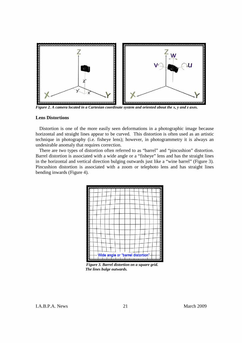
Camera Position and Orientation
The camera’s position in 3D space is defined as the x, y and z coordinates where it islocated from an object in the photograph and its orientation is simply how the camera is“angled” or rotated about each of the x, y and z axes (Figure 2).
I.A.B.P.A. News 21 March 2009
Figure 2. A camera located in a Cartesian coordinate system and oriented about the x, y and z axes.
Lens Distortions
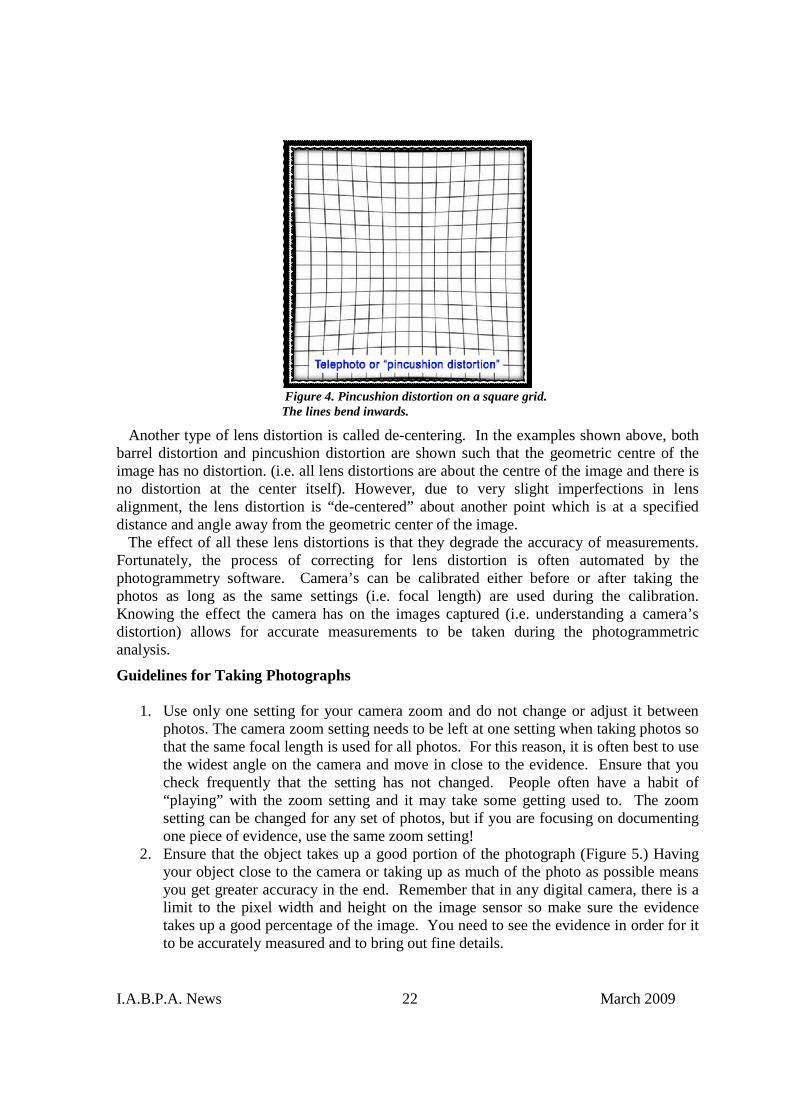
Distortion is one of the more easily seen deformations in a photographic image becausehorizontal and straight lines appear to be curved. This distortion is often used as an artistictechnique in photography (i.e. fisheye lens); however, in photogrammetry it is always anundesirable anomaly that requires correction.
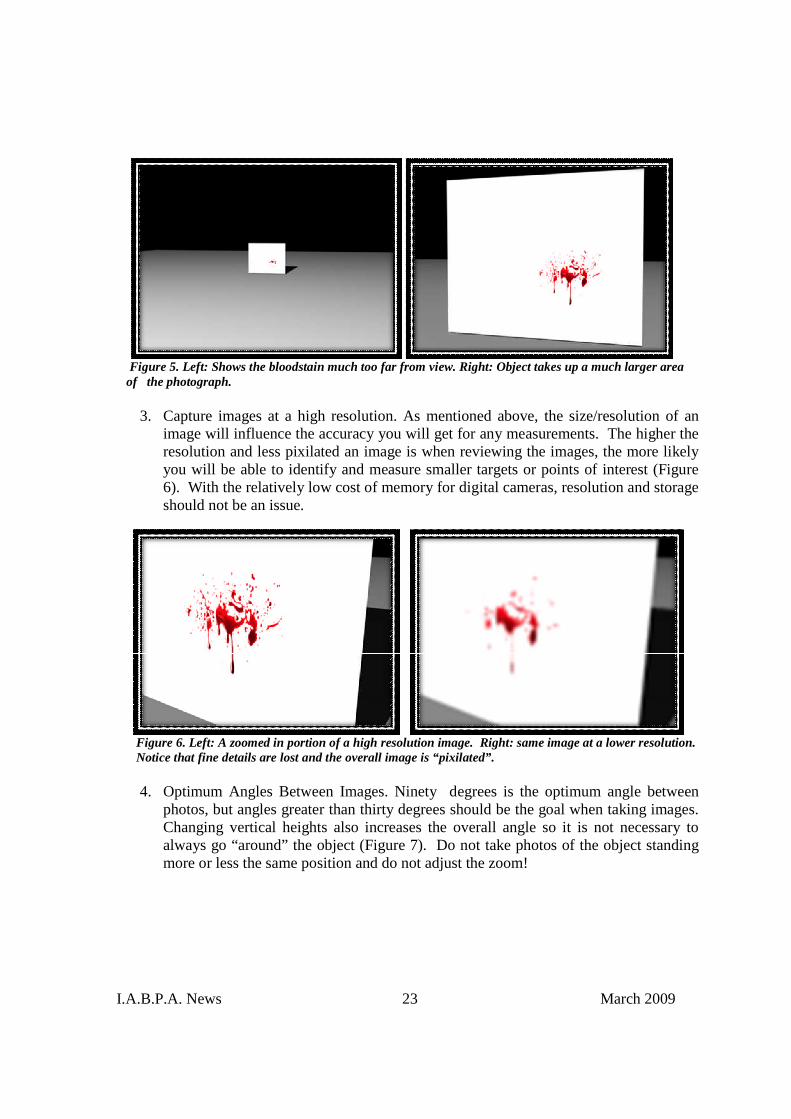
There are two types of distortion often referred to as “barrel” and “pincushion” distortion.Barrel distortion is associated with a wide angle or a “fisheye” lens and has the straight linesin the horizontal and vertical direction bulging outwards just like a “wine barrel” (Figure 3).Pincushion distortion is associated with a zoom or telephoto lens and has straight linesbending inwards (Figure 4).
Figure 3. Barrel distortion on a square grid.The lines bulge outwards.
I.A.B.P.A. News 22 March 2009
Figure 4. Pincushion distortion on a square grid.The lines bend inwards.
Another type of lens distortion is called de-centering. In the examples shown above, bothbarrel distortion and pincushion distortion are shown such that the geometric centre of theimage has no distortion. (i.e. all lens distortions are about the centre of the image and there isno distortion at the center itself). However, due to very slight imperfections in lensalignment, the lens distortion is “de-centered” about another point which is at a specifieddistance and angle away from the geometric center of the image.
The effect of all these lens distortions is that they degrade the accuracy of measurements.Fortunately, the process of correcting for lens distortion is often automated by thephotogrammetry software. Camera’s can be calibrated either before or after taking thephotos as long as the same settings (i.e. focal length) are used during the calibration.Knowing the effect the camera has on the images captured (i.e. understanding a camera’sdistortion) allows for accurate measurements to be taken during the photogrammetricanalysis.
Guidelines for Taking Photographs
1. Use only one setting for your camera zoom and do not change or adjust it betweenphotos. The camera zoom setting needs to be left at one setting when taking photos sothat the same focal length is used for all photos. For this reason, it is often best to usethe widest angle on the camera and move in close to the evidence. Ensure that youcheck frequently that the setting has not changed. People often have a habit of“playing” with the zoom setting and it may take some getting used to. The zoomsetting can be changed for any set of photos, but if you are focusing on documentingone piece of evidence, use the same zoom setting!
2. Ensure that the object takes up a good portion of the photograph (Figure 5.) Havingyour object close to the camera or taking up as much of the photo as possible meansyou get greater accuracy in the end. Remember that in any digital camera, there is alimit to the pixel width and height on the image sensor so make sure the evidencetakes up a good percentage of the image. You need to see the evidence in order for itto be accurately measured and to bring out fine details.
I.A.B.P.A. News 23 March 2009
Figure 5. Left: Shows the bloodstain much too far from view. Right: Object takes up a much larger areaof the photograph.
3. Capture images at a high resolution. As mentioned above, the size/resolution of animage will influence the accuracy you will get for any measurements. The higher theresolution and less pixilated an image is when reviewing the images, the more likelyyou will be able to identify and measure smaller targets or points of interest (Figure6). With the relatively low cost of memory for digital cameras, resolution and storageshould not be an issue.
Figure 6. Left: A zoomed in portion of a high resolution image. Right: same image at a lower resolution.Notice that fine details are lost and the overall image is “pixilated”.
4. Optimum Angles Between Images. Ninety degrees is the optimum angle betweenphotos, but angles greater than thirty degrees should be the goal when taking images.Changing vertical heights also increases the overall angle so it is not necessary toalways go “around” the object (Figure 7). Do not take photos of the object standingmore or less the same position and do not adjust the zoom!

I.A.B.P.A. News 24 March 2009
Figure 7. The images above show a good separation of angles between each camera view.
5. Use the Ring Method to take photos of any objects. The ring method is nothing morethan taking photos around an object in a circular fashion and in regular intervals of 90degrees or less (Figure 8). Normally, if one were to take 12 photos around abloodstain located on the ground, then one would divide the spaces evenly (about 30degrees). This ensures there is good overlap of photos. For areas of particularimportance take a minimum of 3 photos from different angles.
Figure 8. Use the “ring” method and take as many photos as necessaryin a circular fashion around the object. Having more photos is alwaysbetter (always strive for a minimum of 3).
6. Mark a minimum of 7 visible targets on important areas. Where possible, it is best toinclude a minimum of 7 targets around the evidence of interest. The greater thenumber of targets that can be marked and referenced between the set of photos, themore accurate the results should be. The best targets are high contrast (e.g. black carwith white target dots). Targets can be as simple as printing out a set of black circulardots on white paper and placing them around the bloodstained area (Figure 9). Also,
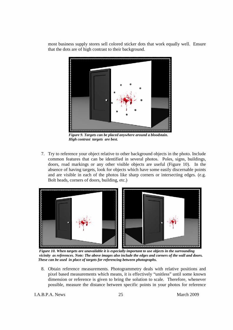
I.A.B.P.A. News 25 March 2009
most business supply stores sell colored sticker dots that work equally well. Ensurethat the dots are of high contrast to their background.
Figure 9. Targets can be placed anywhere around a bloodstain.High contrast targets are best.
7. Try to reference your object relative to other background objects in the photo. Includecommon features that can be identified in several photos. Poles, signs, buildings,doors, road markings or any other visible objects are useful (Figure 10). In theabsence of having targets, look for objects which have some easily discernable pointsand are visible in each of the photos like sharp corners or intersecting edges. (e.g.Bolt heads, corners of doors, building, etc.)
Figure 10. When targets are unavailable it is especially important to use objects in the surroundingvicinity as references. Note: The above images also include the edges and corners of the wall and doors.These can be used in place of targets for referencing between photographs.
8. Obtain reference measurements. Photogrammetry deals with relative positions andpixel based measurements which means, it is effectively “unitless” until some knowndimension or reference is given to bring the solution to scale. Therefore, wheneverpossible, measure the distance between specific points in your photos for reference

I.A.B.P.A. News 26 March 2009
(Figure 11). Try to gather measurements over larger areas. Do not try to measuresmall items in the photograph! Also, try to gather several measurements in differentdirections. These can all be used to verify the accuracy of the photogrammetryanalysis in the future.
Figure 11. The height of the door is a good reference measurement since itspans a large section of the photograph.
By taking a few extra photos with photogrammetry in mind, bloodstain measurements areeffectively captured and stored to be recalled at any time in the future. Digital photographsare cheap and can still provide accurate measurements years after an accident has occurred.The value of the photos is increased significantly, especially when the outcome of a trialdepends on the information contained in the images.
References
1. Luhmann, T., Robson, S, Kyle, S., Harley, I., Close Range Photogrammetry:Principles, Techniques and Applications, Whittles Publishing Co., Dunbeath,Scotland, UK
2. Linder, W., Digital Photogrammetry – A Practical Course, Springer Publishing Co.,Berlin, Heidelberg, New York, NY, 2006
3. EOS Systems, Inc., Photomodeler 6 Training, Collision Investigation Course Notes,Vancouver, Canada, 2007
I.A.B.P.A. News 27 March 2009
An Expiratory Bloodstain Pattern Due to Medical InterventionPeter Lamb and Gillian Leak
Forensic Science ServicesEngland, UK
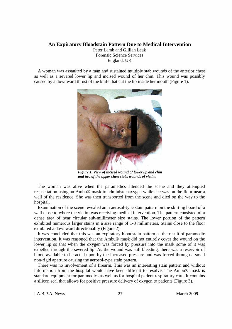
A woman was assaulted by a man and sustained multiple stab wounds of the anterior chestas well as a severed lower lip and incised wound of her chin. This wound was possiblycaused by a downward thrust of the knife that cut the lip inside her mouth (Figure 1).
Figure 1. View of incised wound of lower lip and chinand two of the upper chest stabs wounds of victim.
The woman was alive when the paramedics attended the scene and they attemptedresuscitation using an Ambu® mask to administer oxygen while she was on the floor near awall of the residence. She was then transported from the scene and died on the way to thehospital.
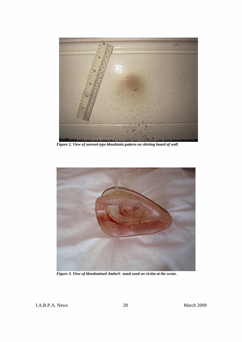
Examination of the scene revealed an n aerosol-type stain pattern on the skirting board of awall close to where the victim was receiving medical intervention. The pattern consisted of adense area of near circular sub-millimeter size stains. The lower portion of the patternexhibited numerous larger stains in a size range of 1-3 millimeters. Stains close to the floorexhibited a downward directionality (Figure 2).
It was concluded that this was an expiratory bloodstain pattern as the result of paramedicintervention. It was reasoned that the Ambu® mask did not entirely cover the wound on thelower lip so that when the oxygen was forced by pressure into the mask some of it wasexpelled through the severed lip. As the wound was still bleeding, there was a reservoir ofblood available to be acted upon by the increased pressure and was forced through a smallnon-rigid aperture causing the aerosol-type stain pattern.
There was no involvement of a firearm. This was an interesting stain pattern and withoutinformation from the hospital would have been difficult to resolve. The Ambu® mask isstandard equipment for paramedics as well as for hospital patient respiratory care. It containsa silicon seal that allows for positive pressure delivery of oxygen to patients (Figure 3).
I.A.B.P.A. News 28 March 2009
Figure 2. View of aerosol-type bloodstain pattern on skirting board of wall.
Figure 3. View of bloodstained Ambu® mask used on victim at the scene.
I.A.B.P.A. News 29 March 2009
ABSTRACTS OF RECENT BPA RELATED ARTICLES PUBLISHEDIN THE SCIENTIFIC LITERATURE
Rian K. Morgan-Smith, Douglas A. Elliot, Hussain Adam. Enhancement of Aged Shoeprintsin Blood, Journal of Forensic Identification Vol. 59 (1), 2009\45-50
Abstract:
Shoeprints in blood deteriorate over time, even in indoor or sheltered environments. Of the reagents tested,ninhydrin was the best reagent for treating aged impressions on paper substrates. On wooden and linoleumsubstrates, amido black was the best of the reagents tested.
Eric L. Ellis, Tim P. Wong, Scott W. Bowers. Locating Latent Bloodstains. Journal ofForensic Identification Vol. 59 (1), 2009 pp. 59-64
Abstract:
This stabbing case illustrates the usefulness of Bluestar® Forensic Latent Detection Reagent for the detection oflatent bloodstain evidence.
Tontarski, K.L., Hoskins, K.A., Wadkins, T.G., Brun-Conti, L. and Michaud, A.L. ChemicalEnhancement techniques of Bloodstain Patterns and DNA Recovery after Fire Exposure. J.Forensic Sci., 2009, Vol. 54, No.1
Abstract:
It is common in forensic casework to encounter situations where the suspect has set a fire to cover up ordestroy possible evidence. While bloodstain pattern interpretation, chemical enhancement of blood, andrecovery of deoxyribonucleic acid from bloodstains is well documented in the literature, very little informationis known about the effects of heat or fire on these types of examinations. In this study, a variety of known typesof bloodstain patterns were created in a four-room structure containing typical household objects andfurnishings. The structure was allowed to burn to flashover and then it was extinguished by firefighters usingwater. Once the structure cooled over night, the interior was examined using a bright light. The bloodstainswere evaluated to see if the heat or fire had caused any changes to the patterns that would inhibit interpretation.Bloodstain patterns remained visible and intact inside the structure and on furnishings unless the surface thatheld the blood was totally burned away. Additionally, a variety of chemical techniques were utilized to bettervisualize the patterns and determine the possible presence of blood after the fire. The soot from the fire formeda physical barrier that initially interfered with the chemical enhancement of blood. However, when the soot wasremoved, using water or alcohol, the chemicals used, fluorescein, luminol, Bluestar®, and Hemastix®,performed adequately in most of the tests. Prior to DNA testing, the combined phenolphthalein/tetramethylbenzidine presumptive test for blood was conducted in the laboratory on samples recovered from the structurein an effort to access the effectiveness of using this type of testing as a screening tool. Test results demonstratedthat reliance on obtaining a positive presumptive test for blood before proceeding with DNA analysis couldresult in the failure to obtain useful typing results. Finally, two DNA recovery methods (swabbing the stain pluscutting or scraping the stain) were attempted to evaluate their performance in recovering samples in an arsoninvestigation. Recovery of DNA was more successful in some instances with the swabbing method, and in otherinstances with the cutting/scraping method. Therefore, it is recommended that both methods be used. For themost part, the recovered DNA seemed to be unaffected by the heat, until the temperature was 800° or greater.At this temperature , no DNA profiles were obtained.
I.A.B.P.A. News 30 March 2009
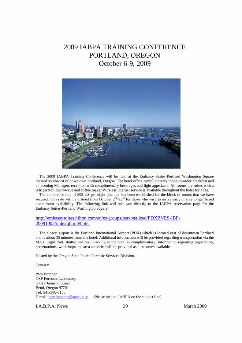
2009 IABPA TRAINING CONFERENCEPORTLAND, OREGON
October 6-9, 2009
The 2009 IABPA Training Conference will be held at the Embassy Suites-Portland Washington Squarelocated southwest of downtown Portland, Oregon. The hotel offers complimentary made-to-order breakfast andan evening Managers reception with complimentary beverages and light appetizers. All rooms are suites with arefrigerator, microwave and coffee maker Wireless internet service is available throughout the hotel for a fee.
The conference rate of $98 US per night plus tax has been established for the block of rooms that we havesecured. This rate will be offered from October 2nd-12th for those who wish to arrive early or stay longer basedupon room availability. The following link will take you directly to the IABPA reservation page for theEmbassy Suites-Portland Washington Square:
http://embassysuites.hilton.com/en/es/groups/personalized/PDXBVES-IBP-20091002/index.jhtml#hotel
The closest airport is the Portland International Airport (PDX) which is located east of downtown Portlandand is about 35 minutes from the hotel. Additional information will be provided regarding transportation via theMAX Light Rail, shuttle and taxi. Parking at the hotel is complimentary. Information regarding registration,presentations, workshops and area activities will be provided as it becomes available.
Hosted by the Oregon State Police Forensic Services Division.
Contact:
Pam BordnerOSP Forensic Laboratory63319 Jamison StreetBend, Oregon 97701Tel: 541-388-6150E-mail: [email protected] (Please include IABPA on the subject line)

I.A.B.P.A. News 31 March 2009
Organizational Notices
Moving Soon?
All changes of mailing address need to be supplied to our Secretary Norman Reeves. Each quarterNorman forwards completed address labels for those who are members. Do not send change ofaddress information to the NEWS Editor. E-mail your new address to Norman Reeves at:
[email protected] Reeves
I.A.B.P.A.12139 E. Makohoh Trail
Tucson, Arizona 85749-8179Fax: 520-760-5590
Membership Applications / Request for Promotion
Applications for membership as well as for promotion are available on the IABPA website:IABPA Website: http://www.iabpa.org
The fees for application of membership and yearly dues are $40.00 US each. If you have notreceived a dues invoice for 2009 please contact Norman Reeves. Apparently, non US creditcards are charging a fee above and beyond the $ 40.00 membership/application fee.Your credit card is charged only $40.00 US by the IABPA. Any additional fees areimposed by the credit card companies.
IABPA now accepts the following credit cards:
Discover MastercardAmerican Express Visa

I.A.B.P.A. News 32 March 2009
Training Opportunities
April 20-24, 2009
Basic Bloodstain Pattern Analysis CourseBlutspureninstituteUsingen, Germany
Instructors: Dr. Silke Brodbeck and Martin EversdjikContact: Dr. Silke Brodbeck
Tel: +49-170-84 84 248Fax: +49-6081-14879
May 11-15, 2009
Advanced Bloodstain Pattern Analysis andExpert Witness Workshop
Miami, Florida
Presented by the Specialized Training Unit at theMetropolitan Police Institute of the Miami-Dade
Police DepartmentDoral, Florida
Contact: Toby L. Wolson, M.S., F-ABCMiami-Dade Police Department
Crime Laboratory Bureau9105 NW 25th StreetDoral, Florida 33172Voice: 305-471-3041Fax: 305-471-2052
E-mail: [email protected]
May 25-29, 2009Math and Physics for Bloodstain Analysis
CourseOntario Police College
Alymer, Ontario, Canada
Course Coordinator: Brian AllenE-mail: [email protected]
Further information: http://www.opconline.ca
June 1-5, 2009Bloodstain Evidence Institute
Corning, New York
Contact: Professor Herbert Leon MacDonellDirector
Laboratory of Forensic SciencePost Office Box 1111
Corning, New York 14830Tel: 607-962-6581
E-mail: [email protected]
June 8-12, 2009
Basic Bloodstain Pattern Analysis CourseElmira College
Elmira, New York
Contact: Paul Erwin KishForensic Consultant & Associates
P.O. Box 814Corning, New York 14830
Tel: 607-962-8092E-mail: [email protected]
June 15-19, 2009
Advanced Bloodstain Pattern Analysis CourseElmira College
Elmira, New York
Instructors: Paul Erwin Kish and Stuart JamesContact: Paul Erwin Kish
Forensic Consultant & AssociatesP.O. Box 814
Corning, New York 14830Tel: 607-962-8092
E-mail: [email protected]
July 13-17, 2009
Advanced Bloodstain Pattern Analysis CourseLondon, England
Instructors: Paul E. Kish and Stuart H. JamesContact:
Anthony LarkinE-mail: [email protected]

I.A.B.P.A. News 33 March 2009
August 31-September 4, 2009
Advanced Bloodstain Pattern Analysis CourseOntario Police College
Alymer, Ontario, Canada
Course Coordinator: Brian AllenE-mail: [email protected]
Further information: http://www.opconline.ca
September 21-25, 2009
Bloodstain Evidence InstituteCorning, New York
Contact: Professor Herbert Leon MacDonellDirector
Laboratory of Forensic SciencePost Office Box 1111
Corning, New York 14830Tel: 607-962-6581
E-mail: [email protected]
Training Announcements for the issue of the June 2009IABPA News must be received before May 15th, 2009

I.A.B.P.A. News 34 March 2009
Editor’s Corner
This issue introduces the membership to our newly elected officers for 2009 with theirphotograph and short bio. On behalf of the membership, I welcome our new President, IrisDalley, Vice president of Region I, Carolyn Gannett, Vice president of Region III, Todd A.Thorne and Sergeant at Arms, Jeff Scozzafava.
I am receiving a number of requests for copies of articles that have been published in theIABPA NEWS from college libraries and individuals. Some of the requested articles date backas far as 1989. I was recently informed by our new President, Iris Dalley that an article publishedin the June 2002 issue of the NEWS, entitled Extreme Temperature Effects on BloodstainPattern Analysis authored by Tom Brady, John Tigmo and Grant Graham, Sr. was used in a trialin Texas. It is encouraging that articles published in the NEWS are continuing to be of interestboth at the college level and in the courts.
The first announcement of the 2009 IABPA Training Conference is in this issue of the NEWS.Please send in your presentation abstracts and workshop requests to Pam Bordner. I am planningon hosting the Bring Your Own Case session again which is well attended each year. Thosepresentations should be limited to 15 minutes to allow enough time for all those wishing todiscuss an interesting case.
Paul Kish informed me that there was an excellent response to the Research into the ErrorRates Associated with Bloodstain Pattern Analysis online survey. This survey was composed ofvery basic bloodstain patterns for recognition prepared by Breeanna Meneses and Brian J.Gestring of the Forensic Science Program at Cedarcrest College in Allentown, Pennsylvania. Theresults of that survey will be published in a future issue of the NEWS when they becomeavailable.
Finally, I would again encourage the membership to submit research articles and case reportsfor peer review and publication in the NEWS.
Stuart H. JamesEditorIABPA NEWS
E-mail: [email protected]
I.A.B.P.A. News 35 March 2009
Past Presidents of the IABPA
V. Thomas Bevel 1983-1984Charles Edel 1985-1987Warren R. Darby 1988Rod D. Englert 1989-1990Edward Podworny 1991-1992Tom J. Griffin 1993-1994Toby L. Wolson, M.S. 1995-1996Daniel V. Christman 1997-1998Phyllis T. Rollan 1999-2000Daniel Rahn 2001-2002Bill Basso 2002-2006
Associate Editors of the IABPA News
L. Allyn DiMeoBarton P. Epstein
Paul E. KishDaniel MabelJon J. Nordby
Alexei PaceJoseph Slemko
Robert P. SpaldingT. Paulette SuttonTodd A. Thorne
The IABPA News is published quarterly in March, June, September, and December. 2009. The International Association of BloodstainPattern Analysts. All rights are reserved. Reproduction in whole or in part without written permission is prohibited.