table of contents - bloodstain pattern analysis publications... · the medically normal clotting...
TRANSCRIPT


Table of Contents
2012 IABPA Officers 1
President’s Message 2
Medical and Forensic Aspects of Blood Clot Formation
in the Presence of Saliva – A Preliminary Study
Celestina Rossi, Misty Holbrook,
Stuart H. James and Dan Mabel 3
An Interesting Bloodstain 13
Abstracts of Papers Presented at the 2012 IABPA
Training Conference in Tucson, Arizona 14
IABPA Distinguished Member Award 22
Recent BPA Articles in the Scientific Literature 23
Organizational Notices 24
Training Opportunities 24
Editor’s Corner 26
Publication Committee/Associate Editors 27
Past Editors of the IABPA News/Journal of Bloodstain
Pattern Analysis 27
Past Presidents of the IABPA 27

Journal of Bloodstain Pattern Analysis 1 Vol. 28, No. 3 September 2012
2012 IABPA Officers
PRESIDENT
Todd A. Thorne tthorne @kenosha.org
Vice President, Region I Vice President, Region II
Carolyn Gannett Leah Innocci [email protected] [email protected]
Vice President, Region III Vice President, Region IV
Rex T. Sparks Anthony Mangione [email protected] [email protected]
Vice President, Region V Vice President, Region VI
Peter Lamb Brett McCance
[email protected]. [email protected]
Secretary / Treasurer Sergeant at Arms Norman Reeves Nicholas A. Paonessa [email protected] [email protected]
Immediate Past President Historian
Iris Dalley Stuart H. James [email protected] [email protected]

Journal of Bloodstain Pattern Analysis 2 Vol. 28, No. 3 September 2012
President’s Message Greeting to you all! I hope this finds each and every one of you well.
With the passing our educational conference, signs of the end of yet another year are present. I enjoyed speaking with many of you, on a range of very interesting topics. Thanks to all of you that were able to attend and participate in the educational goals of the IABPA.
2012 has been a productive year for the IABPA. Your board has been working hard to update and improve several areas of the organization. Please look to the website for updated information on the bylaws, certification research, education as well as other committee actions.
The web site (web management system) has been up and running seemingly effortless thanks to the dedicated service of Joe Slemko. Joe has opted to hand off the web master responsibilities to Jon Thomas. Should you have any questions concerning the website, please contact Jon. Thanks Joe for being instrumental in getting our web off the ground.
Our Journal has been running solidly since its creation, but Stuart is always looking for more articles, research and legal updates. Please consider publishing with the IABPA Journal first! As always, feel free to get involved in our organization, there is always plenty to do. 2013 will bring the continuation and completion of several major projects started by your board of directors.
It has been an absolute honor serving with this board throughout my time as your president. I look forward to continued progress. Blessings, Todd

Journal of Bloodstain Pattern Analysis 3 Vol. 28, No. 3 September 2012
Medical and Forensic Aspects of Blood Clot Formation in the Presence of Saliva – A Preliminary Study
Celestina Rossi
1, Misty Holbrook
2, Stuart H. James
3 and Daniel Mabel
4
Abstract
Clinical tests for the determination of clotting or coagulation times for therapeutic monitoring of anti-coagulant therapy utilize relatively non-invasive venipuncture or lancet piercing techniques. The medically normal clotting times for human blood should only be used as a guideline when considering clotting times in forensic casework. Environmental factors such as temperature and various substrates where blood has accumulated may shorten clotting times. Medical conditions and the presence of anticoagulants in the blood that extend clotting times may be an unknown factor.
Trauma to the body and organs will release high levels of tissue factor (TF) as a result of the injury and this may shorten normal clotting times. Research into the medical and scientific literature has indicated that saliva contains high levels of TF as well and will accelerate the clotting time of blood. This paper describes experiments to demonstrate that a mixture of saliva and blood will accelerate the clotting time of blood on external substrates of porcelain and vinyl tiles.
Introduction
The observation of coagulated or clotted bloodstains and patterns at scenes has been documented by bloodstain pattern analysts in case work. In general, the presence of partially clotted or fully formed blood clots within bloodstain patterns and the presence of clotted impact spatter have been associated with a time lapse between initial bloodshed and the formation of the clots. A normal range of 3-15 minutes for visible clots to appear has been utilized in BPA case work.
1 This normal range was based upon the three-tube Lee-White clotting test performed at 37.5
degrees centigrade that historically was utilized in hospital and clinical laboratories2. However,
the normal ranges for the Lee-White test vary as low as 5-8 minutes3.This method has been
largely discontinued in the medical arena and replaced with more sophisticated techniques.4
In a forensic situation where injury has caused bloodshed, the bloodstain pattern analyst must consider external factors that could affect the length of time required to form a clot. In 2001, Laber and Epstein published a research study entitled Substrate Effects on the Clotting Time of Human Blood in the Journal of the Canadian Society of Forensic Science.
5 They utilized 10mL
of freshly drawn human blood applied to substrates of human skin, glass, wool-polyester cloth, paper and wood. They noted that clotting times varied among individuals and the surface upon which the blood was deposited. Though not addressed in their paper, they recognized that environmental factors such as temperature and humidity as well as other conditions and circumstances will also have an effect on the clotting times of human blood.
A blood clot is formed by a complex mechanism involving the plasma protein fibrinogen, platelets, and other clotting factors. Tissue factor (TF) is released as a result of tissue damage. Tissue factor (TF) combines with Factor VII, creating a protease that acts on Factor X. In turn, Factor X binds to and activates Factor V, creating prothrombinase that converts prothrombin (or Factor II) into thrombin. Thrombin cleaves fibrinogen and also activates Factor XII, which acts on the fibrinogen to form fibrin to create an insoluble meshwork that is integral to clotting. For fibrin to be available whenever an injury occurs, it is constantly being transported through the body by the circulating blood in a soluble form called fibrinogen. Fibrinogen must change into an insoluble form whenever clotting becomes necessary. The clotting cascade is demonstrated in Figure 1.
____________________________________________________________________
1Montgomery County Sheriff’s Office, Conroe, Texas
2Kentucky State Police Northern Regional Forensic Laboratory, Cold Spring, Kentucky
3James and Associates Forensic Consultants, Inc., Fort Lauderdale, Florida
4 Forensic Scientist, Cuyahoga County Regional Forensic Science Laboratory, Cleveland, Ohio

Journal of Bloodstain Pattern Analysis 4 Vol. 28, No. 3 September 2012
Subsequently, the blood clot begins to retract causing a separation of the remaining liquid portion that is referred to as serum (Figure 2). Factors affecting the time for clot retraction to occur include blood volume, surface texture, temperature and humidity. An image of a blood clot taken with a scanning electron microscope is demonstrated in Figure 3.
Figure 1. Diagram of the Clotting Cascade indicating the contribution of thromboplastin (TF from tissues).
Figure 2. Appearance of clotted blood and serum separation around the edges of the clot.

Journal of Bloodstain Pattern Analysis 5 Vol. 28, No. 3 September 2012
Figure 3. Formation of blood clot taken with a Hitachi Field Emission Scanning Electron Microscope at 1445x. The red concave discs are red blood cells; the blue cellular elements are activated platelets and the yellow strands are fibrin. (Copyright Dennis Kunkel Microscopy, Inc., Kailua, Hawaii). In normal healthy individuals, the time between initiation of the bleeding to the appearance
of visible clot formation requires approximately 3-15 minutes in a clinical setting depending on the particular clotting time test used. There are a variety of clinical tests for the determination of clotting or coagulation times and a wide range of normal values. The whole blood is obtained by venipuncture or lancet puncture. The prothrombin and partial thromboplastin tests are performed on plasma not whole blood and the results are reported in seconds. The average normal values have been established for hospital and clinical laboratories by the testing of large sample populations in controlled laboratory studies as shown in Table 1.
6-7 The values
may vary depending on the diagnostic reagents used for the tests and the procedure utilized for the specific tests.
Table 1. Average normal values for clinical coagulation tests.
Test Normal Value
Prothrombin Time 9.5-12.0 seconds
Partial Thromboplastin Time 60-85 seconds
Activated Partial Thromboplastin Time 20-35 seconds
Duke Bleeding Time (Lancet in Ear lobe) 1.0-3.0 minutes
Ivy Bleeding Time (Incision in Forearm) Less than 9.0 minutes
Lee-White Clotting time 3.0-15.0 minutes
There are numerous medical conditions and diseases that will lengthen the blood clotting
time of individuals including: Anemia Clotting factor deficiencies such as hemophilia Low platelet count (normal range is 150,000 – 400,000/mcL) Liver disease (alcoholic cirrhosis, hepatitis)

Journal of Bloodstain Pattern Analysis 6 Vol. 28, No. 3 September 2012
Vitamin K deficiency Malignant tumors Leukemia Systemic lupus erythematosus Chemotherapy
Prescribed anticoagulant medications for so-called “blood thinning” reduce the risk of recurrent strokes and heart attacks, and maintain patients at a controlled and lengthened clotting time. Common anticoagulant therapy medications are:
Warfarin - Coumadin® Heparin Enoxaparin - Lovenox® (clot blaster)
Patients prescribed these therapeutic medications are monitored and tested on a regular basis
to ensure that the intended lengthened clotting times are not exceeded. Aspirin will also increase the clotting time.
1 Many adults are advised by their doctors to ingest a therapeutic
dose of 81 mg of aspirin per day to reduce the risk of stroke or heart attack. It has been reported in Circulation – The Journal of the American Heart Association that the statin drug Simvastatin®, which acts to prevent coronary artery disease by reducing the cholesterol level also lengthens the clotting time of blood. Reportedly, this drug depresses the activation of prothrombin, Factor V and Factor XIII.
8 There are medical and physiological conditions that
have the effect of shortening the clotting time of blood which is referred to as hypercoagulability. High platelet count (in excess of 400,000 mcL) and excessive Tissue Factor (TF) will shorten the clotting time
2.
Cerebrospinal fluid (CSF) is one of the triggers that may accelerate clotting. Head injuries or injuries to the spinal canal potentially release CSF into the site of injury. As a result, the clotting times in such instances may be shortened. A review of the scientific literature revealed studies that indicated an acceleration of the clotting time of blood when mixed with saliva due to a high level of tissue factor (TF) in saliva
9-16.
It was reported in 1938 in the American Journal of Physiology by A.J. Glazko and D.M. Greenberg that “it is well known that the addition of saliva to blood will accelerate its coagulation. The results obtained indicate that saliva contains a factor which acts like the thromboplastic material present in blood platelets, brain and lung tissue and is probably protein in nature.”
9 In 1939, Volker reported in the American Journal of Orthodontics that “reports of
earlier investigators that saliva hastens blood coagulation have been substantiated by the use of a more accurate apparatus for recording blood clotting time.”
10 In 1960, Doo and Lee reported
in the Yomsei Medical Journal that the “accelerating effect of saliva on the clotting of blood seems to be due to the augmentation of thromboplastic activity. Human saliva accelerates the prothrombin time as much as snake venom does.”
11 In 1973, in the Journal of Neurosurgery,
Keimowitz and Annis reported a case of disseminated intravascular coagulation in a patient with massive brain trauma. It was suggested that the condition was caused by the liberation of thromboplastin.
12 In 2005, the journal, Neurology India, published an article by Pathak et. al.
describing that tissue thromboplastin (TTP) also called tissue factor (TF) is an integral membrane protein contributing to coagulopathy (clotting) including disseminated intravascular clotting after trauma to the brain which is a rich source of TF.
13 In 2011, Berckmans et. al.
reported in Blood – The Journal of the American Society of Hematology “that saliva is known to contain Tissue Factor (TF) that may be associated with cell-derived vesicles to facilitate coagulation when saliva directly contacts blood.”
14 Also in 2011, Gross reported in the
American Journal of Hematology that Tissue Factor on microvesicles in saliva can cause blood to clot. This may provide the basis for the wound-licking reflex.
15
The nature and location of the blood-letting injury can also affect the amount of tissue thromboplastin (TF) released. Lacerations of the skin and organs will release higher concentrations of TF and shorten clotting times. It is also known that brain contains high levels

Journal of Bloodstain Pattern Analysis 7 Vol. 28, No. 3 September 2012
of TF that may affect clotting times in a similar manner with injuries that produce an intermixing of brain tissue and blood, such as is seen in beating and gunshot wounds of the head. Sharp force injuries inflicted with knives, razors and glass may involve less tissue damage and may not release a large amount of TF and resultant clotting times may not be shortened. Rapid bleeding may extend the time required for clotting.
4 In one author’s experience, a victim
was beaten about the head and then dunked repeatedly in a toilet in an attempt to elicit a statement. Continually wetting the area of injury will also extend the length of time required for a clot to form.
In 1957, Nour-Eldin and Wilkinson reported the results of their experiments in their article, The Blood Clotting Factors in Human Saliva published in the Journal of Physiology article
16.
“For two normal donors, a range of clotting times from 2.5- 4.0 minutes was obtained when 1.0 volume of saliva was mixed with 9.0 volumes of blood. The control clotting times performed on 9.0 volumes of whole blood mixed with 1.0 volume of 0.9% NaCl ranged from 5.5–6.0 minutes. In addition, he utilized three donors with hemophilia, two with Christmas disease and one with thrombocytopenia. He reported clotting times in the range of 2.5-4.0 minutes for the saliva/blood mixtures and a clotting time range of 4.5-20.5 minutes for the donor whole blood controls. Experiments to Determine the Effect of Human Saliva on the Clotting time of Freshly Drawn Human Blood Purpose
Generated by an interest in the article by Nour-Eldin16
, an experiment was designed to compare the clotting time of freshly drawn human blood to the clotting time of freshly drawn human blood mixed with human saliva when applied to a non-porous surface. Two sets of experiments were conducted. Group I was conducted by Celestina Rossi and Group II by Misty Holbrook. Blood was drawn from ten volunteers for each experiment. The temperature and humidity of the experiment area were documented. The age and sex of the volunteers and any medication taken were also recorded (Tables 2 and 3). Materials Smooth porcelain and vinyl tiles Vacutainer® blood drawing apparatus Micro-pipettes Human saliva Freshly drawn human blood Glass rods Timer Methodology
1. Saliva was collected from ten volunteers and ~1.0 ml volume placed into clean test tubes.
2. Two tubes of blood were obtained via venipuncture from the ten volunteers. 3. One tube (~10.0 ml) of blood was poured onto a smooth tile. 4. The second tube of blood (~9.0 ml) was added to the tube containing ~1.0 ml of saliva
from the same individual and then poured onto a second smooth tile. 5. The blood on the tiles was observed at 15.0 second intervals for the appearance of
visible clots by gently drawing a glass rod through the blood. 6. The time of the appearance of clotting was recorded and the results for whole blood and
whole blood mixed with saliva were compared.

Journal of Bloodstain Pattern Analysis 8 Vol. 28, No. 3 September 2012
Experimental Conditions
Table 2. Group I Experimental Conditions (Porcelain Tiles)
Table 3. Group II Experimental Conditions (Vinyl Tiles)
Results
Table 4. Clotting Time Results from Group I
Donor Age
Whole Blood (~10.0 ml) Clotting Time In Minutes.
Whole Blood (~9.0ml) + Saliva (~1ml) Clotting Time In Minutes.
Medication Taken
1 Female 34 15.20 3.80 Zyrtek - D
2 Female
23 10.65 5.05 birth control
3 Female
35 12.13 3.92 birth control
4 Male 35 9.05 7.08 ** Splenectomy**
5 Female 38 16.00 2.90 birth control
6 Male 47 10.75 3.18 none
7 Male 43 11.15 3.47 none
8 Male 42 9.17 3.50 Accutane
9 Female 40 11.92 4.07 birth control
10 Male 29 10.50 3.65 B-12
Condition
Group I Range Mean
Standard
Deviation Min Max
Age 23 47 24 36.60 7.04
Ambient Temp (°C) 70.20 74.00 3.80 71.49 1.19
Tile Temp (°C) 70.00 73.00 3.00 71.40 0.99
Humidity 47% 62% 15% 58% 5%
Condition
Group II Range Mean
Standard
Deviation Min Max
Age 28 51 23 38.60 6.85
Ambient Temp (°C) 75.20 75.20 0.00 75.20 0.00
Tile Temp (°C) 75.20 75.20 0.00 75.20 0.00
Humidity 60% 60% 0% 60% 0%

Journal of Bloodstain Pattern Analysis 9 Vol. 28, No. 3 September 2012
Table 5. Clotting Time Results from Group II
Donor Age
Whole Blood (~10.0 ml) Clotting Time In Minutes.
Whole Blood (~9.0ml) + Saliva (~1ml) Clotting Time In Minutes.
Medication Taken
11 Female 38 16.22 4.60 Birth control
12 Male 42 13.73 3.00 None
13 Male 46 20.55 3.72 None
14 Female 51 13.20 3.15 None
15 Male 35 14.97 2.17 Statin
16 Male 28 12.87 6.23 None
17 Male 42 9.25 4.63 None
18 Male 32 9.93 2.50 None
19 Female 34 12.60 3.62 Birth control
20 Female
38 14.80 2.05 None
Discussion
The results of the experiments demonstrated that mixtures of saliva and blood significantly accelerated the clotting of blood and the clotting time was reduced as observed by Nour-Eldin and Wilkinson. The experiments differ from those conducted by Nour-Eldin since the blood and blood/saliva samples were poured onto surfaces of tile and porcelain rather than conducted in test tubes at 37.5 degrees Centigrade. The clotting times in Group I were conducted at room temperatures that ranged from ranged from 72.2 - 74 degrees Fahrenheit and relative humidity range of 47-62 percent. This was due to the experiments being conducted over a period of a few weeks. The clotting times in Group II were conducted at a room temperature of 75.2 degrees Fahrenheit and a relative humidity of 75 percent. Group II was completed in one day. Lee-White clotting time tests were usually performed in test tubes at 37.5 degrees Centigrade. However, it is known that Lee-White clotting time tests performed at room temperature increased the clotting times.
2 The age and gender of the group volunteers and medications
taken did not appear to be a significant factor in acceleration of the clotting times of the blood/saliva mixtures.
It was noted that a 34-year-old male donor in Group I had undergone the removal of his spleen for the treatment of G6PD anemia and idiopathic thrombocytopenia purpura (low platelet count). He was receiving post-surgical platelet transfusions and his platelet count would sometimes reach 450,000 mcL. His whole blood clotting time of 9:03 minutes was only shortened to 7:05 minutes by the addition of saliva to his blood. It is not known if his condition and infusion of platelets was a factor in producing the close clotting times for his blood and blood/saliva mixture.
For Group I, the clotting times for whole blood ranged from 9.05 to 16.00 minutes and from 2.09 to 7.08 minutes for the blood/saliva mixtures. For Group II, the clotting times for whole blood ranged from 9.25 to 20.55 minutes and from 2.05 to 6.03 minutes as demonstrated in tables 6 and 7.
According to a research paper by Sutton, Studies on Blood Coagulation and the Effect of Digitalis published in the Journal of the American Heart Association in 1950, “The coagulation time of an individual must differ by at least twice the standard deviation from the mean in order to be considered probably abnormal.”
17 Applied to the current study, normal whole blood
coagulation times would range at least from a minimum of 7.20 minutes to a maximum of

Journal of Bloodstain Pattern Analysis 10 Vol. 28, No. 3 September 2012
18.27 minutes. The blood/saliva mixture coagulation times would range at least from a minimum of 1.30 minutes to a maximum of 6.32 minutes. These calculations were based on the mean clotting times of Groups I and II averaged together, from Tables 6 and 7. Based on these limits, which reveal great individual variation in blood coagulation time, the ranges of error do not intersect. Because the range of blood/saliva clotting times fall outside the expected range of whole blood clotting times, the data can be considered to represent a statistically significant variation from the normal. The bar graph in table 8 demonstrates the acceleration of the clotting times with the saliva/blood mixtures compared to the whole blood clotting times of the combination of Groups I and II.
Table 6. Group I and II Whole Blood (~10.0 mL) Coagulation Time Ranges in Minutes
Group Min Max Range Mean Time Standard Deviation
I 9.05 16.00 6.95 11.67 2.34
II 9.25 20.55 11.30 13.81 3.20
Mean of Groups I and II 9.15 18.28 9.13 12.74 2.77
Table 7. Groups I and II Whole Blood (~9.0 mL) + Saliva (~1mL) Coagulation Time
Ranges in Minutes
Group Min Max Range Mean
Time Standard Deviation
I 2.90 7.08 4.18 4.06 1.21
II 2.05 6.23 4.18 3.57 1.30
Mean of Groups I and II 2.48 6.66 4.18 3.81 1.26

Journal of Bloodstain Pattern Analysis 11 Vol. 28, No. 3 September 2012
Table 8. Bar Graph of the Effects of Human Saliva on Human Blood Clotting Time
Conclusions
The results of the experiments indicated that mixtures of saliva and blood significantly accelerated the clotting time of blood as demonstrated by Nour-Eldin and Wilkinson in their article, “The Blood Clotting Factors in Human Saliva” published in the Journal of Physiology in 1957. Therefore, the estimation of the elapsed time for the formation of blood clots in pooled blood or clotted spatters at scenes involving facial injuries and injuries to the airway will be reduced in the presence of tissue factor (TF). This set of experiments has provided another factor to be considered for the estimation of clotting times in forensic casework and re-enforces the need for conservative conclusions. Another factor to consider is that the clotting times recorded in these experiments were based upon the “appearance of visible clots” which is a subjective visualization.
A similar approach should be applied to the estimation of clotting time for injuries that produced mixtures of blood with brain, spinal fluid and other organ tissues that also contain high levels of TF.
Venipuncture is a minimally traumatic procedure and does not release excessive tissue factor (TF). Therefore, clotting time tests performed with freshly drawn human blood from a volunteer via venipuncture would not be valid for comparison to forensic casework where there
0.00
5.00
10.00
15.00
20.00
25.00
1 2 3 4 5 6 7 8 9 10 11 12 13 14 15 16 17 18 19 20
Tim
e in
Min
ute
s
Groups I and II
Effects of Human Saliva on Human Blood Clotting Time
Whole Blood(10 mL)Clotting Time
Whole Blood(9 mL) +Saliva (1 mL)Clotting Time

Journal of Bloodstain Pattern Analysis 12 Vol. 28, No. 3 September 2012
are mixtures of blood with tissues or saliva. This is an important area for consideration of additional research and experimentation. The porcelain and vinyl tiles were chosen for this set of experiments since they are common floor coverings. The effect of wood and other surfaces with various textures should be studied in the future. Experiments should also be conducted to determine the effect of high levels of tissue thromboplastin (TF) on the acceleration of clot retraction and serum separation. The degree of the acceleration of clotting time of saliva mixed with blood of individuals taking anticoagulant medication should also be explored. References
1. James, S.H. , Kish, P.E. and Sutton, T. P., Principles of Bloodstain Pattern Analysis – Theory and Practice, Taylor and Francis – CRC Press, Boca Raton, Florida, 2005.
2. Marshall, J., Fundamental Skills for the Laboratory Professional, US Army Medical Department Center and School, Fort Sam Houston, Texas, 1993.
3. Guyton, A. Textbook of Medical Physiology, Philadelphia, Pennsylvania, 1976 4. Wonder, A.Y., Blood Dynamics, Academic Press, San Diego, California, 2001. 5. Laber, T.L. and Epstein, B.P., Substrate Effects on the Clotting Time of Human Blood, Can. Soc. Forens.
Sci., Vol. 34, No. 4 (2001) pp. 209-214. 6. Malarkey, L.M. and McMorrow, M.E., Laboratory Tests and Diagnostic Procedures – 2
nd Edition, Mosby
Company, St. Louis, Missouri, 2001. 7. United States National Library of Medicine, Bethesda, Maryland, 2011. 8. Undas, A., Brummel, K.E., Musial, J., Mann, K.G. and Szczeklik, A., Simvastatin® Depresses Blood
Clotting by Inhibiting Activation of Prothrombin, Factor V and Factor VIII and by Enhancing Factor Va Inactivation, Circulation, Journal of the American Heart Association, 2001 Vol. 103.
9. Glazko, A.J. and Greenberg, D.M., The Mechanism of the Action of Saliva in Blood Clotting Time, American Journal of Physiology, 125, 1938, pp. 108-112.
10. Volker, J.F., The Effect of Saliva on Blood Clotting Time, American Journal of Orthodontics and Oral Surgery, Vol. 25, No. 3, March, 1939, pp. 277-281.
11. Doo, H.K. and Lee, P.H., Effect of Human Saliva on Blood Clotting time, Yomsei Medical Journal, Vol. 1, 1960, pp. 17-21.
12. Keimowitz, R.M. and Annis, B.L., Disseminated Intravascular Clotting Time Associated with Massive Brain Injury, Journal of Neurosurgery, August 1973, Vol. 39, No. 2.
13. Pathak, A., Dutta, S., Marwaha, N, Singh, D., Varma, N., and Mathuriya, N. Change in Tissue Thromboplastin Content of Brain Following Trauma, Neurology India, June 2005, Vol. 53, Issue 2.
14. Berckmans, R., Sturk, A., Schaap, M.C.L. and Nieuwland, R., Cell-derived Vesicles Exposing Coagulant Tissue Factor in Saliva, Blood, Journal of the American Society of Hematology, On-line ISSN 1528-0020, 2011.
15. Gross, P.L., Salivary Microvesicles Clot Blood, Blood, Journal of the American Society of Hematology 2011, 117(11).
16. Nour-Eldin, F. and Wilkinson, J.F., The Blood Clotting Factors in Human Saliva, Journal of Physiology, 136, 1957, pp. 324-332.
17. Sutton, G.C., Studies on Blood Coagulation and the Effect of Digitalis, Circulation, Journal of the American Heart Association, 1950, Vol. 2, pp. 271-277.
Journal of Bloodstain Pattern Analysis 13 Vol. 28, No. 3 September 2012
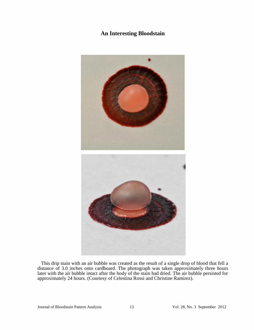
An Interesting Bloodstain
This drip stain with an air bubble was created as the result of a single drop of blood that fell a distance of 3.0 inches onto cardboard. The photograph was taken approximately three hours later with the air bubble intact after the body of the stain had dried. The air bubble persisted for approximately 24 hours. (Courtesy of Celestina Rossi and Christine Ramirez).

Journal of Bloodstain Pattern Analysis 14 Vol. 28, No. 3 September 2012
ABSTRACTS OF PAPERS PRESENTED AT THE 2012 IABPA TRAINING CONFERENCE IN TUCSON, ARIZONA
Machete and Rum Party at a Homeless Campsite Stuart H. James James and Associates Forensic Consultants, Inc. Fort Lauderdale, Florida USA
On the date of January 3
rd 2012 at approximately 1:32 AM the Sheriff’s Office received a
911 call and responded to a wooded area on the west coast of Florida. When deputies arrived, they were met by a homeless man who led them to a camping area that he shared with two other homeless men. A deceased man was on the ground near the campsite area. Reportedly, the three men were drinking rum and cooking a meal over a campfire. An altercation ensued between two of the individuals when the cooking grill was knocked over. This resulted in the death of one of them due to sharp force injuries inflicted with a machete that included severing of the right carotid artery and jugular vein. Reportedly, the third individual witnessed the altercation and called 911. The homeless man who reportedly wielded the machete was subsequently arrested and charged with second degree murder. The individual who claimed that he witnessed the homicide and called 911 claimed that he only assisted with the dragging the body to its final location for burial was arrested later and charged with being a principal to second degree murder. This presentation will demonstrate projected stains and an interesting transfer pattern on clothing worn by the two defendants. Medical and Forensic Aspects of Blood Clot Formation in the Presence of Saliva – A Preliminary Study Celestina Rossi Montgomery County Sheriff’s Office Conroe, Texas USA Stuart H. James James and Associates Forensic Consultants, Inc. Fort Lauderdale, Florida USA
Clinical tests for the determination of clotting or coagulation times for therapeutic monitoring of
anti-coagulant therapy utilize relatively non-invasive venipuncture or lancet piercing techniques. The medically normal clotting times for human blood should only be used as a baseline when considering clotting times in forensic casework. Factors such as high temperature and rough surface texture where blood has accumulated will shorten clotting time. Medical conditions and the presence of anticoagulants in the blood that extend clotting times may be an unknown factor.
Trauma to the body and organs will release high levels of tissue factor (TF) in response to the injury and this may shorten normal clotting times. Research into the medical and scientific literature has indicated that saliva contains high levels of TF as well and will accelerate the clotting time of blood. This presentation describes experiments to demonstrate that a mixture of saliva and blood will accelerate the clotting time of blood on external surface substrates of porcelain and vinyl tiles.

Journal of Bloodstain Pattern Analysis 15 Vol. 28, No. 3 September 2012
A Study of Errors in Area of Origin Evaluations Young il Seo National Forensic Service Yangsan Gyeongnam South Korea
An area of origin can be evaluated using straight-line geometry and trigonometric functions.
Both methods assume blood droplets travel in straight-line trajectories. However, blood droplets follow a parabolic trajectory due to gravity and air resistance. So errors occur when evaluating an area of origin using straight-line geometry and trigonometric functions.
In our experiment we measured errors in an area of origin evaluations with respect to distances and developed an improved method to evaluate an area of origin including gravity and air resistance. Furthermore we compared accuracies between stringing method and forensic software application (Hemospat®) method. A Novel and Objective Method for Determining the Impact Velocity of Bloodstains Nick Laan MSc
Netherlands Forensic Institute The Hague van der Waals-Zeeman Institute, University of Amsterdam Dr. Karla de Bruin
Netherlands Forensic Institute The Hague Prof. Dr. Daniel Bonn van der Waals-Zeeman Institute, University of Amsterdam
We show how the impact velocity of a bloodstain can be determined by means of general fluid dynamic principles and how this can be used to improve methodology for the region of origin determination on crime scenes. When finding an impact pattern on a crime scene, the investigator wants to know where the region of origin was at the time of the bloodletting event, for purposes of crime scene reconstruction or confirmation of witness testimony. To determine the region of origin, usually the stringing method is used, either with real or virtual strings. The stringing method is based on the assumption that the flight path is a straight line. However, ballistic objects like blood droplets are not projected through the air in a straight line but rather follow a curved trajectory due to gravity and air resistance. De Bruin [1] measured that the further the blood source was located from the pattern, the larger the deviation was between the determined region of origin and the actual origin. The vertical deviation could be as large as 45 cm, which could be the distinction between the person sitting or standing. In addition, only upward directed bloodstains can be used for a reliable region of origin estimation, while on the crime scene often a majority of bloodstains are directed downward. Our novel method relies on a correct mechanical and fluid dynamical description of the flight path and impact event. In this way, also downward directed bloodstain can be taken into account, thus increasing the amount of bloodstains which can be analyzed.
Four parameters are required to unambiguously describe the path of a blood drop which accounts for gravity and air resistance: 1) position 2) impact angle 3) volume and 4) impact velocity. In a previous study we have shown that, under certain circumstances, the original volume of the droplet can be determined from a dried stain [2]. Our laboratory experiments show how the impact velocity of a droplet is related to bloodstain size and volume based on

Journal of Bloodstain Pattern Analysis 16 Vol. 28, No. 3 September 2012
previous research done by Clanet et al. [3] and Bartolo et al. [4]. We measured this for various surfaces of different surface roughness and wettability. Based on the relations found, we were able to predict the impact velocity as a function of stain size, stain volume, and type of surface. References: 1. de Bruin, K.G., R.D. Stoel, and J.C.M. Limborgh, Improving the Point of Origin
Determination in Bloodstain Pattern Analysis. Journal of Forensic Sciences, 2011. 56(6): p. 1476-1482.
2. Laan, N., et al., Volume determination of bloodstains by means of optical coherence tomography. Journal of Forensic Sciences, Submitted.
3. Clanet, C., et al., Maximal deformation of an impacting drop. Journal of Fluid Mechanics, 2004. 517: p. 199-208.
4. Bartolo, D., et al., Dynamics of Non-Newtonian Droplets. Physical Review Letters, 2007. 99(17): p. 174502.
Point of Pivot of a Simple Cast-off Celine Nicloux French Forensic Institute (Gendarmerie Nationale) Seine Saint Denis France
Is it possible to mathematically calculate the point of pivot of a cast off? A pivot is the part
constituting the support or the extremity of the axis around which an element turns. When a bloody object is in motion, blood drops are released. The point around which the object is in motion is the point of pivot. Depending on the movement, it could be for example a wrist, elbow, shoulder position or an arm which holds the object in movement during the cast-off. This study is a beginning of a scientific research trying to find an answer to this question. The Characteristics of Blood on “Wicking” Fabrics
Richard Tewes Pioneer Forensics Loveland, Colorado, USA
This presentation illustrates the “Wicking Effect” of blood on certain modern fabrics. Under
Armour® Dri-Fit® and Climacool® are just a few of the new generations of clothing and fabrics that report the ability to move moisture from the body and repel exterior moisture. How does this effect blood? We will explore the appearance and changes from an internal bleeding simulation to in-flight stain impact and appearance. High magnification video and imaging are used to help display the actions in these modern fabrics.
Visualization of Cast-off Patterns
Andy Maloney FORident Software, Inc. Ottawa Canada
Currently cast-off patterns are identified at the scene and then documented using photos and a description of the pattern. Often this is the only information included in a bloodstain report which makes information about the pattern difficult to communicate to others.

Journal of Bloodstain Pattern Analysis 17 Vol. 28, No. 3 September 2012
Standing at the scene, though, an investigator can visualize roughly where in the room the person was standing when they were swinging the bloodied object which created the pattern, as well as the approximate plane of motion of the swing. So how do we record, analyze and present this information to communicate to others - particularly those that have not been at the scene?
This talk will outline a method of analyzing cast-off patterns to produce an approximate plane of motion suitable for a 3D scene reconstruction. This presentation is based on the article "Visualization of Cast-off Patterns Using 3D Modelling Software" published in the Journal of the Association for Crime Scene Reconstruction. A PDF of the article is available here: http://hemospat.com/research.php
Developing the Analysis of Environmentally Altered Bloodstains
Hester Miles University College London, United Kingdom
Environmentally altered bloodstains are bloodstains that can be considered to have been in some way altered by certain elements or fluctuations in their surrounding environment. These elements or fluctuations are either natural or anthropogenic in origin. Alterations could be forced, for example, by freeze-thaw cycling, stain generation on frozen surfaces, introduction of bleach to stains, washing or burning. Exposure to these elements or fluctuations may alter a stains appearance, persistence, internal composition or behavior (i.e. drying time), all of which have implications for subsequent stain analysis and crime scene reconstruction. For example, alterations to stain appearance may have consequences for stain identification and lead to possible misidentification. Environmental distortions of drying time may influence the temporal reconstruction of a bloodletting event or series of events. Enabling and enhancing the accuracy of stain identification and interpretation for environmentally altered bloodstains is therefore an important aspect of Blood Pattern Analysis, particularly at environmentally exposed crime scenes or in cases where perpetrators have made evasive attempts to remove or alter evidence.
Having outlined the context and importance of conducting empirical research into environmentally altered bloodstains, the methodology and results of an ongoing research project, aimed at developing methods of identifying and interpreting these stains will be presented. “I Have Information about My Dead Wife…” A Look at Clothing Examinations and Experimentation
Brandi Caron State Police Crime Laboratory Augusta Maine, USA
On a frozen January morning in Maine, police were advised to check on the well-being of a young, married, pregnant woman. When detectives arrived at the residence, they made a gruesome discovery.
The husband was in custody and statements made through his attorneys as well as subsequent explanations for the forensic findings furthered this investigation. Ultimately an experiment was conducted and the findings proved helpful to the prosecution. This presentation will reinforce the importance of laboratory examination of clothing; namely the bloodstain pattern interpretation and the effectiveness of well-designed experimentation.

Journal of Bloodstain Pattern Analysis 18 Vol. 28, No. 3 September 2012
Proving a Priest Killed a Nun
T. Paulette Sutton Forensic Consultant, Memphis, Tennessee, USA
In 1980, Sister Margaret Ann Pahl was found murdered in the sacristy of Toledo’s Mercy
Hospital. At the conclusion of the investigation, no one was charged with her death. Twenty six years later the case was reopened. In addition to the usual problem associated with a cold case, the only DNA isolated in this case could potentially be used to create reasonable doubt. While this presentation will discuss the forensic findings, the process of putting the pieces together will be emphasized. The primary objective will be to demonstrate that nothing can replace team work-- traditional police investigative procedures, forensic science, and a good quarterback in the prosecutor’s office. Double International Homicide- an Independent Review
Gillian Leak Principal Forensic Services, Ltd Wetherby West Yorkshire England, United Kingdom
In June 2006, two children were found dead in their cots in The Netherlands. They had both died as a result of multiple stab wounds. The older child had also sustained many defence wounds to her hands. The mother of the children had raised the alarm. She claimed she was working in the kitchen when a man forced his way into the flat, disarming her of a knife she was carrying. In the struggle she sustained minor cuts to her neck and abdomen. She couldn’t recall what happened next, except that the man threw the knife onto the hallway floor as he left. She admits that she then entered her daughter’s bedroom, without putting on the light, and stroking her daughter’s face that seemed upset and was murmuring lightly. She didn’t enter the baby boy’s room. After lengthy police investigations, the mother was arrested on suspicion of killing both of her children. A trial was held, however the Judges were unable to reach a verdict. Gillian was asked by a Judge to review the case. This presentation will cover some of the observations made in what proved to be quite a complex case. At the end of her investigations, Gillian, together with the scientist for the defence, was called to testify at a re-trial held in Amsterdam in 2010.
Comparison of Saliva Tests for Identification of Expectorated Blood Spatter
Si-Keun Lim National Forensic Service Yangsam Gyeongnam South Korea
Identification of expectorated blood spatter is important for bloodstain analysis and crime scene reconstruction because it is difficult to distinguish expectorated spatter from impact spatter. In this study, four saliva test methods which included RSID-Saliva® (Independent Forensics), SALIgAE® (Abacus Diagnostics), Phadebas® Forensic Press Test (Magle Life Sciences), and Blue-starch agarose plates were compared to determine the best methods to identify expectorated blood spatter. RSID-Saliva® kit from Independent Forensics showed the highest sensitivity and no inhibition of blood. Expectorated blood spatter was successfully identified in homicide case occurred at April this year.

Journal of Bloodstain Pattern Analysis 19 Vol. 28, No. 3 September 2012
“How Much Blood is That & How Did it Get There?” Erin Sims Lincoln Police Department Lincoln, Nebraska USA
Bloodstain Pattern Analysts are asked these questions by Detectives, Prosecuting Attorneys, and other Law Enforcement Officials who are attempting to determine if a victim was alive, unconscious, or deceased; during, or as a result of a series of events occurring in a criminal offense. Researchers and Analysts have found that even at pristine crime scenes, blood loss amounts cannot be accurately determined given the number of variables involved. Ms. Sims will present an example of a Homicide case in which these questions were posed. The evidence showed that someone had attempted to clean-up or destroy blood evidence at four separate ‘scenes’. The suspect, after being made aware of the remaining blood evidence, formulated a defense for the presence of the bloodstains as being made during his heroic attempt to fight off ‘the real killer’. Ms. Sims conducted two case specific research projects to use in comparing scene evidence to the research results. This evidence was used by investigators, prosecutors, and ultimately the jury to let them decide for themselves: “How much blood is that and how did it get there?”
Bloodstain Pattern Documentation - A New Approach Michael Perkins Metropolitan Police Department, Las Vegas, Nevada USA
Bloodstain pattern documentation is an important preliminary step in the reconstruction of events where bloodshed occurs. This lecture illustrates new methods of documentation using laser levels, digital and laser measuring devices, and prepared worksheets. This lecture consists of a PowerPoint presentation which will show the various techniques. This approach greatly reduces the chances of cross contamination of the evidence as well as unnecessary biohazard exposure to the crime scene investigator. Mapping of the bloodstain patterns is done with readily available and inexpensive labels, with greater accuracy than traditional methods. Handouts will be available of the various worksheets used for the bloodstain analysis.
Rodney Robert Cocking Case Mitchell W. Dinterman Forensic Sciences Division Maryland State Police Pikesville, Maryland USA The victim in this case was a nationally renowned child psychologist and author. Rodney Cocking worked in Washington, D.C., and had residences in both northern Virginia and Maryland. His residence in Maryland was located in a very rural and secluded area in Carroll County, approximately forty miles west of Baltimore City. His life partner reported him missing when he failed to return home from his office after several days. His partner advised investigators that he did not immediately report him missing as it was not that unusual for him to stay over at his residence in northern Virginia given its close proximity to his office. The investigators, suspecting that the partner may have been involved with the disappearance, obtained search warrants for his residence in Maryland. Upon serving the warrant, a thorough search revealed what investigators believed were bloodstains related to gunshots. Examination

Journal of Bloodstain Pattern Analysis 20 Vol. 28, No. 3 September 2012
of these stains led the investigation in a different direction eventually guiding the investigators to the real suspect.
A 400-year-old Royal Crime Scene – A Special Crime Scene Reconstruction Andre Hendrix Chief Crime Scene Unit Zeeland Police The Netherlands
At the museum "Het prinsenhof" in Delft, one of the most dramatic events in Dutch history took place: The murder on Prince William of Orange, Father of the Nation, on July 10th 1584. At that place, the place WoO thought to be safe, two bullet holes still remains. No doubt about the killer, but still some questions.... Are these bullet holes real? What was the position of the killer and the victim at the time of the attack? Could William speak his famous last words (My God, My God, have pity with me and my poor people)?
Advanced Forensic methods, such as 3D scanning technology, firing tests and trace evidence at the crime scene, supplemented with research into building records and historic resources, provide answers to previously unsolved questions. The results of the research have shed a different light on the murder of the Father of the Nation.
The Bloodstain Pattern Analyst – Looking After Ourselves Joe Slemko Edmonton, Alberta, Canada
A bloodstain pattern analyst finds the data that forms the foundation of their scientific
opinion within the results of violent and traumatic interactions between human beings. BPA is conducted in a ‘human environment’ and not within the clinical, sterile environment of a laboratory. As a result, the human aspects of a violent occurrence may directly or indirectly, have a profound effect on an analyst. During this presentation the speaker will discuss some of his personal experiences, observations and research regarding an issue that is all too often, ‘hidden in the closet.’
80 Degrees Junk Science - Real Science Lynne D. Herold, Ph.D. Scientific Services Bureau Los Angeles County Sheriff’s Department Los Angeles, California USA
It seems not that long ago that bloodstain pattern analysis was being questioned as having a
scientific basis and its admissibility challenged with every case going to trial. Has the pendulum swung the other way? Is the court actually demanding that bloodstain pattern analysis be presented for a just and fair trial, and for the trier of fact (jury) to be able to reach a verdict based on physical evidence?
This presentation is the case review of People of the State of California vs. Kevin Bennett. Kevin Bennett, accused of committing a murder in 2001, was serving a term of 51 years to life following a jury conviction of second degree murder. In December, 2010, the US Court of Appeals for the Ninth Circuit issued a reverse and remand with a conditional writ of habeas corpus after determining that the defense counsel was deficient and objectively unreasonable. The appellate court specifically cited the failing of counsel to consult with an expert, ask appropriate questions about the bloodstains present, and/or present bloodstain pattern evidence

Journal of Bloodstain Pattern Analysis 21 Vol. 28, No. 3 September 2012
that appeared to corroborate the defendant’s self-defense testimony in his version of events. The case was retried in October 2011, with bloodstain pattern evidence presented by the defense but not the prosecution. The original second degree murder conviction reduced to manslaughter with 25 years sentence.
Dan Rahn Grant Recipient Research Project Elisabeth Williams University of Auckland/ESR Forensic Christchurch Science Centre New Zealand
The aim of this project is to develop a mechanical device capable of generating and projecting reproducible blood droplets at a range of controlled sizes, velocities and projection angles relevant to cast-off pattern formation. Preliminary research in the area of cast-off creation indicates that these stains may yield more information about the position, action and physical characteristics of an assailant than is currently thought. In order to thoroughly investigate this hypothesis, cast-off drops must be studied in reproducible fashion to explore their relationship with biomechanical actions. The construction of such a mechanical device will facilitate a detailed analysis of the fluid dynamics of blood drops in flight and during impact. These mechanical characteristics can be compared between drops with upward, downward and horizontal flight paths. Two high speed digital video cameras will be used to film the flight and impact of droplets. The ability to generate and analyse blood drop dynamics in such a controlled manner lends itself to the development of statistical models which can aid in the presentation of objective bloodstain evidence.
Industrial Death Investigation All Wrapped Up Norman Reeves BPA Consulting Tucson, Arizona USA
This presentation is a civil wrongful death case involving the issues of place and
circumstances surrounding the death at a metal shop lathe. While the civil attorneys had issues in the case not directly related to the examination, the location and position of the victim at the time of the accident was relevant to the case and evaluations by the medical examiners and other experts. The victim was decapitated as a result of the accident and video, OSHA, and activities of the company played a role in the examination.

Journal of Bloodstain Pattern Analysis 22 Vol. 28, No. 3 September 2012
Martin Eversdijk Receives the IABPA Distinguished Member Award
Martin Eversdijk was the recipient of the 12
th IABPA Distinguished Member Award at the
2012 Conference in Tucson, Arizona. The award is given in recognition of outstanding contributions to the discipline of bloodstain pattern analysis and to the IABPA.
Martin is a forensic professional and serious crime scene coordinator for the forensic department of the Amsterdam-Amstelland Regional Police in The Netherlands. He started working as a CSI in 1993 and his interest in this field developed into extensive work and research in blood enhancement and search techniques. Important outcomes resulting from this research, like the application of Luminol/Lumiscene with a special air compressor, are still in use and formed the basis for blood search techniques in the Netherlands. For 7 years Martin was a staff member and trainer at the Institute for Criminal Investigations and Crime Science, the national training center for CSI’s in the Netherlands, developing and teaching courses in Bloodstain Pattern Analysis. He is an internationally known lecturer on the subject of BPA and contributor of new methods. He has educated hundreds of students from 19 countries. He has done bloodstain crime scene work internationally and has given expert opinion evidence. Martin is an author of various articles and textbook chapters on the topic of bloodstain patterns and has been an (board) member of SWGSTAIN. Since 2011 Martin and his companion Rene Gelderman, also a forensic professional and serious crime scene coordinator, have their own forensic training institute providing several different levels of BPA courses.
Martin Eversdijk

Journal of Bloodstain Pattern Analysis 23 Vol. 28, No. 3 September 2012
Recent BPA Related Articles in the Scientific Literature
Kuula, J., Pölönen, I., Puupponen, H.,Selander, T., Reinikainen, T., Kalenius, T. and Saari, S. Using VIS/NIR and IR Spectral Cameras for Detecting and Separating Crime Scene Details, SPIE Defence, Security + Sensing, Baltimore, Maryland, April 2012. Stene, I. and Adair, T., The Survival of Neat and Cleaned Blood after the Application of Wallpaper, J. Assoc, Crime Scene Reconstruction, 2012, 18(3), pp. 21-28. Li, B., Beveridge, P., O’Hare, W.T., and Islam, M., The Estimation of the Age of a Bloodstain using Reflectance Spectroscopy with a Microspectrophotometer, Spectral Pre-processing and Linear Discriminant Analysis, Forensic Science International, 212, (2011), pp. 198-204. Farrar, A., Porter, G. and Renshaw, A., Detection of Latent Bloodstains Beneath Painted Surfaces using Reflected Infrared Photography, J. Forensic Sci., September 2012, Vol. 57, No. 5. pp. 1190-1198. Gardner, Ross M., Maloney, Michael and Rossi, Celestina, “A Crime Scene Investigator’s Method for Documenting Impact Patterns for Subsequent Off-Scene Area-of-Origin Analysis” Journal of Forensic Identification, Vol.62, No. 4, July/August 2012
Association for Crime Scene Reconstruction 2013 Conference The 2013 ACSR conference will be held in Atlanta, Georgia from February 7 to 10,
2013. The conference program is still in development and will involve 3½ days of presentations. There are numerous openings for research presentations, case studies, and workshops. There will be three rounds of workshops scheduled, two day-time and one night-time. Tentatively these include: Practical Application of Event Analysis, Alternate Light Source Utilization, Laser Trajectory Documentation, Electro-static Dust-print Lifters, and Laser Scanning stations with both the Leica and Faro systems. The conference will be held at the Hilton Garden Inn, one mile south of Hartsfield Jackson Airport. The room rate is set at $99.00 per night. As part of the registration fee, continental breakfast will be provided Tuesday through Friday and lunch will be provided Tuesday through Thursday. Once the contract is finalized, ACSR will provide a link for registration at www.acsr.org. Look for updates as the year progresses. If you have an interest to present, please contact Conference Chair Ross M. Gardner at [email protected] for further information

Journal of Bloodstain Pattern Analysis 24 Vol. 28, No. 3 September 2012
Organizational Notices
Moving Soon?
All changes of mailing address need to be supplied to our Secretary Norman Reeves. Each quarter
Norman forwards completed address labels for those who are members. Do not send change of address information to the Bloodstain Digest Editor. E-mail your new address to Norman Reeves at:
Norman Reeves
I.A.B.P.A.
12139 E. Makohoh Trail
Tucson, Arizona 85749-8179
Fax: 520-760-5590
Membership Applications / Request for Promotion
Applications for membership as well as for promotion are available on the IABPA website:
IABPA Website: http://www.iabpa.org
The fees for application of membership and yearly dues are $40.00 US each. If you have not received a dues invoice for 2013 please contact Norman Reeves. Apparently, non US credit cards are charging a fee above and beyond the $ 40.00 membership/application fee. Your credit card is charged only $40.00 US by the IABPA. Any additional fees are imposed by the credit card companies.
IABPA now accepts the following credit cards:
Discover MasterCard
American Express Visa
Training Opportunities
October 22-26, 2012 Advanced BPA Course
Nieuw-Vennep The Netherlands
For further information contact:
Loci Forensics, B.V.
Flierveld 59
2151 LE Nieuw-Vennep
The Netherlands
Martin Eversdijk
Tel: +31639455563
Renѐ Gelderman
Tel: +31639455562
Fax: +31(0)20-8907749
E-mail: [email protected]
______________________

Journal of Bloodstain Pattern Analysis 25 Vol. 28, No. 3 September 2012
November 26-30, 2012 Visualization of Latent Bloodstain Course
Nieuw-Vennep The Netherlands
For further information contact:
Loci Forensics, B.V.
Flierveld 59
2151 LE Nieuw-Vennep
The Netherlands
Martin Eversdijk
Tel: +31639455563
Renѐ Gelderman
Tel: +31639455562
Fax: +31(0)20-8907749
E-mail: [email protected]
December 3-7, 2012
Basic Bloodstain Pattern Analysis Workshop Presented by the Specialized Training Unit at the Miami-Dade Safety Training Institute,
Doral, Florida
Contact: Toby L. Wolson, M.S., F-ABC
Miami-Dade Police Department
Forensic Services Bureau
9105 N.W. 25th
Street
Doral, Florida 33172
Voice: 305-471-3041
Fax: 305-471-2052
E-mail: [email protected]
December 3-7, 2012
Advanced Bloodstain Pattern Analysis Course
Hosted by the Johnson County Sheriff’s Office Criminalistics Laboratory
Olathe, Kansas
Instructors: Paul Erwin. Kish and Stuart H. James
Contact: Paul Erwin Kish
Forensic Consultant and Associates
Tel: 607-962-8092
Fax: 607-962-2093
E-mail: [email protected]
Articles and training announcements for the December 2012 issue of the Journal of Bloodstain Pattern Analysis must be received before November 30, 2012

Journal of Bloodstain Pattern Analysis 26 Vol. 28, No. 3 September 2012
Editor’s Corner
The 2012 IABPA Conference held in Tucson, Arizona October 2-6 was well attended with
102 participants that represented countries that included the USA, Canada, England UK, The Netherlands, France, South Africa, Australia, New Zealand, the Republic of the Philippines and Korea. There were excellent presentations given and I have included the abstracts of their content in this issue of the Journal. I invite the speakers to submit articles of their presentations for peer review and publication in our Journal.
The highlight of the Conference was the awarding of IABPA Distinguished Member status to Martin Eversdijk of the Netherlands. He was the 12
th member to receive this honor. Martin is
well deserving of this award and on behalf of the membership and personally, I congratulate him for his accomplishments in the discipline of bloodstain pattern analysis and his dedication to the IABPA.
As Historian of the IABPA, I am collecting photographs and memorabilia from the 2012 conference as well as past conference. I am condensing the prior conference notebooks into one or two binders that will reflect the speakers, their presentations and activities. The plan is to organize the materials so that they can be displayed at future conferences. Please send me copies of photographs and other materials that would be of interest to the membership.
The December issue of the Journal has space for submitted articles of research and case reports. The abstracts of papers presented at the 4
th European IABPA Conference to be held in
Edinburgh, Scotland, UK November 12-14, 2012 will be published in that issue.
Stuart H. James Editor [email protected]

Journal of Bloodstain Pattern Analysis 27 Vol. 28, No. 3 September 2012
Publication Committee
Associate Editors
Kacper Choromański Barton P. Epstein Carolyn Gannett
Paul E. Kish Daniel Mabel Jon J. Nordby Jon Thomas
T. Paulette Sutton Todd A. Thorne
Past Editors of the IABPA News/Journal of Bloodstain Pattern
Analysis
Anita Y. Wonder 1984-1985 Norman Reeves 1986-1989 David Rimer 1990-1996 Toby L. Wolson 1997-2000 Paul E. Kish 2001-2003 Stuart H. James 2004-current
Past Presidents of the IABPA
V. Thomas Bevel 1983-1984 Charles Edel 1985-1987 Warren R. Darby 1988 Rod D. Englert 1989-1990 Edward Podworny 1991-1992 Tom J. Griffin 1993-1994 Toby L. Wolson, M.S. 1995-1996 Daniel V. Christman 1997-1998 Phyllis T. Rollan 1999-2000 Daniel Rahn 2001-2002 Bill Basso 2002-2006 LeeAnn Singley 2007- 2008
The Journal of Bloodstain Pattern Analysis published quarterly in March, June, September, and December.
2012. The International Association of Bloodstain Pattern Analysts. All rights are reserved. Reproduction in
whole or in part without written permission is prohibited.

