t -t ad-a241 877t -t ad-a241 877 illl c~i ad__ grant no: damd17-91-z-1030 title: 4th annual trauma...
TRANSCRIPT
T -T
AD-A241 877illl c~i
AD__
GRANT NO: DAMD17-91-Z-1030
TITLE: 4TH ANNUAL TRAUMA ANESTHESIA AND CRITICAL CARE SYMPOSIUM
PRINCIPAL IN' -STIGATOR: Christopher M. Grande, M.D.
CONTRACTING ORGANIZATION: International Trauma Anesthesiaand Critical Care Society
MIEMSS Anesthesiology Department22 South Greene StreetBaltimore, Maryland 21201
REPORT DATE: July 1, 1991
TYPE OF REPORT: Published Proceedings
PREPARED FOR: U.S. ARMY MEDICAL RESEARCH AND DEVELOPMENT COMMANDFort Detrick, Frederick, Maryland 21702-5012
DISTRIBUTION STATEMENT: Approved for public release;distribution unlimited
The findings in this report are not to be construed ai atofficial Department of the Army position unless so desigri.ted byother authorized documents.
91-12958
1 July 1991 Published Proceedings (4/15/91 - 6/15/91)
4th Annual Trauma Anesthesia and Critical Care Grant No.Symposium DAMD7-91-Z-1030
(Held May 2-5, 1991)
Christopher M. Grande, M.D.
International Trauma Anesthesia and Critical CareSociety, MIEMSS Anesthesiology Department
22 south Greene StreetBaltimore, Maryland 21201
U.S. Army Medical Research and Development CommandFort DetrickFrederick, Maryland 21702-5012
Approved for public release; distribution unlimited
Symposium, Anesthesia, Trauma, RA2
Unclassified Unclassified N/A N/A

* ~4th Annu~al
May 2-5, 1991Baltimore, Maryland, USA
Sponsored by
The International Trauma Anesthesia and Critical Care Society
00
I "g :,1
0
0

SYMPOSIUM EXECUTIVE COMMITTEE
DIRECTOR
Christopher M. Grande, MDSpecial Consultant and Chief, Special ProjectsDepartment of AnesthesiologyThe Shock Trauma Center, MIEMSSUniversity of Maryland Medical SystemCaptain, Medical Corps, USAR,Flight Surgeon and Diving Medical Officer11th Special Forces Group (Airborne)Ist T. ,," pecal FoccesExecutive Director, ITACCSEditor, Trauma AnesthesiaEditor, Overview of Trauma Anesthesiaand Critical Care, Critical Care Clinics
Baltimore, Maryland
ASSOCIATE DIRECTORS
Peter J.F. Baskett, MB, BCh. FFARCSConsultant AnesthetistDepartment of AnesthesiologyFrenchay HospitalPresident, Association of Anaesthestist ofGreat Britain and IrelandPresident, World Association for Emergencyand Disaster MedicineChairman, Bylaws Committee, ITACCSLieutenant Colonel, RAMCAuthor, Medicine for DisastersAuthor, Resuscitation HandbookBristol, EnglandUnited Kingdom
Brian F. Condon, MDColonel Medical Corps, USAD-irector, Anesthesia and Operative ServicesWalter Reed Army Medical CenterAssociate Professor of AnesthesiologyUniformed Services University of theHealth SciencesConsultant for Anesthesiology to theSurgeon General, U.S. Army
Washington, DC

Wolfgang F. Dick, MD, PhD, FFARCS (Hon)Professor and Chairman for the Clinic ofAnaesthesiologyUniversity Hospital Mainz, GermanyVice-President, European Academy ofAnaesthesiology
Medical Director, University Hospital MainzSecretary, German Association of Intensive Careand Emergency MedicineCo-Editor, Der AnasthesistManaging Editor, Notfallmedizin(Emergency Medicine)
Advisory Board, Current Anaesthesia PracticeAuthor/Editor, Paediatric Anaesthesia(Kinderanasthesie)
German National Representative, ITACCSMainz, Germany
Elizabeth A.M. Frost, MDProfessor of AnesthesiologyAlbert Einstein College of Medicine/Montefiore Medical CenterDirector, Division of NeuroanesthesiaEditorial Board, Anesthesiology NewsEditor, ITACCS NewsletterNew York, New York
Adolph H. Giesecke, MDJenkins Professor and ChairmanDepartment of AnesthesiologyUniversity of TexasSouthwestern Medical SchoolParkland Memorial HospitalVice President, ITACCSEditor, Anesthesia for Surgery of TraumaDallas, Texas
Vladimir Kvetan, MDAssociate Professor of Anesthesiology andCritical Care Medicine
Director, Division of Critical Care MedicineAlbert Einstein College of Medicine/Montefiore Medical Center
Editor, Disaster Manaqement, CriticalCare ClinicsChairman, ITACCS Disaster CommitteeBronx, New York
Colin A.B. McLaren, MB, ChB, FFARCSAir Commodore, Royal Air ForceConsultant Advisor AnaestheticsRoyal Air ForceKelvin House, LondonVice President, Association of Anaesthetists,Great Britian and IrelandLondon, EnglandUnited Kingdom
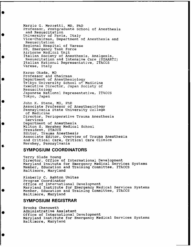
Marzio G. Mezzetti, MD, PhDProfessor, Postgraduate School of Anesthesiaand Resuscitation
University of Pavia, ItalyVice-Chairman, Department of Anesthesia andResuscitation
Regional Hospital of VaresePR, Emergency Task ForceAirborne Medical UnitItalian Society of Anesthesia, Analgesia,Resuscitation and Intensive Care (SIAARTI)Italian National Representative, ITACCSVarese, Italy
Kazuo Okada, MDProfessor and ChairmanDepartment of AnesthesiologyTeikyo University School of MedicineExecutive Director, Japan Society ofResuscitologyJapanese National Representative, ITACCSTokyo, Japan
John K. Stene, MD, PhDAssociate Professor of AnesthesiologyPennsylvania State University Collegeof Medicine
Director, Perioperative Trauma AnesthesiaServices
Department of AnesthesiaMilton S. Hershey Medical SchoolPresident, ITACCSEditor, Trauma AnesthesieAssociate Editor, Overview of Trauma Anesthesiaand Critical Care, Critical Care ClinicsHershey, Pennsylvania
SYMPOSIUM COORDINATORS
Terry Slade YoungDirector, Office of International DevelopmentMaryland Insitute for Emergency Medical Services SystemsMember, Education and Training Committee, ITACCSBaltimore, Maryland
Kimberly C. Ashton UnitasProgram CoordinatorOffice of International DevelopmentMaryland Institute for Emergency Medical Services SystemsMember, Education and Training Committee, ITACCSBaltimore, Maryland
SYMPOSIUM REGISTRAR
Brooks ChenowethAdministrative AssistantOffice of International DevelopmentMaryland Institute for Emergency Medical Services SystemsBaltimore, Maryland

AD HOC ADVISOR
Williamd R. Anderson, CPAExecutive DirectorShock Trauma Associates, PABaltimore, Maryland
We would also like to gratefuly acknowledge Leanne Allgaier forher assistance in the production of the Symposium Series.
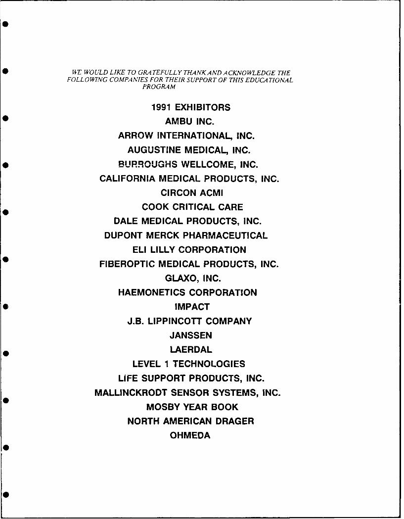
* WE WOULD LIKE TO GRATEFULLY THANKAND ACKNOWLEDGE THEFOLLOWING COMPANIES FOR THEIR SUPPORT OF THIS EDUCATIONAL
PROGRAM
1991 EXHIBITORSAMBU INC.
ARROW INTERNATIONAL, INC.
AUGUSTINE MEDICAL, INC.BURROUGHS WELLCOME, INC.
CALIFORNIA MEDICAL PRODUCTS, INC.
CIRCON ACMI
COOK CRITICAL CARE
DALE MEDICAL PRODUCTS, INC.
DUPONT MERCK PHARMACEUTICAL
ELI LILLY CORPORATION
FIBEROPTIC MEDICAL PRODUCTS, INC.
GLAXO, INC.HAEMONETICS CORPORATION
IMPACT
J.B. LIPPINCOTT COMPANY
JANSSEN
LAERDAL
LEVEL 1 TECHNOLOGIES
LiFE SUPPORT PRODUCTS, INC.
MALLINCKRODT SENSOR SYSTEMS, INC.MOSBY YEAR BOOK
NORTH AMERICAN DRAGER
OHMEDA
ORGANON, INC.
PANAMERICAN TRAUMA SOCIETY
PREFERRED PHYSICIANS MUTUALSIEMENS
TONOMETRICS, INC.
U.S. ARMY BIOMEDICAL RESEARCH AND
DEVELOPMENT LABORATORY
U.S. ARMY MEDICAL DEPARTMENT
W.B. SAUNDERS
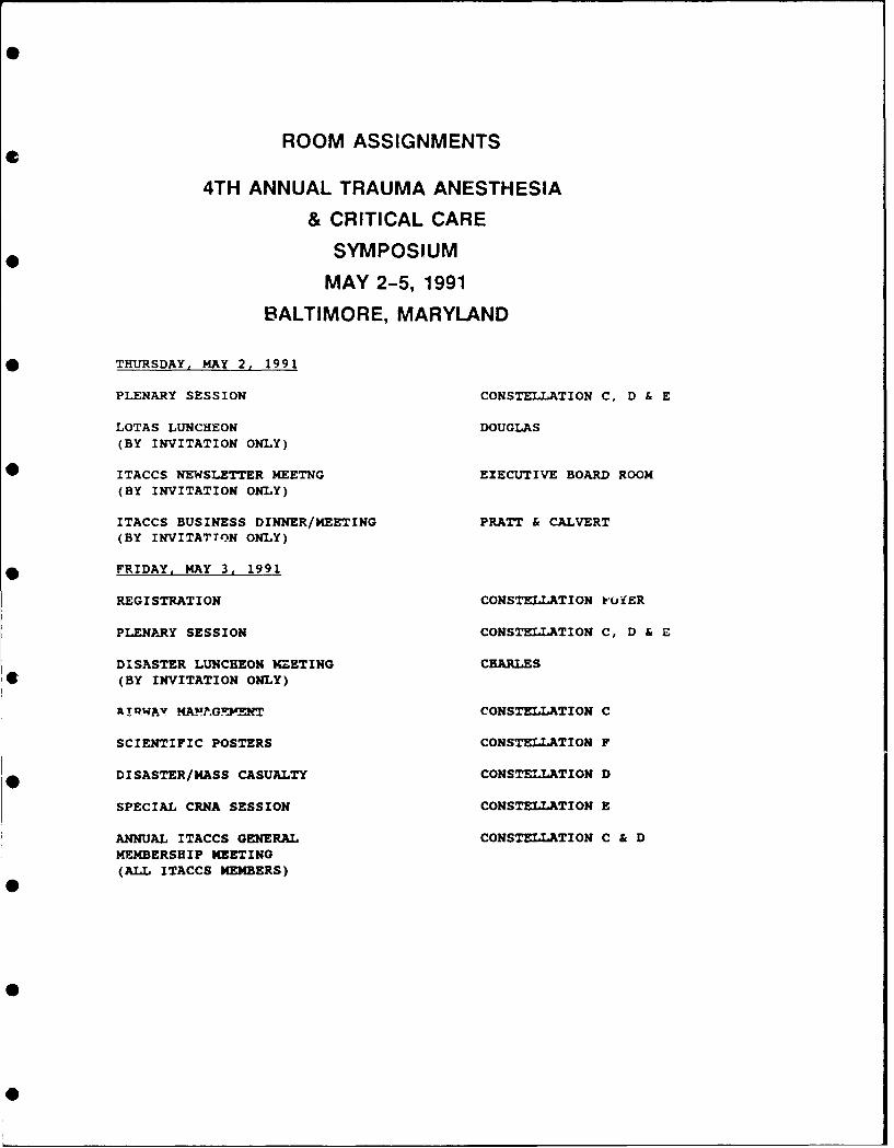
ROOM ASSIGNMENTS
4TH ANNUAL TRAUMA ANESTHESIA
& CRITICAL CARE
SYMPOSIUM
MAY 2-5, 1991
BALTIMORE, MARYLAND
THURSDAY, MAY 2, 1991
PLENARY SESSION CONSTELLATION C, D & E
LOTAS LUNCHEON DOUGLAS
(BY INVITATION ONLY)
ITACCS NEWSLETTER MEETNG EXECUTIVE BOARD ROOM(BY INVITATION ONLY)
ITACCS BUSINESS DINNER/MEETING PRATT & CALVERT(BY INVITA7TON ONLY)
FRIDAY, MAY 3, 1991
REGISTRATION CONSTELLATION ru'ER
PLENARY SESSION CONSTELLATION C, D & E
DISASTER LUNCHEON MEETING CHARLES
(BY INVITATION ONLY)
ATRWAV MA_ .G-FENT CONSTELLATION C
SCIENTIFIC POSTERS CONSTELLATION F
DISASTER/MASS CASUALTY CONSTELLATION D
SPECIAL CRNA SESSION CONSTELLATION E
ANNUAL ITACCS GENERAL CONSTELLATION C & D
MEMBERSHIP MEETING
(ALL ITACCS MEMBERS)

SATURDAY, MAY 4, 1991
REGISTRATION CONSTELLATION FOYER
TEXTBOOK MEETING DOUGLAS(BY INVITATION ONLY)
PLENARY SESSION CONSTELLATION C, D & E
DISASTER LUNCHEON MEETING DOUGLAS(BY INVITATION ONLY)
ANESTHESIA EQUIPMENT CONSTELLATION C
SCIENTIFIC ABSTRACTS CONSTELLATION E
DISASTER/MASS CASULATY CONSTELLATION D
SPECIAL CRl.A SESSION BALTIMORE BALLROOM
5TH ANNUAL ORGANIZING COMMITTEE MEETING DOUGLAS(BY INVITATION ONLY)
SUNDAY, MAY 5, 1991
PLENARY SESSION CONSTELLATION A & B
0o
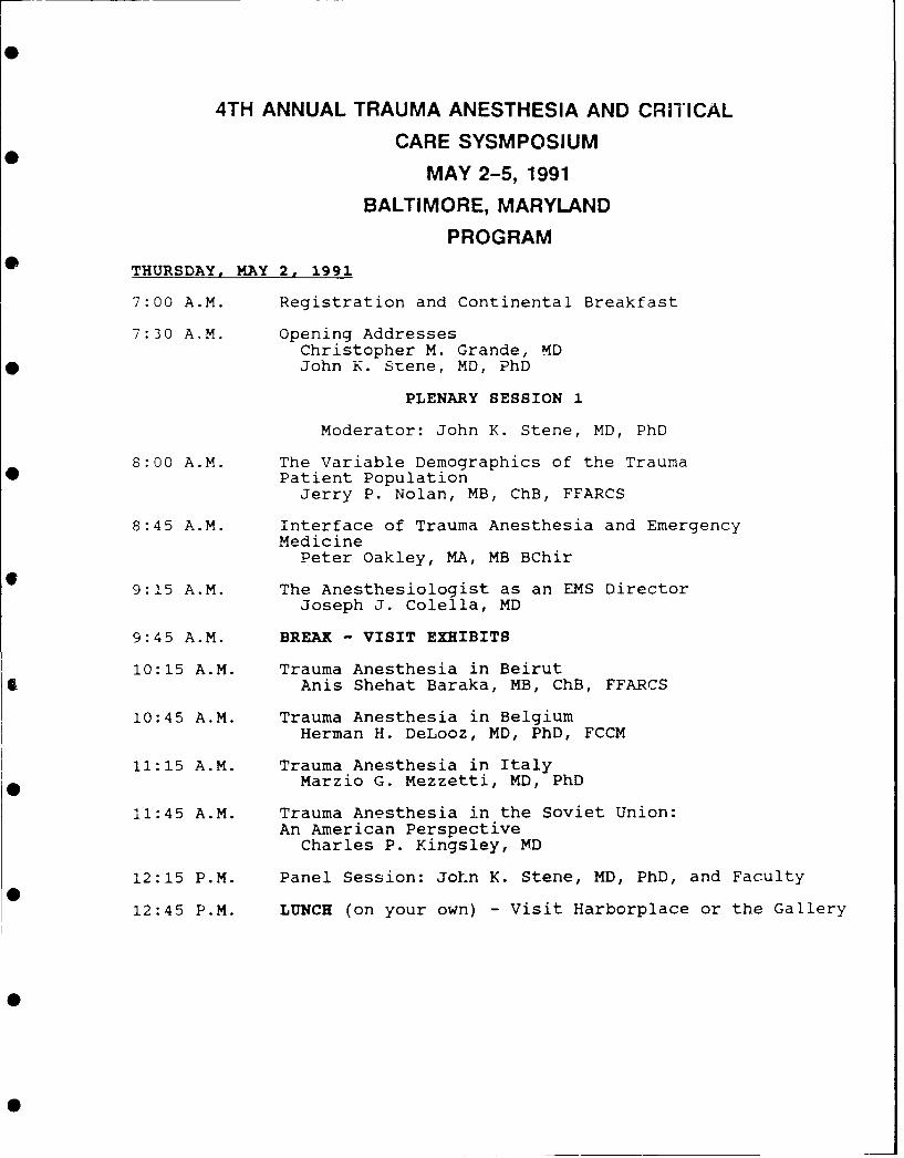
4TH ANNUAL TRAUMA ANESTHESIA AND CRITICAL
CARE SYSMPOSIUM
MAY 2-5, 1991
BALTIMORE, MARYLANDPROGRAM
THURSDAY, MAY 2, 1991
7:00 A.M. Registration and Continental Breakfast
7:30 A.M. Opening AddressesChristopher M. Grande, MDJohn K. Stene, MD, PhD
PLENARY SESSION 1
Moderator: John K. Stene, MD, PhD
8:00 A.M. The Variable Demographics of the TraumaPatient Population
Jerry P. Nolan, MB, ChB, FFARCS
8:45 A.M. Interface of Trauma Anesthesia and EmergencyMedicine
Peter Oakley, MA, MB BChir
9:15 A.M. The Anesthesiologist as an EMS DirectorJoseph J. Colella, MD
9:45 A.M. BREAK - VISIT EXHIBITS
10:15 A.M. Trauma Anesthesia in BeirutAnis Shehat Baraka, MB, ChB, FFARCS
10:45 A.M. Trauma Anesthesia in BelgiumHerman H. DeLooz, MD, PhD, FCCM
11:15 A.M. Trauma Anesthesia in ItalyMarzio G. Mezzetti, MD, PhD
11:45 A.M. Trauma Anesthesia in the Soviet Union:An American Perspective
Charles P. Kingsley, MD
12:15 P.M. Panel Session: John K. Stene, MD, PhD, and Faculty
12:45 P.M. LUNCH (on your own) - Visit Harborplace or the Gallery

THURSDAY. MAY 2. 1991 cont.
PLENARY SESSION 2
Moderator: Elizabeth A.M. Frost, MD
2:00 P.M. Perioperative Anesuhetic Management of MaxillofacialTrauma
Alexander W. Gotta, MD
2:45 P.M. Perioperative Anesthetic Management of Orthopedic Trauma
Andrew D. Rosenberg, MD
3:30 P.M. BREAK - VISIT EXHIBITS
4:00 P.M. Perioperative Anesthetic Management of OpthalmologicTraumaMargaret M. Libonati, MD
4:45 P.M. Malignant Hyperthermia in the Trauma PatientShiela Muldoon, MD, PhD
5:30 P.M. Panel Session: Elizabeth A.M. Frost, MD, and Faculty
6:00 P.M. Tour of the R Adams Cowley, MD Shock Trauma CenterChet I. Wyman, MDMary R. Wright
7:30 P.M. Annual ITACCS Business Meeting(Board of Directors and Committee Members Only)
FRIDAY, MAY 3. 1991
7:00 A.M. Continental Breakfast
PLENARY SESSION 3
Moderator: Wolfgang F. Dick, MD, PhD, FFARCS (Hon)
7:30 A.M. Alcohol and Drug Abuse in the Trauma Patient PopulationEdward G. Pavlin, MD
8:15 A.M. Economics of Trauma Anesthesia and Critical CareZvi J. Herschman, MD
8:45 A.M. Monitoring for Trauma Anesthesia
Kevin K. Tremper, MD, PhD
9:30 A.M. BREAK - VISIT EXHIBITS
10:00 A.M. Pharmacokinetic Alterations Secondary to TraumaLeo H.D.J. Booij, MD, PhD

FRIDAY. MAY 3, 1991 cont.
10:30 A.M. Perioperative Management of Hypothermia in theTrauma Patient
Edward G. Pavlin, MD
11:15 A.M. Perioperative Use of Regional Anesthesia in theTrauma PatientAlasdair Dow, MB, ChB, MCRP (U.K.) FFARCS, DA
12:00 NOON Panel Session: Wolfgang F. Dick, MD, PhD, and Faculty
12:30 P.M. LUNCH (on your own) - Visit Harborplace or the Gallery
2:00 P.M. SIMULTANEOUS BREAKAWAY SESSIONS
I. Airway Management-Case Discussions/Skill StationsII. Scientific PostersIII. Disaster/Mass Casualty/Military Anesthesia: Part IIV. Special CRNA Session: Part I
3:30 P.M. BREAK - VISIT EXHIBITS
4:00 P.M. Simultaneous Sessions Continued
5:00 P.M. Day's Adjournment
5:30 P.M. Annual .TACCS General Membership Meeting(All ITACCS Members)
7:30 P.M. Social Event - National Aquarium in Baltimore
SATURDAY, MAY 4, 1991
8:00 A.M. Continental Breakfast - Visit Exhibits
PLENARY SESSION 4
Moderator: Pierre Carli, MD
8:30 A.M. Perioperative Anesthetic Management of Chemical,Biological and Nuclear Warfare Casualties
David J. Baker, M Phil, DM, FFARCS
9:30 A.M. Perioperative Anesthetic Management of the Burn PatientAnne J. Sutcliffe, BSc, FFARCS, LRPS
10:30 A.M. BREAK - VISIT EXHIBITS
11:00 A.M. Perioperative Management of Organ DonorsAke N.A. Grenvik, MD, PhD
11:45 P.M. Panel Session: Pierre Carli, MD, and Faculty

SATURDAY, MAY 4, 1991 cont.
12:15 P.M. LUNCH (on your own) - Visit Harborplace or the Gallery
2:00 P.M. SIMULTANEOUS BREAKAWAY SESSIONS
I. Anesthesia Equipment: "Hands-On"II. Scientific Abstracts
III. Disaster/Mass Casualty/Military Anesthesia: Part IIIV. Special CRNA Session: Part II
3:00 P.M. BREAK
4:00 P.M. Simultaneous Sessions Continued
5:00 P.M. Day's Adjournment
SUNDAY, MAY 5, 1991
7:00 A.M. Continental Breakfast
PLENARY SESSION 5
Moderator: Levon M. Capan, MD
7:30 A.M. Trauma Anesthesia: The Maryland ApproachChet I. Wyman, MD
8:00 A.M. Quality Assessment and Risk Control in Trauma AnesthesiaBrian G. McAlary, MD
8:45 A.M. Postoperative Pain Management for Trauma Patients
Roger S. Cicala, MD
9:30 A.M. BREAK
10:00 A.M. Critical Care Transport of the Pediatric Trauma PatientJames I. Gilman, MD
10:45 A.M. Complications of Trauma: Coagulopathy and Embolism
Levon M. Capan, MD
11:30 A.M. Panel Session: Levon M. Capan, MD, and Faculty
12:00 NOON Closing Remarks - Symposium AdjournmentChristopher M. Grande, MD

FACULTY
Kenneth J. Abrams, M.D.Chief ResidentDepartment of AnesthesiologyAlbert Einstein College of Medicine/Montefiore Medical CenterPending Coordinator, Trauma AnesthesiaServices
Bronx Municipal Hospital CenterEditorial Board, ITACCS NewsletterBronx, New York
Robert Akins, CRNATechnical Director,Department of AnesthesiologyUniversity of Kentucky Medical CenterDivision of Trauma
Lexington, Kentucky
David J. Baker, M Phil, DM, FFARCSSurgeon Commander, Royal NavyDepartment of AnaestheticsRoyal Naval HospitalGibraltar, United Kingdom
Russell Baker, CRNAAssociate Director, Nurse AnesthesiaThe Shock Trauma Center, MIEMSSUniversity of Maryland Medical SystemsBaltimore, Maryland
Anis Shehat Baraka, MB, ChB, DA, MDProfessor and CharimanDepartment of AnesthesiologyAmerican University of BeirutMember, WFSA CPR CommitteeMember, Pan Arab Scientific Committee ofAnesthesiology and Intensive CareEditor-in-Chief, Middle East Journal ofAnesthesiologyBeirut, Lebanon
Charles R. Barton, CRNAAssistant Professor,Department of Nurse Anesthesia EducationUniversity of Kansas Medical CenterChairman, CRNA Liasion Committee, ITACCSKansas City, Kansas

Peter J.F. Baskett, MB, BCh, FFARCSConsultant AnesthetistDepartment of AnaesthesiologyFrenchay HospitalPresident, Association of Anaesthetists ofGreat Britain and IrelandPresident, World Association for Emergencyand Disaster MedicineChairman, Bylaws Committee ITACCSLieutenant Colonel, RAMCAuthor, Medicine for DisastersAuthor, Resuscitation HandbookBristol, EnglandUnited Kingdom
Elizabeth C. Behringer, MDInstructor in AnesthesiaDepartment of AnaesthesiaHarvard Medical SchoolDirector, Post Anesthesia Care UnitAssistant in Anesthesia, Department ofAnesthesiaMassachusetts General HospitalMember, Education and Training Committee,ITACCS
Boston, Massachusetts
Nicholas G. Bircher, MDAssistant Professor of AnesthesiologyInvestigator, International Resuscitation ResearchCenter, University of PittsburghGeneral Member of the Board of Directors,ITACCSLieutenant Commander, Medical Corps, USNRAuthor, Cardiopulmonary Cerebral ResuscitationEditor, Emergency Medicine: The EssentialUpdatePittsburgh, Pennsylvania

Leo H.D.J. Booij, MD, PhDProfessor and Chairman of the Institute forAnaesthesiologyUniversity Hospital-NijmegenMember of the National Health Council(Gezondheidsraad)
Vice-President, National Anaesthesia Educationand Examination Committee
Vice-President, Dutch Anesthesia Qualityand Safety Committee
Member, International Task Force on Safety inAnaesthesiaPresident, International and NationalScientific Committee-10th World Congressof Anaesthesiologists, 1992
The Hague, The NetherlandsDutch National Representative, ITACCSNijmegen, The Netherlands
Enrico M. Camporesi, MDProfessor and ChairmanDepartment of AnesthesiologyState University of New YorkHealth Science CenterChairman, ITACCS Research CommitteeSyracus, New York
Levon M. Capan, MDAssociate Professor of Anesthesiology
0 New York University Medical CenterDirector of AnesthesiologyBellevue Hospital CenterGeneral Member of the Board of Directors,ITACCSEditor, Trauma: Anesthesia and PerioperativeCareNew York, New York
Pierre Carli, MDProfessor of AnesthesiologyUniversity of Paris, VVice-Chairman, SAMU of ParisDirector, Department of Anesthesiology andCritical CareHospital NeckerFrench National Representative, ITACCSParis, France
Roger S. Cicala, MDAssistant Professor of AnesthesiologyUniversity of Tennessee, MemphisDirector of Trauma AnesthesiaRegional Medical Center at MemphisMemphis, Tennessee
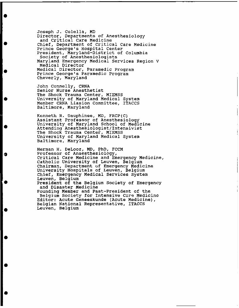
Joseph J. Colella, MDDirector, Departments of Anesthesiologyand Critical Care Medicine
* Chief, Department of Critical Care MedicinePrince George's Hospital CenterPresident, Maryland-District of ColumbiaSociety of Anesthesiologists
Maryland Emergency Medical Services Region VMedical DirectorMedical Director, Paramedic ProgramPrince George's Paramedic ProgramCheverly, Maryland
John Connelly, CRNASenior Nurse AnesthetistThe Shock Trauma Center, MIEMSS
• University of Maryland Medical SystemMember CRNA Liasion Committee, ITACCSBaltimore, Maryland
Kenneth R. Dauphinee, MD, FRCP(C)Assistant Professor of AnesthesiologyUniversity of Maryland School of Medicine
* Attending Anesthesiologist/IntensivistThe Shock Trauma Center, MIEMSSUniversity of Maryland Medical SystemBaltimore, Maryland
Herman H. DeLooz, MD, PhD, FCCM* Professor of Anaesthesiology,
Critical Care Medicine and Emergency Medicine,Catholic University of Leuven, BelgiumChairman, Department of Emergency MedicineUniversity Hospitals of Leuven, BelgiumChief, Emergency Medical Services SystemLeuven, BelgiumPresident of the Belgium Society of Emergencyand Disaster MedicineFounding Member and Past-President of theBelgium Society for Intensive Care MedicineEditor: Acute Geneeskunde (Acute Medicine),Belgian National Representative, ITACCS
* Leuven, Belgium
Wolfgang F. Dick, MD, PhD, FFARCS (Hon)Professor and Chairman for the Clinic ofAnaesthebinlogyUniversity Hospital Mainz, GermanyVice-President, European Academy ofAnaesthesiology
Medical Director, University Hospital, MainzSecretary, German Association of Intensive Careand Emergency MedicineCo-Editor, Kliniche Anaesthesiologie andIntensivtherapie
Co-Editor, Der AnasthesistManaging Editor, Notfallmedizin(Emergency Medicine)
Section Editor, Current Opinion inAnaesthesiologiologyAdvisory Board, Current Anaesthesia PracticeAuthor/Editor, Paediatric Anaesthesia(Kinderanasthesie)
Author/Editor, Regional Anaesthesia andObstetrics (Regionalanasthesie inder Geburtshilfe)Author, Anaesthesia Notebook (AnesthesiaMerkbuch)German National Representative, ITACCSMainz, Germany
Yoel Donchin, MDSenior Lecturer in AnesthesiaHead, Trauma UnitHadassah University HospitalSpecial Advisor for Anesthesia to the SurgeonGeneral, Israeli Defense Forces (IDF)Lietenant Colonel, Medical Corps, IDFIsraeli National Representative, ITACCSJerusalem, Israel
Alasdair Dow, MB, ChB, MRCP (U.K.) FFARCS, DAVisiting Assistant ProfessorDepartment of AnesthesiologyUniversity of Maryland School of MedicineAttending AnesthesiologistThe Shock Trauma Center, MIEMSSUniversity of Maryland Medical SytemsMember, By-Laws Committee, ITACCSEditorial Board, ITACCS NewsletterBaltimore, Marlyand
Elizabeth A.M. Frost, MDProfessor of AnesthesiologyDirector, Division of NeuroanethesiaAlbert Einstein College of Medicine/Montefiore Medical Center
Editorial Board, Anesthesiology NewsEditor, ITACCS NewsletterNew York, New York
Adolph H. Giesecke, MDJenkins Professor and ChairmanDepartment of AnesthesiologyUniversity of TexasSoutherwestern Medical SchoolParkland Memorial HospitalVice President, ITACCSEditor, Anesthesia for the Surgery of TraumaDallas, Texas
James I. Gilman, MDProfessor of Anesthesiology and PediatricsUniversity of Colorado Health Sciences CenterDirector, Intensice Care Unit ifChildren's Hospital, DenverAssociate Director, AnesthesiologyFormer Director, Pediatric TransportDenver, Colorado
Alexander W. Gotta, MDProfessor of Clinical AnesthesiologyVice Chairman for Academic AffairsDepartment of AnesthesiologyState University of New YorkHealth Science Center at BrooklynBrooklyn, New York
Christopher M. Grande, MDSpecial Consultant and Chief, Special ProjectsDepartment of Anesthesiology,The Shock Trauma Center, MIEMSSUniversity of Maryland Medical SystemCaptain, Medical Corps, USARFlight Surgeon and Diving Medical Officer11th Special Forces Group (Airborne)1st U.S. Army Special ForcesExecutive Director, ITACCSEditor, Trauma AnesthesiaEditor, Overview of Trauma Anesthesiaand Critical Care, Critical Care Clinics
Baltimore, Maryland
Ake N.A. Grenvik, MD, PhDProfessor of Anesthesiology and SurgeryUniversity of Pittsburgh School of MedicineDirector, Multidisciplimary Critical CareMedicine Training ProgramUniversity of Pittsburgh Medical CenterPast President and Founding Member, Societyof Critical Care Medicine
Associate Editor, Textbook of Critical CareSeries Editor, Contemporary Management inCritical Care Medicine
Pittsburgh, Pennsylvania
Kathleen Hartman, CRNAStaff Nurse AnesthetistThe Shock Trauma Center, MIEMSSUniversity of Maryland Medical SystemBaltimore, Maryland
Zvi J. Herschman, MDAssistant Professor of Anesthesiologyand SurgeryMount Sinai Medical School, New York CityDirector of Surgical Intensive Care UnitCity Hospital Center at Elmhurst, New York CityAssociate Medical Director, Departmentof Respiratory Therapy
City Hospital Center Elmhurst, New York CityConsultant in Toxicology, New York CityPoison Control Center
Elmhurst, New York
Mahmood Jaberi, MDAssistant Professor of AnesthesiologyUniversity of Maryland School of MedicineAttending Anesthesiologist,The Shock Tr-uma Center, MIEMSSUniversity of Maryland Medical SystemBaltimore, Maryland
Murray A. Kalish, MDAssistant Professor of AnesthesiologyUniversity of Maryland School of MedicineAttending AnesthesiologistThe Shock Trauma Center, MIEMSSUniversity of Maryland Medical SystemPresident, Maryland-District ofColumbia Society of AnesthesiologistsBaltimore, Maryland
Vladimir Kvetan, MDAssociate Professor of Anesthesiology andCritical Care MedicineDirector, Division of Critical Care MedicineAlbert Einstein College of Medicine/Montefiore Medical Center
Editor, Disaster Management,Critical Care ClinicsChairman, ITACCS Disaster CommitteeBronx, New York
Charles P. Kingsley, MDChief, Department of Clinical InvestigationStaff Anesthesiologist,Brooke Army Medical CenterAssociate Professor of AnesthesiologyAssistant Clinical ProfessorUniversity of Texas at San Antonio,Health CenterMajor, U.S. ArmySan Antonio, Texas
Margaret M. Libonati, MDAssociate Director, AnesthesiologyMedical Director, Day Surgery UnitWills Eye HospitalPhiladelphia, Pennsylvania
Robert Loeb, MDAssistant ProfessorDepartment of AnesthesiologyUniversity of California, Davis Medical CenterSacramento, California
Colin F. Mackenzie, MDAssociate Professor and Director of ResearchDepartment of AnesthesiologyUniversity of Maryland School of MedicineAttending AnesthesiologistThe Shock Trauma Center, MIEMSSUniversity of Maryland Medical SystemEditor, Chest Physiotherapy in the ICUBaltimore, Maryland
Brian G. McAlary, MDAssistant Professor of AnesthesiologyUniversity of Maryland School of MedicineAttending Anesthesiologist andEducation DirectorDepartment of Anesthesiology
The Shock Trauma Center, MIEMSSUniversity of Maryland Medical SystemBaltimore, Maryland
Marzio G. Mezzetti, MD, PhDProfessor, Postgraduate School of Anesthesiaand ResuscitationUniversity of Pavia, ItalyVice-Chairman, Department of Anesthesia andResuscitation
Regional Hospital of VaresePR, Emergency Task ForceAirborne Medical UnitItalian Society of Anesthesia, Analgesia,Resuscitation and Intensive Care (SIAARTI)Italian National Respresentative, ITACCSVarese, Italy
T. Michael Moles, MBBS, DTMH FFARCS, FC Anaes, FHKC AnaesPresident,Hon9 Kong College of AnaesthesiologistsChairman, International CoordinationCommittee, ITACCS
Member, CPCR Committee, WFSAMajor, RAMC, 10 Bn Parachute Regiment, TAVRAuthor, Management of the Injured PatientEditor, Recent Advances in Anaesthesia,Immediate and Critical Care
Hong Kong
Shiela Muldoon, MD, PhDProfessor and ChairpersonDepartment of AnesthesiologyUniformed Services University of theHealth SciencesBethesda, Maryland

Mark T. Murphy, MDFellow in Trauma Anesthesia and Critical CareMassachusetts General HospitalHarvard Medical SchoolMajor, Medical Corps, USARBrigade Surgeon, 187th Infantry BrigadeMember, Education and Training Committee,ITACCSBoston, Massachusetts
Jerry P. Nolan, MB, ChB, FFARCSClinical InstructorDepartment of AnesthesiologyUniversity of Maryland School of MedicineAttending AnesthesiologistThe Shock Trauma Center, MIEMSSUniversity of Maryland Medical SystemRegistrar in AnaesthesiaBristol, Royal InfirmaryMember, Education and Training Committee,ITACCS
Editorial Board, ITACCS NewsletterBaltimore, Maryland
Peter Oakley, MA, MBB, Chir, MRCGP, FC AnesWellcome Trust Research Fellow and HonorarySenior RegistrarNuffield Department of AnaestheticsJohn Radcliffe HospitalOxford UniversityMember, Executive Committee,British Trauma SocietyOxford, England
Michael J. Parr, MB, ChB. MRCP(U.K.), FFARCS, DA
Visiting Assistant ProfessorDepartment of AnesthesiologyUniversity of Maryland School of MedicineAttending AnesthesiologistThe Shock Trauma Center, MIEMSSUniversity of Maryland Medical SystemBaitimore, Maryland
Edward G. Pavlin, MDAssociate Professor of AnesthesiologyUniversity of Washington School of MedicineAttending AnesthesiologistHarborview Medical Center/Trauma CenterSeattle, Washington
Clayton Petty, MDProfessor of AnesthesiologyUniformed Services University of theHealth SciencesAttending AnesthesiologistNational Naval Medical CenterCaptain, Medical Corps, USNRBethesda, Maryland
Christopher Romanowski, CRNAStaff Nurse AnesthetistThe Shock Trauma Center, MIEMSSUniversity of Maryland Medical SystemBaltimore, Maryland
Andrew D. Rosenberg, MDAssociate Chairman and AttendingAnesthesiologist
Hospital for Joint Disease-Orthopedic InstituteClinical InstructorDepartment of AnesthesiologyNew York University School of MedicineNew York, New York
John K. Stene, MD, PhDAssociate Professor of AnesthesiologyPennsylvania State UniversityCollege of MedicineDirector, Perioperative Trauma AnesthesiaServices
Department of AnesthesiaMilton S. Hershey Medical SchoolPresident, ITACCSEditor, Trauma AnesthesiaAssociate Editor, Overview of Trauma Anesthesiaand Critical Care, Critical Care ClinicsHershey, Pennsylvania
Gregory Stocks, CRNAStaff Nurse AnesthetistThe Shock Trauma Center, MIEMSSUniversity of Maryland Medical SystemBaltimore, Maryland
Anne J. Sutcliffe, BSc, FFARCS, LRPSConsultant AnaesthetistBirminghi.m Accident Hospital, EnglandMember, Council of Association of Anaesthetistsof Great Britian and IrelandAuthor, Handbook of Emergency AnesthesiaBritish National Representative, ITACCSBirmingham, EnglandUnited Kingdom
Pat Taub, CRNAAssociate Director, Nurse AnesthesiaThe Shock Trauma Center, MIEMSSUniversity of Maryland Medical qq*y 4-Baltimore, Maryland
Kevin K. Tremper, MD, PhDProfessor and ChairmanDepartment of AnesthesiologyUniversity of MichiganEditorial Board, Critical CareEditorial Board, Critical MonitoringEditor, Oxygen Monitoring, InternationalAnesthesiology Clinics
Ann Arbor, Michigan
Mary R. WrightQuality Assessment CoordinatorDepartment of AnesthesiologyThe Shock Trauma Center, MIEMSSUniversity of Maryland Medical SystemBaltimore, Maryland
Chet I. Wyman, MDClinical InstructorDepartment of AnesthesiologyUniversity of Maryland School of MedicineAttending PhysicianDepartment of AnesthesiologyDepartment of Critical Care MedicineFranklin Square Hospital Center,Attending AnesthesiologistThe Shock Trauma Center, MIEMSSUniversity of Maryland Medical SystemMember, ITACCS Newsletter CommitteeEditorial Board, ITACCS NewsletterBaltimore, Maryland

PLENARY SESSION LECTURES
SESSION I
THURSDAY, MAY 2, 1991
8:00 A.M. - 12:45 P.M.
Moderator: John K. Stene, M.D., Ph.D.
VARIABLE DEMOGRAPHICS OF THE TRAUMA PATIENT POPULATION
Jerry P. Nolan, MB ChB FCAnaes
I. Introduction
II. Source of information
III. Trauma Epidemiology: An International Perspective
A. Motor vehicle accidents
B. Homicide
Suicide
1V. Trauma Epidemiology: The United States
A. Trauma versus other cases of death
B. The chronological pattern of trauma deaths
C. Mechanisms of trauma deaths
D. Variation in population subgroups
1. Sex
2. Race
E. Geographical variation in the rates of trauma
F. Epidemiology of non-fatal injuries
V. Epidemiology of selected causes of trauma
A. Motor Vehicle Accidents
1. Age
2. Sex
3. Type of vehicle
4. Factors influencing the incidence of motor vehicleaccidents
a. Alcohol

b. Seatbelt use
c. Airbag
d. Motorcycle helmets
B. Homicide
1. Age
2. Sex
3. Geographical variation
4. Race
C. Suicide
1. Method
2. Age
3. Sex
0. Falls
VI, The MIEMSS Statistics
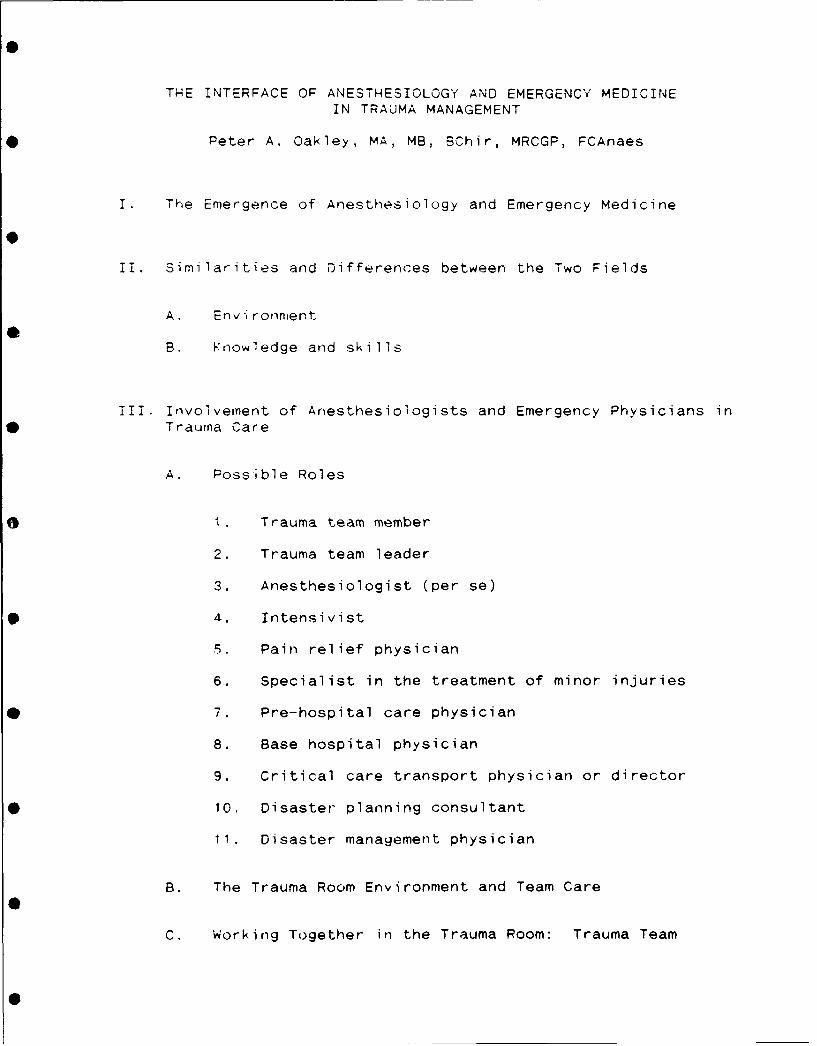
THE INTERFACE OF ANESTHESIOLOGY AND EMERGENCY MEDICINEIN TRAUMA MANAGEMENT
Peter A. Oakley, MA, MB, SChir, MRCGP, FCAnaes
I. The Emergence of Anesthesiology and Emergency Medicine
Ii. Similarities and Differences between the Two Fields
A. Env -ironment
B. Knowledge and skills
III. Involvement of Anesthesiologists and Emergency Physicians in
Trauma Care
A. Possible Roles
i. Trauma team member
2. Trauma team leader
3. Anesthesiologist (per se)
4. Intensivist
5. Pain relief physician
6. Specialist in the treatment of minor injuries
7. Pre-hospital care physician
8. Base hospital physician
9. Critical care transport physician or director
10, Disaster planning consultant
11. Disaster management physician
B. The Trauma Room Environment and Team Care
C. Working Together in the Trauma Room: Trauma Team
Members
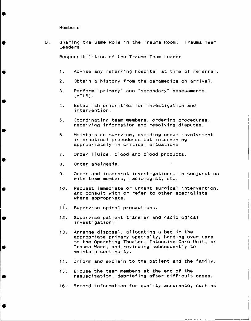
D. Sharing the Same Role in the Trauma Room: Trauma TeamLeaders
Responsibilities of the Trauma Team Leader
1. Advise any referring hospital at time of referral.
2. Obtain a history from the paramedics on arrival.
3. Perform "primary" and "secondary" assessments(ATLS).
4. Establish priorities for investigation andintervention.
5. Coordinating team members, ordering procedures,receiving information and resolving disputes.
6. Maintain an overview, avoiding undue involvementin practical procedures but interveningappropriately in critical situations
7. Order fluids, blood and blood products.
8. Order analgesia.
9. Order and interpret investigations, in conjunctionwith team members, radiologist, etc.
10. Request immediate or urgent surgical intervention,and consult with or refer to other specialistswhere appropriate.
11. Supervise spinal precautions.
12. Supervise patient transfer and radiologicalinvestigation.
13. Arrange disposal, allocating a bed in theappropriate primary specialty, handing over careto the Operating Theater, Intensive Care Unit, orTrauma Ward, and reviewing subsequently tomaintain continuity.
14. Inform and explain to the patient and the family.
15. Excuse the team members at the end of theresuscitation, debriefing after difficult cases.
16. Record information for quality assurance, such as
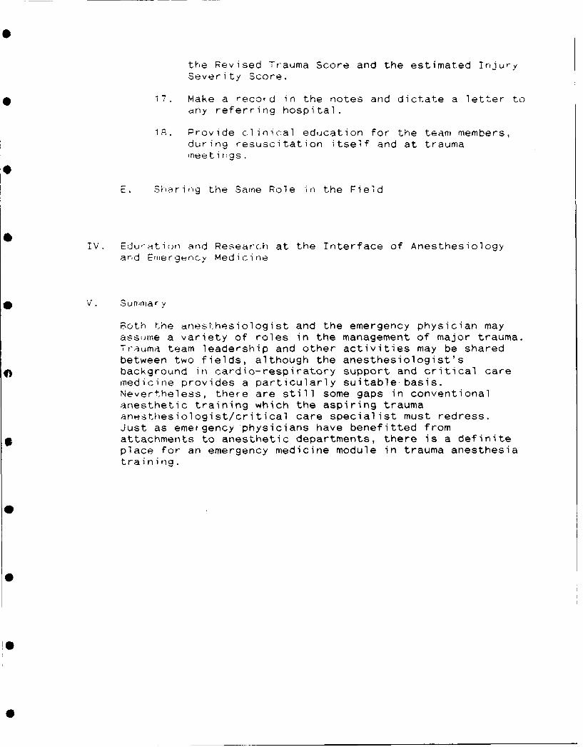
the Revised Trauma Score and the estimated InjurySeverity Score.
17. Make a record in the notes and dictate a letter toany referring hospital.
1A. Provide clinical education for the team members,
during resuscitation itself and at traumaIfeet i rl's.
E. Shari,,q the Same Role in the Field
IV. Edu.-etiun and Research at the Interface of Anesthesiologyand Emergercy Medicine
v. Summary
Roth the anesthesiologist and the emergency physician mayasstime a variety of roles in the management of major trauma.Trauma team leadership and other activities may be sharedbetween two fields, although the anesthesiologist'sbackground in cardio-respiratory support and critical caremedicine provides a particularly suitable basis.Nevertheless, there are still some gaps in conventionalanesthetic training which the aspiring traumaanesthesiologist/critical care specialist must redress.Just as emergency physicians have benefitted fromattachments to anesthetic departments, there is a definiteplace for an emergency medicine module in trauma anesthesia
tra i n i rg.

THE ANESTHESIOLOGIST AS AN EMS DIRECTOR
Joseph J. Colella, Jr., M.D.
I. Introduction
A. History and development of EMS systems
B. Organization
II. Medical Accountability
A. off-line medical direction
B. On-line medical direction
C. Certification
D. Professional stress syndrome ("burn-out")
III. Prehospital Intervention
A. Drug therapy
B. Monitoring
C. Technical intervention
1. Airway management and intubation
2. MAST and volume resuscitation
IV. Medical Incident Reporting
A. Quality assurance
B. Retrospective medical control
C. Decertification and counselling

V. TeachinG Initiatives
A. Basic and advanced concepts
B. Continuing medical education (CHE) requirements
C. Operating room training in intubation
VI. Community Service Orientation

TRAUMA ANESTHESIA IN BEIRUT
Anis Baraka, M.B.B.Ch., D.A., D.M., M.D., F.C.Anaesth. (Hon.)
I. Problems related to the civil war itself
II. Personnel deficiency (death, exodus, isolation and nervousexhaustion).
III. Deficiency of supplies, particularly oxygen and nitrousoxide.
IV. Workload.
V. Problems of anesthesia in seriously traumatized patientssuch as:
a. Respiratory failure
b. Tetanus
c. "Full stomach"
VI. Special problems
a. Postoperative respiratory failure
B. "etanus
c. Gas gangrene
d. Decompression sickness

TRAUMA CARE IN BELGIUM
Herman H. DeLocz, M.D., Ph.D., FCCM
Historical background
A. Emergency telephone exchanges
B, The law 0f july 1964
1. Evolution _f the system
4, Ambulances
B. Crew
C. Hospital-based emergency facilities
I:1. The "Leuven" system
A. The basic goal
B. The first "Golden Hour"
1. The first witness
2. The EMS system
3. The emergency physician in pre-hospital traumacare
4. The Department of Emergency Medicine TraumaProtocol
5. The Observation Care Unit
- short term hospitalization
- short term intensive care
6. Disaster medicine
IV. Problems and remedies
V. Conclusions
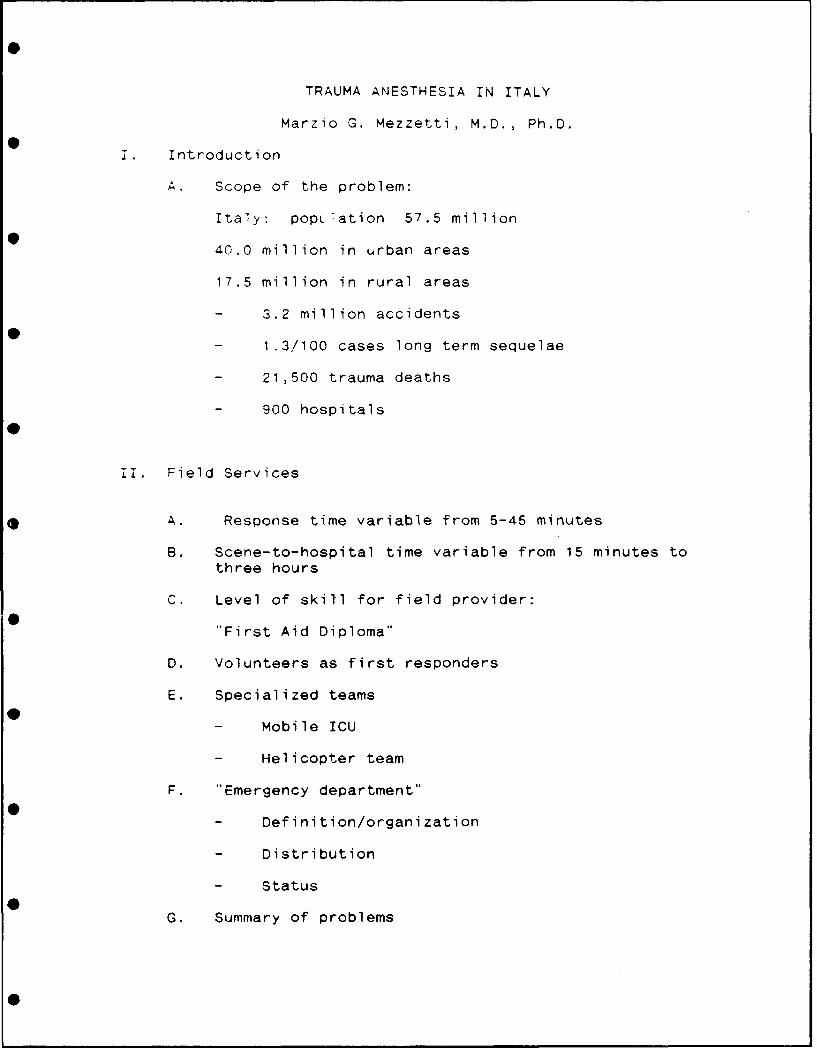
TRAUMA ANESTHESIA IN ITALY
Marzio G. Mezzetti, M.D., Ph.D.
i. Introduction
A. Scope of the problem:
italy: popLation 57.5 million
40.0 million in urban areas
17.5 million in rural areas
- 3.2 million accidents
- 1.3/100 cases long term sequelae
- 21,500 trauma deaths
- 900 hospitals
.ii. Field Services
A. Response time variable from 5-45 minutes
B. Scene-to-hospital time variable from 15 minutes tothree hours
C. Level of skill for field provider:
"First Aid Diploma"
D. Volunteers as first responders
E. Specialized teams
- Mobile ICU
- Helicopter team
F. "Emergency department"
- Definition/organization
- Distribution
- Status
G. Summary of problems

Differences in regional organization
Lack of "echelons of care" in peripheral areas
Retrospective study:
a. 17% of major trauma deaths preventable
b. Treatment in the field vs. "scoop and run"
c. Different levels of experience
III. Role of the Anesthesiologist
A. Specialist in anesthesiology and resuscitation
B. Involved in prehospital medicine
IV. Recent Issues
1. Need for adequacy of care
2. Economic costs
3. Trauma center vs. traditional hospital
V. How ITACCS will contribute to improve the situation?

TRAUMA ANESTHESIA IN THE SOVIET UNION:AN AMERICAN PERSPECTIVE
Charles P. Kingsley, M.D.
i. Introduction
A. General perspective
1. Natural disasters
2. Humanitarian visits
3. Military operations
II. Mission
A. Planning
1. Casualties
2. Supplies and equipment
3. Transportation
III. Local Constraints
A. International transportation
B. Local transportation
C. Language
D. Equipment
E. Supplies
F. Blood and fluids
G. Electricity
H. Repair capabilities
I. Recovery care
J. Local customs
K. Communications
IV. Intensive Care Medicine

A. Fxdfclte
A. Field facilities
B. Fiel facilities n
PLENARY SESSION LECTURES
SESSION II
THURSDAY, MAY 2, 1991
2:00 P.M. - 6:00 P.M.
Moderator: Elizabeth A.M. Frost, M.D.
PERIOPERATIVE ANESTHETIC MANAGEMENT OF MAXIL10=ACIAL TRAUMA
Aleaxandr- W. Gotta, M.i-i.
I. Normal Anatomy
7) ivision of faal skeletorn in-lo thirds
C, Mand i bl e
T: 71 K~~im of F:orc-e Dispersion
.. Vulnerahilitv of mandible
B, Vuineri-bility of maxilla
C. N.itwure of blow determines fracture site and extent
TT. nifinn Fakciai Fractures
A. L+-ort I
B1. LeFort II
C. LeFort III
D. Mandibular fractures
F. Maxillary fractures
F. Possible involvement of base of skull and cranial vault
IV. Preoperative Evaluation
A. Common Concurrent Medical Problems
1 . Myocardial infarct
2. Stroke
3. Drug Abuse
4. Alcoholism
5. Neurologic disorder
B. Common Concurrent Surgical Problems
1. Fracture of the skull
2. Intracranial hemorrhage
3. Subdural hematoma
4. Fracture of the cervical spine
5. Fat embolism
6. Pneumothorax
7. Flail chest
8. Cardiac tamponade
9. Cardiac hematoma
10. Ruptured spleen or liver
C. Necessary Laboratory and X-ray Data
V. Airway Assessment
A. Technique of oral intubation
B. Technique of nasal intubation
1. Guided
2. Blind
a. Indications
C. Superior Laryngeal Nerve Block
1. Anatomy
2. Technique
3. Indications
4. Contraindications
VII. Tracheostomy

A. Indications
S. Contraindications
VITI. Choice of Anesthetic Agent and Technique
A. Intravenous Agents
1 . Narcotics
2. Ketamine
R. inhalation Agents
1. Alkanes
a. Cardiac arrhythmias
2. Ethers
0.. Local Anesthetics
PERIOPERATIVE ANESTHETIC MANAGEMENT OF ORTHOPEDIC TRAUMA
Andrew D. Rosenberg, M.D.
I. Pelvic Injuries
A. Mechanism of Injury
B. Associated problems
1. Hemorrhage
2. Pelvic organ injuries
3. Coagulopathies
4. Pelvic thrombosis
C. Case Approach
1. General vs. regional or combination
2. Monitoring ("A"-line, central monitoring)
3. SSEP
4. Cell Saver
5. Urine output
6. Role of MAST
7. Postoperative pain management
II. Hip Fractures
A. Dislocations and fracture dislocations
B. Proximal femoral fractures
1. Site of Injury
a. Subcapital
b. Transcervical
C. Trochanteric
d. Subtrochanteric
2. Patient Characteristics
a. Age
b. Concurrent diseases
c. Changes in the elderly (pharmacology andphysiology of aging)
3. Case approach
a. Perioperative checklist
b. Preview of systems
c. Role of anesthetic technique
d. Timing of surgery
e. Perioperative management
1. Hypotension
2. Hypertension
3. Cement
4. Pulmonary embolism considerations
f. Postoperative management
III. Thromboembolism
A. Incidence
B. Prevention
1. Medication
2. Lower limb compression devices
IV. Compartment Syndrome
V. Long Bone Fractures
A. Stabilization
B. Fat embolism
1. Incidence
2. Pathophysiology
3. Presentation
4. Diagnosis and treatment
VI. Cervical Spine
A. Incidence
B. Anatomy
C. Injuries
D. Principles of management
1. Stabilization
2. Securing an airway
3. Maintaining adequate ventilation
a. C5 and above vs. C5 and below
b. Pa02, C02
4. Maintaining adequate BP
5. Medications (steroids, succinylcholine)
6. Spinal shock
7. Respiratory considerations:
Pulmonary edema
8. Circulation:
a. Autonomic hyperflexia
9. Gastrointestinal
10. Case Approach
a. Fiberoptic
b. SSEP
c. Monitoring
d. Agents
e. Positioning
f. Postoperative considerations
VII. Thoracic Spine
A. 1. General considerations
2. Associated injuries
B. Case approach
(see cervical spine injuries)
PERIOPERATIVE ANESTHETIC MANAGEMENT OF OPHTHALMOLOGIC TRAk?1A
Margaret M. Libonati, M.D.
I. Introduction
A. Incidence
B. Etiology
C. Types of Injuries
D. Factors determining visual outcome
II. Intraocular Pressure Physiology
A. Intraocular contents
B. Normal pressures and variations
C. Major factors affecting pressure during surgery
1. Aqueous humor secretion
2. Vitreous volume
3. Choroidal blood volume
4. Extraocular muscle tone
III. History and Physical Examination
A. General exam
B. Eye exam
C. Lab test
IV. Preoperative Preparation
A. Sedation
B. Pain control
C. Premedication
D. Fasting
V. Surgical Management
VI. Anesthetic Management
A. Monitoring
B. Anesthetic Agents
k, Muscle Relaxants
I. Non-depolarizing
2. Depolarizing
D. Anticholinergic
E, Anesthetic Adjuncts
F. Guides to management
VII. Potential Problems
A. Pediatric patients
B. Geriatric patients
,* VIII.Conclusion
S0
MALIGNANT HYPERTHERMIA IN THE TRAUMA PATIENT
Sheila M. Muldoon, M.D.
I. Introduction
A. Definition of the syndrome
B. Spectrum identified
iI. Prevalence and Incidence of MH
III. Clinical Syndrome
A Fulminant episode
IV. Site of Defect In Skeletal Muscle
V. Genetics
VI. Management
A. Patient with fulminant episode
B. Patient with documented history of MH
C. Patient with a questionable history of MH
VII. Testing
A. Evaluation of sus.ceptibility, standard tests
B. Future testing
VIII.Referral Centers
A. Acute crisis
B. Consultation
PLENARY SESSION LECTURES
SESSION IIIFRIDAY, MAY 3, 19917:30 A.M. - 12:30 P.M.
Moderator: Wolfgang F. Dick, M.D., Ph.D.

ALCOHOL AND DRUG ABUSE IN THE TRAUMA PATIENT POPULATION
Edward G. Pavlin, M.D.
I. Introduction
II. The Influence of Alcohol and Drugs on Trauma
A. Associated incidence on:
1. Homicide
2. Vehicular accident
3. Industrial trauma
B. Effect of alcohol and drugs on outcome from trauma
C. The association with head injuries
III. Physiologic Interactions of Acute Intoxication
A. Alcohol
B. Narcotics
C. Cocaine and other stimulants
IV. Aesthetic Drug Interactions
A. Alcohol
B. Narcotics
C. Cocaine and other stimulants
V. Anesthetic Considerations in the Patient with AcuteIntoxication
VI. Conclusion
ECONOMICS OF TRAUMA ANESTHESIA AND CRITICAL CARE
Zvi Herschman, M.D.
I. Introduction
II. Eight viewpoints on trauma care:
A. Anesthesia providing organization (APO)
B. Government
C. Private insurance companies (PIC
D. Privately-paying patient
E. "Charity" hospital (CH?
F. Individual clinician
G. Trauma patient
H. Public opinion
T11. Three economic s:,narios
A. Fully ' -ivate - no governmental outlays for patientcare
1. Viewpoints
A.
B.
C.
D.
E.
F.
G.
H.
B. Nationalized health care (fully funded by thegovernment)
1. Viewpoints
A.
B.
C.
D.
E.
F.
G.
H.
C. Current system (government and private payment)
1. Viewpoints
A.
B.
C.
D.
E.
F.
G.
H.
2. RBRVS
a. Effects on current anesthesia income
b. What we can do to make the system work forUS.
i. Manpower strategies

MONITORING FOR TRAUMA ANESTHESIA
Kevin K. Tremper, Ph.D., M.D.
Introduction
A. 1. Approximately 50 million traumatic injuries occur in
the United States each year.
2. 100,000 of these injuries are fatal and ten millionothers result in permanent disability
3. Trauma is the leading cause of death for patients under30 years of age
B. 1. Guidelines for monitoring trauma patients are similarto those for monitoring any acutely ill or anesthetizedpatient
2. Emphasis is placed on some of the specialconsiderations one might have when monitoring anacutely injured patient
3. Primary differences involve the necessity of rapidevaluation and treatment compounded by the inability ofobtaining an adequate history, which underscores thenecessity for comprehensive and aggressive monitoring
4. Review of monitoring will follow the "ABC's" of acutepatient evaluation:
a. Airway: monitors of oxygenation
b. Breathing: monitors of ventilation
c. Circulation: monitors of cardiovascular status
I
II. Airway/Oxygenation Monitors
A. 1. Subjective assessment of skin color has been used fordecades
2. It has been demonstrated to be unreliable even withtrained personnel in a controlled setting
B. 1. Ear oximetry was the first noninvasive continuousmonitor devised to objectively evaluate a patient'soxygenation. Several of its technical shortcoming havebeen resolved with the development of the pulseoximeter

C. Pulse Oximeter
1. In less than a decade the pulse oximeter as a monitorof arterial hemoglobin saturation (Sa02) has becomeubiquitous throughout the hospital. Its usefulness inmonitoring the trauma patient is a logical consequenceof its demonstrated usefulness in the operating roomand the intensive care unit
2. Several physiologic and technical limitations:
a. Sa02 as a measure of oxygenation does notcompletely assess pulmonary oxygen exchange
b. Oxyhemoglobin dissociation desaturation curve isflat above an oxygen tension of 100 torr
c. Substantial alveolar-arterial oxygen gradient mayexist due to pulmonary pathology without evidenceof desaturation
d. Saturation measurements are excellent forassessing adequacy of hemoglobin saturation butthey do not rule-out pulmonary disease
e. Oxygen transport requires not only saturatedhemoglobin, but an adequate hemoglobinconcentration itself
f. Relatively insensitive to anemia down tohematocrits as low as 10% thus, independentmeasure of hemoglobin or hematocrit is alsonecessary
3. How a pulse oximeter derives its estimates of Sa02
a. The pulse oximeter transilluminates tissue withlow wavelengths of light (red and infrared). Thedevice measures the ratio of the pulsatileabsorbance in red light to the pulsatileabsorbance in infrared light. This ratio is thenempirically calibrated to Sa02
b. Calibration curves all range from a ratio of 0.4when the saturation is nearly 100% to a ratio of3.4 when the saturation is approximately 0%.
c. When the absorbance ratio equals one thesaturation is approximately 85%
d. Artifacts are related to the production of a false
ratio thus reporting a false saturation:
i. Substantial light or motion artifact which
may occur in an emergency room setting.
ii. Artifacts may be minimized by covering thesensor and holding the sensor site still toobtain a true tissue absorption signal
iii. Uses only two wavelengths:
Adult blood is composed of four types of
hemoglobin: oxyhemoglobin, reducedhemoglobin, methemoglobin, andcarboxyhemoglobin. These last two speciesare in low concentration except in pathologicstates.
The pulse oximeter deals with blood as though
it contains only oxyhemoglobin and reducedhemoglobin and therefore will produce errorsin the presence of either carboxyhemoglobinor methemoglobin
Carboxyhemoglobin is red (as isoxyhemoglobin) and the pulse oximeter reportsapproximately the sum of oxyhemoglobin andcarboxyhemoglobin. A blood sample must besent to a laboratory for study by amultiwavelength co-oximeter
Methemoglobin is dark brownish and abso'bsboth red and infrared light greatly Itspresence forces the light absorbance ratiotoward unity and therefore the pulseoximeter's saturation estimate towards 85%
D. Other oxygen monitors have been used to assess thetrauma patient. Each continuously monitors oxygenationmore at the tissue level than that of the arterialblood:
1. Transcutaneous oxygen sensor (Ptc02)
2. Conjunctival oxygen sensor (Pcj02)
3. Oximetric pulmonary artery catheter (Sv02)
E. 1. Arterial blood gas samples are the "gold standard"
of assessing oxygenation, ventilation and acidbase status.
2. Recent development in miniature optode technologymay allow continuous arterial blood gas monitoringin the future. Currently, these devices areexperimental.
III. Breathing/Ventilation and C02 Exchange
A. Can be subjectively assessed by chest movement andausculation
B. Confirmed only by arterial blood gas analysis for C02.
C. Two noninvasive monitors of carbon dioxide. Althougheach of these monitors displays C02 data, each ismeasuring C02 at different locations and is, therefore,a different physiologic variable:
1. Capnometer (ETC02)
2. Transcutaneous carbon dioxide monitor (PtcC02)
3. Normal capnograpm is the tracing of expired C02from the airway
4. a. ETC02 is measured from the sample gas at theend of expiration. End-expired gas isalveolar gas coming from both well perfusedalveoli and alveolar dead space (non-perfusedalveoli).
b. Usually assumed that the C02 concentration ofpure alveolar gas is very close to thearterial C02 tension.
c. Since alveolar dead space contains no carbondioxide he ETC02 concentration willreprese art erial C02 only when there is noalveolar dead space: the difference betweenan arterial C02 value and the ETC02 value isdue to the presence of alveolar dead space.
d. The trauma patient may be hemodynamicallyunstable and therefore have non-uniform
, pulmonary perfusion. The V/Q mismatchproduces alveolar dead space and thereforeproportionate increases in arterial C02 andETC02 differences
e. Extreme cases (cardiac arrest): No pulmonaryperfusion, the entire lung is alveolar deadspace and therefore the ETC02 value will betheoretically "0". This dependence of ETC02on pulmonary blood flow has been used as away of assessing the adequacy ofcardiopulmonary resuscitation.
f. ETC02 value increases to approach thearterial C02 value as the patient's cardiac
output increases. In this way, the ETC02 isbeing used primarily as a perfusion monitor
5. a. It has been stated that the presence of acapnograph is the most reliable way to assessendotracheal (as opposed to esophageal)intubation. The observation of repeatednormal capnographs with each breath is anessential feature of endotracheal intubationin a hemodynamically stable patient.
b, Confusion may arise when the patient ishemodynamically unstable and has been maskventilated with significant gastric
distention. In this situation, there may besignificant carbon dioxide in the stomach,
and therefore allowing the appearance of aninitial capnogram upon an esophagealintubation. This capnograph amplitude shoulddecrease rapidly with each breath as the C02is washed-out of the stomach.
c. Could be confused with a proper endotrachealintubation in a patient who is in cardiacarrest and therefore not perfusing the lungs.
d. In the arrest situation it is important toconfirm intratracheal intubation by methodsincluding direct visualization, chestobservation and ausculation, gastricauscultation, pulse oximeter observation, andintraarterial blood gas measurement
O D. 1. Transcutaneous carbon dioxide monitor is usefulfor monitoring changes in arterial C02 in thecritically ill
2. Not been applied in the trauma setting due to thetechnical limitations of calibration and warm-uptime.
E. Intraarterial optode may also measure arterial C02 butas stated above it is still in the experimental phaseof development.
F. The adequacy of ventilation in the trauma patientshould be assessed by clinical observation, continuouscapnography, and arterial blood sampling for arterialC02 measurement.
IV. Circulation/Cardiovascular Function
A. Begins with evaluation of pulses, blood pressure, ECG,
urine output and cardiac output
B. Immediate assessment of blood pressure does not insureadequate blood volume and cardiac output but it doesimply perfusion of the heart and brain.
C. After blood pressure and cardiac rhythm are establishedit is important to ascertain the adequacy of
intravascular volume and blood flow. In the emergencysituation this is not always easy. Routine methodsused to measure are:
1. Urine output
2. Postural blood pressure changes
3. Presence of a metabolic acidosis on arterial bloodgas analysis.
D. Several continuous monitoring devices have recentlybeen used to assess peripheral perfusion and cardiac
* cwtput"
1. Transcutaneous and conjunctival oxygen tensionmonitors:
a. Demonstrated to follow changes in cardiacoutput during shock and resuscitation fromshock.
b. Measure oxygen on the skin surface andconjunctival surface thus they are dependentupon arterial blood oxygenation and tissueperfusion.
C. Both have limitations of requiringcalibration and the transcutaneous oxygensensor requires 10 to 15 minutes of warm-upon the patient's skin surface.
d. Both have been demonstrated to be useful inevaluating acute trauma patient
2. Oximetric pulmonary artery catheter:
a. Provides
i. Intermittent thermodilution cardiacoutput measurement
ii. Continuous pulmonary artery pressures
iii. Mixed-venous oxygen saturation
b. Supplies continuous and intermittentcomprehensive hemodynamic and oxygentransport evaluation. Therefore, the "goldstandard" in monitoring the hemodynamicstatus of any patient.
c. Disadvantages: requires the placement of apulmonary artery catheter which may not bepractical in many situations and too timeconsuming in others.
E. Noninvasive cardiac output by thoracic bioimpedance:
1. Advocated as a possible means of monitoring the
trauma patient.
2. Advantages: noninvasive and continuous and at thesame time being easy to apply to the patient
3. Limitations: requires eight ECG electrodes to beplaced on the patient and can be disabled bysignificant patient motion artifact.
F. Precordial and transesophageal doppler have also beenadvocated as noninvasive measure of cardiac output.Neither of these techniques has been applied to thetrauma patient
G. Myocardial Contusion, Myocardial Ischemia
1. ECG: one of the mainstays in monitoring. It hasbeen demonstrated to be relatively insensitive inthe detection of myocardial contusion orconcussion.
2. CPK-MB fraction: greater than 5% is a myocardialconcussion, a contusion is defined as a concussionwith an abnormal echocardiogram.
3. 2D Echocardiography: It has been found thatapproximately 40% of all trauma patients withapparent chest trauma have a myocardial contusionor concussion, while less than 50% oill bedetected by nonspecific ECG changes.
A. It is currently recommended that CPK-MB fractionsbe measured on admission and every six hours forthp first 24 hours to rule out myocardial damage.
V. Conclusions
1, Lack of history and preparation mandates intitial andcontinuous monitoring of ABC's
2. Continuous assessment of ABC's using monitoring devicesallows any serious changes to be detected early enoughto implement appropriate intervention.
PHARMACOKINETICS ALTERATIONS SECONDARY TO TRAUMA
Leo H.D.J. Booij, M.D., Ph.D.
I. Introduction
A. Pharmacodynamics versus pharmacokinetics
B. Pharmacokinetics for nonpharmacokineticists
C. What to do the various parameters mean
II. Influence of trauma on pharmacokinetic parameters
A. Initial volume of distribution
B. Half-life times
C. Plasma Clearance
1. redistribution
2. liver function
3. renal function
III. Clinical Implications
PERIOPERATIVE MANAGEMENT OF HYPOTHERMIA IN THE TRAUMA PATIENT
Edward G. Pavlin, M.D.
I. Introduction
II. Hypothermia in Trauma
A. Incidence
B. Contributing factors
C. Associated features
D. Outcome
II. Physiologic Effects of Hypothermia
A. Cardiovascular system
B. Central nervous system
C. Distribution of fluids
D. Hematologic considerations
E. Shivering
IV. Interaction with Anesthetic Drugs
A. Anesthetic agents
B. Fixed agents
C. Muscle relaxants
D. Cardiovascular drugs
V, Prevention and Treatment of Hypothermia
A. Surface warming
B. Core warming
C. Treatment of complications
VI. Conclusion
REGIONAL ANESTHESIA IN THE TRAUMA PATIENT
Alasdair Dow, MB, ChB, MRCP, FFARCS
I. Introduction
A. History of regional anesthesia
B. i. Aims
ii. Benefits
ii. Disadvantages
II. Specific Controverseries
A. Epidural/spinal vs. general anesthesia
B. Choice of drugs for epidural/spinal space
C. Epidural vs. intrapleural analgesia for chest trauma
D. Risks of regional anaesthesia and compartment syndrome
E. Upper airway analgesia
III. Possible Future Advances in Regional Anesthesia
IV. Summary
Id
I!
PLENARY SESSION LECTURES
SESSION IV
SATURDAY, MAY 4, 1991
8:30 A.M. - 12:15 P.M.
Moderator: Pierre Carli, M.D.

PERIOPERATIVE ANESTHETIC MANAGEMENT OF CHEMICAL, BIOLOGICAL ANDNUCLEAR WARFARE CASUALTIES
David J. Baker, M Phil, MD, FFARCS
I. Introduction
A. History and proliferation: Recent use and the current
threat
B. The role of the anaesthetist
C. Why link nuclear, biological and chemical injury?
D. An integrated approach to wounding
II. Nuclear Events
A. Nuclear explosion: rapid rclease of light, heat, blast
energy and radiation
B. Reactor meltdown: release of heat and radiation
C. Nuclear injuries: burns, blast injury, penetrating and
blunt missile injury
D. Radiobiological syndromes: CNS, gastrointestinal,hemopoietic
E. Implications for the anesthetist: high casualty load,
breakdown of facilities,intercurrent respiratory and
electrolyte problems.
III. Definitions of Chemical and Biological Agents
A. Classical definitions of chemical and biological agents
1. The unifying concept of the "toxic agent"
2. The BCW spectrum of toxic agents of warfare
B. Characteristics nf toxic agents
1. Operational - persistency
2. Pathophysiological
a. Toxicity
b. Latency
c. Transmissibility
IV. Special Techniques in Dealing with Toxic Wounding
A. Protection - individual and collective
B. Detection
C. Decontamination
V. Properties of Some Important Agents
A. Lung damaging aqents
1. Properties of phosgene
a. Mechanisms of action
b. Signs and symptoms
c. Treatment
B. Vesicant agents
1. Sulphur and nitrogen mustard
a. Absorption and toxicity
b. Pathology
c. Signs and symptoms
d. Respiratory effects
e. Management
C. Nerve agents
1. Structure and properties
a. Toxicology - pharmacology
b. Interactions with acetylcholinesterase
2. Organ effects
a. The eye
b. Respiratory system
c. Skeletal neuromuscular system
0 ! !! !!i w

3. Signs and symptoms
a. Vapor exposure
b. Liquid exposure
4. Diagnosis
5. Management
a. Carbamate pretreatment
b. First aid
c. Medical treatment
i. Ventilation
ii. Cholinergic blockade
iii. Enzymes reactivation
iv. Benzodiazepines
6. Long-term management and prognosis
D. Cyanide agents
1. Protection
2. Pathophysiology
a. Signs and symptoms
b. Treatment
E. Toxins
1. Classification
a. Neurotoxins
b. Botulinum toxin
c. Signs and symptoms
d. Treatment
VI. Sites of Injury for Toxic Agents
A. Organ systems affected by toxic agents
1. Skin
I
2. Viscera
3. Blood and cell respiration
4. Central nervous system
5. Peripheral nervous system
6. Eyes
B. The respiratory tract
C. Respiratory center
1. Muscles of respiration
2. Nasopharynx
3. Larynx
4. Large airways
5. Small airways
6. Alveoli
D. The importance of toxic injury to the respiratorysystem
VII. Anesthesia and Toxic Agent Casualties
1. Types of casualty
2. Preoperative assessment
3. Induction
4. Maintenance of anesthesia
5. Recovery
VII. Pyridostigmine Pretreatment and General Anesthesia
A. Pharmacology of pyridostigmine
1. Protective action against nerve agents
2. Side effects
B. Interactions between pyridostigmine and anestheticdrugs

1. Premedications
2. Induction agents
C. Neuromuscular blockers
1. Depolarizing
2. Nondepolarizing
3. Effects on "train-of-four" monitoring
IX. Anesthesia and Biological Warfare
A. Classification
1. Use of biological agents
2. Routes of entry and disease patterns
3. Problems of latency
B. Protection
1. Physical
2. Medical
C. Anesthetic problems with biological agents
X. Complications of Toxic Injury
A. Respiratory
1. Airway damage
2. Alveolar damage
B. Neurological
XI. Conclusions
I

PERIOPERATIVE MANAGEMENT OF BURN PATIENTS
Anne J. Sutcliffe, BSc, FFARCS, LRPS
I. Introduction
II. Pathophysiology of Burn Injury
A. Microvascular integrity
B. Cardiovascular changes
c. Metabolic changes
d. Immunological changes
e. Altered response to pharmacological agents
III. Clinical Assessment of the Burn Injury
A. Cutaneous burns
B. Respiratory burns
C. Electric burns
D. Burns in association with other injuries
IV. Resuscitation of the Burned Patient
A. Control of the airway
B. Difficulty with breathing
C. Fluid resuscitation
D. Monitoring the adequacy of resuscitation
V. Failure to Achieve Adequate Resuscitation
VI. The Advantages and Disadvantages of Early SurgicalIntervention
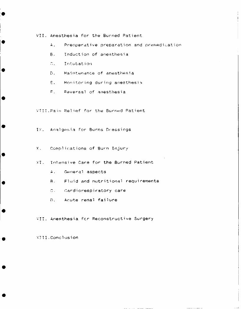
VII. Anesthesia for the Burned Patient
A. Preoperative preparation and prte-rie-diiation
B. Induction of anesthesia
k I n tuat ion'
D) Mainte-nance of anesthes--ia
P . Monitoring during anesthesia~
P Reversal of anesthesia
VTII.Paini Relief for the Burri-~d Patient
TX . Analgc-ia ror Burns Diress-ings
(. cornr)l i(at ions of Burn Injury
1;T. Iote nsive Care for the Burned Patient
A. Gt-neral aspects
* . Fluid and nutritional requirements
(.. ardiorespiratory care
t). Acute renal failure
';I!. Anesthesia fcr Reconstructive Surgery
* ?'TTI.Concltusion

PERIOPERATIVE MANAGEMENT OF ORGAN DONORS
* Ake Grenvik, M.D.
I. Historical background
II. Current transplantation frequency
III. Organ needs vs supply
IV. Organ donor categories
V. Evolution of brain death concept
VI. Donor management in the ICU and OR
Vii. Organ procurement and preservation
VIII. Allocation of organs
IX. US transplant organization
X. Future expectations
XI. Ethical problems
PLENARY SESSION LECTURES
SESSION V
SUNDAY, MAY 5,1991
7:30 A.M. - 12:00 P.M.
Moderator: Levon M. Capan, M.D.
0l p I" • L • - • - " -"t r "' - , i r 1 -- T
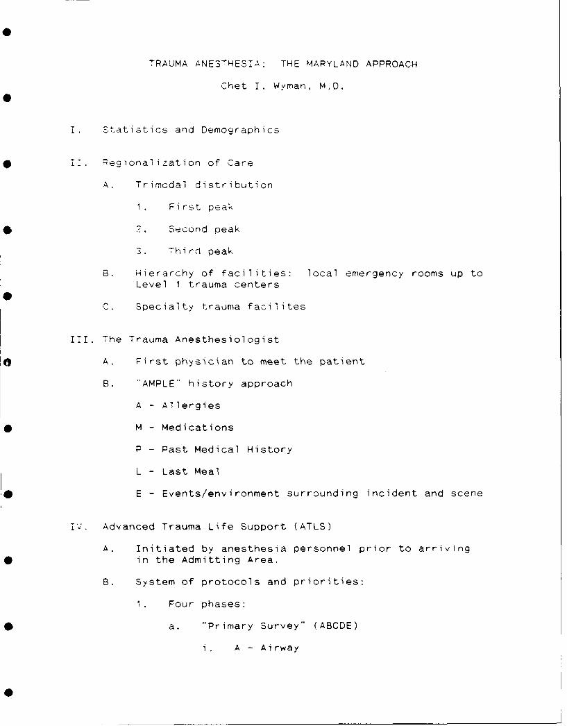
TRAUMA ANESTHESIA: THE MARYLAND APPROACH
Chet I. Wyman, M.D.
i. Statistics and Demographics
*K. Regionalization of Care
A. Trimodal distribution
1. First peak
2. Second peak
3. Third peak
B. Hierarchy of facilities: local emergency rooms up toLevel 1 trauma centers
C. Specialty trauma facilites
iiI. The Trauma Anesthesiologist
A. First physician to meet the patient
B. "AMPLE" history approach
A - Allergies
M - Medications
P - Past Medical History
L - Last Meal
E - Events/environment surrounding incident and scene
i.. Advanced Trauma Life Support (ATLS)
A. Initiated by anesthesia personnel prior to arrivingin the Admitting Area.
B. System of protocols and priorities:
1. Four phases:
a. "Primary Survey" (ABCDE)
i. A - Airway

I
ii. B - Breathing
iii. C - Circulation
iv. 0 - Disability (Mini-Neuro Exam)
v. E - Exposure
b. "Resuscitation Phase"
Sc. "Secondary Survey"
d. "Definitive Care Phase"
e. Anesthesiologist is involved in each phase ofthe perioper'tive care of the trauma patient
V. A~r'iy Management
A, The first priority
* B. Three objectives:
1. Secure an intact airway
2. Protect a jeopardized airway
* 3. Provide an airway if there is not. one.
C. Most early deaths in trauma are due to faulty airwaymanagement, either through inexperience or, moreimportantly, faulty judgement.
S1). Manual in-line cervical stabilization (A.K.A."traction", "immobilization")
E. MIEMSS prospective study, documented neurologic injuryriot found
• r. Surgical airway
V!, Ventilation
• VII. Circulation
A. Recognize shock
B. Establish IV lines
• C. High volume fluid warmer/infusers
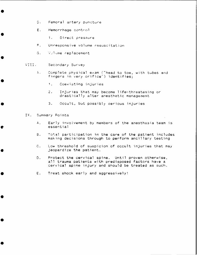
D. Femoral artery puncture
E. Hemorrhage control
1. Direct pressure
F. Unresponsive volume resuscitation
G. V_,Ime replacement
VIII. Secondary Survey
A, Complete physical exam ("head to toe, with tubes andfingers in very orifice") identifies;
1. Coexisting injuries
2. Injuries that may become life-threatening ordrastically alter anesthetic management
3. Occult, but possibly serious injuries
IX. Summary Points
A. Early involvement by members of the anesthesia team isessential
B. To tal participation in the care of the patient includesmaking decisions through to perform ancillary testing
C. Low threshold of suspicion of occult injuries that mayjeopardize the patient.
D. Protect the cervical spine. Until proven otherwise,all trauma patients with predisposed factors have acervical spine injury and should be treated as such.
E. Treat shock early and aggressively!

QUALITY ASSESSMENT AND RISK CONTROLIN TRAUMA ANESTHESIA
Brian McAlary, M.D.
I. Introduction
A. Role of CQI Program
1. Trauma "Prevention"
2. Targeted Education
3. Interdepartmental Dialogue
B. Role of Risk Control
1. Cost Control
2. Stress Reduction
II. Features of any valid program
A. Commitment to Excellence vs. Acceptable
1. ability to anticipate problems
2. ability to anticipate and reward excellence
B. On going vs. Sporadic Monitoring
1. Relevant Generic Screening
2. Meaningful and Changing Indications
a. thresholds for evaluation
3. Ability to detect low incidence events
4. Capacity to identify trends
C. Involvement of all levels
1. Reporting
2. Review / Analysis
D. Free of Negative Association
1. Anonymous review
2. Instructional emphasis

3. Confidential reporting and filing
4. Easy appeal mechanism
5. Courteous feedback to reporting source
E. Compliance with External Review Bodies
1. Federal
2. State
3. JCAHCO
4. Professional Liability Insurer
F. Meaningful Solutions
1. Effective Administrative Changes
2. Extradepartmental responsiveness
3. Decreased incidence of unwanted occurrences
4. Contribution to credentialing
G. Credibility
1. Intradepartmental
2. Interdepartmental
III. Practical Procedural Concerns
A. Effective Data Collection
1. Involvement of Companion Departments
a. PACU
b. Biomedical
c. Pharmacy
2. Generic Screening Tools
3. Use of Problem Identification Form
a. Description of event or concern
b. List of alternative recommendations
c. Preferred "solution"

d. Option of feedback vs. anonymity
4. Retrospective reviews
B. CQI Coordinator
1. Independent Authority
a. Standard response letters
b. Requesting screening assistance
2. Liaison between departmental and external elements
a. Notification of concern
b. Follow up to CQI Committee
c. Notification of Risk Manager
3. Liaison with CQI Committee and department members
a. Requests for clarification of events/actionstaken
b. Accelerates urgent reviews
c. Obtain needed medical records
d. Confidential filing
C. Role of CQI Committee
1. Objective review of all input
2. Makes recommendations
a. refer to Medical Staff bodies
b. policy change
c. counseling/commendation
d. reprimand/suspension
e. no further review
3. Action roles
a. Refers for inclusion in CME
b. Instructional letters
c. Notification of other departments
d. Notification of Risk Manager
IV. Outcome Analysis
A. Problem "solved"
1. Document in CQI minutes
2. Delete indicator
3. "Thank you" letters
B. Problem "remains"
1. Repeat educational effort
2. Attempt alternative solution
3. Notify Director, etc. of residual difficulty
V. Trauma Specific Indicators
A. Pre Hospital Care
1. The requisite "big four"
2. Proper ET placement
B. Admission Phase
1. Use of In line stabilization
2. Appropriate use of blood products
3. Availability of support services
C. Intra-operative Phase
1. Frequency of repeat laboratory studies
2. Temperature control efforts
D. Post-operative Phase
1. Need for prolonged or repeated intubation
2. Fluid management
PERIOPERATIVE PAIN MANAGEMENT FOR TRAUMA PATIENTS
Roger S. Cicala, M.D.
I. Pathophysiology of Acute Pain
II. Principles of Pain Management
1II, Methods of Acute Pain Management
A. Parenteral medications: narcotic analgesics
B. Parenteral medications: non-narcotic agents
C. Epidural analgesia
1. Catheter placement
2. Epidural administration of narcoLic analgesics
3. Epidural administration of local anesthetics
4. Selection of agent and monitoring for epidural
analgesia
D. Intrathecal analgesia
E. Intrapleural analgesia
F. Peripheral nerve blocks
G. Cryoanalgesia
E. Transcutaneous electrical nerve stimulation
III. Analgesia for Patients with Specific Types of Injury
A. Thoracic injuries
B. Femoral fracture
C. Ther-mal injuAries
Iv. Chronic Pain
*A. Sympathetically mediated pain syndromes
G. Post amputation pain
CRITICAL CARE TRANSPORT OFTHE PEDIATRIC TRAUMA PATIENT
James I. Gilman, M.D.
I. Introduction - Pediatric Perspective
II. Scene Response
A. Assessment
1. Airway/Ventilation/Oxygen
2. CNS/Level of Consciousness
3. Cardiovascular/Pulses
B. CPR/ABC's
C. Vascular Access/Fluids
D. Temperature Control
E. Monitors
1. ECG
2. Pulse Oximeter
3. Temperature
F. Specific Injuries
1. Head
2. Neck
3. Airway
4. Chest
5. Abdomen
6. Extremities
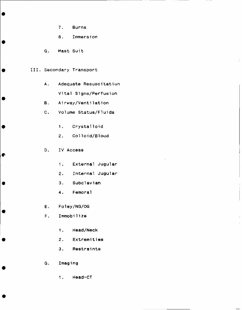
7. Burns
8. Immersion
G. Mast Suit
III. Secondary Transport
A. Adequate Resuscitation
Vital Signs/Perfusion
B. Airway/Ventilation
C. Volume Status/Fluids
1. Crystalloid
2. Colloid/Blood
D. IV Access
1. External Jugular
2. Internal Jugular
3. Subclavian
4. Femoral
E. Foley/NG/OG
F. Immobilize
1. Head/Neck
2. Extremities
3. Restraints
G. Imaging
1. Head-CT

2. Neck-X-Ray/MRI
3. Chest-AP/LAT X-Ray - CT
4. Abdomen - CT, X-ray, DPL
H. Drugs
1. Muscle relaxants
pancuronium
2. Analgesia/narcotics
3. Sedation
4. Seizure control
5. Pressors
I. Vehicle Considerations
1. Ambulance
2. Rotorcraft
3. Fixed wing
4. Parents

COMPLICATIONS OF TRAUMA: COAGULOPATHY AND EMBOLISM
Levon M. Capan, M.D.
I. Coagulopathy
A. Physiology of coagulation
B. Phases of coagulation abnormalities after trauma
C. Causes of coagulopathy in trauma
D. Diagnosis
E. Management
II. Deep Vein Thrombosis
A. Diagnosis
B. Prevention
C. Pharmacologic prevention
D. Treatment
III. Pulmonary Embolism
A. Mechanism
B. Clinical manifestations
C. Diagnostic tests
D. Treatment
E. Anesthetic considerations
SIMULTANEOUS
BREAKAWAY SESSIONS
FRIDAY, MAY 3, 1991

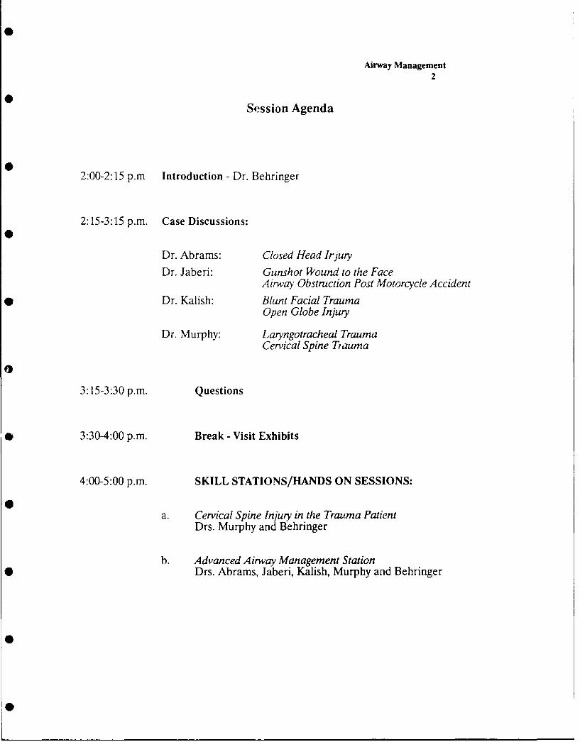
SIMULTANEOUS BREAKAWAY SESSIONS
I. Airway Management-Case Discussions/SkilI Stations
Friday, May 3, 1991
2- 5 p.m.
Moderator: Elizabeth C. Behringer, M.D.
0
Panelists: Kenneth J. Abrams, M.D.
Mahmood Jaberi, M.D.
Murray A. Kalish, M.D.
Mark T. Murphy, M.D.
0
Airway Management
2
Session Agenda
2:00-2:15 p.m Introduction - Dr. Behringer
2:15-3:15 p.m. Case Discussions:
Dr. Abrams: Closed Head Irjury
Dr. Jaberi: Gunshot Wound to the FaceAirway Obstruction Post Motorcycle Accident
* Dr. Kalish: Blunt Facial TraumaOpen Globe Injury
Dr. Murphy: Laryngotracheal TraumaCervical Spine Tauma
3:15-3:30 p.m. Questions
3:30-4:00 p.m. Break - Visit Exhibits
4:00-5:00 p.m. SKILL STATIONS/HANDS ON SESSIONS:
a. Cervical Spine Injury in the Trauma PatientDrs. Murphy and Behringer
b. Advanced Airway Management StationDrs. Abrams, Jaberi, Kalish, Murphy and Behringer

* Airway Management
3
AIRWAY MANAGEMENT: CASE DISCUSSIONS

Airway Management
4
CLOSED HEAD INJURYKenneth J. Abrams, M.D.
A. Case Stem:
A 32-year-old male unbelted passenger involved in a head on collision with a
telephone pole. At scene, reportedly alert, AOB, obvious right leg deformity. VS at
scene: BP 140/90, P 110, RR 24. Transported to ER with 02 via FM, 18 G. RPIV,
Philadelphia collar in place, Thomas splint, on backboard.
On arrival in ER, found to be stuporous. VS: BP 150/95, P 118, RR 28,
GCS= 11.
The anesthesiologist responds with the trauma team.
B. Guidelines for Discussion:
1. Anesthetic considerations in patients with closed head injury.
2. Criteria for early intubation.
3. Intubation techniques.
a. What options exist?b. Sequence for intubationc. Useful pharmacology
4. Failed intubation management
C. References:
1. Abrams KJ, Nolan JP, Grande CM. Trauma anesthesia and critical care: Anesthesiology'sB oldest speciality reborn. Anesth Clin. 1991; 9:2, in press.
2. Capon LM. Airway management. In: Capon LM, Miller SM, Turndorf H, eds. Trauma Anes-thesia and Intensive Care. Philadelphia: JB Lippincott, 1991:43-81.
3. Frost EAM. Perioperative anesthetic management of central nervous system trauma. in: SteneJK, Grande CM, eds. Trauma Anesthesia. Baltimore: Williams & Wilkins, 1991:250-265
4. Frost EAM. Anesthesia for patients with 'iead trauma. In: Rogers MC, ed. Current Practice inAnesthesiology. Philadelphia: BC Decker, 1991, in press.
5. Grande CM, Stene JK, Bernhard WN. Airway management: considerations in the traumapatient. Crit Care Clin. 1990; 6:1.
6. Howard RA, Matjasko MJ. Management of intracranial hypertension in severe head injury. In:Matjasko MJ, Shin B, eds. Problems in Anesthesia, 1990; 4:3.
7. Paly D, Wolf A, Matjasko MJ. Management of severe head injury: The anesthesiologist's role.Probl Anesth. 1990; 4:1.
8. Tabaddor K, Frost EAM. The management of head injury. In: Frost EAM, ed. Clinical Anes-thesia in Neurosurgery. Boston: Butterworth-! ieinemann, 1991:403-438.
Airway Management5
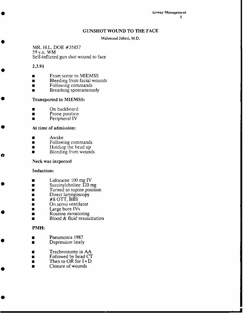
GUNSHOT WOUND TO THE FACEMahmood Jaberi, M.D.
MR. H.L. DOE #3585759 y.o. WMSelf-inflicted gun shot wound to face
2.3.91
a From scene to MIEMSSN Bleeding from facial woundsH Following commands* Breathing spontaneously
Transported to MIEMSS:
E On backboardE Prone positionE Peripheral IV
40 At time of admission:
0 Awake0 Following commandsa Holding the head up0 Bleeding from wounds
Neck was inspected
Induction:
N Lidocaine 100 mg IV* 0 Succinylcholine 120 mg
0 Turned to supine positionN Direct laryngoscopya #80TT, BBSa On servo ventilator* Large bore IVs
0 N Routine monitoring0 Blood & fluid resuscitation
PMH:
0 Pneumonia 1987* Depression lately
* Tracheostomy in AAa Followed by head CT0 Then to OR for I+ D* Closure of wounds
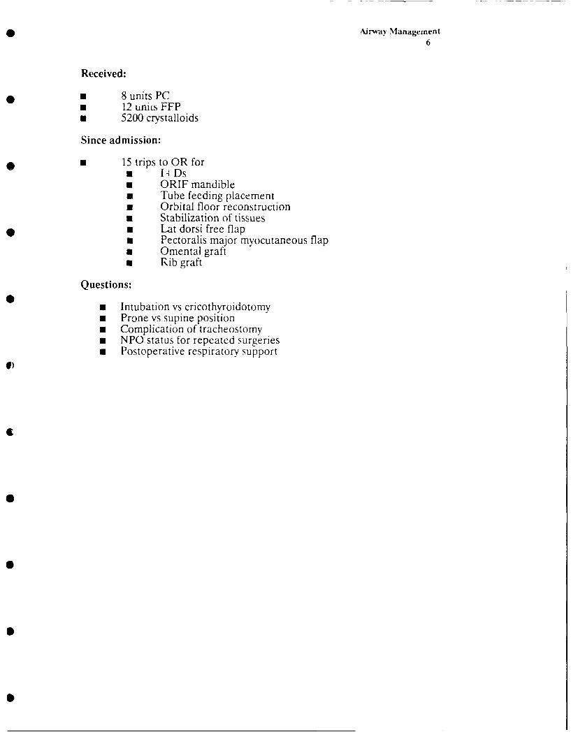
Ai-,w-ay Management6
Received:
S8 units PC0 12 units FFPa 5200 crystalloids
Since admission:
* 15 trips to OR fora I Ds* ORIF mandible• Tube feeding placementa Orbital floor reconstructiona Stabilization of tissues• a Lat dorsi free flapa Pectoralis major myocutaneous flapa Omental graft• Rib graft
Questions:
" Intubation vs cricothyroidotomy* Prone vs supine position" Complication of tracheostomv" NPO status for repeated surgeries" Postoperative respiratory support

Airway NManagerm-t
7
AIRWAY OBSTRUCTION POST MOTORCYCLE ACCIDENT
Mahmood Jaberi, M.D.
MR. D.R. DOE #3627522 y.o. WMAdmitted at 1506 (3.16.91)
0 MCA vs carN Ejected0 Neck hit the side mirror* No helmet0 Semiconscious
0 Brought to MIEMSS on backboard• 02 mask• Spont. resp.N IV fluid
At the time of admission:
" Awake" Oriented X1* GCS 14/15* BP: 162/94" HR: 104/min" RP- 20/min
" No scalp lac* PEARL (2 mm)" Blood in mouth & nares* Facial lac (R) mental area* Mandibular tenderness
* Avulsed teeth (incisors)" Cut upper lip* Hard palate laceration" Tongue laceration 5 cm top, 1 cm underneath
* Neck: V-shaped superficial lac" Trachea midline" No hematoma" No hoarseness" No airway obstruction" No spine tenderness
Other findings:
" Abrasion (L) arm, deltoid area" Otherwise negative
" Admission ABG on 21% F10 2 : 89/7.39/39/99/-5/24" H/H: 13/38.9" Lytes: Normal* Coag: Normal

Airway Management
8
PMH:
" Not remarkable
SH:
" Drug abuse
" Moderate alcohol* Smoker: one PPD
X-rays
" C-spine X-ray: Normal" T-spine: Normal* L-spine: Normal" Head CT: Normal
1800 (3.16.91)
" Local anesthesia & IV sedation* Closure of wounds" Archbars & elastic bandage* Stable vital signs" Spontaneous respiration" Patent airway
0715 (3.17.91)
" Complains of shoulder pain* Received Demerol 75 mg IM
1200 (3.17.91)
" Dyspnea" Sense of air hunger" Diffuse chest pain" Dizziness" Confused at times
" Archbars in place with bands" Patent nares" PEARL" Good air exchange at bases" Diffuse wheezing upper airway" SagO: 94%* ABG: 77/7.38/45
" Dry" Well perfused" Lethargic* T wave changes on lead III

Airway Management
9
Impression:
m Partial airway obstruction, secondary to swelling
Orders:
* NPO* 450 head up position
* Hold narcotics* 0 4 lit/min nasal cannula* 1emove elastic bands
1300 (3.17.91)
* SaO 2 =100%a 1110Crz alet" Good memory* Elastic bands removed* Clear resp sounds* Oropharynx: minimal swelling" Uvula in midline
1630 (3.17.91)
" 15% 0-, by mask• SaG 2 = 99)%
0100 (3.18.91)
* Deterioration of mental status" Lethargic" Disoriented* Confused" Mildly combative" No drooling" Comfortable in upright position
* SaO2 : 99-100%" BP: 130/85" HR: 94/min* RR: 26/min
" HEENT: 3-4 mm nares" (R) perimandibular hematoma, extending with soft tissue swelling* Decreased breath sounds at bases" ABG: 159-7.36-40
" Partial airway obstruction at level of epiglottis secondary to soft tissue swelling

Airway Management10
" Nasal intubation" Sedation" On ventilator" TV950x SIMV 10" BP: 170/85 to 130/80" HR: 107 to 94" ABG on 50% Fi02: 228/7.35/44/99/24" Lytes: Normal" Coag: Normal
0900 (3.18.91)
" Subepiglottic edema per x-ray• TMax 100.2
" CT scan" Significant bleeding from the tongue" Many areas of hematoma* No hyoid bone fracture
" Tracheostomy with #8 Shiley fenestrated tube* In OR under 02-air, fentanyl, norcuron, pentothal
Lateral x-ray of neck: 1250 (3.18.91)
a Marked inferior displacement of hyoid bone and a large soft tissue mass im-pinging upon the anterior aspect of the hypopharynx, completely occluding theairway
Mandibular series:
a Demonstrates a vertical fracture through the (R) mandibular ramus with milddestruction at the fracture site
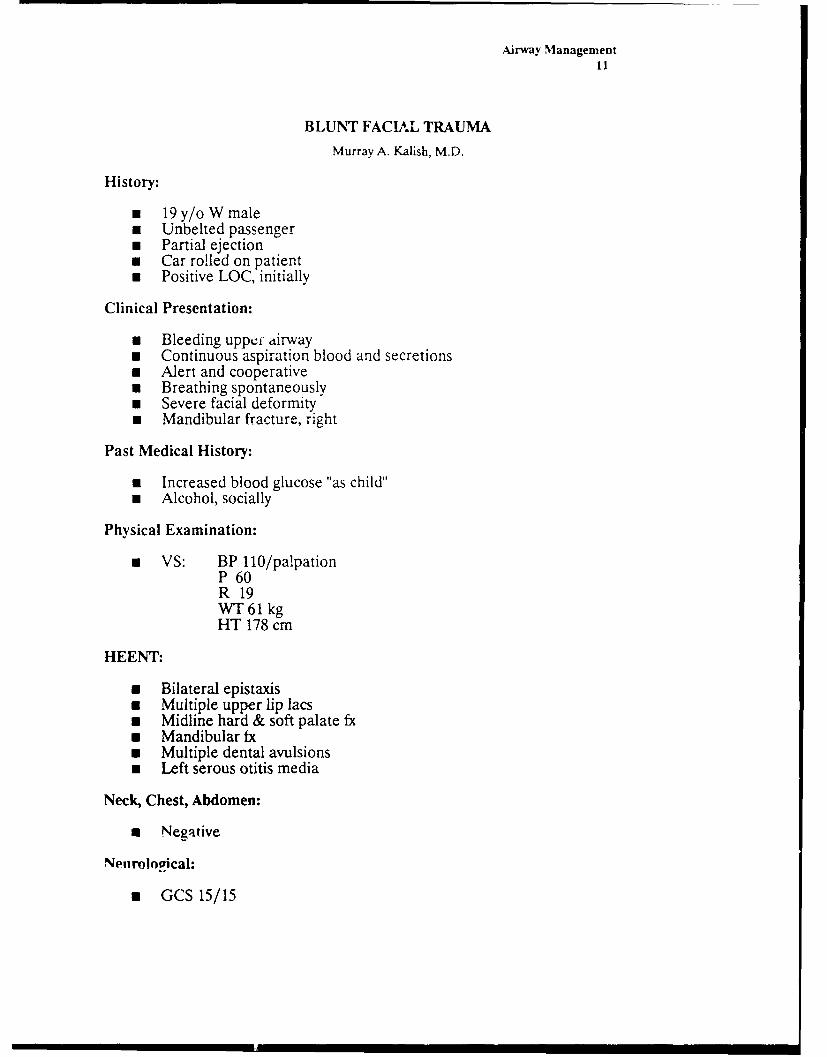
Airway Management11
BLUNT FACLtL TRAUMA
Murray A. Kalish, M.D.
History:
" 19 y/o W male" Unbelted passenger" Partial ejection* Car rolled on patient" Positive LOC, initially
Clinical Presentation:
* Bleeding uppet dirway" Continuous aspiration blood and secretions" Alert and cooperative" Breathing spontaneously" Severe facial deformity" Mandibular fracture, right
Past Medical History:
* Increased blood glucose "as child"" Alcohol, socially
Physical Examination:
N VS: BP 110/palpationP 60R 19WT61 kgHT 178 cm
HEENT:
" Bilateral epistaxis" Multiple upper lip lacs" Midline hard & soft palate fx" Mandibular ix" Multiple dental avulsions" Left serous otitis media
Neck, Chest, Abdomen:
a Negative
Nenrolo cal:
n GCS 15/15

Airway Management
12
Lab:
" H/H 11.5/34.8" K 2.8" Glucose 303
X-ray Studies:
" C-spine, CXR negative* CT head & face:
* Bilateral LeFort III" Impingement right optic nerve on bony fragment" Anterior & posterior sinus fx with pneumocephalus" Hard palate fx* NOE fx
Ophthalmology Consult:
* Poor blink & enopthalmos OS" Decrease visual acuity OS, ? macular edema" Ptosis OD* Diplopia due to decrease EOM, trauma
Procedures: Took 22 hours
" Tracheostomy" Dental impressions* Placement of arch bars" Intermaxillary fixation* Bicoronal incision" ORIF bil. naso-orbital-ethmoid fx* ORIF bil. LeFort I" Reconstruction orbital floors & medial walls with plates & rib grafts" Repair facial lacs" ORIF split palate* ORIF bil. zygoma
Anesthesia:
" Rapid sequence technique* In-line neck stabilization" Thiopental, succinylcholine, IV lidocaine" Maintenance: vecuronium, fentanyl, midazolam -- isoflurane, sufentanil drip
Hospital Course:
* Multiple additional anesthetics via tracheostomy" Washouts, IMF adjustments, placement ungual splint, dental extraction,
evacuation scalp hematoma

Airway Management13
OPEN GLOBE INJURYMurray A. Kalish, M.D.
History:
* 17 y.o. BM* Unbelted driver* Positive LOC* Difficult extrication
Clinical Presentation:
" Combative" Mild respiratory difficulty* Facial pain
Past Medical History:
" Asthma-like symptoms, 1985" PCP, cocaine, marijuana* Smokes cigarettes" Occasional alcohol intake
Physical Examination:
VS:* BP 80/40-100/70" P 100" R 18 mild respiratory difficulty* WT 70 kg" HT 175 cm
HEENT:
* Lac. left lateral upper & lower eyelid" Lac. left temporal-parietal area" Lac. facial ner'e
Neck, Chest, Abdomen:
* Negative
Extremities:
0 Lac. right ankle & left wrist
Neurological:
* GCS 12/15
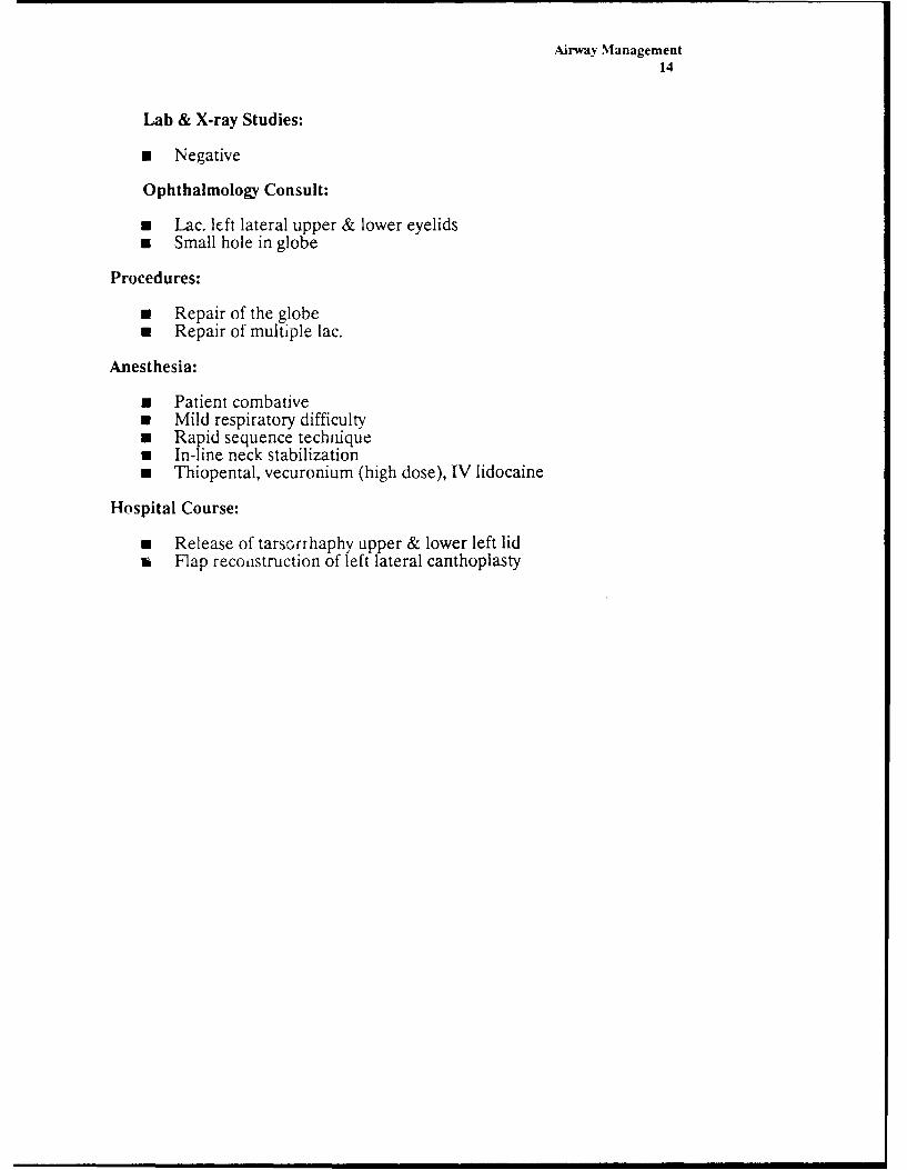
Airway Management14
Lab & X.ray Studies:
n Negative
Ophthalmology Consult:
" Lac. left lateral upper & lower eyelids* Small hole in globe
Procedures:
" Repair of the globe" Repair of multiple lac.
Anesthesia:
* Patient combative" Mild respiratory difficulty" Rapid sequence technique" In-line neck stabilization* Thiopental, vecuronium (high dose), IV lidocaine
Hospital Course:
* Release of tarsorrhaphy upper & lower left lidh Flap recotistruction of left lateral canthoplasty

Airway Management
15
LARYNGOTRACHEAL TRAUMA
Mark T Murphy, M.D.
A 43 y.o. intoxicated male was the unrestrained driver of an open jeep. He
was ejected from the vehicle after striking a tree at a high rate of speed. In the ER, the
patient was obviously intoxicated, following commands and speaking in a grating voice
about "getting even." His vital signs were stable with normal respirations. Physical
exam was significant for an open right femur fracture and a left radial fracture. His
neck was nontender and his cervical spine was "cleared" radiographicaily.
He was taken to the OR for orthopedic intervention. After preoxygenation, a
rapid sequence induction with cricoid pressure was performed. The cords were well
visualized. However, a "clot" was noted between the vocal cords during laryngoscopy.
An endotracheal tube would not pass. Desaturation began.
Discussion:
E Differential diagnosis
0 Laryngotracheal injuries
N PresentationN IncidenceE Management
0 Treatment decisions/ protocols
0 Head & neck position0 Direct oral intubation0 TTJVa Flexible fiberoptic bronchoscope0 Surgical cricothyroidotomy/tracheostomy
References
1. A TLS Program Instructors Manual, Chicago, American College of Surgeons, 1988.2. Mathisen D, GrUo, H. Laryngotracheal trauma. Ann Thorac Surg. 1987; 43:2-54-62.3. Fuhrman G, et al. Blunt laryngeal trauma: classification and management protocol. J Trauma.
1990; 30(1):87-92.
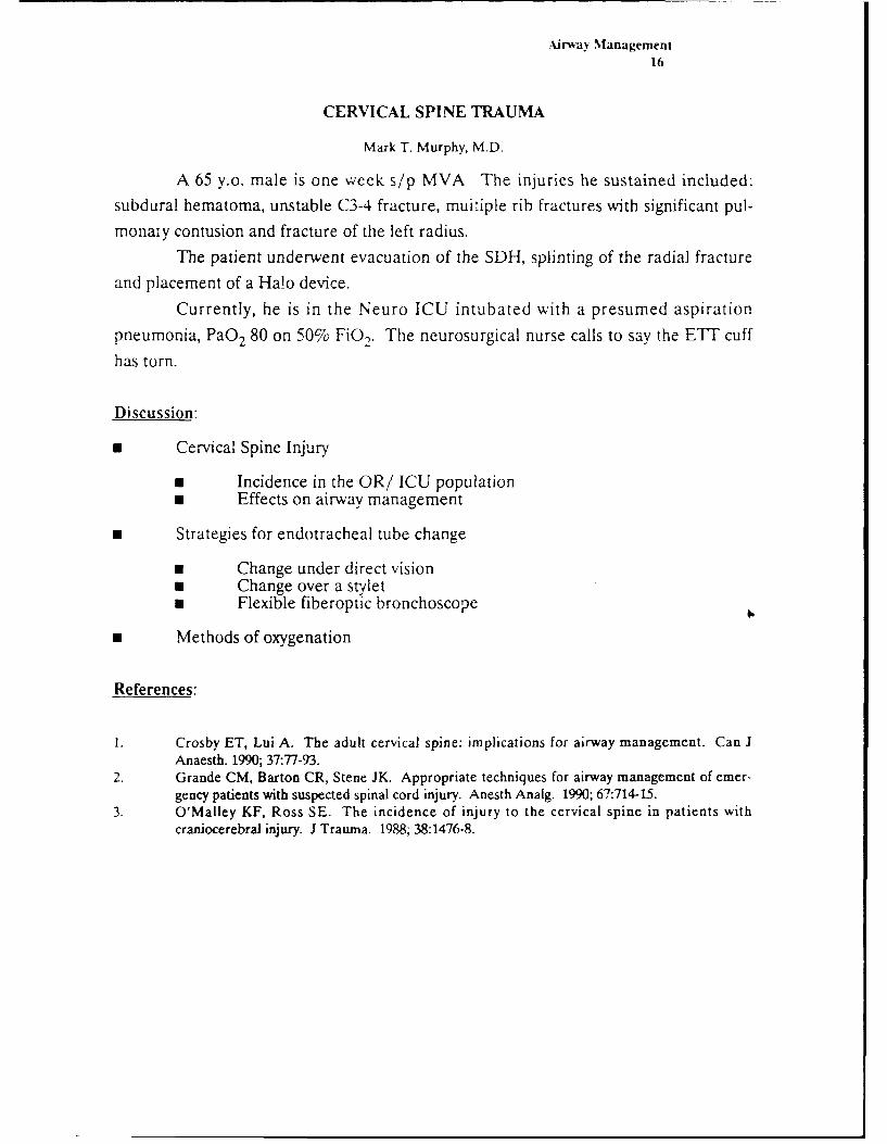
.Air'way Management
16
CERVICAL SPINE TRAUMA
Mark T. Murphy, M.D.
A 65 y.o. male is one week s/p MVA The injuries he sustained included:
subdural hematoma, unstable C3-4 fracture, mui:iple rib fractures with significant pul-
monay contusion and fracture of the left radius.
The patient underwent evacuation of the SDH, splinting of the radial fracture
and placement of a Halo device.
Currently, he is in the Neuro ICU intubated with a presumed aspirationpneumonia, PaO 2 80 on 50% FiO 2. The neurosurgical nurse calls to say the ETT cuff
has torn.
Discussion:
* Cervical Spine Injury
* Incidence in the OR/ICU population* Effects on airway management
* Strategies for endotracheal tube change
N Change under direct vision• Change over a stylet* Flexible fiberoptic bronchoscope to
* Methods of oxygenation
References:
1. Crosby ET, Lui A. The adult cervical spine: implications for airway management. Can JAnaesth. 1990; 37:77-93.
2. Grande CM, Barton CR, Stene JK. Appropriate techniques for airway management of emer-gency patients with suspected spinal cord injury. Anesth Analg. 1990; 67:714-15.
3. O'Malley KF, Ross SE. The incidence of injury to the cervical spine in patients withcraniocerebral injury. J Trauma. 1988; 38:1476-8.

SCIENTIFIC PosUrEs
FRIDAY, MAY 3, 1991
2:00 P.M. - 5:00 P.M.
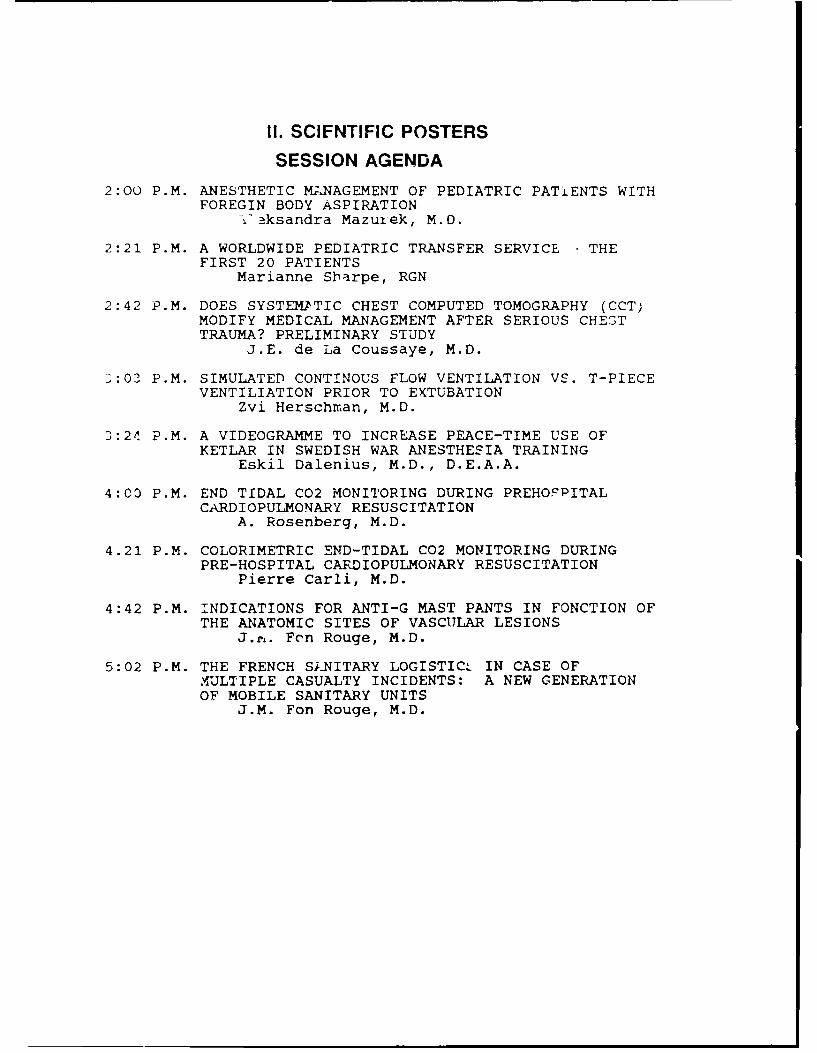
II. SCIFNTIFIC POSTERS
SESSION AGENDA
2:00 P.M. ANESTHETIC MANAGEMENT OF PEDIATRIC PATIENTS WITHFOREGIN BODY ASPIRATION
i-Bksandra Mazurek, M.D.
2:21 P.M. A WORLDWIDE PEDIATRIC TRANSFER SERVICE THEFIRST 20 PATIENTS
Marianne Sbarpe, RGN
2:42 P.M. DOES SYSTEMPTIC CHEST COMPUTED TOMOGRAPHY (CCT)MODIFY MEDICAL MANAGEMENT AFTER SERIOUS CHESTTRAUMA? PRELIMINARY STUDY
J.E. de La Coussaye, M.D.
3:03 P.M. SIMULATED CONTINOUS FLOW VENTILATION VS. T-PIECEVENTILIATION PRIOR TO EXTUBATION
Zvi Herschman, M.D.
3:24 P.M. A VIDEOGRAMME TO INCREASE PEACE-TIME USE OFKETLAR IN SWEDISH WAR ANESTHESIA TRAINING
Eskil Dalenius, M.D., D.E.A.A.
4:00 P.M. END TIDAL C02 MONITORING DURING PREHOPPITALCARDIOPULMONARY RESUSCITATION
A. Rosenberg, M.D.
4.21 P.M. COLORIMETRIC END-TIDAL C02 MONITORING DURINGPRE-HOSPITAL CARDIOPULMONARY RESUSCITATION
Pierre Carli, M.D.
4:42 P.M. INDICATIONS FOR ANTI-G MAST PANTS IN FONCTION OFTHE ANATOMIC SITES OF VASCULAR LESIONS
J.m. Fon Rouge, M.D.
5:02 P.M. THE FRENCH SANITARY LOGISTICL IN CASE OFXAULTIPLE CASUALTY INCIDENTS: A NEW GENERATIONOF MOBILE SANITARY UNITS
J.M. Fon Rouge, M.D.
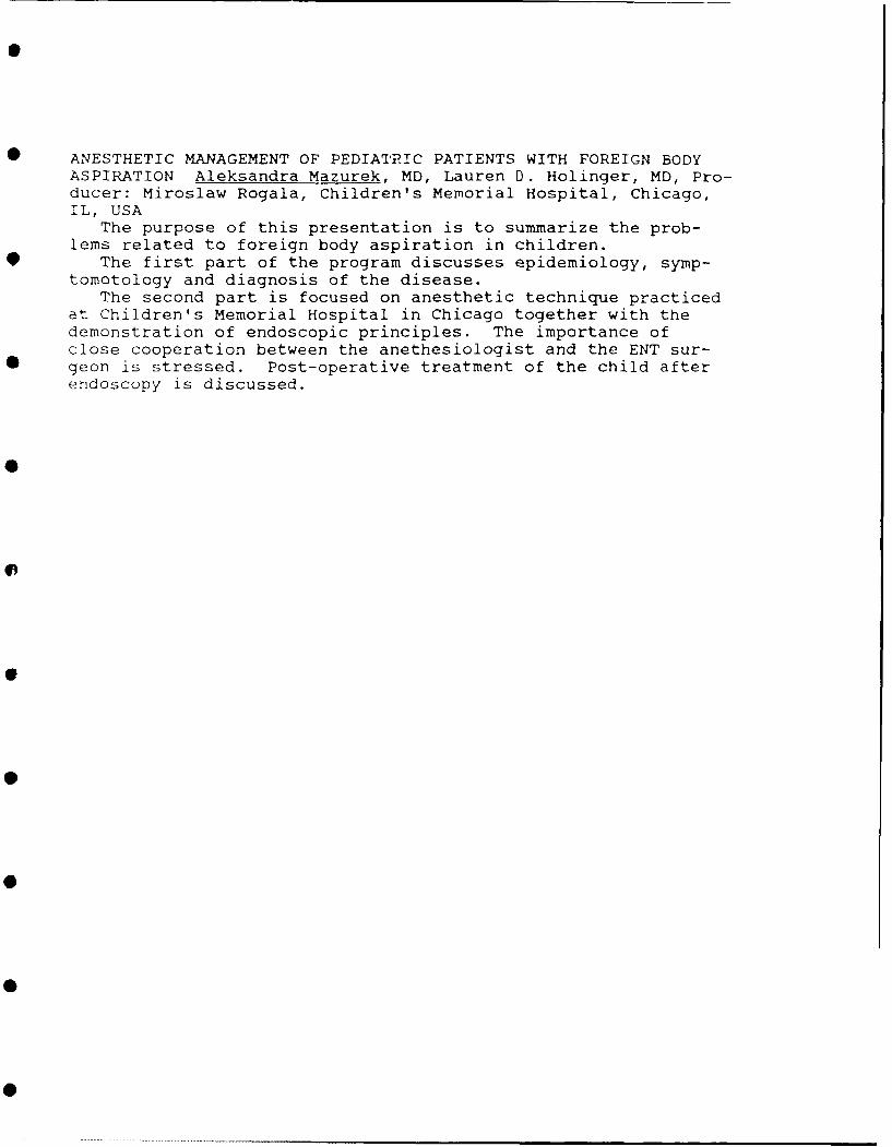
ANESTHETIC MANAGEMENT OF PEDIATRIC PATIENTS WITH FOREIGN BODYASPIRATION Aleksandra Mazurek, MD, Lauren 0. Holinger, MD, Pro-ducer: Miroslaw Rogala, Children's Memorial Hospital, Chicago,IL, USA
The purpose of this presentation is to summarize the prob-lems related to foreign body aspiration in children.
The first part of the program discusses epidemiology, symp-tomotology and diagnosis of the disease.
The second part is focused on anesthetic technique practicedat Children's Memorial Hospital in Chicago together with thedemonstration of endoscopic principles. The importance ofclose cooperation between the anethesiologist and the ENT sur-geon is stressed. Post-operative treatment of the child afterendoscopy is discussed.
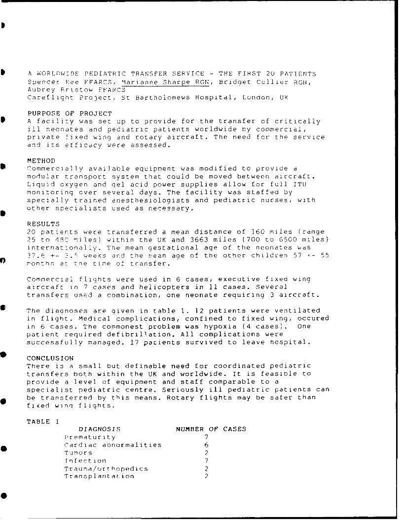
A WORLDWIDE PEDIATRIC TRANSFER SERVICE - THE FIRST 20 PATIENTSSpencer Kee FFARCS, Marianne Sharpe RGN, Bridget Collier RGN,Aubrey Pristow FFARCSCareflight Project, St Bartholomews Hospital, London, UK
PURPOSE OF PROJECTA facility was set up to provide for the transfer of criticallyill neonates and pediatric patients worldwide by commercial,private fixed wing and rotary aircraft. The need for the serviceand its efficacy were assessed.
METHODCommercially available equipment was modified to provide amodular transport system that could be moved between aircraft.Liquid oxygen and gel acid power supplies allow for full ITUmonitoring over several days. The facility was staffed byspecially trained anesthesiologists and pediatric nurses, withother specialists used as necessary.
RESULTS20 patients were transferred a mean distance of 160 miles (range25 to 4RC miles) within the UK and 3663 miles (700 to 6500 miles)internationally. The mean gestational age of the neonates was37.6 +- . weeks and the mean age of the other children 57 - 55months at the time of transfer.
Commercial flights were used in 6 cases, executive fixed wingaircraft in 7 cases and helicopters in 11 cases. Severaltransfers used a combination, one neonate requiring 3 aircraft.
The diaonoses are given in table 1. 12 patients were ventilatedin flight. Medical complications, confined to fixed wing, occuredin 6 cases. The commonest problem was hypoxia (4 cases). Onepatient required defibrillation. All complications weresuccessfully managed. 17 pacients survived to leave hospital.
CONCLUSIONThere is a small but definable need for coordinated pediatrictransfers both within the UK and worldwide. It is feasible toprovide a level of equipment and staff comparable to aspecialist pediatric centre. Seriously ill pediatric patients canbe transferred by this means. Rotary flights may be sater thanfixed winq flights.
TABLE 1DIAGNOSIS NUMBER OF CASES
Prematurity 7Cardiac abnormalities 6Tumors 2Infection 7Trauma/orthopedics 2Transplantation 2

DOES SYSTEMATIC CHEST COMPUTED TOMOGRAPHY (CCT) MODIFYMEDICAL MANAGEMENT AFTER SERIOUS CHEST TRAUMA ? PRELIMINARYSTUDY. J.E. de La Coussave M.D.*, J.J. Estorc M.D.*, J.M. Teissier M.D.**, R.Cohendy M.D.*, P. Richard M.D.*, A. Proust M.D.*, L. Metge M.D.**, F.M. LopezM.D.**, J.J. Eledjam M.D.** Department of Anesthesiology, Critical Care and Emergency.** Department of Radiology. University Hospital Nimes, France.
Patients with serious chest trauma require a clinical and radiological evaluationin emergency room. The aim of the study was to precise whether a systematic CCTcould modified the medical management of these patients.
Methods : In a prospective preliminary study were included patients with seriouschest trauma, as defined by 2 or more preradiologic criteria : blunt trauma, morethan 3 rib fractures, flail chest, pleural abnormality, acute respiratory distress,penetrating injury, PaO2 < 60 mmHg, heart injury (ECG, echocardiography).Exclusion criteria were : depressed level of consciousness, emergency laparotomyor other major surgery, unstable hemodynamic state. Every patient had 4 portablefilms in emergency room - high and low voltage AP films, anterior and posteriorlateral views. CCT scan was performed (Elscint 2002) from the level of the lungapices through the diaphragm.
Results Twenty nine paticnts were included : 24 after traffic accidents, 5 afterpenetrating injuries.
Type of lesion Chest x-ray CCT Modification inmanagement
..........................................................................................
Rib fracture 124 136 No *Flail chest 13 17 NoSternal fracture 5 9 NoRet-osternal hematoma 1 3 NoHemothorax 10 32 NoPneumothorax 18 21 Yes tLung contusion 0 12 NoDiaphragm rupture 2 2 NoMediastinum widening 12 12 No -
* additional rib fractures were found in the same patients t one patientrequiring anesthesia for peripheral surgery : chest tube ; - none patient hadaortic injury, none patient with mediastinal abnormality on CCT had a normalmediastinum on plain film.
Discussion/Conclusion : Except in mediastinal widening where CCT was moreaccurate than plain film, CCT did modify the management of only one of the 29patients. CCT showed small pleural effusions and minimal atelectatic areas thatnever required any specific treatment. In conclusion, CCT induced only 1modification in the management of serious chest trauma after initial clinicalevaluation and chest X-ray with 4 films.
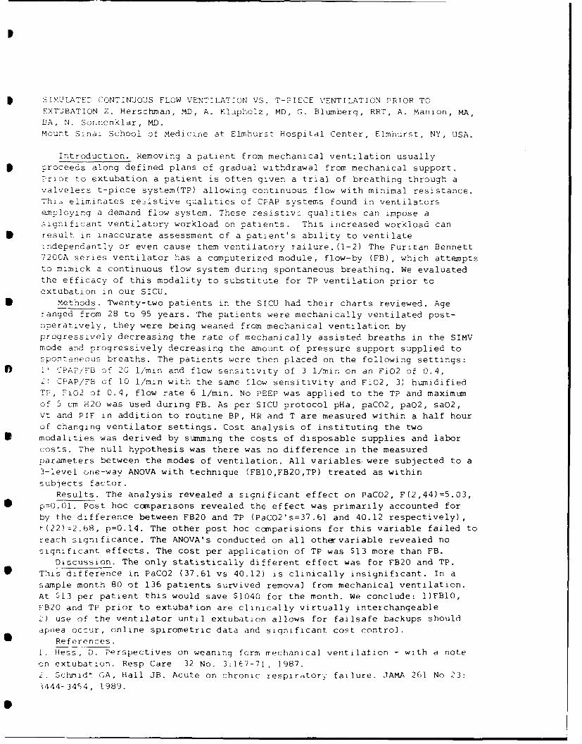
SIMULATED CONTINUOUS FLOW VENTILATION VS. T-PIECE VENTILATION PRIOR TO
EXTUBATION Z. Herschman, MD, A. Klapholz, MD, G. Blumberg, RRT, A. Manion, MA,
HA, N. Sonnenklar, MD.
Mount Sinai School of Medicine at Elmhurst Hospital Center, Elmhurst, NY, USA.
Introduction. Removing a patient from mechanical ventilation usuallyproceeds along defined plans of gradual withdrawal from mechanical support.Prior to extubation a patient is often given a trial of breathing through avalveless t-piece system(TP) allowing continuous flow with minimal resistance.Thi- eliminates resistive qualities of CPAP systems found in ventilatorsemploying a demand flow system. These resistive qualities can impose asignificant ventilatory workload on patients. This increased workload canresult in inaccurate assessment of a patient's ability to ventilateindependantly or even cause them ventilatory tailure. (1-2) The Puritan Bennett7200A series ventilator has a computerized module, flow-by (FB), which attemptsto mimick a continuous flow system during spontaneous breathing. We evaluatedthe efficacy of this modality to substitute for TP ventilation prior to
extubation in our SICU.Methods. Twenty-two patients in the SICU had their charts reviewed. Age
ranged from 28 to 95 years. The patients were mechanically ventilated post-operatively, they were being weaned from mechanical ventilation byprogressively decreasing the rate of mechanically assisted breaths in the SIMVmode and progressively decreasing the amount of pressure support supplied tospontaneous breaths. The patients were then placed on the following settings:0 PAPiFB of 20 I/min and flow sensitivity of 3 1/min on an FiO2 of 0.4,
2)TAP/FB of 10 I/min with the same flow sensitivity and FiO2, 3) humidifiedTP, Fi02 of 0.4, flow rate 6 1/min. No PEEP was applied to the TP and maximumof 5 cm H20 was used during FB. As per SICU protocol pHa, paCO2, paO2, saO2,Vt and PIF in addition to routine BP, HR and T are measured within a half hourof changing ventilator settings. Cost analysis of instituting the twomodalities was derived by summing the costs of disposable supplies and laborcosts. The null hypothesis was there was no difference in the measuredparameters between the modes of ventilation. All variables were subjected to a3-level one-way ANOVA with technique (FB10,FB20,TP) treated as within
subjects factor.Results. The analysis revealed a significant effect on PaCO2, F(2,44)=5.03,
p=0.01. Post hoc ccnparisons revealed the effect was primarily accounted forby the difference between FB20 and TP (PaCO2's=37 .61 and 40.12 respectively),t(22)=2.68, p=0.14. The other post hoc comparisions for this variable failed toreach significance. The ANOVA's conducted on all othervariable revealed nosignificant effects. The cost per application of TP was $13 more than FB.
Discussion. The only statistically different effect was for FB20 and TP.This difference in PaCO2 (37.61 vs 40.12) is clinically insignificant. In asample month 80 of 136 patients survived removal from mechanical ventilation.At $13 per patient this would save $1040 for the month. We conclude: I)FBlO,FB20 and TP prior to extubation are clinically virtually interchangeable,) use of the ventilator until extubation allows for failsafe backups shouldapnea occur, online spirometric data and significant cost control.
References.1. Hess, D. Perspectives on weaning form mechanical ventilation - with a note
on extubation. Resp Care 32 No. 3:167-71, 1987.2. Schmidt GA, Hall JB. Acute on chronic respiratory failure. JAMA 261 No 23:
3444-3454, 1989.
A VIDEOGRAMME TO INCREASE PEACE-TIME USE OF
KETALAR IN SWEDISH WAR ANESTHESIA TRAINIV-
Dalenius F. M.D., D.E.A.A., Lagerholm G., Lindahl C.,
Nathanaelsson K., Norberg K.-A., Rydberg R.
R & D Dept (War), Medical Board of the Armed Forces;
Board of Health & Welfare; Parke-Davis/Warner Lambert
Scandinavia Ltd; Cue Productions Ltd; Sweden.
In the planning for a war, Sweden probably is hampered
by the fact that we have had peace for over 200 years, and
so we have to draw heavily on the experience of other
countries. Our planning presupposes a total integration of
the civilian and military health care resources, of which
the former will bear approx 70 % of the total surgicalworkload, supplemented by field hospitals etc.
The war situation that is judged to bear most resemblan-
ces to the swedish is that faced by the british in the Falk-
lands conflict, both in terms of resources and climate. We
.jase our military anesthesia service on a draw over machine
similar to the Penlon Triservice with OMV vaporizer, and at
present with halothane as the principal drug. The possibility
of a serious oxygen shortage is foreseen.
Therefore, a very important drug to include in the planning is
ketamine, but at present the peace-time use of this is minute
in Sweden as in many other countries. A videogramme was there-
fore produced jointly by the military and civilian Medical
Boards, for use in military as well as civilian training of
both physician anesthetists and nurse anesthetists, to make
them realize that ketamine has many uses in civilian practise
and to increase their openmindedness toward this drug.
This videogramme, in its international version, will be
shown and the results in Sweden discussed.
END TIDAL C02 MONITORING DURING PREHOSPITALCARDIOPULMONARY RESUSCITATION A.Rozenbera. M.D, P.Carli, M.D,O.Lamour, M.D., M.Bousquet, M.D.. A.Derossi. M.D.Department of Anesthesiology H6pital Necker (Pr Barrier) Paris, France
The monitoring of cardiopulmonary resuscitation (CPR) and advanced cardiac lifesupport (ACLS) in the prehospital setting was until recently mainly clinical. End TidalCarbon Dioxide partial pressure (ETCO2) monitoring had been used for the treatment ofcardiac arrest in emergency rooms and hospital wards (1). The aim of this study was toevaluate capnometry in the prehospital setting and compare ETC02 values obtained insuccessfully resuscitated and non resuscitated patients.
Methods: with Institutional approval 25 consecutive patients were prospectivelystudied on field after cardiac arrest. CPR was begun by Emergency Medical Techniciansand continued with ACLS according to American Heart Association protocols by amedical resuscitation team (including at least one anesthesiologist). As soon as thepatient was intubated, ETC02 was continuously recorded by a portable infraredcapnometer (POET Criticare System Inc. ). ETCO2 data were analysed by amicrocomputer. In order to provide a strict ventilatory control the patient wasimmediatly connected to a portable mechanical ventilator Patients age, sex, initialcardiac rythm , continuous EKG and circumstances were recorded. A chart ofresuscitations efforts with precise timing of drugs administration, clinical status of thepatient, duration of prehospital treatment was established. Special attention wasfocused on bicarbonates administration responsible of misleading ETCO2 increaseduring CPR (2). The patient was considered as successfully resuscitated if he returned tospontaiieous circulation (ROSC) and was alive with stable blood pressure on admission tothe hospital. Patients were shared in groups according to the success of resuscitation :groupl resuscitated, group 2 nonresuscitated . Statistical analysis used unpairedstudent's t test for continuous variables and data were expressed as mean ± SD.
Results: One patient (sucessfully resuscitated) was excluded of the study because of therupture of the cuff of the endotracheal tube. The 24 remaining patients were 10 in groupI and 14 in group 2. The two groups were similar except for the initial cardiac rythm(significantly more VF in group 1). The maximum ETCO2 value recorded was higher ingroup I patients (58 ± 14 mmHg) as compared to group 2 (21 ± 5 mmHg) p < 0.001.Maximum ETCO2 was observed in group 1 just before the return of spontaneouscirculation, time to reach this maximum value was 92 ± 302 sec from the beguinning ofthe rtcording in this group. Initial ETC02 value was higher in group 1 (20 ± 8mmHg) ascompared to group 2 (13 ± 8mmHg) p < 0.05
We conclude that in the prehospital setting ETCO2 monitoring is simple to perform andis a guide to resuscitation effort.References :1- Sanders A.B., Kern KB., Otto C.W. End-tidal carbon dioxyde monitoring during CPR: a
pronostic for survival. JAMA 1989, 262:1347-512- Garnett A.RT.. Ornato J.P.. Gonzales E.R.. Hohnson EB. End-tidal carbon dioxydemonitoring during CPR JAMA, 1987. 257: 512 - 515
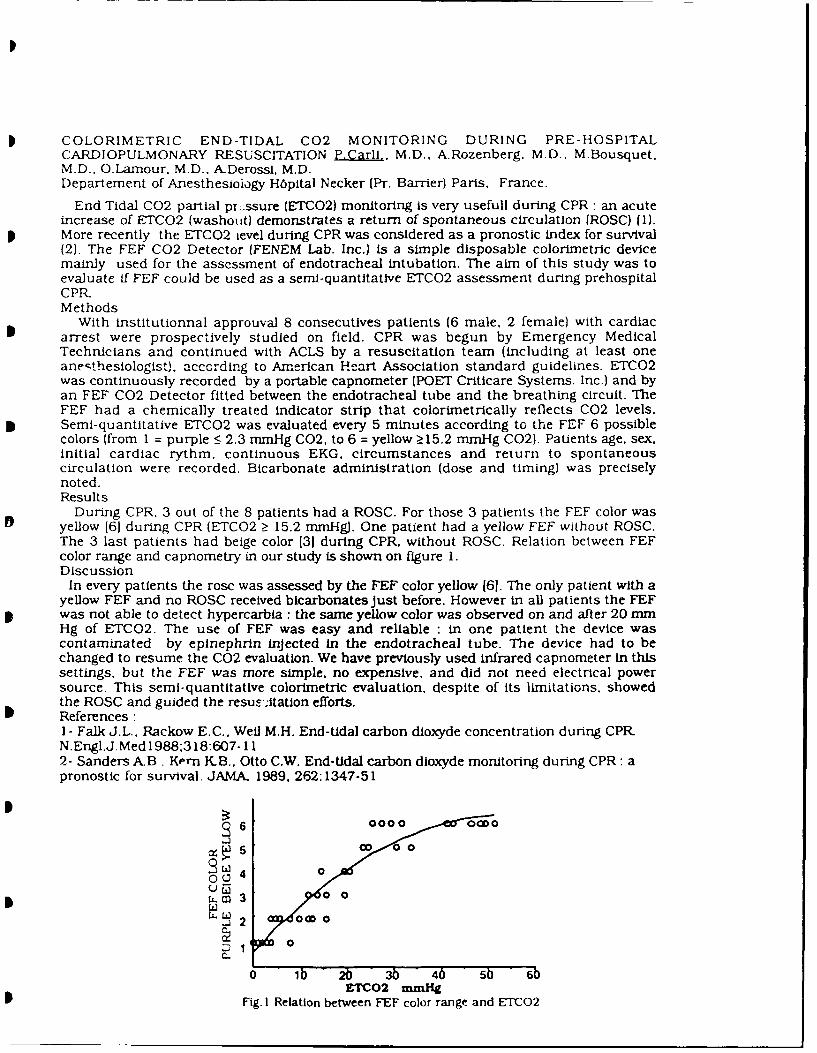
COLORIMETRIC END-TIDAL C02 MONITORING DURING PRE-HOSPITALCARDIOPULMONARY RESUSCITATION P.Carli., M.D.. A.Rozenberg, M.D.. M.Bousquet,M.D.. O.Lamour. M.D.. A.Derossi, M.D.Departement of Anesthesioiogy H6pital Necker (Pr. Barrier) Paris. France.
End Tidal C02 partial pr:ssure (ETCO2) monitoring is very usefull during CPR: an acuteincrease of ETCO2 (washout) demonstrates a return of spontaneous circulation (ROSC) (1).More recently the ETCO2 ievel during CPR was considered as a pronostic index for survival(2). The FEF C02 Detector (FENEM Lab. Inc.) is a simple disposable colorimetric devicemainly used for the assessment of endotracheal intubation. The aim of this study was toevaluate if FEF could be used as a semi-quantitative ETCO2 assessment during prehospitalCPR-Methods
With institutionnal approuval 8 consecutives patients (6 male. 2 female) with cardiacarrest were prospectively studied on field. CPR was begun by Emergency MedicalTechnicians and continued with ACLS by a resuscitation team (including at least oneanesthesiologist), according to American Heart Association standard guidelines. ETCO2was continuously recorded by a portable capnometer (POET Criticare Systems. Inc.) and byan FEF C02 Detector fitted between the endotracheal tube and the breathing circuit. TheFEF had a chemically treated indicator strip that colorimetrically reflects C02 levels.Semi-quantitative ETCO2 was evaluated every 5 minutes according to the FEF 6 possiblecolors (from 1 = purple < 2.3 mmHg C02, to 6 = yellow >15.2 mmHg C02). Patients age, sex,initial cardiac rythm, continuous EKG, circumstances and return to spontaneouscirculation were recorded. Bicarbonate administration (dose and timing) was preciselynoted.Results
During CPR. 3 out of the 8 patients had a ROSC. For those 3 patients the FEF color wasyellow (61 during CPR (ETCO2 > 15.2 mmHg). One patient had a yellow FEF without ROSC.The 3 last patients had beige color 131 during CPR. without ROSC. Relation between FEFcolor range and capnometry in our study is shown on figure 1.Discussion
In every patients the rosc was assessed by the FEF color yellow 161. The only patient with ayellow FEF and no ROSC received bicarbonates Just before. However in all patients the FEFwas not able to detect hypercarbia : the same yellow color was observed on and after 20 mmHg of ETCO2. The use of FEF was easy and reliable : in one patient the device wascontaminated by eplnephrin Injected in the endotracheal tube. The device had to bechanged to resume the C02 evaluation. We have previously used infrared capnometer in thissettings, but the FEF was more simple. no expensive, and did not need electrical powersource. This semi-quantitative colorimetric evaluation, despite of its limitations, showedthe ROSC and guided the resusItaton efforts.References :1- Falk J.L.. Rackow E.C.. Wel M.H. End-tidal carbon dioxyde concentration during CPR-N.Engl.J.Med 1988:318:607-112- Sanders A.B, Kern K.B.. Otto C.W. End-tidal carbon dioxyde monitoring during CPR: apronostic for survival. JAMA. 1989. 262:1347-51
5 00 06 o
1 0oUr
C..
0 Ib ' 2b 3b 46 -5b + 6bETCO2 mmHg
Fig. I Relation between FEF color range and ETCO2
INDICATIONS FOR ANTI-G MAST PANTS
IN FONCTION OF THE ANATOMIC SITES OF VASCULAR LESIONS.
J.M. Fon rouge MD*, G. Leclerc MD*, J.M. Fontanella MD**,F. Lakdja MD***, S. Tarti~re MD*, M. Sabathie MD****
*SAMU 93 Assistance Publique des Hopitaux de Paris .- --
**SAMU 03 MONTLUCON *** Fondation Bergonie **** Universit6 Bordeaux
The use of MAST pants or flying sit anti-gravity equipment is frequent inthe emergency extra-hospital care of severe collapsus patientt.The role of this material is to force back blood from the lower limbstowards the systemic circulation and exert a counter abdominal pressure.This permits an increase in the pre-charge by elevating the central venouspressure. The elevation of the pre-charge pressure appears to be related tothe post-charge when the abdominal counter pressure is from 60 to 80 Tor.The increase in the systemic arterial resistance can exclude the peripheralvascular sector by clamping the aorta below the diaphragm (this proceduremaintains the arterial pressure and conserves the optimal cerebralvascularization.
The human body can be divided into two zones1) A compressible zone lower limbs or abdomen,2) An incompressible zone (sub and supra diaphragmic thoracic area).If the compression of the lower limbs is not considered when the patientpresents a hypo-volumic collapsus, abdominal compression is required whenthe clinical diagnosis is in favour of hemorrhaging.Three different actions using MAST anti-G pants can be defined according tothe hemorrhage site :- In sub-ombilical abdominal lesions involving major arteries or veins,aneurysm of the aorta, extra uterin hemorrhaging the anti-G MAST pantsprove to be a vital equipment with few negative effects.- In lesions that are supra ombilical and below the diaphragm (spleenand liver) the abdominal compression will act indirectly on bleeding,limiting the area of blood collection and pushing the visceral tissuesagainst the diaphragm. The consequences are a counter pressure that willlimit bleeding. The negative effects of this technic are primarily on themechanical aspects of breathing. Assisted ventilation with trachealintubation should be systematically initiated particularly when theincrease in gastric pressure can result in bronchial inhalation, a gastricaspiration tube must be placed. It is necessary to consider a partial orcomplete diaphragmic rupture and the risk of having an intra-abdominalorgans herniation.- In lesions that are sub-costal and supra-diaphragmatic three diagnosesmay be considered :
- ANEURYSM OF THE THORACIC AORTA = ABSOLUTE CONTRA-INDICATION OFANTI-G MAST PANTS.
- RUPTURE OF 0ESOPHAGIAL VARICES = ABSOLUTE CONTRA-INDICATION OFANTI-G MAST PANTS because the veinous drainage is sub-diaphragmic.
- Hemothorax when the drainage is associated with an auto transfusionthe indication of anti-G MAST pants is extremely controversial.
In conclusion, anti-G MAST pants represent the technical material of choicein emergency extra-hospital care for patient in severe collapsus. It isimportant not to underestimate the patients' status even when there appearsto be a dramatic improvement. When a lesion is suspected to be severe athoracic exploration in order to clamp the aorta must be considered and theappropriate vascular filling (liquid support) during the hypo-volimic phaseinduced by the decompression effects of the anti-G MAST pants.
0

LA LOGISTIQUE SANITAIRE FRANCAISE FACE A UN AFFLUX DEBLESSES: UNE NOUVELLE GENERATION DE POSTES
SANITAIRES MOBILES.
HUFNAGEL G.*, LALANDE G.', BOLLOTTE B.', FONROUGE J.M.7, FONTANELLA J.M.*,
* SAMU 03 Centre hospitalier de Montlugon03109 MONTLUCON cedex FRANCE
**SAMU 9311 Centre hospitalier Avicenne
93009 BOBIGNY Cedex FRANCE
Resume
Les auteurs rappellent Ihistorique de la stratgie fran(aise de medicalisation des urgences etils dLcrivent les moyens materiels nouveaux mis A la disposition des 6quipes de S.A.M.U. pour
* faire face A un grand nombre de blesses en situation d'exception (accidents routiers graves,ferrovieres, a~dens, catastrophes naturelles, attentats, etc...). Les postes sanitaires mobiles(P.S.M.) de nouvelle generation sont oprationnels depuis la fin des annees 1980. Ce sont deveritables stocks logistiques, dissociables en quatre lots polyvaents (4*25 blesses graves) etdeux lots de base (2*200 blesses graves). La couverture du territoire national est en cours der~alisation et comportera A terme 30 P.S.M.. ILs sont congus pour equiper un ou plusieurspostes m6dicaux avances, pour renforcer un potentiel hospitaier en cas de catastrophe ouencore pour 6tre utilis6 dans le cadre de I'aide mdicale intemationale. En cas de conflitl'ensemble des P.S.M. constitue une des bases de [a defense sanitaire natinnals piacee sousrautont6 du haut fonctionnaire de ddfcnse au ministbre de la sm-,'wt$ Un P.S.M. comporte presde 6 tonnes de mLdicaments, solutes, pansements et de matriel m6dico-chirurgicalconditionnds en 150 conteneurm comportant une identification graphique. La gestioninformatis_%e de4 stocks permet I entretien et [a rotation des matriels p6rissables. Le P.S.M. estcompl~t6 par une remorque, une tente gonflable avec eclairage, chauffage et un lot tactique deradiophonie lui assurant autonomie et mobilite.
THE FRENCH SANITARY LOGISTICS IN CASE OF MULTIPLE CASUALTY
INCIDENTS: A NEW GENERATION OF MOBILE SANITARY UNITS
Summary
The authors recall the history of the French strategy in the medical care of emergencies outsidethe hospital and they describe the new equipment available to SAMU teams to cope with alarge number of injuned persons in exceptional situations such as road, air or railway accidents,natural catastrophes, criminal attempts...This is a new generation of mobile sanitary units operational since the end of the 80'es. Theyare genuine logistic stocks separable in 4 multi-usage packs (4*25 seriously injuried persons)and 2 base units (2*200 seriously injuried persons). The covering of the French territory isrunning its course and will be made up of 30 mobile sanitary units. They have been designed toallow for medical actions on the spot, to reinforce hospital potentials in case of disasters, or tobe used in an international frame. In case of an armed struggle the whole lot of mobile sanitoryunits is one of the bases of the national sanitary defence under the authority of the high officialof defence of the Health Ministry. A moble sanitary unit is made up of around 6 tons of drugs,solutions, dressings and surgical materials packaged in 150 boxes with graphic identification.The computerized management of stocks runs the maintenance and the rotation of perishableproducts. The mobile sanitary unit is completed by a trailer, an inflatable tent with lighting,heating and a tactical radio system to ensure autonomy and mobility.
0

DISASTER MASS/CASUALTY/MILITARY
ANESTHESIA: PART I
FRIDAY, MAY 3, 1991
2:00 P.M. - 5:00 P.M.
a
0 i ' I ' " - :
BREAKAWAY SESSION
DISASTER / MASS CASUALTY ,/ MILITARY ANESTHESIA: PART I
Project Leader: M. Parr
Moderatcrs: P. Baskett, Y. Donchin, J. Nolan
The Scenario
The disaster is to take place on the day of the opening gameat the Baltimore Orioles Stadium in central Baltimore. It is awarm clear May day. A crowd of 47,000 spectators is present.Before the game begins, a block of one of the mezzanine (middleleiel) stands, with it's spectators, collapses onto the seatingbelow, a distance of about 28 feet. Casualties include adultsand children, the majorit'/ of survivors coming from the upperstand who are injured as a result of the fall. The spectators inthe lower stand are injured by falling masonry and bodies andmost are dead.
The distribution of casualties according to priorities is:
Priority 1: 32 adults and 11 children (urgent therapy requiredto maintain life)Priority 2: 64 adults and 23 children (urgent therapy requiredbut no immeaiate threat to life)Priority 3: 134 adults (non-urgent therapy required)Priority 4: )100 dead
.100 apparently not needing any therapy
Teams (2 teams of 4)
2 Police officersParamedicFire OfficerDirector of stadium servicesNurse2 Physicians
Within the grounds there are 40 police officers whosedistribution is controlled by a command post in the stadium._,utside the stadium there are a further 20 police officers,mainly situated at the gates. Also, within the stadium are 4nurses in 2 first aid rooms situated on the mezzanine and terracelevels and one doctor who is treating a patient with an asthmaattack. There are 2 paramedic units on the ground outside thestadium and there are also about 100 ushers around the stadium,some of whom have two-way radios.
The major problems are likely to arise with effective crowdcontrol of 47,000 people attempting to leave the ground; themanagement and evacuation of large numbers of casualties and
ensuring adequate EMS access while appreciatina there may bedifficulties with access to the stadium for EMS vehicles.
As the scenario unfolds the leaders .iill introduce eachs de in turn and put the questions to the appropriate
4vduaIs. Discussion will be directed by the leaders to -overtie following points:
Act'Lati-Ir" cI a disaster p'an and ensuring it's efFectiveness.
Command and control, incident command system.
Secur1ng the disaster area to ensue rescuer safety.
Ersuring access for emergency medical services.
Extrication and triage.
Trasportation and evacjation.
Allocat' bns of resources.
Crowd cointrol involving thousands of people.
Tn,"ormation and Public Relations communication.
Councel I i ng.
Lear-ning From experience.
SPECIAL CPNA SESSION: PART IFRIDAY, MAY 3, 1991
2:CD P.M. - 5:00 P.M.
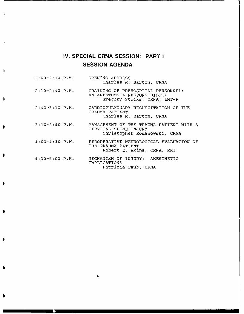
IV. SPECIAL CRNA SESSION: PART I
SESSION AGENDA
2.00-2:10 P.M. OPENING ADDRESSCharles R. Barton, CRNA
2:10-2:40 P.M. TRAINING OF PREHOSPITAL PERSONNEL:AN ANESTHESIA RESPONSIBILITY
Gregory Stocks, CRNA, EMT-P
2:40-3:10 P.M. CARDIOPULMONARY RESUSCITATION OF THETRAUMA PATIENT
Charles R. Barton, CRNA
3:10-3:40 P.M. MANAGEMENT OF THE TRAUMA PATIENT WITH ACERVICAL SPINE INJURY
Christopher Romanowski, CRNA
4:00-4:30 D.M. PEROPERATIVE NEUROLOGICAL EVALUATION OFTHE TRAUMA PATIENT
Robert E. Akins, CRNA, RRT
4:30-5:00 P.M. MECHANISM OF INJURY: ANESTHETICIMPLICATIONS
Patricia Taub, CRNA
I I i
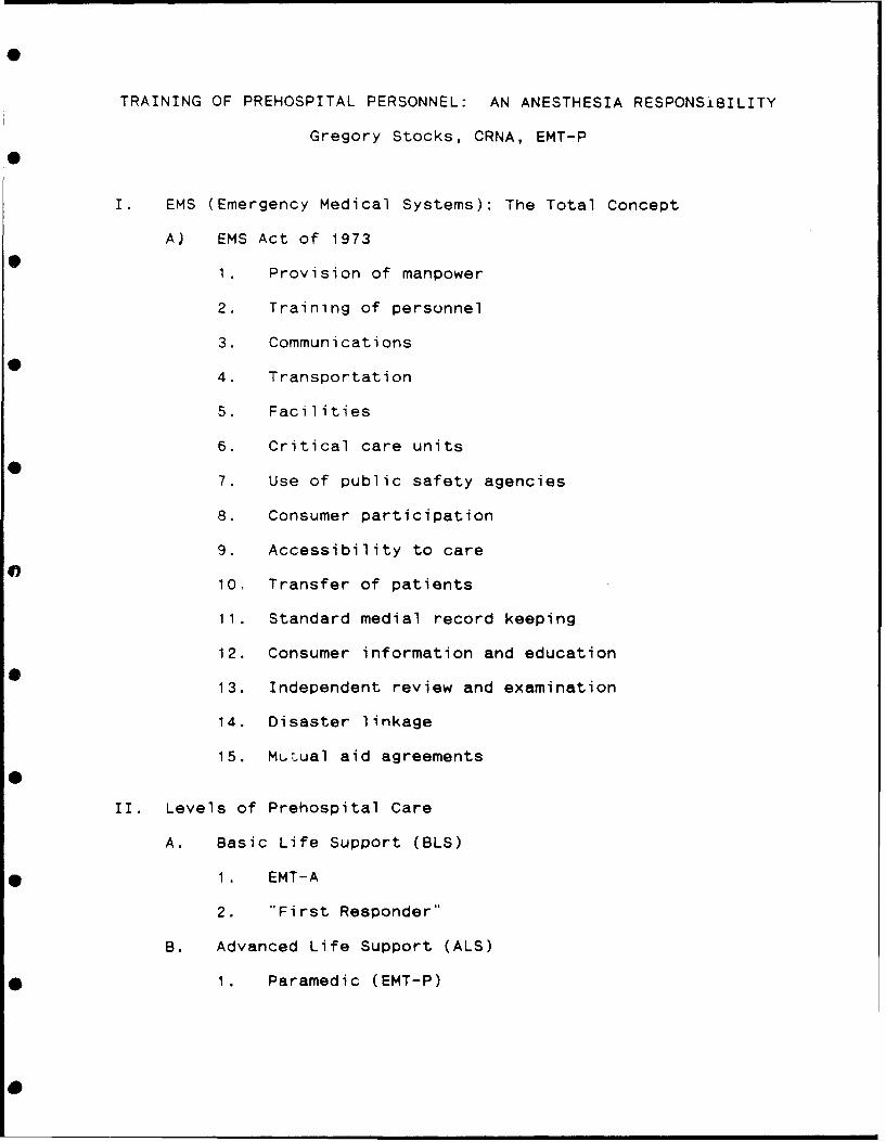
TRAINING OF PREHOSPITAL PERSONNEL: AN ANESTHESIA RESPONSiBILITY
Gregory Stocks, CRNA, EMT-P
I. EMS (Emergency Medical Systems): The Total Concept
A) EMS Act of 1973
1. Provision of manpower
2. Training of personnel
3. Communications
4. Transportation
5. Facilities
6. Critical care units
7. Use of public safety agencies
8. Consumer participation
9. Accessibility to care
10, Transfer of patients
11. Standard medial record keeping
12. Consumer information and education
13. Independent review and examination
14. Disaster linkage
15. Mutual aid agreements
II. Levels of Prehospital Care
A. Basic Life Support (BLS)
1. EMT-A
2. "First Responder"
B. Advanced Life Support (ALS)
1. Paramedic (EMT-P)
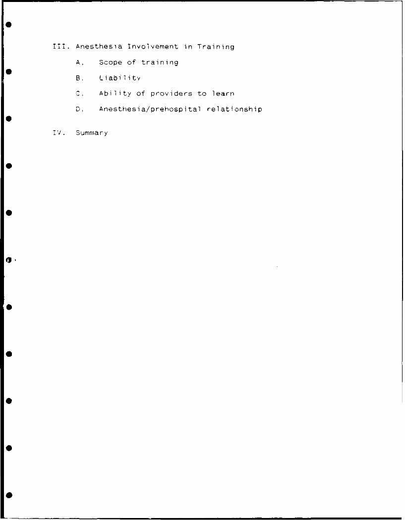
III. Anesthesia Involvement in Training
A. Scope of training
B. Liability
Ability of providers to learn
D, Anesthesia/prehospital relationship
I'). Summary

CARDIOPULMONARY RESUSCITATION OF THE TRAUMA PATIENT
Charles R. Barton, CRNA
I. Introduction
A. Care of the trauma patient is unique
B. Frequently treatment and resuscitation efforts are
required while the diagnosis is still being determined
C. Anesthetic concerns:
1. Assess adequacy of ventilation and perfusion
2. Select proper emergency drugs and anestheticagents/techniques.
II. Early Resuscitation Efforts
A. Early intubation and appropriate ventilation withadequate FI02
B. Adequate intravenous access and appropriate monitoring
C. Assessment and correction of fluid/blood status
D. Relationship of mechanism of injury to current status
E. Assessment and correction of critical factors, (e.g.,tension pneumothorax, pericardial tamponade, disruptionof major blood vessels or airway)
III. Principle Drugs During CPR
1. Epinephrine
2. Lidocaine
3. Sodium bicarbonate
IV. Adjunctive Drugs
A. Antidysrhythmics (procainamide, bretylium)
B. Cardiovascular stimulants (norepinephrine, dopamine,dobutamine, ephedrine, isoproteronol, atropine,

calcium)
C. Vasodilators (nitroprusside, nitroglycerine,
trimethaphan)
D. Beta-blockers (propranol, esmolol)
E. Diuretics (furosemide, ethacrynic acid)
F. Anticonvulsants (barbiturates, benzodiazepines,phenytoin)
V. Fluid Management/Resuscitation
A. Crystalloids, colloids
B. Packed RBCs, component therapy
VI. Invacive Monitoring
A. Arterial line
B. CVP
C. PA catheter
D. Intracranial bolt

HANAGEMENT OF THE TRAUMA PATIENT WITH A CERVICAL SPINE INJURY
Christopher Romanowski, CRNA
I. Mechanism of injury
A. Blunt trauma
1. Motor vehicle accidents (MVA)
2. Fall victim
3. Sports-related injury (e.g., diving,football,etc.)
4. Industrial accident
B. Penetrating trauma
1. Shooting
2. Stabbing
3. Environmental hazards
C. Congenital anomalies
Ti. Classification of C-Spine Injuries
A. Musculoligamentous
B. Vertebral
C. Age groups
III. Signs and Symptoms of C-Spine Injuries
A. Central nervous system (CNS)
B. Cardiac
C. Pulmonary
IV. Protocols and Treatment of C-Spine Injuries
A. CNS
B. Cardiac
0
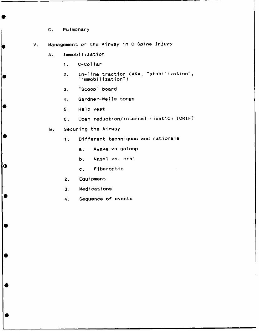
C. Pulmonary
V. Management of the Airway in C-Spine Injury
A. Immobilization
1. C-Collar
2. In-line traction (AKA, "stabilization","immobilization")
3. "Scoop" board
4. Gardner-Wells tongs
5. Halo vest
6. Open reduction/internal fixation (ORIF)
B. Securing the Airway
1. Different techniques and rationale
a. Awake vs.asleep
b. Nasal vs. oral
c. Fiberoptic
2. Equipment
3. Medications
4. Sequence of events

PREOPERATIVE NEUROLOGIC EVALUATION OF THE TRAUMA PATIENT
Robert E, Akins, CRNA, RRT
I. History
A. Events preceding the incident
B. Factors about the changing nature of the patient'sneurologic state,
II. Observation of the Results of the Resuscitation
A. Mental status
B. Respiratory pattern
C. Movement of extremities
III. Physical Exam
A. Scalp
B. Neck
C. Mental status
D. Cranial nerves
E. Motor and sensory system
F. Reflexes
IV. Neck Injury
A. Lateral cervical spine x-ray
B. If the patient is awake and alert range of motion canbe tested.
V. Mental Status Evaluation
A. Speech
B. Memory calculation
C. Spatial orientation
D. Reading

E. Affect
SVT. Cranial Nerve Examination (CN2->12)
A. Monitoring the pupillary response
1. Puril shape
2. Speed of response to light.
B. Cold-water caloric testing (CN3,6,8)
C. Corneal stimulations (CN5,7)
D. Pharyngeal stimulation (CN9,10)
E. Shoulder shrug (CN8)
t-. Tongue stimulation (CN12)
VII. Peripheral Stimulation
A. Sensory and motor fibers
B. Strength of the reaction is graded.
VIII.Tone and Reflexes
A. Confirms existence of neurologic deficit.
IX. Follow-up
A. Repeat every 5 to 10 minutes
B. Prevent permanent neural damage.
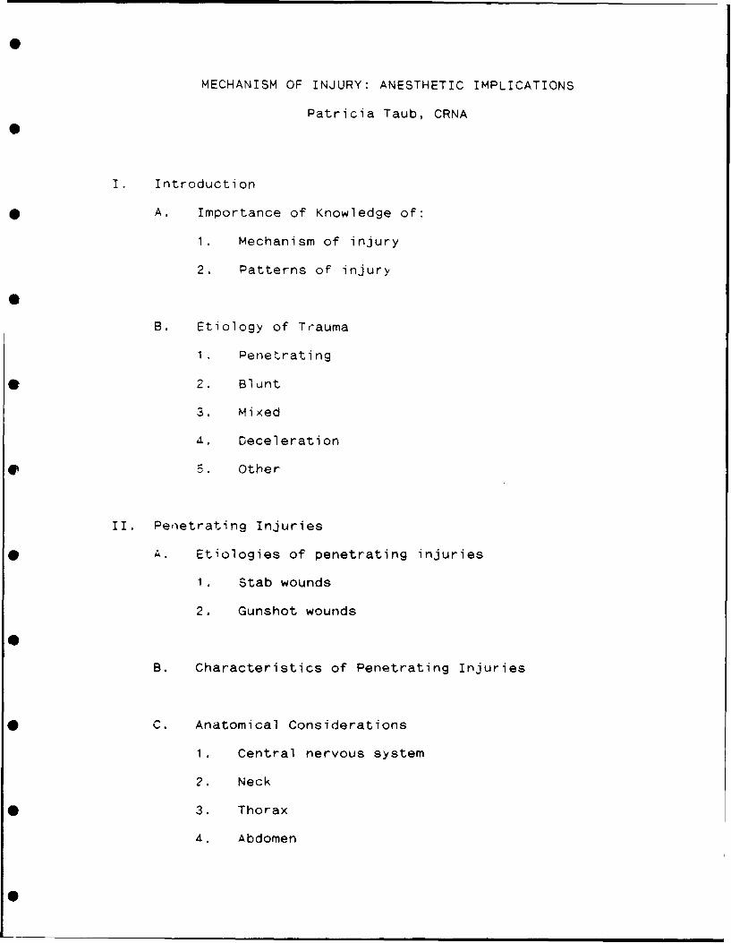
MECHANISM OF INJURY: ANESTHETIC IMPLICATIONS
Patricia Taub, CRNA
I. Introduction
A. Importance of Knowledge of:
1. Mechanism of injury
2. Patterns of injury
B. Etiology of Trauma
1. Penetrating
2. Blunt
3. Mixed
4. Deceleration
5. Other
II. Peoetrating Injuries
A. Etiologies of penetrating injuries
1. Stab wounds
2. Gunshot wounds
B. Characteristics of Penetrating Injuries
C Anatomical Considerations
1. Central nervous system
2. Neck
3. Thorax
4. Abdomen

5. Extremities
!II. "Blunt" Trauma
A. Forces involved
B. indications of Major Blunt Injuries
C. Etiologies of Blunt Trauma
1. Motor vehicle accidents
a. Motorcycle accidents
b. "Pedestrian struck"
2. Deceleration injuries
a. Terminal velocity
D. Anatomical Considerations
1. Central nervous system
2. Neck
3, Thorax
4. Abdomen
a. "Seat belt syndrome"
5. Extremities
IV. Anesthetic Implications
A. Prolonged surgery
B. Impact on body systems
a. cardiovascular
b. respiratory

c. central nervous system
C. Choice of Anesthetic Agents
V. Conclusion/Summary

SIMULTANEOUS
BREAKAWAY SESSIONSSATURDAY, MAY 4, 1991
ANESTHESIA EQUIPMENT: "HANDS-ON"
SATURDAY, MAY 4, 1991
2:00 P.M. - E:00 P.M.
9, 5 " m l ! I' I "l i I

BREAKAWAY SESSION
ANESTHESIA EQUIPMENT: "HANDS-ON"
Faculty: Drs. Petty, Kingsley, Loeb
Objective: To acquaint anesthesia providers with portable andstationary anesthesia machines which are used in caring fortrauma patients.
Dr. Clayton Petty 4i l provide an overall view of desirablesafety features for anesthesia machines. He wil I emphasize thepract;cal features which can be incorporated into fieldanesthesia machines. The United States Anesthesia Field Machinewil, be used to show the principles of conception, d,sign, andmanufacture required to provide an anesthesia apparatus for aspecific need.
Dr. Charles Kingsley will emphasize the simplicity andeffectiveness of portable anesthesia apparatus. He will discussthe elements of draw-over vaporizers. He will delineate theplace of draw-over vaporizers, the practical application of theirlong-term use, and the need for mobility of the units.
Dr. Robert Loeb shall review the present state-of-the-art inportable and stationary ventilators designed for anesthesia. Hewill emphasize working principles of the ventilato-s. Evaluationwill be given of new portai.le ventilors and their practical valuein the field situation.
A "hands-on ' session will allow anesthesia care providers toexamine a wide variety of anesthesia machines. Representativesfrom anesthesia machine manufacturers will have portable andstationary anesthesia machines on display. Drs. Petty, Kingsley,and Loeb will be available to answer questions and assist in thedemonstration of anesthesia machines.

SCIENTIFIC ABSTRACTS
SATURDAY, MAY 4, 1991
2:00 P.M. - 5:00 P.M.

II. SCIENTIFIC ABSTRACTS
SESSION AGENDA
2:00 P.M. SELF-EXTUBATION: A TWELVE-MONTH EXPERIENCE INAN URBAN TRAUMA CENTER
Marc F. Domsky, D.O.
2:11 P.M. THE USE OF A FIBEROPTIC LARGYNGOSCOPE INTRACHEAL INTUBATION OF TRAUMA PATIENTS WITHIMMOBILIZED CERVICAL SPINE
Yves Lambert, M.D.
2:22 P.M. CONTINUOUS ARTERIO-VENOUS REWARDING: A NEWTREATMENT FOR HYPOTHERMIA
Larry M. Gentilello, M.D.
2:33 P.M. COST VERSUS EFFECTIVENESS ? THE FENEM C02CONTROVERSY IN THE UK
D.J. Wilkinson, M.D.
2:44 P.M. THE EFFECTS OF PROPOFOL ON MYOCARDIALCONTRACTILITY AND FUNCTION IN HYPOVOLEMIC SWINE
Charles P. Kingsley, M.D.
2:55 P.M. THE IMPORTANCE OF BASE DEFICIT IN TRAUMA PATIENTSI. McConachie, MB, FFARCS
3:07 P.M. ELECTROLYTE CHANGES AFTER BLUNT TRAUMA AND FLUIDRESUSCITATION
I. McConachie, MB, FFARCS
3:17 P.M. THE EFFECT ON OXYGEN DELIVERY AND CONSUMPTION OFWITHDRAWING NEUROMUSCULAR BLOCKADE IN CRITICALLYILL INTENSIVE CARE PATIENTS
Kenneth J. Power, M.D.
3:28 P.M. HYPOMAGNESEMIA IN SICU SUBPOPULATIONSZvi Herschman, M.D.
4:00 P.M. ENHANCED EFFICACY OF REMOVAL OF THEOPHYLLINE BYCAVHD
William Chiang, M.D.
4:11 P.M. EFFECTS OF PROPOFOL SEDATION ON ICP, CBF VELOCITYAND EEG IN SEVERE HEAD INJURED PATIENTS
Vincenzo Fodale, M.D.
4:22 P.M. JEHOVAH'S WITNESS: THE TRAUMAIan Darling, M.D.

SELF-EXTUBATION: A TWELVE-MONTH EXPERIENCE IN AN URBANTRAUMA CENTERM. Domsky, D.O., K. Dauphinee, M.D., B. McAlary, M.D.Maryland Institute for Emergency Medical Services Systems,Baltimore, MD., USA
Although endotracheal intubation is a common occurrercein the critical care unit, there is a paucity of 'iteratureregarding one of its most life-threatening complications, self-ext-ubation. We retrospectively evaluated the incidence, clinicalfeatures, and outcomes of self-extubations in ourMulti-Trauma Unit. In a 12 month period, 584 mechanicallyventilated patients (Male-421; Mean Age 39.9+19.5 yrs.) wereadmitted. Eighty-one self-extubations ccurred in 73 patients:a 13.9% incidence. Mean intubation period before self-extubation was 7 days. A disproportionate number (64.2%) ofself-extubi.tions occurred between the hours of 7pm and 7am.While the nurse to patient ratio remained the same during alltimes, there were fewer hospital personnel present during boththe evening and night shifts.
Ninety-one percent of the self-extubated patients receivedintravenous sedation within 4 hours of the event, and fifty-fourpercent of the patients were restrained. None of the patientshad received muscle relaxants. Nine of the self-extubations (11%)appeared to have been accidental, occurring while patients werecoughing or during patient repositioning, while the other seven-ty-two appear to have been secondary to patient effort. There wasa significant difference in the number of patients with adiagnosed head injury GCS<12 and CAT scan confirmed)in theself-extubated group versus those in the non-self-extubated group(56.8% vs. 43.7% respectively p<.03). Of the eight patients thatextubated themselves twice, five had head injuries. Fourteen ofthe sixteen self-extubations in this group occurred between thehours of 7pm and 7am. Forty-eight of the eighty-one episodes ofself-extubation required re-intubation (59.2%) for respiratoryinsufficiency that was clinically evident within 30 minutes;another two patients were re-intubated within 6 hours. Only onepatient developed an arrhythmia with associated hypotension postextubation.
In conclusion, self-extubations are more likely to occur betweenthe hours of 7pm and 7am, and in those patients with a diagnosedhead injury. Since most patients had been restrained and/or givensedation, we also concluded that extubations will occur despitethese precautions. The majority of our self-extubated patientsrequired re-intubation almost immediately for impending respiratoryarrest. There were no deaths and only one arrhythmia. Ourexperience suggests that although self-extubation is a seriouscomplication, it can be managed safely with few negative sequelae.

THE USE OF A FIBEROPTIC LARGYNGOSCOPE IN TRACHEAL INTUBATIONOF TRAUMA PATIENTS WITH IMMOBILIZED CERVICAL SPINE. Y Lambert, M.D.,and B. Riou, M.D.Departments of Anesthesiology, C.H.U. Henri Mondor, Cr6teil, and C.H.U. Piti6-
0 Salp6tri~re, Paris, France
Trauma patients with unstable cervical spine are at high risk of spinal cord injury, whichrequires cervical spine immobilization until definitive surgical fixation. Tracheal intubationis often difficult in these patients. We describe the use of a new fiberoptic laryngoscope intrauma patients with immobilized cervical spine.
Ten consecutive trauma patients were selected. All patients were scheduled fororthopedic surgery because of an unstable cervical spine, and were treated, using cervicalspine immobilization (halovest or hard collar). Anesthesia was induced with thiopental and
0 fentanyl, and muscle relaxation was achieved using vecuronium bromide. Directlaryngoscopy was difficult leading to the use of the fiberoptic laryngoscope (Bullard, CirconACMI, Stamford, CT). In the first 6 patients we used a stylet to band the tracheal tube justabove its cuff, reproducing the curve of the laryngoscope blade. Intubation with thistechnique follows five steps : 1) introduction of the laryngoscope blade and visualisation of
o the vocal cords ; 2) introduction of the tube with its stylet just above the laryngoscopeblade; 3) placement of the tube between the vocal cords under fiberoptic vision ; 4)adequate placement of the tube into the trachea ; 5) withdrawal of the stylet and thelaryngoscope. A special introducing stylet (Lis-A adult introducing stylet, Circon ACMI)was used in the four last patients. Before tracheal intubation, preoxygenation was
0 administered. Each attempt lasted no longer than 2 min, and further attempts wereseparated by face mak ventilation with oxygen for at least 1 min.
Tracheal intubation was successful at the first attempt (duration 35 to 105 sec) except in
one patient. In this patient, tracheal intubation was unsuccessful at the two first attempts,because adequate placement of the tube into the trachea, the stylet being maintained, wasimpossible. At the third attempt (35 sec), tracheal intubation was successful using the nasalroute under the fiberoptic laryngoscope control. One patient had uncomplete and anotherpatient had complete neurologic deficit prior to induction of anesthesia. The deficit wasjudged unchanged in the recovery room in these two patients, and no neurological deficitoccurred in the 8 remaining patients. No complication was observed and no recall oftracheal intubation was noted. Tracheal intubations with the introducing stylet which wasused in the four last patients, were considered to be easier than those previously performed.
The fiberoptic laryngoscope enables a rapid, non-invasive, and oral technique for
tracheal intubation, without modilization of the cervical spine. Its other advantages are a* lower cost and a higher rubustness compared with fiberoptic bronschospe.
0
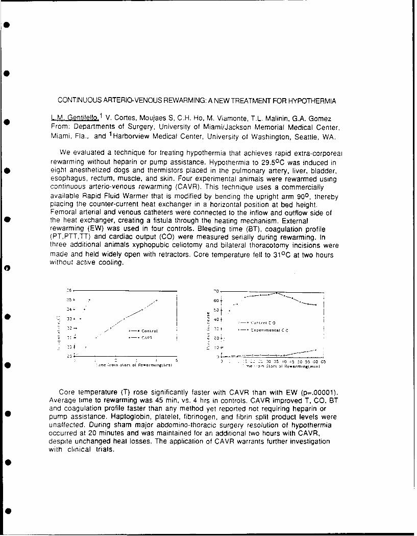
CONTINUOUS ARTERIO-VENOUS REWARMING: A NEW TREATMENT FOR HYPOTHERMIA
L.M. Gentilello.1 V. Cortes, Moujaes S, C.H. Ho, M. Viamonte, T.L. Malinin, G.A. GomezFrom: Departments of Surgery, University of Miami/Jackson Memorial Medical Center,Miami, Fla., and 1 Harborview Medical Center, University of Washington, Seattle, WA.
We evaluated a technique for treating hypothermia that achieves rapid extra-corporealrewarming without heparin or pump assistance. Hypothermia to 29.50C was induced ineight anesthetized dogs and thermistors placed in the pulmonary artery, liver, bladder,esophagus, rectum, muscle, and skin. Four experimental animals were rewarmed usingcontinuous arerio-venous rewarming (CAVR). This technique uses a commerciallyavailable Rapid Fluid Warmer that is modified by bending the upright arm 900, therebyplacing the counter-current heat exchanger in a horizontal position at bed height.Femoral arterial and venous catheters were connected to the inflow and outflow side ofthe heat exchanger, creating a fistula through the heating mechanism. Externalrewarming (EW) was used in four controls. Bleeding time (BT), coagulation profile(PT,PTT,TT) and cardiac output (CO) were measured serially during rewarming. Inthree additional animals xyphopubic celiotomy and bilateral thoracotomy incisions weremade and held widely open with retractors. Core temperature fell to 310C at two hourswithout active cooling.
0-
25 i. 60±
2 4" -0
233 . t 40 f- -- Control C 0
22'-. '- Control 20 + . I o xpertmental C 0
2: CAVR -=
S2 .--1 30 35 1O 15 0O 55 30 65I'inc Ntomn Lart of Il 11an ingitirs) me 1-, SLart of iRcwormingtnin)
Core temperature (T) rose significantly faster with CAVR than with EW (p=.00001).Average time to rewarming was 45 min, vs. 4 hrs in controls. CAVR improved T, CO, BTand coagulation profile faster than any method yet reported not requiring heparin orpump assistance. Haptoglobin, platelet, fibrinogen, and fibrin split product levels wereunaffected. During sham major abdomino-thoracic surgery resolution of hypothermiaoccurred at 20 minutes and was maintained for an additional two hours with CAVR,despite unchanged heat losses. The application of CAVR warrants further investigationwith clinical trials.
COST VERSUS EFFECTIVENESS ?THE FENEM C02 CONTROVERSY IN THEUKWilkinson DJ, Hayes M, Denman WT. Department of Anaesthesia,St. Bartholomews Hospital, London ECIA 7BE. UK.
The Fenem end tidal C02 detector has been recently shown torapidly and successfully differentiate between oesophagealand tracheal intubation(l). It has also been shown to be avaluable aid in the management of cardiopulmonary resuscit-tation, by providing an indication of the effectivenss ofchest compressions and also the return of spontaneous circu-lation(2). Its light weight and simple design make it ofparticular value for paramedical use at all resuscitationsoutside the hospital where more conventional capnography isimpractical. The Fenem end tidal C02 detector is a colori-metric monitor which, when exposed to carbon dioxide in ex-pired respiratory gas, becomes more acidic and the indicatorchanges colour. Problems with its use have occurred whendrugs have been directly instilled into the tracheal tubeduring cardiopulmonary resuscitation(3) (In accordance withthe British Resuscitation Council guidelines). Both adrena-line (1:10000, Ph - 3.33 and lignocaine (PH 1.72) caused apermanent colour change of the indicator which was identicalto that produced by carbon di xide. Its routine use in resus-itation in the UK has been limited by its cost (£14 (25$) perunit) at a time of considerable financial restraint in ourhealth service. To increase its availability in our teachinghospital to every resuscitation box and in the Accident andEmergency Department would necessitate an initial outlay of£2000 and then a further £3920 per year, as we deal with app-roximately 280-300 resuscitations per year. This cost was con-sidered excessive by hospital administrators and health careprofessionals alike. We believe this financial argument is in-valid. By indicating successful tracheal intubation in theoperating room and effective external cardiac massage duringresuscitation it may help to reduce hypoxic insults whichcan lead to vegetative survival. The cost of caring for veg-etative survival being astronomically more than the cost ofthis device.(1)Denman W, Hayes M, Higgins D, Wilkinson DJ. The Fenemcarbon dioxide device. Anaesthesia 1990; 45: 465-467(2)Higgins D, Hayes M, Denman W, Wilkinson DJ. Effectivenessof using end tidal carbon dioxide concentration to monitorcardiopulmonary resuscitation. Br Med J 1990; 300: 581(3)Muir JD, Randalls PB, Smith GB. end tidal C02 detector formonitoring cariopulmonary resuscitation (Letter) British Med-ical Journal 1990; 31: 41

THE EFFECTS OF PROPOFOL ON MYOCARDIAL CONTRACTILITY AND F[INCTION IN HYPOVOLEMIC SWINEJ. Baeuerte, M.D., C.P. Kingsley, M.D., J.A. Ward, Ph.D., D. William, and J. HindsAnesthesia Service and Department of Clinical Investigation,
* Brooke Army Medical Center, Fort Sam Houston, Texas 78234-6200
Introduction. Anesthetic induction is a crucial aspect of trauma patient care and a significant challengefor anesthesia providers. Propofot is a new intravenous anesthetic with the advantages of rapid, smoothemergence from anesthesia, short duration and lack of accumulation. This study assesses the effects ofpropofoL on myocardial contractility, afterload and cardiac output by measurement of the end-systolicpressure-dimension relationship (1) and hemodynamics in a hypovotemic swine model.
Methods. Under an approved animal use protocol, ten Yorkshire swine weighing 15-25 kg were anesthetized* and instrumented with arterial and pulmonary artery catheters for pressure, hemodynamic and blood gas
monitoring. Through a median sternotomy, sonomicrometer crystals were placed in the anterior and posteriorLeft ventricular walt (AP dimension) and a 5 mm Konigsberg pressure transducer was placed through the apexof the left ventricle (LV). An umbilical tape was placed around the inferior vena cava (IVC) to varypreload. The animals were splenectomized to minimize the effects of autotransfusion. The instrumentationwas exteriorized and the incisions approximated. Signals were amplified using a polygraph recorder, A/Dconverted at 200 Hz and analyzed on an IBM-AT compatible computer using locally developed software.Pressure-dimension (PD) loops were obtained while occluding the IVC. End-systolic elastance tEes) wascalculated as the slope of the linear regression line through the series of end-systolic pressure-dimensionpoints during the IVC occlusion. Baseline control (BC) hemodynamics and PD loops were obtained. Animalswere then hemorrhaged to a MAP of 40 mniHg and allowed to recover for 10 minutes to simulate a hypovolernicstate. New Ees and hemodynamic measurements were then made at this new hemorrhage control state (HC).Propofol (5 mg/kg) (2) was given as an i.v. bolus. Pressure-dimension (PD) loops and hemodynamic data weremeasured at 1, 5, 15 and 30 minutes (P1, P5, P15 and P30) after injection. At the end of the experiment,animals were euthanized with T-61. Data was analyzed with a repeated measures ANOVA and a Bonferronicorrected t-test (two-tailed). A p value <0.05 was considered statistically significant.
Results. Table 1 summarizes the results. The changes in Ees were statistically significant between theBC and HC states and the HC and P1 states. Mean arterial pressure (MAP) was significantly decreased at PI,P5 and P15. Cardiac index (CI) and HR were significantly different between the HC and BC states. The HRat P15 and P30 was also significantly different from the HC value. Systemic vascular resistance (SVR) andCI were not changed significantly as a result of propofoL adninistration.
Discussion. This study indicates that myocardial contractility as assessed by a load insensitive index,Ees, in hypovoemic swine is initially depressed by propofot and returns to hemorrhage control values. Meanarterial pressure (MAP) is significantly reduced Dy propofol although there is no net effect on C!. Thisstudy supports the notion that propofol is myocardial depressant as assessed by a load-insensitive measureand a reduction in dosage or avoidance of propofol in the acutely hypovolemic patient is advised.
Table 1. Effect of propofol on myocardial contractility and function in hypovolemic swine (N=10)
Ees SVR MAP CI HRmmHgmm mmig/mL ml mflg 1/min m
2 beats/min
BC 23.8* 51.2 88.0 2.7' 124.1*HC 48.2 62.3 72.1 1.8 141.9PI 29.1* 39.9 40.0* 1.5 126.7P5 34.9 44.6 46.4* 1.5 133.2P15 32.4 48.0 52.9* 1.6 123.4'P30 40.2 50.7 56.9 1.7 120.8'
MSr 199.74 413.86 202.48 0.13 194.240 Significantly different from HC valuesNote: Mean residual sum of squares (MSr) is equivalent to a pooled variance
within groups.
References.1. Suga H, Sagawa K, Shoukas AA: Load independence of the instantaneous pressure-volume ratio of the canineleft ventricle and effects of epinephrine and heart rate on the ratio. Circ Research 32:314-322, 1973.2. Glen JB: Animal studies of the anesthetic activity of ICI 35,868. Br J Anaesth 52:731-741, 1980.
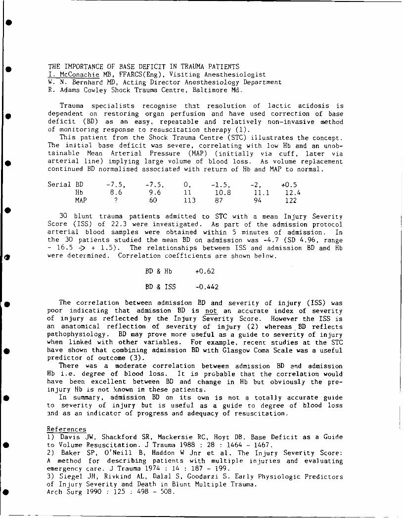
THE IMPORTANCE OF BASE DEFICIT IN TRAUMA PATIENTSI. McConachie MB, FFARCS(Eng), Visiting AnesthesiologistW. N. Bernhard MD, Acting Director Anesthesiology DepartmentR. Adams Cowley Shock Trauma Centre, Baltimore Md.
Trauma specialists recognise that resolution of lactic acidosis isdependent on restoring organ perfusion and have used correction of basedeficit (BD) as an easy, repeatable and relatively non-invasive methodof monitoring response to resuscitation therapy (1).
This patient from the Shock Trauma Centre (STC) illustrates the concept.The initial base deficit was severe, correlating with low Hb and an unob-tainable Mean Arterial Pressure (MAP) (initially via cuff, later viaarterial line) implying large volume of blood loss. As volume replacementcontinued BD normalised associated with return of Hb and MAP to normal.
Serial BD -7.5, -7.5, 0, -1.5, -2, +0.5Hb 8.6 9.6 11 10.8 11.1 12.4MAP ? 60 113 87 94 122
30 blunt trauma patients admitted to STC with a mean Injury SeverityScore (ISS) of 22.3 were investigated. As part of the admission protocolarterial blood samples were obtained within 5 minutes of admission. Inthe 30 patients studied the mean BD on admission was -4.7 (SD 4.96, range- 16.5 -> + 1.5). The relationships between ISS and admission BD and Hbwere determined. Correlation coefficients are shown below.
BD & Hb +0.62
BD & ISS -0.442
The correlation between admission BD and severity of injury (ISS) waspoor indicating that admission BD is not an accurate index of severityof injury as reflected by the Injury Severity Score. However the ISS isan anatomical reflection of severity of injury (2) whereas BD reflectspathophysiology. BD may prove more useful as a guide to severity of injurywhen linked with other variables. For example, recent studies at the STChave shown that combining admission BD with Glasgow Coma Scale was a usefulpredictor of outcome (3).
There was a moderate correlation between admission BD and admissionHb i.e. degree of blood loss. It is probable that the correlation wouldhave been excellent between BD and change in Hb but obviously the pre-injury Hb is not known in these patients.
In summary, admission BD on its own is not a totally accurate guideto severity of injury but is useful as a guide to degree of blood lossand as an indicator of progress and adequacy of resuscitation.
References1) Davis JW, Shackford SR, Mackersie RC, Hoyt DB. Base Deficit as a Guideto Volume Resuscitation. J Trauma 1988 : 28 : 1464 - 1467.2) Baker SP, O'Neill B, Haddon W Jnr et al. The Injury Severity Score:A method for describing patients with multiple injuries and evaluatingemergency care. J Trauma 1974 : 14 : 187 - 199.3) Siegel JH, Rivkind AL, Dalal S, Goodarzi S. Early Physiologic Predictorsof Injury Severity and Death in Blunt Multiple Trauma.Arch Surg 1990 : 125 : 498 - 508.

ELECTROLYTE CHANGES AFTER BLUNT TRAUMA AND FLUID RESUSCITATIONI.McConachie MB, FFARCS(Eng), Visiting AnesthesiologistW.N. Bernhard MD, Acting Director Anesthesiology DepartmentR. Adams Cowley Shock Trauma Centre, Baltimore Md.
Biochemical changes were investigated in 30 blunt injury patientsadmitted to the Shock Trauma Centre (STC) with a mean Injury Severity Scoreof 22.3. Blood samples for analysis were drawn within 5 minutes afteradmission and after 12 hours of resuscitation. Mean volumes of crystalloid(Normal Saline and Ringer's lactate) and packed red cells administeredwere each 15.6L. Samples were analysed for Na, K, Cl, Urea and Glucose.Results are shown as Mean
SDRange. Normal STC range is also shown.
Admission Post Resuscitation Normal Range
Na 137.8 139.3 135 - 148 mmol/l2.47 3.45131 - 142 139 - 149
K 3.89 4.05 3.6 - 5.0 mmol/l0.427 0.6713.1 - 4.8 2.9 - 5.6
C1 107.2 112.1 98 - 108 mmol/l5.04 5.2198 - 120 99 - 124
Urea 15.04 12.61 6 - 20 mg/dl5.49 5.018 - 25 5 - 26
Glucose 185.1 167.0 70 - 110 mg/dl69.0 38.7895 - 396 104 - 291
There were no clinically significant changes in Na, K, Cl or Ureafollowing injury in these patients. Previous studies have suggested thatabnormalities of K are common. Blood sugar was elevated after injury whichis expected from the hormonal and metabolic changes following injury.
Following resuscitation with large volumes of Na containing crystalloidsand blood, Na was still normal. Mean K was normal but the range showsthat individual patients had mild abnormalities unlikely to be clinicallyimportant. However, mean Cl after resuscitation was clearly highpresumably due to the high chloride content in N saline. Hyperchloraemiais known to contribute to metabolic acidosis and may be limited bysubstituting other more balanced fluids than N Saline, such as Ringer'sLactate.
In summary, electrolyte changes after blunt injury and resuscitationwith large volumes of electrolyte containing fluid were uncommon in thisstudy and are probably of little clinical significance.
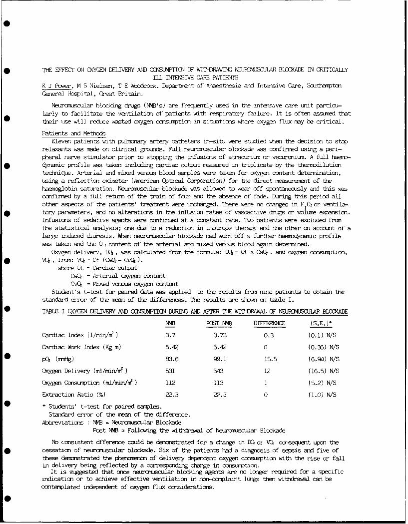
01
THE TECT ON OXYQ DELIVEY AND CU'ZW 4TION OF WIThERAWIG NE I BLOJ(ADE IN CRITICALLYILL INTENSIVE CARE PATIEN\TS
K J Power, M S Nielsen, T E Woodcock. Department of Anaesthesia and Intensive Care, Southampton
General Hospital, Grkeat Britain.
Neuramrcular blocking drugs (NvB's) are frequently used in the intensive care unit particu-larly to facilitate the ventilation of patients with respiratory failure. It is often assumed thattheir use will reduce wasted oxygen coumption in situaticns where oxygen flux may be critical.
Patients and MethodsEleven patients with pulmnary artery catheters in-situ were studied when the decision to stop
relaxants was made on clinical grounds. Full neu sular blockade was ocnfired using a peri-pheral nerve stimulator prior to stopping the infusicns of atracurium or vecurcrium. A full haemo-dynamic profile was taken including cardiac output measured in triplicate by the thenrodilutiontechnique. Arterial and mixed venous blood samples were taken for oxygen content determination,using a reflection oxureter (American Optical Corporation) for the direct measuremrit of thehaemoglobin saturation. Neurcmuscular blockade was allowed to wear off spontaneously and this wasccnfirmed by a full retzn of the train of four and the absence of fade. During this period allother aspects of the patients' treatment were unharnged. There were no changes in F 02 or ventila-tory parameters, and no alterations in the infusion rates of vasoactive drugs or volume expansion.Infusians of sedative agents were continaed at a constant rate. Two patients were excluded fromthe statistical analysis; a-e due to a redicticn in irmtrope therapy and the other on account of alarge induced diuresis. When neuramiscular blockade had worn off a further haemodynaic profilewas taken and the 0 2 content of the arterial and mixed venous blood again determined.
Oxygen delivery, [O) , was calculated from the formila: D() = Qt x CaO . and oxygen onsumption,V* W, from: Vc Qt (CaC,- CvC ).
where Qt = Cardiac outputCa = Arterial oxygen contentCv( = Mixed venous oxygen ccntent
Student's t-test for paired data was applied to the results from nine patients to obtain thestandard error of the mean of the differ--es. The results are shown on table I.
TABLE I OXYGEN DELIVERY AND CaLJION URING AND AFTEUR TE WITHDRAWAL OF NBUtCK4JSM AR BL03KADE
NB POST NMB DIFFERECE (S.E.)*
Cardiac Index (I/min/m2) 3.7 3.73 0.3 (0.1) N/S
Cardiac Work Index (Kg m) 5.42 5.42 0 (0.36) N/S
pQ (mm*g) 83.6 99.1 15.5 (6.94) N/S
Oxygen Delivery (mI/min/m2) 531 543 12 (16.5) N/S
Oxygen Consurption (ml/min/m2) 112 113 1 (5.2) N/S
Extraction Ratio (%) 22.3 22.3 0 (1.0) N/S
* Students' t-test for paired samples.
Standard error of the mean of the difference.Abbreviations : WB = Neur=oaAar Blockade
Post NMB = Following the withdral of Neuramuoular Blockade
No consistent dfferene could be demontrated for a change in DQ0 or V5 ocrsequIt upon the
cessation of neurmucular blocade. Six of the patients had a diagnosis of sepsis and five ofthese demonstrated the phenomero of delivery dependant oxygen ccnstwtin with the rise or fallin delivery being reflected by a corresponding charge in acnsuptimn.
It is suggested that once neuramscular blocking agents are no longer required for a specificindication or to achieve effective ventilation in non-ocrplaint lungs then withdrawal can beconterplated independent of oxygen flux cosiderations.
L
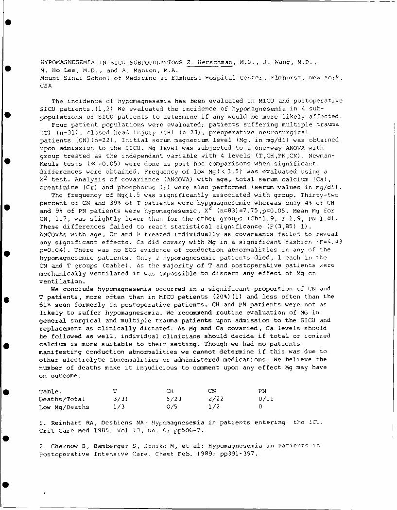
HYPOMAGNESEMIA IN SICU SUBPOPULATIONS Z. Herschman, M.D., J. Wang, M.D.
M. Ho Lee, M.D., and A. Manion, M.A.
Mount Sinai School of Medicine at Elmhurst Hospital Center, Elmhurst, New York,
USA
The incidence of hypomagnesemia has been evaluated in MICU and postoperative
SICU patients.(l,2) We evaluated the incidence of hypomagnesemia in 4 sub-
populations of SICU patients to determine if any would be more likely affected.
Four patient populations were evaluated; patients suffering multiple trauma
(T) (n-31) , closed head injury (CH) (n=23) , preoperative neurosurgicalpatients (CN) (n=22) . Initial serum magnesium level (Mg, in mg/dl) was obtained
upon admission to the SICU. Mg level was subjected to a one-way ANOVA with
group treated as the independant variable 4ith 4 levels (T,CH,PN,CN) . Newman-
Keuls tests (- =0.05) were done as post hoc comparisons when significant
differences were obtained. Frequency of low Mg ( < 1.5) was evaluated using aX2 test. Analysis of covariance (ANCOVA) with age, total serum calcium (Ca)
creatinine (Cr) and phosphorus (P) were also performed (serum values in mg/dl).
The frequency of Mg<!.5 was significantly associated with group. Thirty-two
percent of CN and 39% of T patients were hypomagnesemic whereas only 4% of CH*27and 9% of PN patients were hypomagnesemic, X (n=83)=7 .75,p=0.05. Mean Mg for
CN, 1.7, was slightly lower than for the other groups (Ch=l.9, T=1.9, PN=l.8).
These differences failed to reach statistical significance (F(3,85) 1).
ANCOVAs with age, Cr and P treated individually as covariants failed to reveal
any significant effects. Ca did covary with Mg in a significant fashion (F=4.43
p=0.0 4 ). There was no ECG evidence of conduction abnormalities in any of the
hypomagnesemic patients. Only 2 hypomagnesemic patients died, 1 each in the
CN and T groups (table). As the majority of T and postoperative patients were
mechanically ventilated it was impossible to discern any effect of Mg on
ventilation.
We conclude hypomagnesemia occurred in a significant proportion of CN and
T patients, more often than in MICU patients (20%)(1) and less often than the
61% seen formerly in postoperative patients. CH and PN patients were not as
likely to suffer hypomagnesemia. We recommend routine evaluation of MG in
general surgical and multiple trauma patients upon admission to the SICU and
replacement as clinically dictated. As Mg and Ca covaried, Ca levels shouldbe followed as well, individual clinicians should decide if total or ionizedcalcium is more suitable to their setting. Though we had no patientsmanifesting conduction abnormalities we cannot determine if this was due toother electrolyte abnormalities or administered medications. We believe thenumber of deaths make it injudicious to comment upon any effect Mg may have
on outcome.
Table. T CH CN PNDeaths/Total 3/31 5/23 2/22 0/11Low Mg/Deaths 1/3 0/5 1/2 0
i. Reinhart RA, Desbiens NA: Hypomagnesemia in patients entering the ICU.Crit Care Med 1985; Vol 13, No. 6; pp5 06- 7 .
2. Chernow B, Bamberger S, Stoiko M, et al: Hypomagnesemia in Patients in
Postoperative Intensive Care. Chest Feb. 1989; pp 39 1 - 397 .

ENHANCED EFFICACY OF REMOVAL OF THEOPHYLLINE BY CAVHD W. Chiang,MD,Z.Herschman,
1MD. NYC Poison Control Center, New York, NY, USA.
Introduction.Continuous arterio-venous hemofiltration-dialysis (CAVHD) hasbeen proven effective in managing fluids and electrolytes in the critically ill.
Recent reports have demonstrated therapeutic substances can be removed by CAVHDhowever the low hourly clearance limits its applicability to treating drugintoxication.(1,2)
Methods.Heparinized canine blood was mixed with aminophylline (approximately
30mg in 0.6 1) and held at a height of 100cm. After arteriovenous-filtratepriming with 1.0 1 of heparinized normal saline(NS) and either NS or 25%
albumin to prime the dialysate-filtrate side of the cartridge(approximately50cc) the blood was infused through the hemofilter (Amicon 20) in the usual
fashion. Samples for theophylline concentration were taken towards the end ofthe infusion from arterial (A), venous (V) , and the filtrate (F) ports. The
venous blood was then returned to the arterial side and reinfused, no effortwas made to replace volume lost as filtrate. This process was repeated 5 to 6
times. Sieving coefficient(SC) was calculated as 2F/(A+V). Statistical analysis
was performed using a single-tailed t-test with P<0.5 indicating statistical
significance.Rcsults.Results are summarized in table 1. For all episodes albumin as
dialysate was much more efficient at removing theophylline than was NS. Thedifference between sieving coefficients was statistically significant. This isespecially true if the first set of values are disregarded as these values seem
to be out of character with the rest. The reason for this disparity is likely
a result of residual priming solution in the system.
Discussion.Albumin may enhance drug removal by increasing protein bindingsites or by removing more tainted filtrate. The filtrate concentrations of
theophylline were greater. Though not quantified the volume of filtrate from
the albumin treated filter was about 3 times greater. We conclude the efficacyof CAVHD for theophylline removal can be enhanced using albumin as the dialy-
sate.
References. 1. Domoto DT, Brown, WW, Bruggensmith P. Removal of Toxic Levels
of N-Acetylprocainamide with Continuous Arteriovenous Hemofiltration or
Continuous Arteriovenous Hemodiafiltration. Ann Intern Med 1987;106:550-552.
2. Golpher TA, Bennett WM. Drug Removal by Continuous Arteriovenous Hemofiltra-
tion. Med Toxicology 1988;3:341-349.
Table 1.Arterial Venous Ultrafiltrate SC
Al 16.5 V1 32.3 Fl 7.8 .32
A2 18.2 V2 19.1 F2 17.1 .94
A3 22.2 V3 21.7 F3 12.8 .58
A4 21.2 V4 21.1 F4 13.4 .63
A5 21.0 V5 21.8 F5 13.9 .65
Alb Al 38.1 AlbVl 32.4 AlbFl 11.3 .32
Alb A2 28.4 AlbV2 28.0 AlbF2 29.3 1.04
Alb A3 28.8 AlbV3 27.6 AlbF3 33.1 1.17
Alb A4 27.2 AlbV4 26.0 AlbF4 33.0 1.24
Alb A5 26.2 AlbV5 25.9 AlbF5 29.5 1.13
Alb A6 26.5 AlbV6 25.9 AlbF6 29.9 1.14
Means of the SC for the 2 dialysate solutions are 0.62±0.22 & 1.01±0.34 for
saline and albumin respectively, p<O.05. If the first set of results for saline
& albumin are eliminated, means becomes 0.70±0.16 and 1.14±0.07 respectively,
p<0.001.
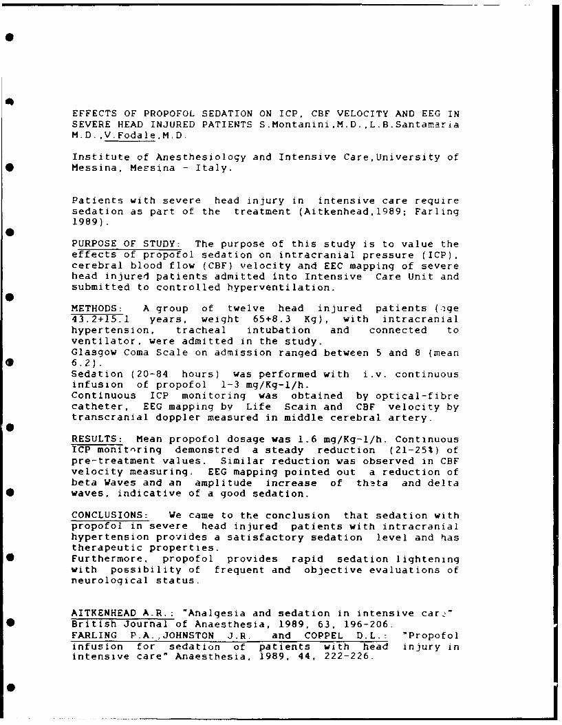
EFFECTS OF PROPOFOL SEDATION ON ICP, CBF VELOCITY AND EEG INSEVERE HEAD INJURED PATIENTS S.Montanini.M.D.,L.B.SantamariaM.D.,V.FodaleM.D.
Institute of Anesthesiology and Intensive CareUniversity ofMessina, Mersina - Italy.
Patients with severe head injury in intensive care requiresedation as part of the treatment (Aitkenhead,1989; Farling1989).
PURPOSE OF STUDY: The purpose of this study is to value theeffects of propofol sedation on intracranial pressure (ICP),cerebral blood flow (CBF) velocity and EEC mapping of severehead injured patients admitted into Intensive Care Unit andsubmitted to controlled hyperventilation.
METHODS: A group of twelve head injured patients (,ige43.2+15.1 years, weight 65+8.3 Kg), with intracranialhypertension, tracheal intubation and connected toventilator, were admitted in the study.Glasgow Coma Scale on admission ranged between 5 and 8 (mean6.2).Sedation (20-84 hours) was performed with i.v. continuousinfusion of propofol 1-3 mg/Kg-i/h.Continuous ICP monitoring was obtained by optical-fibrecatheter, EEG mapping by Life Scain and CBF velocity bytranscranial doppler measured in middle cerebral artery.
RESULTS: Mean propofol dosage was 1.6 mg/Kg-i/h. ContinuousICP monitoring demonstred a steady reduction (21-25%) ofpre-treatment values. Similar reduction was observed in CBFvelocity measuring. EEG mapping pointed out a reduction ofbeta Waves and an amplitude increase of thata and deltawaves, indicative of a good sedation.
CONCLUSIONS: We came to the conclusion that sedation withpropofol in severe head injured patients with intracranialhypertension provides a satisfactory sedation level and hastherapeutic properties.Furthermore, propofol provides rapid sedation lighteningwith possibility of frequent and objective evaluations ofneurological status.
AITKENHEAD A.R.: "Analgesia and sedation in intensive carn"British Journal of Anaesthesia, 1989, 63, 196-206.FARLING P.A.,JOHNSTON J.R. and COPPEL D.L.: "Propofolinfusion for sedation of patients with head injury inintensive care" Anaesthesia, 1989, 44, 222-226.

JEHOVAH'S WITNESS: THE TRAUMAIan DarlinG. M.D., Jagdip B. Shah, M.D., Richard L. Keenan, M.D.
Department of Anesthesiology, Medical College of Viiginia, Richmond, VA 23298, USA
Patient is a 25 year old female Jehovah's Witness presented to the Emergency Room afterbeing involved in a motor vehicle accident. Initial blood pressure in the ER was 60 mmHgand HR 140/min. She was alert, oriented, and refused blood. Oxygen by mask and fluidresuscitation with crystalloid was started through a 16 g I.V. and an 8.5 Fr RSCV cordis,the systolic BP rose to 110. Neck injury was rulec: out by x-ray. A DPL was grosslypositive and the patient was taken immediately to the OR. On the way to OR she was askedrepeatedly if she would accept blood and refused; however, she would accept "'iumansynthetic protein". She was induced in rapid sequence with cricoid pressure and oxygen,etomidate, sucinyl choline, lidoceine and intubated Standard and customary monitors wereapplied. For maintenance of anesthesia fentany!, midazolam and vecuronium were used.Five minutes after induction she went into EMD. Chest compressions were started andepinephrinc 1 mg was administered. Albumin 5% was administered aggressively and after 5minutes BP was noted in the 70's The abdomen was opened with gross blood observed. Acomplex tear of the right lobe of toe liver with almost complete fracture line was identified.At this time BP again began to drop, a second 8.5 F cordis was placed and continued colloidresuscitive measures administered. The aorta was cross clamped just below the diaphragmand the liv,,r packed off with return of BP to the 100's. Several attempts to remove theaortic cros clamp were made with resulting BP's in the 50's and 60's. Dopamine wasstarted and atropine, NaHCO3 and Ca chloride administered. ABG; PaO2 344 mmHg, PaCo2 30
mmHg, PH 7.08, Hg 1.7 gm/dl. It was decided that the liver could not be repaired withoutfatal blood loss, therefore, the abdomen was packed and closed. BP on leaving the OR wasstill in the 60's. Total fluids administered in the OR were 7750 ml, 5% albumin and3000 ml LR. The patient wec ventilated in the 7'ACU. A repeat Hgb was 0. The patientexpired after 2.5 hours in the PACU with phenylnephrine and dopamine drip. Exanguinationcaused her ueath in less than four hours following fatal injury.
Treatment usually not accepted by Jehovah's Witnesses 1) blood; 2) auto transfusion; 3)a!bumin; 4) fresh frozen plasma; 5) platelets; 6) cryoprecipitate; 7) stromal free Hgb.
Treatment acceptable to Jehovah's Witnesses 1) synthetic colloid; 2) fluosol;3) oyperbaric oxygen with or without hypothemia; 4) MAST Trousers.
Additional pharmacological acceptable modalities 1) Erythropoetion; 2) DDAVP;3) ,"tamin K; 4) amino caproic acid.
These people claim to be religious but not litiginous. They certainly pose a challenge asthey seek modern medical assistance. Meticulous surgical technique aimed at saving eachdrop of blood is probably the single most important aspect of a good outcome.
DISASTER MASS/CASUALTY/MILITARY
ANESTHESIA: PART II
SATURDAY, MAY 4, 1991
2:00 P.M. - 5:00 P.M.

BREAKAWAY SESSION
DISASTER / MASS CASUALTY / MILITARY ANESTHESIA: PART II
Project Leader: A. Dow
Moderators: K. DauphineeV. KvetanM. Moles
Description:
The session is to examine the primary and secondaryresponses to a mock, table-top disaster. The disaster is to be atrain wreck, that occurs somewhere outside of Baltimore, andinvolves a considerable number of injured people (both adults andchildren).
There will be two "teams", who will provide suggestions andideas for the management of the disaster as it unfolds on thescreen before them. Each of these teams will consist of 5 healthcare professionals, who are either playing their own, or anotherrole, from real life. the two teams are not in directcompetition with each other, but it is intended that theiranswers should complement each other.
The scenario, and the questions that accompany it, will bepresented by three team "leaders", who will take it in turn toquestion the team layers. The team leaders are all people whohave extensive experience in disaster medicine. The projectleader will control the flow of the disaster, and will beresponsible for starting and ending the proceedings at theappropriate time and will control the double slide projector thatis used to present the events of this mock disaster.
The intention of the session is to convey some of theproblems that can occur during a real disaster, and to show how aplanned approach to management can result in a better outcome forthe injured. There are no "right" or "wrong" answers that theteam players are expected to provide, merely some guidelinesalong which they may go. This should be an instructive anduseful exercise for the audience and the team players, and willprovide some suggestions for future mass casualty management.
SPECIAL CRNA SESSION: PART II
SATURDAY, MAY 4,1991
2:00 P.M. - 5:00 P.M.

IV. SPECIAL CRNA SESSION: PART II
SESSION AGENDA
2:00-2:30 P.M. ADVANCED AIRWAY INTERVENTIONS IN THETRAUMA PATIENT
John Connelly, CRNA
2:30-3:00 P.M. NEW AGENTS, NEW CHOICES FOR MANAGINGPOST ACUTE TRAUMA SURGERY PATIENTS
Charles R. Barton, CRNA
3:00-3:30 P.M. THORACIC SPINAL INJURIES OF THE TRAUMA PATIENTKathleen A. Hartman, CRNA
3:30-4:00 P.M. BREAK
4:00-4:30 P.M. FAMILY FOCUS IN PAIN MANAGEMENT OF THETRAUMA PATIENT
C. Russel Baker, CRNA
4:30-5:00 P.M. METHODS OF CAPNOGRAPHYRobert Akins, CRNA, RRT
5:00 P.M. CLOSING REMARKSCharles R. Barton, CRNA
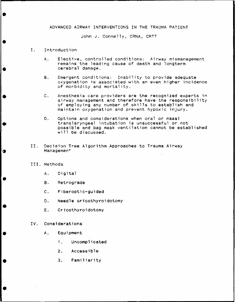
ADVANCED AIRWAY INTERVENTIONS IN THE TRAUMA PATIENT
John J. Connelly, CRNA, CRTT
I. Introduction
A. Elective, controlled conditions: Airway mismanagementremains the leading cause of death and longtermcerebral damage.
B. Emergent conditions: Inability to provide adequateoxygenation is associated with an even higher incidenceof morbidity and mortality.
C. Anesthesia care providers are the recognized experts inairway management and therefore have the responsibilityof employing any number of skills to establish andmaintain oxygenation and prevent hypoxic injury.
D. Options and considerations when oral or nasaltranslaryngeal intubation is unsuccessful or notpossible and bag mask ventilation cannot be establishedwill be discussed.
II. Decision Tree Algorithm Approaches to Trauma AirwayManagemen*
III. Methods
A. Digital
B. Retrograde
C. Fiberoptic-guided
D. Needle Gricothyroidotomy
E. Cricothyroidotomy
IV. Considerations
A. Equipment
1. Uncomplicated
2. Accessible
3. Familiarity

B. Complications:
1. Esophageal perforation
2. Subcutaneous air
3. Hemorrhage
4. Barotrauma
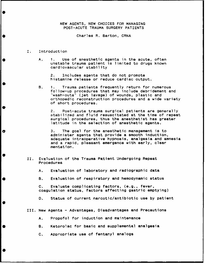
NEW AGENTS, NEW CHOICES FOR MANAGINGPOST-ACUTE TRAUMA SURGERY PATIENTS
Charles R. Barton, CRNA
I. Introduction
A. 1. Use of anesthetic agents in the acute, oftenunstable trauma patient is limited to drugs knowncardiovascular stability
2. Includes agents that do not promotehistamine release or reduce cardiac output.
B. 1. Trauma patients frequently return for numerousfollow-up procedures that may include debridement and"wash-outs" (jet lavage) of wounds, plastic andorthopedic reconstruction procedures and a wide varietyof short procedures.
2. Post-acute trauma surgical patients are generallystabilized and fluid resuscitated at the time of repeatsurgical procedures, thus the anesthetist has greaterlatitude in the selection of anesthetic agents.
3. The goal for the anesthetic management is toadminister agents that provide a smooth induction,adequate intraoperative hypnosis, analgesia and amnesiaand a rapid, pleasant emergence with early, clearmentation.
II. Evaluation of the Trauma Patient Undergoing Repeat
Procedures
A. Evaluation of laboratory and radiographic data
B. Evaluation of respiratory and hemodynamic status
C. Evaluate complicating factors, (e.g., fever,coagulation status, factors affecting gastric emptying)
D. Status of current narcotic/antibiotic use by patient
III. New Agents - Advantages, Disadvantages and Precautions
A. Propofol for induction and maintenance
B. Ketorolac for basic and supplemental analgesia
C. Appropriate use of fentanyl analogs
D. Strategies for reducing post-anesthesia nausea andvomiting
E. Use of current agents and adjuncts in conjunction withnewer agents

THORACIC SPINAL INJURIES OF THE TRAUMA PATIENT
AND PAIN MANAGEMENT
Kathleen A. Hartman, CRNA
I. Introduction
A. Historical perspective
B. Trauma philosophy
C. Current statistics
II. Mechanisms of Injuries
A. Patient profile
III. Surgical Intervention
A. Harrington rod instrumentation
B. Posterior pedicle screw instrumentation
IV. Patient Assessment
A. Hemodynamics
B. Respiratory physiology
V. Anesthetic Considerations
A. Perioperative
1. Cardiovascular considerations
2. Invasive monitoring
B. Postoperative Management
VI. Pain Management
A. Pharmacological considerations
1. Drug choice
2. Drug route
B. Regional Techniques
C. Indications
D. PECA
E. Precautions
VII. Conclusion
FAMILY FOCUS IN PAIN MANAGEMENT OF THE TRAUMA PATIENT
C. Russel Baker, CRNA
I. Introduction
A. Pain is commonly linked to injury and tissue damage.
B. Postulated that the CNS is able to exert control overthe sensory input.
1. Cutaneous sensory input activates the nociceptivesystem
2. Nociceptive input is modified by:
a. Psychological factors
b. Social factors
II. Acute Pain
A. In general there is a relationship between injury and
pain.
1. This relationship is highly variable.
2. Injury may occur without pain and pain may occurwithout injury.
B. Acute pain serves to warn of potential or acute tissuedamage.
1. Severe uncontrolled pain may augment the patient'sphysical problems
2. Severe unabated pain may also cause changes in anindividual behavior.
3. Constant pain inhibits recovery andrehabilitation.
III. Psychological Factors Affecting Acute Pain Responses
A. Pain is always a psychological event.
1. Early acute phase: sensory factors predominate.
2. Over time, persistence of the pain experience isdetermined less by the initial pain producingcause and more by the psychological variables.

3. Pain behaviors 11 be induced, maintained, andreinorced by environmental rewards.
B. Anxiety is the psychological variable most reliably
related to high levels of pain.
1. Trait anxiety
2. State anxiety
3. Trait anxiety seems to be the strongest predictor
of post-operative pain.
IV. Family Systems Theory
A. Provides a frame-work to view symptom development asreflecting disturbances within the family system.
B. Central to systems thinking is the balance betweenforces, when the balances is disrupted, systemsdevelop.
C. Counterbalancing life forces
1. individuality - more intellectual functioning
2. Togetherness - more emotional functioning
D. Fusion in a relationship can both provide a relief fromand be a source of anxiety.
1. The more intense the relationship fusion, the moreunstable the relationship and the more the chronic
anxiety generated by the relationship.
2. Relationships with a high degree of fusiongenerate many problems, little reserve is thenleft to deal with other problems.
3. When the family system's ability to adapt is
exceeded, then acute and chronic symptoms appear.
4. Anxiety can be controlled by diverting attentionto a third party.
5. The grater the anxiety and the greater the un-
differentiation, the greater the likelihood of asystem developing.
E. Better functioning families experience less chronicanxiety, have more emotional reserve, require less useof stabilizing mechanisms, and experience fewer acuteand chronic problems.
V. Family Functioning and Symptom Development
A. Acute disease processes that seem to be influenced by
general family dynamics are:
1 Asthma
Post-cardiotomy psychosis
3. Tuberculosis
4. Abdominal pain in children
5 Accide,.us
6. Chronic pain
B. Family characteristics which contritAte to the onset ofpsychophysiologic disorders:
1. Over involvement
2. Enmeshment
3. Collusion
4. Rigidity
C. Symptoms of the patient may fulfill the emotional needsoF other family members.
D. A patient's illness may stabilize the system byfocusing the family pathology and allowing improvedfunctioning in other areas.
E. Anxiety is related to family functioning. The anxietycan be displaced to a symptom, this lessens anxiety,since energy is invested in the symptom rather than theprecipitating stress.
F. Family's involvement in the development of painbehaviors:
1. Pain behaviors may be learned and other familymembers reinforce the behaviors.
2. Pain used to express a variety of negativeemotions.
3. It is through pain and suffering that caringbehavior is maintained.
G. Pain, as a behavior, is always communication; as a

communication tool the messages are indirect and maskedwith no specific referent pain may be used to express:
1. Tensions and frustrations
2. Mental suffering
3. Anger, powerlessness, martyrdom
4. To punish or be punished
5. To seek or avoid intimacy
H. Poor family functioning is not responsiL fr - poormedical condition or increased levels of p? -; but,that medical condition or pair. .,id :amily functioninginfl iences each other in a circular fashion.
METHODS OF CAPNOGRAPHy
Robert Akins, CRNA, RRT
I. Specifications
A. Concentrations ranging from 0 - 15%
B. Accuracy= +/-10% of reading or +/-3 mmHg
C. Response rapid enough to capture all phases of theventilatory cycle
II. Mass Spectrometers
A. Magnetic sector type (e.g., PPG SARA)
1. Samples are sequenced by a mechanical multiplexer.
2. Electron source ionizes gas
3. Magnetic field creates an ionic "spectrum".
4. Individual collector electrometer plates count thenumber of molecules.
• 5. Counts from all the electrometers are summed andassumed to be the total gas.
6. Only molecules that hit a collector plate will becounted in the summation.
7. Volatile anesthetic agents all have molecularweights such that they would be deflected past theend of the electrometers.
III. Infrared Absorption
A. Polyatomic molecules all have known amounts ofvibrational motion.
B. Black body infrared course, sample cuvette, and
infrared detector, are functional parts.
C Problems
1. Close proximity of the absorptive peaks of carbondioxide and nitrous oxide make detection of each
* gas difficult.
2. Drift radiation emitted from the source must be
0 !
accounted for dynamically.
3. Precision interference filters measure absorptionin narrow bands of the spectrum.
4. Infrared detectors are faster responding than massspectrometers.
5. No physical change in the structure of the
molecule occurs.
IV. Raman Spectroscopy
A. Exciting electrons in molecules to a higher energystate.
B. A particular gas will have a distinctive photonemission.
C. All gases used in anesthesia today have a know Ramanemission spectrum.