systemic phlorizin prevents gold thioglucose necrosis in the ventromedial hypothalamus
TRANSCRIPT
Brain Research Bulletin, Vol. 8, pp. 347-351, 1982. Printed in the U.S.A.
Systemic Phlorizin Prevents Gold Thioglucose Necrosis
in the Ventromedial Hypothalamus’
DANLEY F. BROWN*2 AND JOSEPH M. VILESt
*Molecular, Cellular, and Developmental Biology Program and TDepartment of Zoology Iowa State University, Ames, IA 50011
and *Division of Combat Casualty Care, Letterman Army Institute of Research Presidio of San Francisco, CA 94129
Received 22 June 1981
BROWN, D. F. AND J. M. VILES. Systemic phlorizin prevents gold thioglucose necrosis in the ventromedial hypothala- mus. BRAIN RES. BULL. 8(4) 347-351, 1982.-Intraventrictdar and intrahypothalamic infusions of phlorizin (PHL) are known to cause hyperphagia and to prevent gold thioglucose (GTG) lesion formation in the ventromedial hypothalamus (VMH), respectively. In this study, PHL, administered IP in a large dose (900 m&g), completely inhibited GTG necrosis in the VMH. PHL did not cause excessive urinary excretion of GTG. This evidence suggested that systemic PHL must be injected in a high concentration to alter the hypothalamic response to GTG. In vitro measurements of VMH glucose oxidation substantiated this idea. Only at a high concentration of PHL was glucose oxidation significantly depressed in the VMH (p<O.OOl). Small amounts of PHL elevated VMH glucose oxidation @<O.OOl). Since PHL is an inhibitor of glucose transport, these data support the concept of a hypothalamic glucostatic modulation for the control of food intake.
Phlorizin Gold thioglucose VMH glucose oxidation Glucostatic modulation of food intake
PHLORIZIN (PHL) is a well known inhibitor of glucose transport in a variety of tissues [12,16]. Infusions of small amounts of PHL into the cerebral ventricles cause hyper- phagia and body weight gain [81. However, intraperitoneal (IP) injections of PHL have no effect on eating 181. Glick and Mayer [8] suggested that PHL was affecting cerebral glucoreceptors involved with the control of food intake. Others [9,17] have suggested that the ventromedial hypo- thalamus (VMH) possesses these presumptive glucorecep- tors, since this area has been implicated as a major center for food intake control.
Gold thioglucose (GTG), when injected into mice IP, will cause an obesity syndrome [2]. This obesity development has been attributed to destruction of the VMH by GTG [ 131. Debons et al. [5] demonstrated that unilateral intrahypo- thalamic injections of PHL into the VMH could abolish GTG lesion formation on the side of the brain receiving the PHL. These investigators also showed, by autoradiography, that cells in the VMH bound tritiated PHL [51. These data sup- ported the idea that the VMH contained glucosensitive cells, which were involved with the control of food intake.
Recently, Debons and co-workers [6] have clearly
demonstrated that GTG necrosis in the VMH is highly spe- cific for the area, with neural components as the primary GTG target tissue. They have suggested that GTG could be used as a probe for hypothalamic function. In this study, we used the experimental method of Glick and Mayer [8], in- volving parenteral injections of PHL, and increased the dose of PHL. Using GTG as a probe, changes in the VMH were observed by light microscopy. In addition, the effect of PHL on VMH glucose oxidation in vitro was assessed.
METHOD
CFl female mice (Charles River Breeding Laboratories), weighing 20-25 g, were used in this study. The mice were maintained at 23”C, with a 12 hour light:12 hour dark photo- period, and given Teklad Mouse and Rat Diet and tap water freely.
PHL (900 m&g) was injected IP into mice 2 hours before GTG (300 m&g) was administered IP. Since PHL is only slightly soluble in cold water, the suspension was heated to solublize the PHL and injected. Dissolving PHL in hot water
‘The opinions or assertions contained herein are the private views of the authors and are not to be construed as official or as reflecting the views of the Department of the Army or the Department of Defense. In conducting the research described in this report the investigators adhered to the “Guide for Laboratory Animal Facilities and Care” as promulgated by the Committee on the Guide for Laboratry Animal Resources, National Academy of Sciences, National Research Council.
*D. F. Brown’s present affiliation is with the Division of Combat Casualty Care, Letterman Army Institute of Research, Presidio of San Francisco, CA 94129. Reprint requests should be sent to this address.
Copyright @ 1982 ANKHO International Inc.-0361-9230/82/040347-05$03.00/O

348 BROWN AND VILES
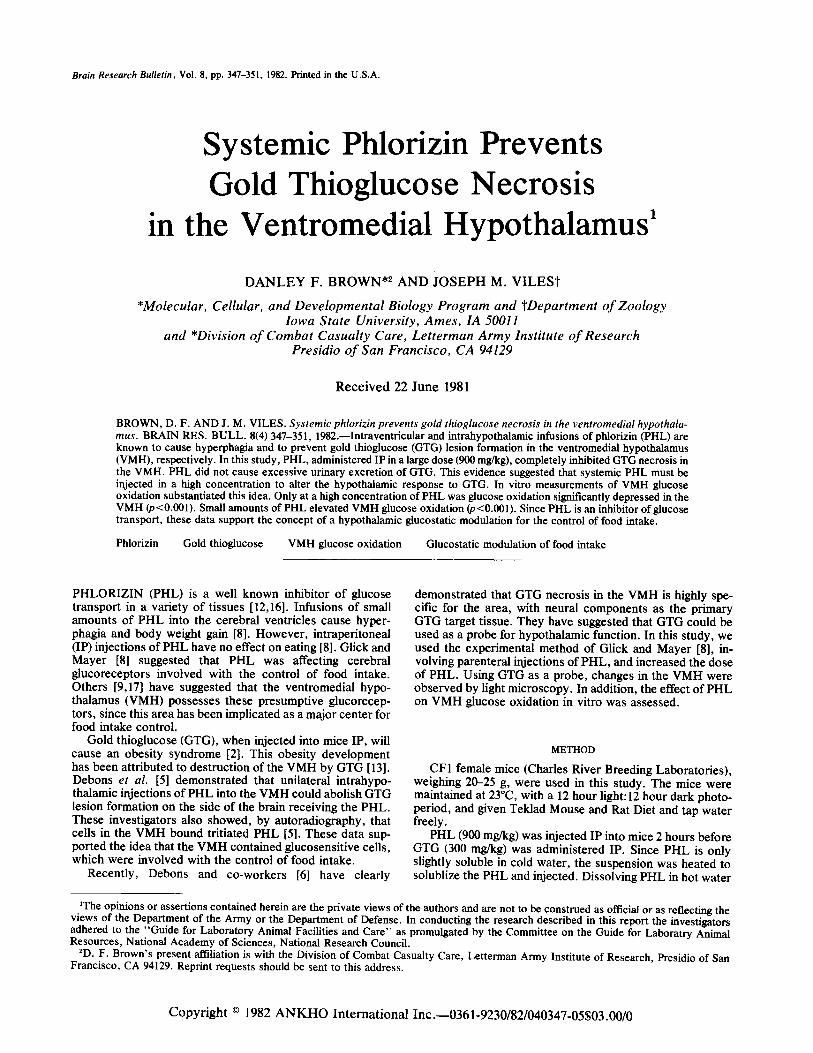
FIG. 1. Coronal sections of mouse brain, showing the VMH at the level of the median eminence, 24 hours after a GTG injection. A. Hot water given IP initially, followed by GTG (300 mgkg). B. PHL (900 mgkg) given IP initially, followed by GTG (300 mglkg). Bars represent 0.1 mm. (90x)
PHLGRIZIN AND GOLD THIOGLUCOSE NECROSIS 349
does not alter the chemical structure of the compound. Hot water injections followed by GTG served as controls.
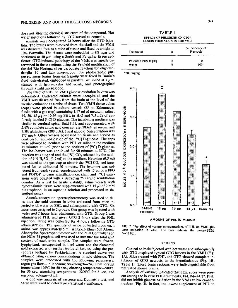
TABLE 1
Animals were decapitated 24 hours after the GTG injec- tion. The brains were removed from the skull and the VMH was dissected free as a cube of tissue and fixed overnight in 10% Formalin. The tissues were embedded in 8% agar and sectioned at 50 pm using a Smith and Farquhar tissue sec- tioner. GTG-induced pathology of the VMH was rapidly de- termined in these sections using the Penfield modification of the de1 Rio-Hortega silver carbonate reaction for oligoden- droglia [lo] and light microscopy. For photographic pur- poses, some brains from each group were fixed in Bouin’s fluid, dehydrated, em~dded in paraffin, sectioned at 7 firn, stained with hematoxylin and eosin, and photographed through a light microscope.
EFFECT OF PHLORIZIN ON GTG* LESION FORMATION IN THE VhfH
Treatment % Incidence of
n Necrosis
Phlorizin (900 m&g) Water
5 0 9 100
*300 mgkg.
The effect of PHL on VMH glucose oxidation in vitro was determined. Untreated animals were decapitated and the VMH was dissected free from the brain at the level of the median eminence as a cube of tissue. Two VMH tissue cubes (caps) were placed in culture vessels (25 ml Erlenmeyer flasks with a gas trap) containing 1.47 ml of medium, saline, 15, 30, 45 fig or 10.66 mg PHL in Hz0 and 7.5 PCi of uni- formly labeled [‘“Cl D-glucose. The incubating medium was similar to cerebral spinal fluid [ll], and supplemented with 2.6% complete amino acid concentrate, 28.6% rat serum, and 1.3% glutathione (200 mM). Final glucose concentration was 172 mg%. Other vessels possessed no tissue and served as controls for auto-oxidation of the [14C1 D-glucose. The caps were allowed to incubate with PHL or saline in the medium 15 minutes at 37°C prior to the addition of [14C] D-glucose. The incubation was continued for 90 minutes at 37°C. The reaction was stopped and the [‘“Cl CO, released by the addi- tion of 9 N HzS04 (0.2 ml) to the medium. Hyamine (0.5 ml) was added to the gas trap to absorb the [‘*Cl CO, and incu- bated for an additional 60 minutes. The hyamine was col- lected from each vessel, supplemented with 1.5 ml of a PPO and POPOP toluene scintillation cocktail, and [14C] emis- sions were counted with a Beckman 250 liquid scintillation counter. As a test for tissue viability, saline vessels with hy~th~amic tissue were supplements with 15 JJ.I of 2 mM dinitrophenol in an aqueous solution and processed as de- scribed above.
Atomic absorption spectrophotometery was used to de- termine the gold content in urine collected from mice in- jected with water or PHL and subsequently with GTG. Six mice were assigned to 2 groups. One group was injected with water and 2 hours later challenged with GTG. Group 2 was administered PHL and given GTG 2 hours after the PHL injection. Urine was collected for 4 hours following GTG administration. The quantity of urine collected from each animal was approximately 5 ml. A Perkin-Elmer 503 Atomic Absorption Spectrophotometer with the 2100 Controller and the HGA-74 graphite cell was used to measure the total gold content of each urine sample. The samples were frozen, lyophylized, resuspended in 1 ml water and the elemental gold extracted with methyl iso-butyl ketone, using the pro- cedures outlined by Perkin-Elmer. A standard curve was obtained using various concentrations of gold chloride. The samples were processed with the following parameters: argon gas flow-15.0 cclmin; wavelength-243.3 nm; drying tern~~tu~ 120°C for 50 sec., charring tern~ratu~~c for 50 set, atomizing temperature-2200°C for 5 set, and injection volume-5 ~1.
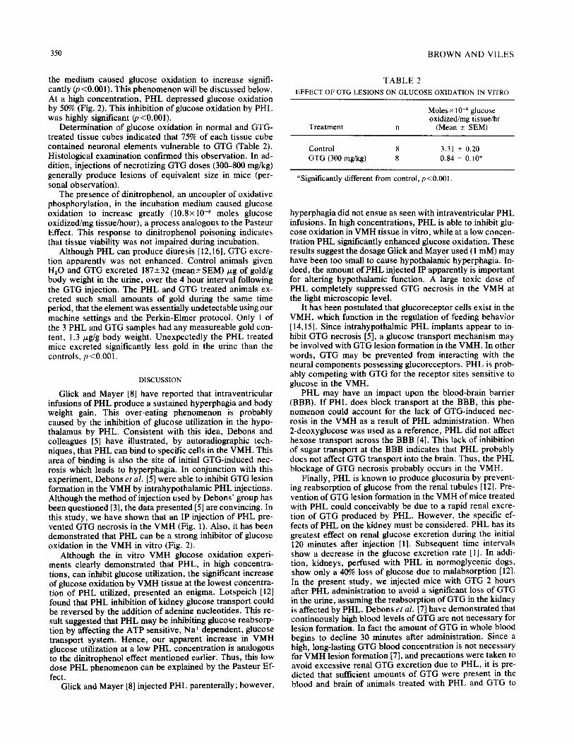
SALINE 15 PS CONTROL
AMOUNT OF PHL IN MEDIUM
FIG. 2. The effect of various concentrations of PHL on VMH &I- case oxidation in vitro. The bars indicate the mean_eSEM. *p<0.001.
RESULTS
Control animals injected with hot water and subsequently with GTG displayed typical GTG lesions in the VMH (Fig. 1A). Mice treated with PHL and GTG showed complete in- hibition of GTG necrosis in the hypothalamus (Fig. 1B; Table 1). These brain sections were ~distin~ishable from untreated mouse brains.
A one way analysis of variance and Dunnett’s test, and t-test were used to determine statistical significance.
Analysis of variance indicated that differences were pres- ent among the in vitro PHL treatments, F(4,16)= 14.27. PHL did not inhibit glucose oxidation in the VMH at low concen- trations (Fig. 2). In fact, the lowest supplement of PHL to
350 BROWN AND VILES
the medium caused glucose oxidation to increase signiti- cantly @ <O.OOl). This phenomenon will be discussed below. At a high concentration, PHL depressed glucose oxidation by 50% (Fig. 2). This inhibition of glucose oxidation by PHL was highly significant @<O.OOl).
Determination of glucose oxidation in normal and GTG- treated tissue cubes indicated that 75% of each tissue cube contained neuronal elements vulnerable to GTG (Table 2). Histological examination confirmed this observation. In ad- dition, injections of necrotizing GTG doses (300-800 mg/kg) generally produce lesions of equivalent size in mice (per- sonal observation).
The presence of dinitrophenol, an uncoupler of oxidative phosphorylation, in the incubation medium caused glucose oxidation to increase greatly (10.8~ 1Oms moles glucose oxidized/mg tissue/hour), a process analogous to the Pasteur Effect. This response to dinitrophenol poisoning indicates that tissue viability was not impaired during incubation.
Although PHL can produce diuresis [12,16], GTG excre- tion apparentlv was not enhanced. Control animals given H,O and GTG excreted 187+32 (meantSEM) pg of gold/g body weight in the urine, over the 4 hour interval following the GTG injection. The PHL and GTG treated animals ex- creted such small amounts of gold during the same time period, that the element was essentially undetectable using our machine settings and the Perkin-Elmer protocol. Only 1 of the 3 PHL and GTG samples had any measureable gold con- tent, 1.3 &g body weight. Unexpectedly the PHL treated mice excreted significantly less gold in the urine than the controls, p<O.OOl.
DISCUSSION
Glick and Mayer [8] have reported that intraventricular infusions of PHL produce a sustained hyperphagia and body weight gain. This over-eating phenomenon is probably caused by the inhibition of glucose utilization in the hypo- thalamus by PHL. Consistent with this idea, Debons and colleagues [5] have illustrated, by autoradiographic tech- niques, that PHL can bind to specific cells in the VMH. This area of binding is also the site of initial GTG-induced nec- rosis which leads to hyperphagia. In conjunction with this experiment, Debons et al. [5] were able to inhibit GTG lesion formation in the VMH by intrahypothalamic PHL injections. Although the method of injection used by Debons’ group has been questioned [3], the data presented [5] are convincing. In this study, we have shown that an IP injection of PHL pre- vented GTG necrosis in the VMH (Fig. 1). Also, it has been demonstrated that PHL can be a strong inhibitor of glucose oxidation in the VMH in vitro (Fig. 2).
Although the in vitro VMH glucose oxidation experi- ments clearly demonstrated that PHL, in high concentra- tions, can inhibit glucose utilization, the significant increase of glucose oxidation by VMH tissue at the lowest concentra- tion of PHL utilized, presented an enigma. Lotspeich [12] found that PHL inhibition of kidney glucose transport could be reversed by the addition of adenine nucleotides. This re- sult suggested that PHL may be inhibiting glucose reabsorp- tion by affecting the ATP sensitive, Na+ dependent, glucose transport system. Hence, our apparent increase in VMH glucose utilization at a low PHL concentration is analogous to the dinitrophenol effect mentioned earlier. Thus, this low dose PHL phenomenon can be explained by the Pasteur Ef- feet .
Glick and Mayer [8] injected PHL parenterally; however,
TABLE 2 EFFECT OF GTG LESIONS ON GLUCOSE OXIDATION IN VITRO
Molesx 10.” glucose oxidizedimg tissueihr
Treatment n (Mean t SEM)
Control 8 3.31 t 0.20 GTG (300 m&g) 8 0.84 -t O.lO*
*Significantly different from control, p<O.OOl
hyperphagia did not ensue as seen with intraventricular PHL infusions. In high concentrations, PHL is able to inhibit glu- cose oxidation in VMH tissue in vitro, while at a low concen- tration PHL significantly enhanced glucose oxidation. These results suggest the dosage Glick and Mayer used (1 mM) may have been too small to cause hypothalamic hyperphagia. In- deed, the amount of PHL injected IP apparently is important for altering hypothalamic function. A large toxic dose of PHL completely suppressed GTG necrosis in the VMH at the light microscopic level.
It has been postulated that glucoreceptor cells exist in the VMH, which function in the regulation of feeding behavior [14,15]. Since intrahypothalmic PHL implants appear to in- hibit GTG necrosis [5], a glucose transport mechanism may be involved with GTG lesion formation in the VMH. In other words, GTG may be prevented from interacting with the neural components possessing glucoreceptors. PHL is prob- ably competing with GTG for the receptor sites sensitive to glucose in the VMH.
PHL may have an impact upon the blood-brain barrier (BBB). If PHL does block transport at the BBB, this phe- nomenon could account for the lack of GTG-induced nec- rosis in the VMH as a result of PHL administration. When 2-deoxyglucose was used as a reference, PHL did not affect hexose transport across the BBB [4]. This lack of inhibition of sugar transport at the BBB indicates that PHL probably does not affect GTG transport into the brain. Thus, the PHL blockage of GTG necrosis probably occurs in the VMH.
Finally, PHL is known to produce glucosuria by prevent- ing reabsorption of glucose from the renal tubules [ 121. Pre- vention of GTG lesion formation in the VMH of mice treated with PHL could conceivably be due to a rapid renal excre- tion of GTG produced by PHL. However, the specific ef- fects of PHL on the kidney must be considered. PHL has its greatest effect on renal glucose excretion during the initial 120 minutes after injection [ 11. Subsequent time intervals show a decrease in the glucose excretion rate [l]. In addi- tion, kidneys, perfused with PHL in normoglycenic dogs, show only a 40% loss of glucose due to malabsorption [l2]. In the present study, we injected mice with GTG 2 hours after PHL administration to avoid a significant loss of GTG in the urine, assuming the reabsorption of GTG in the kidney is affected by PHL. Debons et al. [7] have demonstrated that continuously high blood levels of GTG are not necessary for lesion formation. In fact the amount of GTG in whole blood begins to decline 30 minutes after administration. Since a high, long-lasting GTG blood concentration is not necessary for VMH lesion formation [7], and precautions were taken to avoid excessive renal GTG excretion due to PHL, it is pre- dicted that sufficient amounts of GTG were present in the blood and brain of animals treated with PHL and GTG to
PHLORIZIN AND GOLD THIOGLUCOSE NECROSIS 351
cause VMH necrosis. In an effort to demonstrate definitely that PHL did not cause excessive excretion of GTG in the urine, and thus prevent VMH lesion formation, we measured the total amount of gold in a 4 hour post GTG urine sample, from mice treated with and without PHL. Mice injected with water and GTG excreted s~n~c~tly larger amounts of gold in the urine, compared to PHL and GTG treated mice (p<O.OOl; see Results). This result clearly demonstrates that PHL did not abolish GTG lesion formation in the VMH by increasing the excretion rate of GTG over controls. Fur- thermore, since no VMH lesions developed in PHL treated, GTG challenged mice, PHL apparently affected the VMH directly, thereby preventing any GTG induced damage from occurring in the area.
The evidence reported in this study indicate the PHL dosage used by Glick and Mayer [8] was in fact too small to affect food intake, when given IP. The large IP PHL injec- tion used in this investigation inhibited GTG lesion formation in the VMH. Large doses of PHL also inhibited VMH glu- cose oxidation in vitro. These results substantiate the possi-
ble involvement of a glucoreceptor and/or glucose transport mechanism, in relation to VMH function determined by GTG vulnerability. Moreover, our data support the concept of a glucostatic modulation of feeding behavior [14].
The authors wish to express their deep appreciation to Dr. I. Tabachnick and Dr. E. A. Peets of Schering Corporation, Bloom- field, NJ for supplying us with generous amounts of gold thioglu- case. The technical assistance of Joseph C. White, Judy Brenke and John Tucker, Letterman Army Medical Center, Presidio of San Francisco, CA 94129, for the ator& absorption s~c~photomet~c analysis of gold is recognized. We also thank Susan Davis and Linda Pukajlo for typing the manuscript and Virginia Gildengorin for as- sistance with the statistical analysis. This work was supported by PHS/NIH Grant No. 5 SO5 RRO7034, in part by Letterman Army Institute of Research, Presidio of San Francisco, CA 94129, in part by NIMH Grant No, R03 MH32916, in part by a grant from the Graduate College, Iowa State University, and in part by a gift from Houston Endowment, Inc.
REFERENCES
1. Braun, W., V. P. Whittaker and W. D. Lotspeich. Renal excre- tion of phlorizin and phlorizin glucuronide. Am. J. Physioi. i90: X3-569, 1957.
2. Brecher, G. and S. H. Waxler. Obesity in albino mice due to a single injection of gold thioglucose. Proc. Sot. exp. Biol. Med. 70: 498-501, 1949.
3. Brown, D. F. and J. M. Viles. Inhibition of gold thioglucose lesions by in~y~~~~c saline iniections. Phvsioi. Behav. 25: 179-i81, 1980, Deane, R. and M. B. Segal. The effect of phloridzin, phloretin, and theophylline on the transport of sugars by the choroid plexus. Am. .I. Physioi. 287: 3JP-36P, 1979. Debons, A. F., I. Krimsky, A. Fromand H. Pattinian. Phlorizin inhibition of hypothalamic necrosis induced by gold thioglu- case. Am. J. Physioi. 226: 5X-578, 1974. Debons, A. F., I. Krimsky, A. From, E. Sic&i, M. L. Maayan, K. Fani and F. A. Jimenez. Action of gold thioglucose on pericapillary structures in the ventromedial hypothalamus. J. Path. 129: 73-81, 1979.
7. Debons, A. F., I. Krimsky, H. J. Likuski, A. From and R. J. Cloutier. Gold thioglucose damage to the satiety center: inhibi- tion in diabetes. Am. J. Physioi. 214: 652-658, 1968.
8. Glick, Z. and J. Mayer. Hyperphagia caused by cerebral ven- tricular infusions of phloridzin. Nurrrre 219: 1374, 1%8.
9. Grossman, S. P. The neur~atomy of eating and drinking be- havior. In: Neuroendocrinoiogy, edited by D. T. Krieger and J. C. Hughes. Sunderland, MA: Sinauer Associates, 1980, pp. 131-140.
10. Humason, G. L. Animal Tissue Techniques. San Francisco, CA: W. H. Freeman and Co.. 1972.
11. Jones, M. T., E. Hillhouse and 3. Burden. Secretion of cortico- ttopin releasing hormone in vitro. In: Frontiers in Neuroendo- crinoiogy, vol. 4, edited by L. Martini and W. F. Ganong. New York: Raven Press, 1975, pp. 195-226.
12. Lotspeich, W. D. Phlorizin and the cellular transport of glucose. Harvey Lect. 56: 63-91, l%l,
13. Marshall, N. B., R. J. Barmett and J. Mayer. Hypothalamic lesions in gold thioglucose-injected mice. Proc. SW. exp. Bioi. Med. M 240-244, 1955.
14. Mayer, J. Regulation of energy intake and the body weight: The glucostatic theory and the lipostatic hypothesis. Ann. N.Y. Acad. Sci. 63: U-43, 1955.
15. Mayer, J. The ventromedial glucostatic mechanism as a compo- nent of satiety, Postgrad. Med. 38: AlOl-AlO9, 1%5.
16. Silverman, M. Glucose reabsorption in the kidney. Can. J. Physioi. Pharmac. 59: 209-224, 1981.
I?. Stevenson, J. A. F. Neural control of food and water intake. In: The Hypothaiam~, edited by W. Haymaker, E. Anderson and W. J. H. Nauta. Springfield, IL: C. C. Thomas, 1%9, pp. 524- 621.