systemic mastocytosis with smoldering multiple myeloma ... · systemic mastocytosis with smoldering...
TRANSCRIPT

Case ReportSystemic Mastocytosis with Smoldering Multiple Myeloma:Report of a Case
Sassine Ghanem,1 Gwenalyn Garcia,2 Liu Ying,3 Matthew Hurford,3 and Marcel Odaimi2
1Department of Medicine, Staten Island University Hospital, Northwell Health, 475 Seaview Avenue, Staten Island, NY 10305, USA2Department of Hematology/Oncology, Staten Island University Hospital, Northwell Health, 475 Seaview Avenue, Staten Island,NY 10305, USA3Department of Pathology, Staten Island University Hospital, Northwell Health, 475 Seaview Avenue, Staten Island, NY 10305, USA
Correspondence should be addressed to Sassine Ghanem; [email protected]
Received 10 March 2016; Accepted 4 May 2016
Academic Editor: Junya Kuroda
Copyright © 2016 Sassine Ghanem et al.This is an open access article distributed under theCreativeCommonsAttributionLicense,which permits unrestricted use, distribution, and reproduction in any medium, provided the original work is properly cited.
Systemic mastocytosis (SM) is a disease characterized by a clonal infiltration of mast cells affecting various tissues of the body.It is grouped into six different subtypes according to the World Health Organization classification. It is called indolent systemicmastocytosis (ISM) when there is no evidence of end organ dysfunction, while the presence of end organ dysfunction definesaggressive systemic mastocytosis (ASM). When SM coexists with a clonal hematological disorder, it is classified as systemicmastocytosis with associated clonal hematological nonmast cell lineage disease (SM-AHNMD). Over 80% of SM-AHNMD casesinvolve disorders of the myeloid cell lines. To our knowledge, there are only 8 reported cases to date of SM associated witha plasma cell disorder. We report a patient with ISM who was found to have concomitant smoldering multiple myeloma. Hisdisease later progressed to ASM. We discuss this rare association between SM and a plasma cell disorder, and potential commonpathophysiologic mechanisms linking the two disorders will be reviewed. We also discuss prognostic factors in SM as well as themanagement options considered during the evolution of the patient’s disease.
1. Introduction
Mastocytosis is a disease characterized by the abnormalinfiltration of clonally derived mast cells in different tissuesof the body. When the infiltration is limited to the skin, adiagnosis of cutaneousmastocytosis is made. Extracutaneousinfiltration of clonal mast cells defines systemic mastocytosis(SM). It typically involves the bonemarrow but can also affectany other organ [1].
The classification for systemic mastocytosis was estab-lished by Valent et al. [1] in a consensus proposal and lateradopted in theWorldHealthOrganization 2008 classification[2]. It recognizes six different groups of SM: indolent systemicmastocytosis (ISM), systemic mastocytosis with associatedclonal hematological nonmast cell lineage disease (SM-AHNMD), aggressive systemicmastocytosis (ASM),mast cellleukemia, mast cell sarcoma, and extracutaneous mastocy-toma. The difference between indolent and aggressive SMdepends on the presence of C findings, which indicate organ
dysfunction secondary to excessive mast cell infiltration.C findings include gastrointestinal malabsorption, hyper-splenism, hepatic dysfunction, cytopenias, and pathologicfractures [1, 2].
In a retrospective study of 66 patients with SM, Traviset al. found SM-AHNMD to be the second most commonsubtype (after ISM), with 22 patients affected. Over 80% ofSM-AHNMD cases involved disorders of the myeloid celllines [3].
To our knowledge, there are only 8 reported cases to dateof SM associated with a plasma cell disorder (Table 1) [4–11].We present a patient with ISM with concomitant smolderingmultiple myeloma whose disease later progressed to ASM.
2. Case Presentation
A 59-year-old male with a known history of ISM incidentallyfound on pathologic review after surgery for osteoarthritiswas referred to the hematology clinic for evaluation of
Hindawi Publishing CorporationCase Reports in Oncological MedicineVolume 2016, Article ID 3161768, 6 pageshttp://dx.doi.org/10.1155/2016/3161768

2 Case Reports in Oncological Medicine
Table 1: Previously reported cases of SM with an associated plasma cell disorder.
Reference Age/sex DiagnosisSotlar et al. [4] 70/M SM and multiple myeloma with secondary amyloidosisDu et al. [5] 57/F SM, chronic lymphocytic leukemia, and multiple myelomaJain et al. [6] 64/F ASM and refractory multiple myelomaFilanovsky et al. [7] 76/M ISM and smoldering multiple myelomaMotwani et al. [8] 71/M SM and multiple myelomaStellmacher et al. [9] 51/M SM and multiple myelomaHagen et al. [10] 48/F SM and multiple myelomaPullarkat et al. [11] 84/M SM and monoclonal gammopathy of undetermined significanceSM: systemic mastocytosis; ISM: indolent systemic mastocytosis; ASM: aggressive systemic mastocytosis.
Table 2: Laboratory test results upon initial presentation.
Lab test Result UnitWhite blood cells 5.3 ×10³/mm3
Hemoglobin 14.5 g/dLPlatelet 202 ×103/mm3
Blood urea nitrogen 26 mg/dLCreatinine 1.31 mg/dLAlbumin 3.1 g/dLGlobulin 6.2 g/dLCalcium 8.8 mg/dLSerum proteinelectrophoresis (SPEP) andserum immunofixation
IgG kappa monoclonalprotein
M-spike 3.68 g/dL
suspectedmonoclonal gammopathy.His pastmedical historywas significant for splenectomy. The exact indication for theprocedure was unclear, but it was possibly done for involve-ment with lymphoma versus mastocytosis. He presentedwith suspected monoclonal gammopathy on previously doneblood tests with the results shown in Table 2. His physicalexam revealed no abnormalities.
Repeat serum protein electrophoresis and immunofixa-tion again revealed an IgG kappa monoclonal protein. TheM-spike was 3.8 g/dL. The serum IgG level was elevatedat 4430mg/dL, while IgA and IgM levels were within thenormal range. Serum free kappa light chains were elevated at203.1mg/L with normal free lambda light chains of 9.4mg/Land a free kappa/lambda ratio of 21.61. LDH was normal at145 IU/L and beta-2 microglobulin was elevated at 3.8mg/L.The tryptase level was elevated at 76 ng/mL.
Bone marrow aspiration and biopsy was performed,which showed an overall cellularity of 60–90%. Approxi-mately 50% of the marrow was comprised of nodules ofsmall mononuclear spindle cells (Figure 1(a)). Immunostainswere positive for CD68, CD45, CD117, CD30, CD25, and M-tryptase but negative for CD56, consistent with a mast cellphenotype.
Plasma cells occupied at least 30% of the remainingmarrow (Figure 1(b)).They were CD138 positive by immuno-histochemistry (Figure 1(c)) and kappa restricted by in situhybridization. Flow cytometry of the bone marrow aspirate
detected a monoclonal IgG kappa plasma cell populationcomprising 4.7% of total cells in the sample. Cytogeneticstesting revealed a normal male karyotype negative for muta-tions associated with multiple myeloma. Amyloid stainingwas negative.
Based on the above-mentioned morphology andimmunohistochemical and molecular studies, in addition tothe fact that the patient had no anemia, a normal calciumlevel, and no bone pain, a final diagnosis of ISM associatedwith smoldering multiple myeloma was established. Thepatient was followed up without specific treatment for eithercondition. An abdominal fat pad biopsy was done to ruleout secondary amyloidosis as the cause of his chronic kidneydisease, which came back negative.
Two years later, the patient presented to the emergencydepartment with a 2-day history of generalized weakness,shortness of breath, and melena. A nasogastric lavage wasdone and showed bloody fluid. CBC on admission showedhemoglobin of 5.9 g/dL;WBC and platelet counts werewithinnormal limits. The patient’s coagulation parameters revealeda prolonged PT of>40 secwith an INRof>15 and a prolongedPTT of 65.6 sec. Of note, the patient was on anticoagulationwith warfarin for atrial fibrillation. His transaminases andtotal bilirubin levels were within normal limits.
The patient received packed red cell and fresh frozenplasma transfusions. Emergent upper endoscopy showedgrade IV gastroesophageal varices requiring band ligation.Severe hypertensive portal gastropathy was also found.
The patient had no known history of or obvious riskfactors for liver cirrhosis; hence, workup was initiated forpossible causes of portal hypertension. Ultrasound of theabdomen showed a normal sized liver with smooth contourand normal echogenicity. The portal and hepatic veins wereboth patent. There was small volume abdominal ascites.Screening for viral hepatitis was negative. Serum ceruloplas-min, alpha-1 antitrypsin, and ferritin levels were within nor-mal limits. Testing for c-ANCA, p-ANCA, antimitochondrial,and antismooth muscle antibodies was negative.
Subsequently, liver biopsy was performed, which showedportal and periportal fibrosis with mild inflammation, com-pression of the portal veins, broad fibrous septa, and severeperisinusoidal fibrosis, confirmed by trichrome and reticulinstains. There was focal nodule formation. CD117 immunos-taining demonstrated abundant mast cells in portal tracts.Rare cells were positive for CD25 and mast cell tryptase (Fig-ure 2). Serum tryptase level was 104 ng/mL. These findings
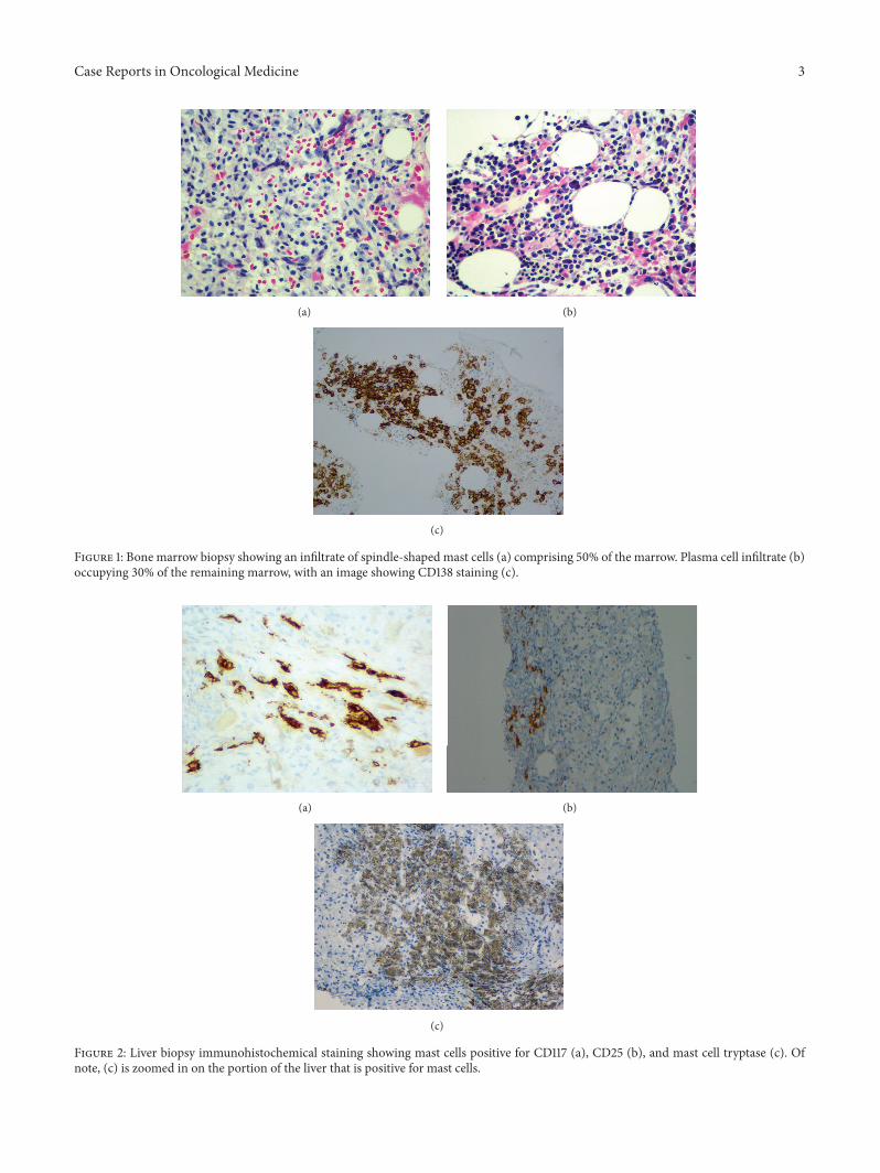
Case Reports in Oncological Medicine 3
(a) (b)
(c)
Figure 1: Bone marrow biopsy showing an infiltrate of spindle-shaped mast cells (a) comprising 50% of the marrow. Plasma cell infiltrate (b)occupying 30% of the remaining marrow, with an image showing CD138 staining (c).
(a) (b)
(c)
Figure 2: Liver biopsy immunohistochemical staining showing mast cells positive for CD117 (a), CD25 (b), and mast cell tryptase (c). Ofnote, (c) is zoomed in on the portion of the liver that is positive for mast cells.

4 Case Reports in Oncological Medicine
MWM
POS
NEG
NT
15182401998
15182401998
KIT-F_782MUT
Figure 3: Sequence analysis of the KIT D816V mutation.
were consistent with mastocytosis involving the liver, result-ing in extensive fibrosis. The patient’s disease had progressedto ASM, and a course of treatment with cladribine asoutpatient was recommended.
Subsequently, the patient followed up at a differentinstitution where further evaluation showed a negative JAK2 mutation and a positive KIT D816V mutation (Figure 3). Akidney biopsy was also performed to rule out secondary amy-loidosis or multiple myeloma as the etiology of his chronickidney disease; this showed interstitial fibrosis and tubularatrophy as well as features of thrombotic microangiopathywithout any amyloid or immune-type deposits. The treatingteam however linked the chronic kidney disease to multiplemyeloma and therefore started himon a treatment regimen ofcyclophosphamide, bortezomib, and dexamethasone as wellas hydroxyurea for the systemic mastocytosis. The patienttolerated therapy and only has complaints of fatigue in thedays that follow the chemotherapy session.
3. Discussion
Criteria for the diagnosis of SM include 1 major criterionand 4 minor criteria. The major criterion is a multifocaldense infiltrate of mast cells (≥15 mast cells in aggregates)in sections from bonemarrow and/or extracutaneous organs.The 4 minor criteria are as follows: (1) >25% of the mast cellsin the infiltrate with spindle-shaped or atypical morphologyor >25% of all mast cells in bone marrow aspirate smearscharacterized as immature or atypical, (2) presence of anactivating point mutation at codon 816 of KIT, (3) expressionof CD2 and/or CD25 in mast cells, and (4) total serumtryptase levels persistently exceeding 20 ng/mL. The first 3minor criteria apply to samples obtained from blood, bonemarrow, or other extracutaneous organs. The last criterionis not valid if there is an associated clonal myeloid disorder.The presence of the major criterion and 1 minor criterion orthe presence of 3 minor criteria is required to establish thediagnosis of SM [2].
On evaluation in our clinic, the patient had a tryptaselevel of 76 ng/mL and a bonemarrow biopsy showing CD25+spindle-shaped mast cells occupying approximately 50% ofthe bone marrow, fulfilling the diagnostic criteria for SM.His bone marrow biopsy also showed CD138+ plasma cellsoccupying 30% of the remaining bone marrow. With thisbone marrow finding along with a serumM-spike of 3.8 g/dLand the absence of end organ damage, the patient wasdiagnosed with concomitant smoldering multiple myeloma.
The coexistence of SM with multiple myeloma placesour patient in the SM-AHNMD subtype. While associ-ated myeloid/myelomonocytic neoplasia in SM-AHNMDaccounts for 82%–89% of SM-AHNMD, associated lympho-proliferative disorders are rare [3, 4]. In their study of 138cases of SM-AHNMD, Pardanani et al. found 7 patients(5.1%) with lymphoma, 5 patients (3.6%) with myeloma, and2 patients (1.5%) with chronic lymphocytic leukemia (CLL)[12].
Since multiple myeloma occurring in an elderly patientis not uncommon, one could argue that the cooccurrenceof SM with smoldering multiple myeloma was a coinci-dence. However, in vitro studies have shown the capacity ofneoplastic mast cells to induce the growth of lymphocyticneoplasms. Tournilhac et al. demonstrated that the humanmast cell line HMC-1 stimulated proliferation of the malig-nant lymphoplasmacytic cells of patients withWaldenstrom’smacroglobulinemia through interactions between CD154 onthe mast cells and CD40 on the lymphoplasmacytic cells [13].In vitro studies also suggest a role of mast cells in the growthof Hodgkin lymphoma via their expression of CD30 ligand[14]. A similar relationship may exist between mast cells andplasma cells as well. Mast cells secrete multiple cytokinesincluding IL-6 and stem cell factor, both of which have beenshown to induce plasma cell proliferation [15–17].
Whether or not the neoplastic mast cells of SM andthe associated hematologic malignancy are derived from thesame clone has been examined. In 2 cases of SM associatedwith lymphoproliferative disorders, the D816V mutation wasdetected in the neoplastic mast cells but not in the malignantlymphocytes, suggesting that the SM and the coexisting lym-phoid malignancy are clonally distinct [18, 19]. In contrast, incases of acute myeloid leukemia, the leukemic translocationt(8;21) can be detected in the malignant myeloid cells as wellas in the neoplastic mast cells. Therefore, they are presumedto have developed from the same clone [20, 21].
When confrontedwith cases of SM-AHNMD, the currentstrategy is to treat each entity on its own as if the otherentity did not coexist in the patient [22–24].With anM-spikegreater than 3 g/dL but no myeloma-defining event such asanemia, hypercalcemia, bony disease, or renal insufficiencysecondary to myeloma, our patient fit into the diagnosisof smoldering multiple myeloma, and management of hisdisease was close clinical monitoring for transformation tosymptomatic multiple myeloma.
In SM, the difference between indolent and aggressivedisease depends on the presence of C findings, which indicateorgan dysfunction related to mast cell invasion. The patient’sleft hip replacement was performed for degenerative jointdisease and no pathological fracture had occurred.Therefore,

Case Reports in Oncological Medicine 5
the finding of mast cells in the surgical specimen wasincidental. In addition, the pathological finding of mast cellson the bone marrow biopsy was also an incidental findingduring workup of the patient’s hypergammaglobulinemia.Symptoms associated with mast cell degranulation, such asurticaria, flushing, or diarrhea, were also not present in thepatient’s history. The patient was therefore considered ashaving ISM.
The clinical presentation of patients with SM is highlyvariable. Thus, decisions regarding whether or not to initiatetreatment are often complex. Patients with ISM rarely exhibitleukemic transformation and their life expectancy is notsignificantly different from the age- and sex-matched USpopulation, as demonstrated in a retrospective study by Limet al. on 342 patients with SM [25]. Thus, the treatmentof ISM is mainly symptom-oriented [26]. Lim et al. alsoidentified advanced age (defined as ≥65 years), weight loss,anemia, thrombocytopenia, hypoalbuminemia, and excessbone marrow blasts as independent adverse prognosticfactors for survival [25]. Similarly, a retrospective studydone by Butterfield and Weiler on 42 elderly patients withSM reported poor survival outcomes in patients with con-comitant thrombocytopenia, leukemias, andmyelodysplasticsyndrome [27]. As our patient was asymptomatic with nopoor prognostic factors, he was not treated for ISM on hisinitial presentation.
With the patient developing esophageal variceal bleedfrom portal hypertension due to a mast cell infiltrated liver,he was reclassified as having ASM, in which the primarygoal of therapy is reducing the mast cell burden. Treat-ment options that have been studied in nonrandomizedtrials include hydroxyurea, interferon-alfa with or withoutprednisone, imatinib, and cladribine. A retrospective studyby Lim et al. on 108 patients with SM favored interferon-alfa and cladribine as first line therapy, as hydroxyurea andimatinib were associated with lower overall response rates[28]. Imatinib is relatively ineffective in patients with SMwhohave amutation ofKITD816V [29].This patient thereforewasa poor candidate for imatinib, and the lack of randomizedcontrolled trials with evidence favoring one treatment overthe others justified the use of hydroxyurea in this patient.A global phase II trial of the use of midostaurin, an oralmultikinase inhibitor of both wild-type and D816-mutatedKIT, is currently underwaywith primary results showing highoverall response rates with reduction in mast cell burden, afavorable safety profile, as well as improvement in symptomsand quality of life [30]. Seeing this as the largest prospectivetrial in advanced systemic mastocytosis, midostaurin maybecome the future standard of care.
In summary, we report the case of a 59-year-old manwho initially presented to our clinic with ISM associatedwith smoldering multiple myeloma. Using the 2008 WHOclassification, he was diagnosed with SM-AHNMD. For bothdiseases, the clinical approach was close follow up until 2years later when the patient progressed to ASM as evidencedby esophageal variceal bleed secondary to portal hyperten-sion from mast cell infiltration of the liver. The patientwas then offered cytoreductive therapy. The association ofplasma cell disorders with systemic mastocytosis is rare
and the neoplastic cells are believed to be derived fromdistinct clones. Some postulate that cytokines secreted by theneoplastic mast cells induce the proliferation of plasma cells.The treatment for each disease is planned as if the other didnot coexist in the same patient.
Consent
A written and signed consent to publish the information wasobtained from the patient prior to submission of the paper.
Competing Interests
The authors declare that there are no competing interestsregarding the publication of this paper.
References
[1] P. Valent, H.-P. Horny, L. Escribano et al., “Diagnostic crite-ria and classification of mastocytosis: a consensus proposal,”Leukemia Research, vol. 25, no. 7, pp. 603–625, 2001.
[2] H. P. Horny, J. M. Bennett, B. J. Bain et al., “Mastocytosis,” inWHO Classification of Tumours of Haematopoietic and Lym-phoid Tissues, S. H. Swerdlow, S. H. Camp, E. Harris et al., Eds.,pp. 54–63, International Agency for Research on Cancer, Lyon,France, 4th edition, 2008.
[3] W. D. Travis, C.-Y. Li, L. T. Yam, E. J. Bergstralh, and R. G.Swee, “Significance of systemicmast cell disease with associatedhematologic disorders,”Cancer, vol. 62, no. 5, pp. 965–972, 1988.
[4] K. Sotlar, W. Saeger, F. Stellmacher et al., “‘Occult’ mastocytosiswith activating c-kit pointmutation evolving into systemicmas-tocytosis associated with plasma cell myeloma and secondaryamyloidosis,” Journal of Clinical Pathology, vol. 59, no. 8, pp.875–878, 2006.
[5] S. Du, H. H. Rashidi, D. T. Le et al., “Systemic mastocytosis inassociation with chronic lymphocytic leukemia and plasma cellmyeloma,” International Journal of Clinical and ExperimentalPathology, vol. 3, no. 4, pp. 448–457, 2010.
[6] P. Jain, S. Verstovsek, R. Z. Orlowski, E. Yap, and H. M. Amin,“An unusual case of aggressive systemic mastocytosis withassociated refractory plasma cellmyeloma,”Clinical Lymphoma,Myeloma and Leukemia, vol. 12, no. 6, pp. 459–462, 2012.
[7] K. Filanovsky, S. Lev, M. Haran et al., “Systemic mastocytosisassociated with smoldering multiple myeloma: an unexpecteddiagnosis in a patient with a rash,” Leukemia & Lymphoma, vol.51, no. 6, pp. 1152–1154, 2010.
[8] P. Motwani, M. Kocoglu, and R. B. Lorsbach, “Systemic mas-tocytosis in association with plasma cell dyscrasias: report of acase and review of the literature,” Leukemia Research, vol. 33, no.6, pp. 856–859, 2009.
[9] F. Stellmacher, K. Sotlar, L. Balleisen, P. Valent, andH.-P. Horny,“Bone marrow mastocytosis associated with IgM kappa plasmacell myeloma,” Leukemia and Lymphoma, vol. 45, no. 4, pp. 801–805, 2004.
[10] W. Hagen, J. Schwarzmeier, S. Walchshofer et al., “A case ofbone marrow mastocytosis associated with multiple myeloma,”Annals of Hematology, vol. 76, no. 3-4, pp. 167–174, 1998.
[11] S. T. Pullarkat, F. Sedarat, R. Paquette, and J. Said, “Systemicmastocytosis with plasma cell dyscrasia: report of a case,”Leukemia Research, vol. 32, no. 7, pp. 1160–1163, 2008.

6 Case Reports in Oncological Medicine
[12] A. Pardanani, K.-H. Lim, T. L. Lasho et al., “Prognosticallyrelevant breakdown of 123 patients with systemic mastocytosisassociated with other myeloidmalignancies,” Blood, vol. 114, no.18, pp. 3769–3772, 2009.
[13] O. Tournilhac, D. D. Santos, L. Xu et al., “Mast cells in Walden-strom’s macroglobulinemia support lymphoplasmacytic cellgrowth through CD154/CD40 signaling,” Annals of Oncology,vol. 17, no. 8, pp. 1275–1282, 2006.
[14] D. Molin, M. Fischer, Z. Xiang et al., “Mast cells express func-tional CD30 ligand and are the predominant CD30L-positivecells in Hodgkin’s disease,” British Journal of Haematology, vol.114, no. 3, pp. 616–623, 2001.
[15] R. Bataille, M. Jourdan, X.-G. Zhang, and B. Klein, “Serumlevels of interleukin 6, a potent myeloma cell growth factor, asa reflect of disease severity in plasma cell dyscrasias,” Journal ofClinical Investigation, vol. 84, no. 6, pp. 2008–2011, 1989.
[16] R. M. Lemoli, A. Fortuna, A. Grande et al., “Expression andfunctional role of c-kit ligand (SCF) in human multiple mye-loma cells,” British Journal of Haematology, vol. 88, no. 4, pp.760–769, 1994.
[17] T. C. Theoharides, K.-D. Alysandratos, A. Angelidou et al.,“Mast cells and inflammation,” Biochimica et Biophysica Acta(BBA)—Molecular Basis of Disease, vol. 1822, no. 1, pp. 21–33,2012.
[18] H.-P. Horny, K. Sotlar, F. Stellmacher, P. Valent, and J. Grabbe,“An unusual case of systemic mastocytosis associated withchronic lymphocytic leukaemia (SM-CLL),” Journal of ClinicalPathology, vol. 59, no. 3, pp. 264–268, 2006.
[19] Y. Kim, L. M. Weiss, Y.-Y. Chen, and V. Pullarkat, “Distinctclonal origins of systemic mastocytosis and associated B-celllymphoma,” Leukemia Research, vol. 31, no. 12, pp. 1749–1754,2007.
[20] V. Pullarkat, V. Bedell, Y. Kim et al., “Neoplastic mast cellsin systemic mastocytosis associated with t(8;21) acute myeloidleukemia are derived from the leukemic clone,” LeukemiaResearch, vol. 31, no. 2, pp. 261–265, 2007.
[21] W. R. Sperr, J. Drach, A. W. Hauswirth et al., “Myelomastocyticleukemia: evidence for the origin of mast cells from theleukemic clone and eradication by allogeneic stem cell trans-plantation,” Clinical Cancer Research, vol. 11, no. 19 I, pp. 6787–6792, 2005.
[22] P. Valent, C. Akin, W. R. Sperr et al., “Mastocytosis: pathology,genetics, and current options for therapy,” Leukemia and Lym-phoma, vol. 46, no. 1, pp. 35–48, 2005.
[23] W. R. Sperr and P. Valent, “Diagnosis, progression patterns andprognostication inmastocytosis,” Expert Review of Hematology,vol. 5, no. 3, pp. 261–274, 2012.
[24] M. Arock and P. Valent, “Pathogenesis, classification and treat-ment of mastocytosis: state of the art in 2010 and future per-spectives,” Expert Review of Hematology, vol. 3, no. 4, pp. 497–516, 2010.
[25] K.-H. Lim, A. Tefferi, T. L. Lasho et al., “Systemic mastocytosisin 342 consecutive adults: survival studies and prognosticfactors,” Blood, vol. 113, no. 23, pp. 5727–5736, 2009.
[26] A. Pardanani, “How I treat patients with indolent and smol-dering mastocytosis (rare conditions but difficult to manage),”Blood, vol. 121, no. 16, pp. 3085–3094, 2013.
[27] J. H. Butterfield and C. R. Weiler, “Systemic mastocytosis in theelderly,”American Journal of Hematology, vol. 88, no. 5, pp. 406–408, 2013.
[28] K.H. Lim,A. Pardanani, J.H. Butterfield, C.-Y. Li, andA.Tefferi,“Cytoreductive therapy in 108 adults with systemic mastocyto-sis: outcome analysis and response prediction during treatmentwith interferon-alpha, hydroxyurea, imatinib mesylate or 2-chlorodeoxyadenosine,” American Journal of Hematology, vol.84, no. 12, pp. 790–794, 2009.
[29] A. Vega-Ruiz, J. E. Cortes, M. Sever et al., “Phase II study ofimatinib mesylate as therapy for patients with systemic masto-cytosis,” Leukemia Research, vol. 33, no. 11, pp. 1481–1484, 2009.
[30] J. Gotlib, H. C. Kluin-Nelemans, T. I. George et al., “Demon-strates a high rate of durable responses in patients withadvanced systemic mastocytosis: results from the fully accruedglobal phase 2 CPKC412D2201 trial,” in Proceedings of the 56thAnnual Meeting of the American Society of Hematology, vol. 124,no. 21, p. 636, San Francisco, Calif, USA, December 2014.