systemic lupus erythematosus: immunopathogenesis of neurologic dysfunction
TRANSCRIPT

Springer Semin Immunopathol (1995) 17:43-60 Springer Seminars in Immunopathology (~) Springer-Verlag 1995
Systemic lupus erythematosus: immunopathogenesis of neurologic dysfunction Patricia M. Moore, Robert P. Lisak
Wayne State University School of Medicine, 4201 St. Antoine, Detroit, MI 48201. USA
Summary. Neurologic complications of systemic lupus erythematous (neuro-SLE) are common. The most frequent manifestations of neuro-SLE are seizures, encephalopa- thy, and behavioral changes, but a wide variety of other neurologic abnormalities af- fecting the central and peripheral nervous system and muscle also occur. Although the prevalence of neuro-SLE is high, the diversity of clinical presentations, the multiple potential etiologies, and the absence of sensitive and specific diagnostic tests render diagnosis difficult. Recent advances in understanding mechanisms of neuronal dys- function combined with advances in imaging techniques, including functional imaging, should help in diagnosis and management. The mechanisms of neurologic injury can be divided into three broad categories. First, neuronal dysfunction may result from direct effects of the immune system on brain cells such as autoantibody binding to cell surface, immune complex deposition with secondary inflammation, and effects of cytokines. Second, immune- mediated injury to supportive structures such as the vasculature may also affect the nervous system by producing ischemia. Finally, the neuraxis may be affected by any one of several immune and non- immune effects of infection, toxins, and metabolic disturbances.
Introduction
Systemic lupus erythematosus (SLE), a multisystem autoimmune disease, is character- ized by persistent circulating autoantibodies and immune complexes. Autoantibodies occur to a wide variety of autoantigens. The current incidence of SLE is 7.4 per 100,000 with a prevalence of 50.8 per 100,000. Most common in young women, the non-white (blacks, Sioux Indians, and Orientals) population appears disproportion- ately affected. Nonetheless, the disease is wide spread [79]. The cause of SLE is unknown. A genetic predisposition is suggested by population, family, and twin stud-
Correspondence to: P.M. Moore

44 P.M. Moore, R.P. Lisak
ies, although SLE does not fit a classic Mendelian genetic model. The concordance rate for monogygotic twins is about 30%, while dizygotic twins have a rate of 5% which does not differ from the frequency among non-twin siblings. The predisposition to SLE is polygenic and as many as four genes appear involved in the expression of human SLE. The genes influencing the expression of SLE govern immune regulation, autoantibody responses, clearance of immune complexes, complement and hormones [33, 100, 106]. Environmental factors such as drugs, chemicals, UV radiation, and infections, are also implicated in triggering disease [38, 90].
SLE most frequently affects the skin, kidneys, musculoskeletal, and nervous sys- tems [108]. The clinical course may be benign or severe, the tempo indolent or fulminant. Diagnosis and treatment may be difficult. The American Rheumatic Asso- ciation criteria for SLE were revised in 1982 and these remain in use [110]. Primarily developed as a classification and research rather than for diagnosis, the low sensitivity and high specificity of the criteria may make definitive diagnosis difficult in individual patients. Recent multicenter studies have attempted to delineate features associated with a virulent clinical course [21]. The clinical spectrum of SLE has evolved over the past three to four decades. Although the involvement of skin and joints continues to be the most common feature, the incidence of lupus nephritis appears to be declining. The reasons are not clear but may be the result of successful treatment strategies. Neuropsychiatric manifestations of the disease have become more prominent over the past 30 years. Whether this is a true increase in incidence (possibly related to a decline in mortality from other causes) or represents improved recognition of neuro-SLE is not known.
Immunopathogenesis of SLE
Autoantibodies, self tolerance and immunoregulation
The hallmark of SLE is excessive autoantibody formation. These antibodies are di- rected against nuclear, cytoplasmic, and cell surface antigens. Certain families of autoantigens are over represented. Characteristically, elevated titers of antibodies di- rected against the native/3-helical form of DNA, which is not immunogenic in normal subjects, occur. Other important antibodies are those to ribonucleoprotein particles and the associated Sm (Smith) antigens involved in processing of nuclear RNA [69]. Studies of the cross- reactivity of anti-DNA and anti-phospholipid antibodies provide further information on the nature of autoantigens in SLE. The high frequency of pub- lic idiotypes (antibodies to shared domains on immunoglobulin molecules) suggests immunologic cross-reactivity may be important in the pathogenesis of disease [43,97]. Autoantibodies that are associated with SLE are listed in Table 1.
Table 1. Autoantibodies associated with SLE
Anti-nuclear, anti-DNA, anti-ribosomal, anti-Ro, anti-La, anti-Sm, antiphospholipid, anti-lymphocytes, anti- collagen, anti-neuroblastoma, anti-brain cortex, anti-cerebellum
Central to the immunologic abnormalities in SLE is the loss of self tolerance. Numerous mechanisms could lead to loss of self tolerance among individuals with SLE and different animals strains [35, 42, 78, 96]. The stimuli for autoantibody formation, whether an antigen-driven process or a result of polyclonal activation,

Systemic lupus erythematosus: immunopathogenesis of neurologic dysfunction 45
remains a matter of controversy. A plausible hypothesis is that initial polyclonal activation is later biased by antigen-driven mechanisms. Overactive B cells, both produce self-stimulatory factors and respond to excessive signals from T cells. The extent of the participation of T cells and macrophages in abnormal B cell activation in SLE is not clear. T cell abnormalities may be critical because synthesis of many of the pathogenic autoantibodies appears to be T cell dependent. An apparent reduction in T cell activity in vitro and increased activity of T cells in vivo remains unexplained. Defects in immune regulation, present in patients and relatives of patients with SLE, contribute to the breakdown of self tolerance and subsequent autoantibody formation [22]. Increasing evidence of a stem cell defect in murine and human SLE could explain the abnormalities in multiple immune cells [59]. Accelerated maturation of T and B cells during development could impair the usual efficient deletion of self- reactive clones in the thymus. A recent study demonstrating that viral infection may limit apoptosis (programmed cell death) of B cells [41] suggests that an acquired mechanism as well as a genetic mechanism [85] contribute to the numerous and persistent autoreactive B cells. Similarly, thymocytes escaping from apoptosis might result in persistent self-reactive T cells, providing T cell help for autoreactive B cells.
Cytokines play a prominent role in the pathogenesis of joint inflammation and destruction in rheumatoid arthritis, and may be similarly important in the pathogenesis of organ damage in SLE Multiple abnormalities in cytokines secretion have been are reported [4, 7, 44a, 70]. SLE B cells spontaneously produce intedeukin-1 (IL-1), IL-4, and IL-6 which stimulate B cells to differentiate into immunoglobulin (Ig)-producing cells [111, 112]. Receptors for IL-1 and IL-4 are increased on B cells in patients with SLE when compared with controls. IL-6 plays a major role in the maturation of activated B cells into Ig-secreting cells. In SLE, transcripts for IL-6 are increased in T cells and monocytes, and IL-6 is detected in the renal lesions. In addition, increased levels of IL-6 occurs in blood and cerebrospinal fluid of patients with neuro-SLE [70]. Abnormalities in both the synthesis and response to IL-2 are also present. Serum IL- 2 is high; the diminished T cell response to exogenous IL-2 in vitro might reflect postactivation refractoriness [ 119]. Increased serum levels of both interferon "7 and c~ are also associated with disease activity in SLE.
Immunopathogenesis of organ~tissue damage
Although the events leading to persistent autoantibody formation are not clear, much is known about events leading to tissue injury in SLE. Established immunopathogenic mechanisms of disease include immune complex deposition and direct antibody- mediated effects. Clinically, the most clearly delineated immunopathogenic effect in SLE is that of immune complex deposition. Many of the target antigens of the autoanti- bodies in connective tissue diseases are intracellular antigens (such as double-stranded DNA, Sm, Ro and other small ribonucleoproteins) and not readily accessible to anti- body binding in vivo. Thus, antibodies to these intracellular antigens are pathogenic largely by their participation in immune complexes deposition [88,97]. The body nor- mally eliminates immune complexes through several mechanisms. Persistent immune complexes (with certain immunochemical characteristics)may result in tissue damage. Deposition of these antibody-antigen complexes in the vasculature results in recruit- ment of the inflammatory cascade via the Fc portion of the antibody molecule [16].

46 P.M. Moore. R.P. Lisak
Immune complex-mediated inflammation results in much of the renal and cutaneous tissue damage in SLE.
The second major immunopathogenic mechanism in SLE is cellular dysfunction or reduction in cell numbers, resulting from antibodies binding directly to their cognate antigens. Anemia, leukopenia, thrombocytopenia, and some coagulopathies appear to result from direct antibody-mediated effects. In addition, many of the immunoreg- ulatory abnormalities in patients with SLE may result from autoantibody-mediated effects on lymphocytes. In general, the titers of anti-lymphocyte antibodies (ALA) correlate with systemic disease activity. ALA react with a variety of plasma mem- brane antigens including surface Ig, major histocompatibility complex (MHC) class I and II molecules, IL-2 receptors, and different isoforms of CD45. This binding is reflected in vitro by several changes in lymphocyte function [121]. The association of disease flares, lymphopenia and various abnormalities of cellular immunity support the clinical relevance of the increased ALA titers.
Murine SLE
Autoantibody profiles similar to those seen in patients with SLE occur spontaneously in some animals [30, 117]. The (NZB/NZW)F1 hybrid, MRL/Ipr, and BXSB mice and animals with graft versus host disease are examples. Studies of these models provides considerable information on the genetic control of autoantibody formation, structure of epitopes recognized by autoantibodies, pathogenicity of autoantibodies, and therapy of disease resulting from autoantibody formation [109].
Murine strains developing clinical and serologic abnormalities similar to human SLE include the frequently studied MRL-lpr/lpr, (NZB/W) F1, and BXSB mice. These animals illustrate the complex interplay of genetic and hormonal influences on disease. A stem cell defect appears present in all of the animals. MRL/Ipr mice develop a generalized autoimmune disease which includes increased autoantibody production, glomerulonephritis, development of lymphadenopathy as well as systemic vasculitis and some elements of the sicca complex. Serologically, the spectrum of autoantibodies include anti-DNA antibodies, and anti-ribonucleo-protein antibodies. The MRL/lpr strain also develop the anti-Sm antibodies which are infrequent (about 30%) but highly specific for SLE in humans. The disease is accelerated by the lpr gene; disease occurs much later in the congenic MRL/++. The lpr gene is also disease accelerating when introduced on other genetic backgrounds. The lpr genetic defect is identified as a defect in the Fas apoptosis gene, resulting in low expression offas mRNA. An intrinsic abnormality in lpr B cells as well as an associated abnormality in lpr T cell appear.
In addition, NZB and NZB/W F1 hybrid mice also spontaneously develop autoim- mune manifestations. The NZB manifests hemolytic anemia, while the NZB/W F1 animals share the glomerulonephritis, hypertension, and cutaneous clinical disease of human SLE. The disease occurs predominantly in female mice and is accelerated by estrogen. In contrast to that seen in the MRL/lpr strain, IgG but not IgM production is T cell dependent in the NZB/W F1 mouse [78].
Autoimmune disease in the BXSB, which occurs in the male sex, depends, on the Y chromosome, rather than hormonal environment. These animals develop their circulating Ig and immune complex disease fairly early in life as do the MRL/Ipr.

Systemic lupus erythematosus: immunopathogeuesis of neurologic dysfunction 47
Neurologic features present in the (NZB/W) F1 and the MRL/lpr mice include behavioral changes, diminished learning of spatial information, and altered socializa- tion. Focal features of CNS vascular disease are not present clinically or histologically. Neuropathologic features vary somewhat among the strains. Scattered foci of plasma cells and lymphocytes in the meninges appear in the MRL/lpr more often than in the other strains. Cerebrovascular disease has not been identified in several series [5, 93]. Autoantibodies can be identified in the brain substance and immune complex are de- posited in the choroid plexus. The potential role of autoantibodies in the pathogenesis of neurologic abnormalities is being explored in several laboratories.
Clinical syndromes in neuro-SLE
The clinical features of Neuro-SLE ra-,ge from overt seizures, encephalopathy, stroke or psychosis to more subtle deficits in cognition (Table 2). These protean abnor- malities represent a major cause of morbidity and mortality in SLE. The neurologic abnormalities often represent a diagnostic and therapeutic challenge. This reflects our incomplete understanding of immunopathogenic mechanisms and our paucity of specific diagnostic studies. Most perplexing are the encephalopathies and behavioral disorders which have been frustratingly difficult to define. Physicians (and family members) recognize decreased cognition, poor attention span, diminished judgment, depression, flattened affect, emotional liability, and difficult (negative) personality features present in patients with SLE. Cognitive impairment appears more common than previously suspected particularly in patients with no overt neurologic abnormal- ities. These appear to be distinct from changes seen in other autoimmune or chronic diseases. Overt symptomatic neurologic disease may appear early in the course of SLE. Recent studies show that cognitive changes are present in 55% of patients with SLE [24]. Behavioral abnormalities are even more perplexing. Although some inves- tigators minimize their relevance, other studies demonstrate a spectrum of changes [47, 50, 80, 102].
Table 2. Clinical features
Seizures Encephalopathies Behavioral changes Focal motor/sensory abnormalities Ataxias Movement disorders Myelopathies Polyradiculopathies Peripheral neuropathies Cranial neurepathies Increased intracranial pressure
Overlapping and perhaps contributing to these cognitive and behavioral derange- ments are the neuro-endocrine features which are emerging as a link between the brain and the immune system in SLE. Activation of the hypothalamic-pituitary-adrenal (HPA) axis [which may result from the action of cytokines, IL-1, IL-6, or tumor necro- sis factor-a (TNF-c0] initiates release of corticotropin-releasing hormone (CRH) into

48 P.M. Moore, R.P. Lisak
the hypophysial-portal system and, thus, activates a counter regulation of inflamma- tion through glucocorticoids. However, other neuropeptides and hormones which are released and participate in the stress response adversely impact learning, memory, and behavior particularly in chronic activation of the HPA axis [107]. In fact, CRH itself may be anxiogenic. In addition the HPA axis also influences neurotransmitter levels. Norepinephrine, which affects lymphocytic function (lymphoid organs are sympathet- ically innovated) as well as CNS and cardiovascular function, participates in a positive feedback loop with the CRH neurons. Other neurotramsmitters, including serotonin and acetylcholine, also respond to HPA axis stimulation, although the effects are less well studied. Thus, the chronic stimulation of the HPA axis which occurs in SLE may set the stage or contribute directly to the neuro-psychiatric manifestations.
Seizures are prominent in SLE and may occur transiently or as a lifelong but controllable abnormality. Both focal and generalized seizures occur. Strokes occur in SLE, but clinical and histologic evidence of ischemia or infarction is far less common than blood flow studies suggest. Cerebral venous thromboses, also manifest in patients with SLE, presents with pleomorphic clinical manifestations [120].
Seizures, encephalopathies, and psychiatric changes are the predominate syn- dromes. Headaches, although frequent, are nonspecific. Cranial neuropathies (includ- ing a painful ophthalmoplegia) may herald disease.
Strokes and transient ischemic episodes may occur at an time during the course of the disease and are not linked to systemic vascular disease. Ataxia and movement disorders (usually chorea) are distinctive, although less common manifestation of CNS involvement. Myelopathies occur in two patterns, an acute syndrome of traverse myelopathy and a slowly progressive vascular myelopathy, which simulates combined system degeneration peripheral neuropathies. Polyradiculopathies, mononeuropathol- ogy multiplex, autonomic neuropathies and myopathies in SLE are all well docu- mented, although the true incidence has not yet been determined [56, 57, 60, 105].
Difficulties in diagnosis and therapy stem from our inability to distinguish specific pathogenic mechanisms and the absence of reliable sensitive and specific diagnostic tests. Encephalopathies may result from autoantibody-mediated neuronal changes, im- mune complex deposition, increased intracranial cytokines such as IL-2, but may also be the secondary features of infections and metabolic changes. Likewise, focal neu- rologic abnormalities can reflect a chronic vasculopathy, an acute coagulopathy, or a fungal meningoencephalitis. There are no immunochemical, radioimaging or neuro- physiologic tests to unequivicaily distinguish among the immunopathogenic mecha- nisms. Computer axial tomography, magnetic resonance imaging, and positron emis- sion tomography may demonstrate discrete abnormalities but the findings correlate poorly with clinical changes. Newer imaging studies and quantitative electrophysi- ologic tests, particularly those combined with measures of cognitive and behavioral function are promising techniques [91].
Immunopathogenesis of neuro-SLE
The immune manifestations of the neurologic syndromes in SLE are diverse. There are several ways to categorize the mechanisms of clinical injury to provide information of the temporal profile on the immunopathogenesis of disease and suggest therapeutic strategies. One useful scheme distinguishes immunologic effects directly affecting nervous system cells (neurons, glial, ependyma) from those immunologic effects on
Systemic lupus erythematosus: immunopathogenesis of neurologic dysfunction 49
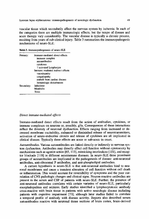
VasCUlar tissue which secondarily affect the nervous system by ischemia. In each of the categories there are multiple immunologic effects, but the tempo of disease and acute therapy vary considerably. The vascular disease is typically a chronic process, resulting from years of sub-clinical injury. Table 3 summarizes the immunopathogenic mechanisms of neuro-SLE.
Table3. Immunopathogenesis of neuro-SLE
Primary: Immune-mediated direct effects immune complex autoantibodies cytokines ? activated lymphocyte
Immune mediated indirect effects vasculopathy coagulopathy emboli from cardiac disease hematologic disturbances
Secondary: Infectious Metabolic Toxic
Direct immune-mediated effects
Immune-mediated direct effects result from the action of antibodies, cytokines, or immune complexes on neurons or, possibly, glia. Consequences of these interactions reflect the diversity of neuronal dysfunction. Effects ranging from increased or de- creased membrane excitability, enhanced or diminished release of neurotransmitters, activation of neuro-endocrine circuits and release of cytokines are all implicated in clinical disease. Typically these effects are acute or sub-acute in onset.
Autoantibodies. Various autoantibodies are linked directly or indirectly to nervous sys- tem dysfunction. Antibodies may directly affect cell function without cytotoxicity by mechanisms such as agonist action [65, 113], mimicking interleukins [101], and recep- tor blockade [118] in different autoimmune diseases. In neuro-SLE three prominent groups of autoantibodies are implicated in the pathogenesis of disease: anti-neuronal antibodies, anti-ribosomal P antibodies, and anti-phospholipid antibodies.
A current hypothesis in neuro-SLE is that anti-neuronal antibodies bind to neu- ronal membranes and cause a transient alteration of cell function without cell death or inflammation. This would account for reversibility of symptoms and the poor cor- relation of CNS pathologic changes and clinical signs. Neuron-reactive antibodies are present in the serum and CSF of patients with neuro-SLE. Further, the presence of anti-neuronal antibodies correlates with certain varieties of neuro-SLE, particularly encephalopathies and seizures. Early studies identified a lymphocytotoxic antibody cross-reactive with brain tissue in patients with active neurologic disease including patients with cognitive impairment [73], although later studies failed to correlate a temporal profile of antibody with disease activity. Reports also described serum autoantibodies reactive with neuronal tissue sections of brain cortex, brain-derived

50 P.M. Moore, R.P. Lisak
plasma membranes and cultured neuroblastoma cells [14, 40, 45, 55]. A variety of target antigens including membrane proteins, membrane glycolipids are described. Access of anti-neuronal antibodies to the CNS may occur by alterations in the blood- brain barrier or by intrathecal synthesis. Evidence for both exists [54, 122]. CSF stud- ies in patients with SLE reveal increased intrathecal Ig synthesis and/or oligoclonal bands [92] occurring in about half of the patients with encephalopathies, seizures and psychiatric abnormalities [39, 122].
Antibodies to ribosomal P proteins, particularly 60S ribosomal subunit phospho- proteins PO, P1 and P2 occur [15]. As intracellular antigens the access of antibodies to ribosomal P protein would be considered problematic, although antibodies to ri- bosomal P protein (38 kDa) were recently shown to also be immunoreactive with cell surfaces including those of neuroblastoma cells [67]. Recent studies have both confirmed [94] and refuted the association between psychoses and depression and anti-ribosomal antibodies [27, 115, 116]. The lower concentration of anti-ribosomal P antibodies in the CSF than in the serum raises some doubts about their role in disease [114].
Anti-phospholipid antibodies (APA), a large group of antibodies, include natural autoantibodies, which may have a physiologic role, as well as pathogenic antibod- ies such as those associated with hemolytic anemia, thrombocytopenia or venous occlusive disease. Even antibodies which are potentially pathogenic may not be so normally because of their association with natural anti-coagulants such as B2GP-1 [1]. In patients with SLE, only transverse myelitis appeared strongly associated with APA in one large series. Distinguishing primary anti-phospholipid syndrome in vas- cular diseases associated with APA from neurologic changes in SLE is under active investigation.
Limitations in interpreting studies correlating antibodies with neurologic disease include the facts that: (1) antibodies are also present in patients without demonstrable CNS involvement, (2) the specificity of the antigen(s) in neuronal tissue is unidenti- fied and are likely heterogeneous, (3) no large, standardized, longitudinal studies exist correlating antibody levels and neuropsychiatric disease in unselected SLE patients [49], and (4) no in vivo effects of this population of antibodies have been demon- strated. Further studies are required to establish the pathogenicity of these antibodies in human disease.
The previously described autoimmune mice, which share antigen specificity with many human autoantibodies, have proved particularly useful in the investigations of neuronal autoantigens. In MRL/lpr and NZB/W F1 mice, serum antibodies bind to brain plasma membranes [82, 87]. Neuron-reactive antibodies are present in the brains of some animals and can be eluted from the parenchyma [82-84]. Early neuropatho- logic studies of murine SLE [5, 93] are joined by studies analyzing neurocognitive abnormalities, autoantibodies, and histopathology [25, 52].
Immune complex. Immune complex-mediated tissue injury is a hallmark of SLE, well established in the pathogenesis of renal and cutaneous disease. There are potentially at least two pathogenic processes attributed to immune complex-mediated injury to the nervous system in SLE: (1) acute inflammation in the choroid plexus or vasculature, and (2) chronic deposition in the intracranial vasculature. In addition, mononeuropathy multiplex is likely similarly mediated.
Much of the interest in immune complexes and neurologic function has developed from the observations that immune complexes appear in the CSF, are deposited in the choroid plexus (a tissue with similarities to the renal glomeruli), and can alter the

Systemic lupus erythematosus: immunopathogenesis of neurologic dysfunction 51
blood-brain barrier when injected into experimental animals [ 104]. However, evidence for immune complex-triggered inflammation in the CNS in humans is scanty. Several patients showed IgG and complement deposits in the choroid plexus post mortem. Two histologic patterns of deposition (membranous and vascular) were identified, both associated with immune complexes in the CSF [95]. CSF DNA-anti-DNA com- plexes, detected in a patient with acute glomerulonephritis and hypocomplementemia, cleared with prednisone therapy [62]. CSF immune complexes of unknown antibody specificity were present in 33 out of 34 patients with CNS-SLE [98]. In most of these patients blood-brain barrier impairment was not present on the basis of Ig/protein ratios in the CSF, although focal or regional defects in the barrier could exist and not be detected by CSF analysis. In other patients loss of integrity of the blood-brain barrier exists but does not correlate with the presence of neurologic abnormalities or the presence of circulating immune complexes [122].
Against immune complex deposition serving as a major cause of neurologic dis- ease in neuro-SLE are (1) the presence of immune complex deposition in the choroid plexus in patients both with and without neurologic symptoms [17], and (2) the paucity of histologic evidence of inflammation. Furthermore, neurologic abnormali- ties are usually not associated with evidence of active elevation of circulating immune complexes and active renal disease, as would be anticipated if immune complex depo- sition in the choroid plexus or vasculature were a prominent cause of encephalopathies or seizures.
Another type of immune complex-mediated injury is more indolent, not always associated with evidence of systemic inflammation, and more difficult to diagnose. Low-level deposition of immune complexes in blood vessels may produce a chronic degenerative vasculopathy rather than acute tissue inflammation. Some of these issues are discussed in indirect immune-mediated effects as discussed below.
Cytokines. Cytokines are prominent in the immunopathogenesis of neurologic dis- ease. Cytokines, small proteins produced by lymphocytes, macrophages, and other cells such as fibroblasts, cause biologic affects in cells with receptors for that partic- ular cytokine. The short distances over which cytokines traditionally manifest their effects and their normal regulatory role complicate research into their pathogenic consequences in the nervous system. Both physiologic and pathologic effects on neu- rologic function may result through several pathways: direct effect of cytokines on function of cells of the nervous system and increased direct effects on endothelial cells which could lead to ischemia.
Evidence that cytokines play a role in normal neuronal function (and therefore may be implicated in disease) begins with histologic localization of cytokines, their receptors, and their antagonists in the neuraxis. IL-1 and IL-1 receptor antagonist co-localize to the hippocampus, hypothalamus, and several other brain regions, form- ing an intrinsic paracrine system. IL-1 activates the hypothalamic-pituitary-adrenal axis, causing release of pituitary hormones and ultimately glucocorticoids [12]. Brain cytokines are synthesized in response to local tissue injury; IL-6 appears with non- specific cerebral injury [77]. Production of CNS cytokines also elicit other cytokines, e.g., the production of IL-6 in response to injections of IL-1 [28]. Thus, cytokines may play a role in the behavioral, cognitive, and neuro-endocrine changes in neuro-SLE.
Activation of cytokines within the CNS reportedly induces profound changes in neural function. Behavioral and cognitive changes induced by cytokines are pertinent to the investigation of neuro-SLE. IL-113 inhibits long-term potentiation in mouse hippocampi with potential effects on memory [61]. Pharmacologic doses of IL-2 in-

52 P.M. Moore, R.P. Lisak
duce fever, slow wave sleep, anorexia, and, in mice, exploring behavior [26, 51]. The neuropathogenic effects of several infectious agents that do not invade the nervous system are attributed to cytokines produced by glial and immune cells or to serum cytokines on areas of the brain with a naturally incomplete blood-brain barrier. Neu- ropeptides such as substance P and substance K (neurotransmitters released at the peripheral terminals of afferent nerves) can stimulate immunocompetent cells as well as neuroglial cells to secrete inflammatory cytokines IL-1, TNF-a and IL-6 [74]. IL-1, TNF-a, and IL-6, in turn, act directly and indirectly on the hypothalamus to stimulate synthesis and secretion of CRH. This is a potent interaction between the neuraxis and the immune system [ 11, 89]. Recent studies on IL-6 suggest the cytokine itself may be associated with neurologic abnormalities.
Another major effect of cytokines in the nervous system is the effect on the vasculature. Despite the potential effects on brain cells and blood vessels, there has been scanty demonstration of increased cytokines in the brain or CSF of patients with SLE. The only reported of abnormal cytokine concentrations in neuro-SLE is for IL-6 which is increased in the CSF [53]. The possible role of cytokines in vascular pathology is discussed in indirect immune-mediated effects.
Activated lymphocytes. Activated lymphocytes may have a pathogenic effort on brain tissue in SLE. Activated immunocompetent cells such as lymphocytes, monocytes, and macrophages can penetrate the blood-brain barrier, lodge in the brain, and secrete their full repertoire of cytokines, leukotrienes, and prostaglandins. B cells present, albeit sparsely, in human and murine CNS may be the source of intrathecal synthesis of Ig. The B cells may be amenable to immunotherapy directed at activation mark- ers. Cell-mediated cytotoxicity, another effector mechanism mediated by a subset of lymphocytes, has not been demonstrated in the nervous system of human or murine SLE.
Indirect immune-mediated effects
Immune-mediated indirect neurologic effects typically result from the action of au- toantibodies, cytokines, immune complexes, or activated lymphocytes on the vascu- lature. Neurologic effects result from tissue ischemia. Tissue ischemia or infarction from occluded blood vessels may develop from several different, indirect immune- mediated effects of disease. This group is distinguished from the first not only in immunopathogenesis but also in clinical presentation and therapy. In these disorders, the immunologic effects on the blood vessel occur over a period of time ranging from months to years. The incidence of stroke in clinical series of SLE ranges from 5 to 20% [34, 66]. Most clearly denoted are thromboses (coagulopathies and hematologic disorders), vasculopathies, with or without hemorrhagic infarcts and emboli from the heart. Arteries, veins, and microvasculature are affected.
Neurologic abnormalities develop when a critical degree of local disease or a se- ries of sub-clinical events (such as mild changes in hemostasis) in abnormal blood vessels occur. Compounding the vascular damage are additional, non-immune abnor- malities such as hypertension. Furthermore, corticosteroid therapy may accelerate the premature atherosclerosis present, at least in the coronary arteries.
Vasculopathy. Vasculopathy refers to structural abnormalities of the blood vessel wall. In patients with neuro-SLE, these changes occur primarily in small but sometimes in medium-sized, vessels. Two recurrent observations are noteworthy. First, degenera-

Systemic lupus erythematosus: immunopathogenesis of neurologic dysfunction 53
tive changes in the blood vessel wall far exceed evidence of inflammation. Second, histologic changes suggest a chronic rather than acute process. In addition, histologic evidence of vasculopathy is more widespread than clinical findings, and also exceeds histologiG evidence of ischemia.
Pathologic studies show endothelial proliferation and medial hyperplasia of small intracortical arterioles and capillaries, petechial hemorrhages, and parenchymal necro- sis with a variable glial reaction [46]. Histologic staining with Masson-Trichrome reveals irregular proliferation of collagen with occasional hyalinization. The frequent association of extravasated blood with occasional hemosiderin-laden macrophages suggests structural incompetence of vascular hemostasis in those vessels [31]. Ab- normalities in microvessels appear in 40-80% of patients, although lesions associated with evidence of tissue infarction are more limited in distribution and extent [58]. Ma- jor vessel thromboses associated with atheroma are occasionally reported. Although CNS vasculitis occurs, it is much less frequent than cutaneous or visceral vasculitis, and is an infrequent cause of neurologic abnormalities.
Numerous mechanisms could be responsible for the vasculopathy but three are considered most likely (1) deposition of circulating immune complexes including DNA-anti-DNA complexes (2) effects of autoantibodies including APA on the vessel wall; and (3) consequences of chronic inflammation.
Clues to the development of an immune complex-mediated degenerative rather than inflammatory vasculopathy are found in animal studies. In murine SLE the du- ration, quantity, and types of circulating immune complexes, as well as the genetic background of the host determine the effects of immune complex deposition on the vasculature. Chronic, low levels of immune complexes are associated with endothelial and intimal proliferation in the vessel wall plus thrombosis [10]. Immune complex- mediated injury also results in the production or accrual of vasoactive substances with a net prothrombotic effect. Evidence that immune complex deposition may be clin- ically relevant is the association of cerebrovascular disease with renal involvement and circulating anti-DNA/DNA complexes [66].
Direct autoantibody effects on the vessel wall may also produce changes. Endothe- lial cells, known regulators of blood flow through their actions on vascular smooth muscle, mediate these changes through vasodilators such as prostaglandin (PG) I1 endothelium-derived relaxing factor and the vasoconstrictor peptide endothelin or polypeptide PDGF [18]. Both anti-endothelial antibodies and APA may result in mild but recurrent injury to the endothelium [19, 44]. Antibodies binding phospholipids in endothelial cell membranes may result in decreased levels of prostacyclin and in- creased platelet aggregation and thrombosis [ 13, 64]. Abnormalities in the endothelial cell in SLE is likely accompanied by changes in blood flow and vessel diameter.
Chronic inflammation may also be injurious. Recent studies suggest serum amyloid A proteins, acute-phase reactants associated with high-density lipoproteins (HDL), displace apolipoprotein A1. This potentially leads to greater deposition of fat in the blood vessel and may result in premature atherosclerosis [8]. Inappropriate activation of adhesion molecules may contribute to the consequences of chronic inflammation [6, 9, 37, 48, 76].
Structural changes in the wall are compounded by coagulation abnormalities, the result of the prothrombotic effect of many cytokines. Subsequent inflammation, tran- sient or recurrent, is controlled by a variety of cell regulatory events, which influ- ence a wide array of cell responses including chemotaxis, apoptosis, differentiation and growth arrest. Intravascular complement activation may also result in occlusive

54 P.M. Moore, R.P. Lisak
vasculopathy [44]. Leukocytes and platelets aggregate in vessel walls secondary to increased adhesiveness rendered by lipopolysaccharide, IL-1, or TNF.
Coagulopathies. The coagulation system is a highly regulated cascade of surface asso- ciated enzymes and cofactors, resulting in the generation of thrombin. The interactions of the coagulant and anticoagulant proteins and the fibrinolytic system occur at the vascular endothelial cell surface. Acquired hypercoaguable states occur with abnor- malities of protein, platelet function or endothelial cell dysfunction. Disorders of fibrin metabolism potentially facilitate deposition of fibrin and platelets on endothelium of small vessels and cardiac valves [86].
APA, often present in patients with SLE and reportedly associated with an in- creased risk of stroke, are the most frequently studied potential cause of stroke. This family of antibodies, including anti-cardiolipin antibody, shares reactivity with nega- tively charged phosphodiester moieties. They are prothrombotic factors in occlusive vascular disease both in the brain and elsewhere. Potential targets of APA are the endothelial cell, prostacyclin, protein C, the protein C-S complex, and platelets. Both acute effects on coagulation and more chronic effects on the vessel wall are possi- ble. The APA manifestations of stroke in SLE may, thus, involve synergism with the vasculopathy or other inflammatory effects.
Thus, APA potentially contribute to stroke by two pathways, interaction with the blood vessel wall and with the coagulation cascade. Numerous studies link APA with stroke [2, 3, 32, 72]. A direct pathogenic role of APA and stroke is, however, not established. The classic features of the anti-phospholipid syndrome are recurrent fe- tal loss, thrombophlebitis, and stroke. Numerous other neurologic abnormalities have been associated with APA. However, in one large series of SLE patients studied prospectively, only the occurrence of transverse myelopathies (not stroke) was statis- tically significantly associated with APA [2]. Also, APA were not found in the CSF of five SLE patients with acute strokes, despite high serum levels of the antibody [32]. In another study only three of the six stroke patients had a serum lupus anticoagulant [120]. Other abnormalities leading to a hypercoaguable state include decreased fib- rinolysis, decreased plasminogen activation, and impaired formation of prostacyclin [108].
Cardiac emboli. In 1924 Libman and Sacks reported four patients with atypical ver- rucous endocarditis. The vegetations consisted of soft clumps of fibrinous material with plasma cells usually found on the chordae tendiae, papillary muscles, or on the valves most frequently the rhitral valve. These vegetations seldom dislodged and em- boli were considered rare. Recently, the potential role of emboli from valvular heart disease has been reevaluated.
In 1988, an autopsy study of 50 patients with SLE identified l0 people with embolic cerebral infarcts, 5 caused by identified Libman-Sacks endocarditis, and 4 from other cardiac sources [29]. Clinical studies, particularly in patients who survive, reveal fewer diagnosed cerebral emboli [36]. An association of mitral valve disease and APA is also noted [63]. The actual incidence of cerebral emboli from the heart in neuro-SLE is not known. As described, the incidence of Libman-Sacks lesions and other mild valvular abnormalities detected by echocardiography is 18% of patients with SLE. There was no occurrence of cerebrovascular disease in 74 patients over 5-period years [36].
Hematologic changes. Thrombotic thrombocytopenic purpura (TTP) presents with wide-spread small vessel lesions in association with the hematologic abnormalities.

Systemic lupus erythematosus: immunopathogenesis of neurologic dysfunction 55
Histologically the brain lesions in the vasculopathy of SLE resemble those of TTP, except those of T I P tend to be all of the same age [58]. Thrombocytopenia appears to result from an antibody-mediated increased destruction of platelets. Underscoring the overlap with the coagulopathies, one of the types of antibodies reported to cause thrombocytopenia is APA [75].
Non-immune mediated effects
Neurologic complications of SLE also result from abnormalities not directly related to the primary disease. Many of these systemic or generalized abnormalities result in neurologic dysfunction, including confusion and seizures, that are clinically indistin- guishable from primary immune-mediated damage. The major etiologies in this group are infections, toxins (including medications), and metabolic derangements. Although survival in SLE has improved over the years, infection remains a leading cause of morbidity and mortali ty in the disease. Meningitis from both bacterial pathogens such as Neisseria and opportunistic infections including Candida and Cryptococcus occur [81]. The spectrum of infections includes bacterial, viral, fungal, and parasitic organ- isms [20, 23, 68, 81,103]. Septic emboli continue to be a cause for concern in patients with fever. The frequency of infections results from the patients ' immunosuppression both by medication and by their underlying disease. The importance of identifying infections was highlighted by work indicating that high-dose corticosteroids did not improve the outcome of neurologic disease in patients with SLE. In fact, patients on high-dose corticosteroids had a higher mortality rate than those on lower doses [99, 123]. Unfortunately, many potentially treatable infections are only diagnosed post mortem.
The toxins most often causing neurologic abnormalities in SLE are medications. Corticosteroids and anti-hypertensive medications can produce neurologic and psychi- atric symptoms. Metabolic abnormalities likely to affect the nervous system include uremia, as well as disorders of calcium, sodium, and magnesium.
References
1. Alarcon-Segovia D, Cabral AR (1994) Antiphospholipid antibodies. Where do they come from? Where do they go? J Rhenmatol 21:982
2. Alarcon-Segovia D, Deleze M, Oria CV, Sanchez-Guerrero J, Gomez-Pacheco L, Cabiedes J, Fernan- dez L, Ponce De Lion S (1989) Antiphospholipid antibodies and the antiphospholipid syndrome in systemic lupus erythematosus. A prospective analysis of 500 consecutive patients. Medicine 68:353
3. Alarcon-Segovia D, Perez-Vazquez ME, Villa AR, Drenkard C, Cabiedes J (1992) Preliminary clas- sification criteria for the antiphospholipid syndrome within systemic lupus erythematosus. Semin Arthritis Rheum 21:275
4. AI-Janadi M, Raziuddin S (1993) B cell hyperactivity is a function of T cell derived cytokines in systemic lupus erythematosus. J Rheumatol 20:1885
5. Alexander EL, Murphy ED, Roths JB, Alexander GE (1983) Congenic autoimmune marine models of central nervous system disease in connective tissue disorders. Ann Neurol 14:242
6. Argenbright LW, Barton RW (1992) Interactions of leukocyte integrins with intercellular adhesion molecule 1 in the production of inflammatory vascular injury in vivo. J Clin Invest 89:259
7. Bankhurst AD (1987) lnterferons and systemic lupus erythematosus. J Rheumatol [Snppl 13] 14:63 8. Beer MC de, Kindy MS, Lane WS, Beer FC de (1994) Mouse serum amyloid A protein (SAAS)
structure and expression. J Biol Chem 269:4661

56 P.M. Moore, R.P. Lisak
9. Belmont HM, Buyon J, Giomo R, Abramson S (1994) Up-regulation of endothelial cell adhestion molecules characterizes disease activity in systemic lupus erythematosus. Arthritis Rheum 37:376
10. Berden JHM, Hang L, McConahey PJ, Dixon FJ (1983) Analysis of vascular lesions in murine SLE. J Immunol 130:1699
11. Besedovsky HO, DelRey AE, Sorkin E (1985) Immune-neuroendocrine interactions. J Immunol 135:750s
12. Besedovsky HO, DelRey A, Sorkin E, Dinarello CA (1986) Immunoregulatory feedback between interleukin-1 and glucocorticoid hormones. Science 233:652
13. Bevilacqua MP, Pober JS, Majeau GR, Cotran RS, Gimbrone MA Jr (1984) Interleukin 1 (IL-1) induces biosynthesis and cell surface expression of procoagulant activity in human vascular endothelial cells. J Exp Med 160:618
14. Bluestein HG (1978) Neurocytotoxic antibodies in serum of patients with systemic lupus erythemato- sus. Proc Natl Acad Sci USA 75:3965
15. Bonfa E, Golombek SJ, Kaufman LD, Skelly S, Weissbach H, Brot N, Elkon KB (1987) Association between lupus psychosis and anti-ribosomal P protein antibodies. N Engl J Med 317:265
16. Boros P, Odin JA, Chen J, Unkeless JC (1994) Specificity and class distribution of FcTR-specific autoantibodies in patients with autoimmuue disease. J Immunol 152:302
17. Boyer RS, Sun NCJ, Verity A, Nies KM, Louie JS (1980) Immunoperoxidase staining of the choroid plexus in systemic lupus erythematosus. J Rheumatol 7:645
18. Brenner BM, Troy JL, Ballermann BJ (1989) Endothelium-dependent vascular responses. J Clin Invest 84:1373
19. Cines DB (1989) Disorders associated with antibodies to endothelial cells. Rev Infect Dis 2 [Suppl 4]:$705
20. Collins DN, Oppenheim IA, Edwards MR (1971) Cryptococcosis associated with systemic lupus erytbematosus. Arch Pathol Lab Med 91:78
21. Combardier C, Gladman DD, Urowitz MB, Caron D, Chang CH (1992) Derivation of the sledai. A disease activity index for lupus patients. Arthritis Rheum 35:630
22. DeHoratius RJ, Pillarisetty R, Messner R, Tallal N (1975) Anti-nucleic acid antibodies in systemic lupus erythematosus patients and their families: incRlence and correlation with lymphocytotoxic an- tibodies. J Clin Invest 56:1149
23. Deleze M, Mintz G, CarmenMejia M (1985) Toxoplasma gondii encephalitis in systemic lupus ery- thematosus: a neglected cause of treatable nervous system infection. J Rheumatol 12:5:994
24. Denburg SD, Carbotte RM, Denburg JA (1987) Cognitive impairment in systemic lupus erythemato- sus: a neuropsychological study of individual and group deficits. J Clin Exp Neuropsychol 9:323
25. Denenberg VH, Sherman GF, Morrison L, Schrott LM, Waters NS, Rosen GD, Behan PO, Galaburda AM (1992) Behavior, ectopias and immunity in BD/DB reciprocal crosses. Brain Res 571:323
26. Denicoff KD, Rubinow DR, Papa MZ, Simpson C, Seipp CA, Lotze M, Chang AE, Rosenstein D, Rosenberg SA (1987) The neuropsychiatric effects of treatment with interleukin-2 and lymphokine- activated killer cells. Ann Intern Med 107:293
27. Derksen RHWM, van Dam AP, Meyling FHJG, Bijlsma JWJ, Smeenk RJT (1990) A prospective study on antiribosomal P proteins in two cases of familial lupus and recurrent psychosis. Ann Rheum Dis 49:779
28. De Simoni MG, Sironi M, De Luigi A, Manfridi A, antovani A, Ghezzi P (1990) Intracerebroventric- ular injection of interleukin 1 induces high circulating levels of interleukin 6. J Exp Med 171:1773
29. Devinsky O, Petito CK, Alonso DR (1988) Clinical and neuropathological findings in systemic lupus erythematosus: the role of vasculitis, heart emboli, and thrombotic thrombocytopenic purpura. Ann Neurol 23:380
30. Dixon FJ (1987) Basic elements of murine systemic lupus erythematosus. J Rheumatol 14:3 31. Ellis SG, Verity MA (1979) Central nervous system involvement in systemic lupus erythematosus: a
review of neuropathologic findings in 57 cases, 1955-1977. Semin Arthritis Rheum 8:212 32. Fields RA, Sibbitt WL, Toubbeh H, Bankhurst AD (1990) Neuropsychiatric lupus erythematosus,
cerebral infarctions, and anticardiolipin antibodies. Ann Rheum Dis 49:114 33. Fronek Z, Timmerman LA, Alper CA, et.al. (1990) Major histocompatibilty complex genes and
susceptibility to systemc lupus erythematosus. Arthritis Rheum 33:1542 34. Futrell N, Millikan C (1989) Frequency, etiology, and prevention of stroke in patients with systemic
lupus erythematosus. Stroke 20:584

Systemic lupus erythematosus: immunopathogenesis of neurologic dysfunction 57
35. Gahring LC, Weigle WO (1990) The regulatory effects of cytokines on the induction of a peripheral immunologic tolerance in mice. J Immunol 145:1318
36. Galve E, Candell-Riera J, Pigrau C, Permanyer-Miralda G, Garcia-Del-Castillo H, Soler-Soler J (1968) Prevalence, morphologic types, and evolution of cardiac valvular disease in systemic lupus erythematosus. N Engl J Med 319:817
37. Gibbons GH, Dzan VJ (1994) The emerging concept of vascular remodeling. N Engl J Med 330:1431 38. Golan DT (1988) Exposure to UV light in the pathogenesis of systemic lupus erythematosus(SLE).
24:360 39. Golombek S J, Elkon KB (1986) Variety of auto-antibodies in the cerebrospinal fluid of lupus patients.
Arthritis Rheum 29:$8 40. Golombek SJ, Graus F, Elkon KB (1986) Autoantibodies in the cerebrospinal fluid of patients with
systemic lupus erythematosus. Arthritis Rheum 29:1090 41. Gregory CD, Dive C, Henderson S, Smith CA, Williams GT, Gordon J, Rickinson AB (1991) Ac-
tivation of Epstein-Barr virus latent genes protects human B cells from death by apoptosis. Nature 349:612
42. Gutierrez-Ramos JC, Andreu JL, Alboran IM de, Rodrignez J, Leonardo E, Kroemer G, Marcus MAR, Martinez-A C (1990) Insights into autoimmunity: from classical models to current perspectives. Immunol Rev ! 18:73
43. Hahn BH, Ebling FM (1984) A public idiotype determinant is present on spontaneous cationic IgG antibodies to DNA from mice of unrelated lupus-prone strains. J lmmunol 133:3015
44. Hammad A, Tsukada Y, Torre N (1992) Cerebral occlusive vasculopathy in systemic lupus erythe- matosus and speculation on the part played by complement. Ann Rheum Dis 51:550
44a. Handwerger B, Rus V, daSilva L. Via C (1994) The role of cyotkines in the immunopathogenesis of lupus. Springer Seminars in Immunopathology 16:153-180
45. Hanly JG, Rajaraman S, Behmarm S, Denburg JA (1988) A novel neuronal antigen identified by sera from patients with system lupus erythematosus. Arthritis Rheum 31:1492
46. Hanly JG, Walsh NMG, Sangalang V (1992) Brain pathology in systemic lupus erythematosus. J Rheumatol 19:732
47. Hanly JG, Walsh NM, Fisk JD, Eastwood B, Hong C, Sherwood G, Jones JV, Jones E, Elkon K (1993) Cognitive impairment and autoantibodies in systemic lupus erythematosus. Br J Rheumatol 32:291
48. Hart MN, Zsuzsanna F, Waldschmidt M, Sandor M (1990) Lymphocyte interacting adhesion molecules on brain microvascular cells. Mol Immunol 27:1355
49. Hay EM, Isenberg DA (1993) Autoantibodies in central nervous system lupus. Br J Rheumato132:329 50. Hay EM, Hnddy A, Black D, Mbaya P, Tomenson B, Bernstein RM, Holt PJL, Creed F (1994) A
prospective study of psychiatric disorder and cognitive function in systemic lupus erythematosus. Ann Rheum Dis 53:298
51. Hellerstein MK, Meydani SN, Meydani M, Wu K, Dinarello CA (1989) Interleukin-l-induced anorexia in the rat. Influence of prostaglandins. J Clin Invest 84:228
52. Hess DC, Taormina M, Thompson J, Sethi D, Diamond B, Rao R, Chamberlain CR, Feldman DS (1993) Cognitive and neurologic deficits in the MRL/Ipr mouse: a clinicopathologic study. J Rheuma- tol 20:610
53. Hirohata S, Miyamoto T (1990) Elevated levels of interleukin-6 in cerebrosl~inal fluid from patients with systemic lupus erythematosus and central nervous system involvement. Arthritis Rheum 33:644
54. Hirohata S, Hirose S, Miyamoto T (1985) Cerebrospinal fluid IgM, IgA, IgG indexes in systemic lupus erythematosus. Arch Intern Med 145:1843
55. How A, Dent PB, Liao S-K, Denburg JA (1985) Antineuronal antibodies in neuropsychiatric system lupus erythematosus. Arthritis Rheum 28:789
56. Hoyle C, Ewing DJ, Parker AC (1985) Acute autonomic neuropathy in association with systemic lupus erythematosus. Ann Rheum Dis 44:420
57. lsenberg DA, Snaith ML (198t) Muscle disease in systemic lupus erythematosus: a study of its nature, frequency and cause. J Rheumatol 8:917
58. Johnson RT, Richardson EP (1968) The neurological manifestations of systemic lupus erythematosus: a clinical-pathological study of 24 cases and review of the literature. Medicine 47:337
59. Jyonouchi H, Kincade PW, Good RA, Fernandes G (1981) Reciprocal transfer of abnormalities in clonable B lymphocytes and myeloid progenitors between NZB and DBA/2 micel. J Immunol 127:1232

58 P.M. Moore, R.P. Lisak
60. Kaell AT, Shetty M, Lee BCP, Lockshin MD (1986) The diversity of neurologic events in systemic lupus erythematosus. Arch Neurol 43:273
61. Katsuki H, Nakai S, Hirai Y, Akaji K, Kiso Y, Satoh M (1990) Interleukin 1B inhibits long-term potentiation in the CA3 region of mouse hippocampal slices. Eur J Pharmacol 181:323
62. Keeffe EB, Bardama El, Harbeck RJ, Pirofsky B, Carr RI (1974) Antibody to deoxyribonucleic acid (DNA) and DNA:anti-DNA complexes in cerebrospinal fluid. Ann Intern Med 80:58
63. Khamashta MA, Hughes GRV (1993) Antiphospholipid antibodies and valve disease in patients with systemic lupus erythematosus. J Am Coll Cardiol 22:1268
64. Khamashta MA, Harris EN, Gharavi AE, Derue G, Gil A, Vazquez JJ, Hughes GRV (1988) Immune mediated mechanism for thrombosis: antiphospholipid antibody binding to platelet membranes. Ann Rheum Dis 47:849
65. Khokher MA, Dandona P, Janah S, Coulston GL (1981) Insulin-like stimulatory effect of human immunoglobulin G on adipocyte lipogenesis. Diabetes 30:1068
66. Kitagawa Y, Gotoh F, Koto A, Okayasu H (1990) Stroke in systemic lupus erythematosus. Stroke 21:1533
67. Koren E, Reichlin MW, Koscec M, Fugat RD, Reichlin M (1993) Autoantibodies to the ribosomal P proteins react with a plasma membrane-related target on human cells. J Clin Invest 89:1236
68. Lee P, Urowitz MB, Bookman AAM, Koehler BE, Smythe HA, Gordon DA, Ogryzlo MA (1977) Systemic lupus erythematosus. A review of 110 cases with reference to nephritis, the nervous system, infections, aseptic necrosis and prognosis. Q J Med 46:1
69. Lerner MR, Boyle JA, Mount SM, Wolin SL, Steitz JA (1980) Are snRNPs involved in splicing? Nature 283:220
70. Linker-Israeli M (1992) Cytokine abnormalities in human lupus. Clin Immunol Immunopathol 63:10 71. Linker-lsraeli M, Deans RJ, Wallace DJ, Prehn J, Ozeri-chen T, Klinenberg JR (1991) Elevated levels
of endogenous IL-6 in systemic lupus erythematosus. J Immunol 147:117 72. Lo S, Salem HH, Howard MA, Oldmeadow MJ, Firkin B (1990) Studies of natural anticoagulant
proteins and anticardiolipin antibodies in patients with the lupus anticoagulant. Br J Haematol 76:380 73. Long AA, Denburg SD, Carbotte RM, Singal DP, Denburg JA (1990) Serum lymphocytotoxic anti-
bodies and neurocognitive function in systemic lupus erythematosus. Ann Rheum Dis 49:249 74. Lotz M, Vaughan JH, Carson DA (1988) Effect of neuropeptides on production of inflammatory
cytokines by human monocytes. Science 241:1218 75. Love PE, Santoro SA (1990) Antiphospholipid antibodies: anticardiolipin and the lupus anticoagulant
in systemic lupus erythematosus (SLE) and in non-SLE disorders. Ann Intern Med 112:682 76. Male D, Pryce G, Hughes C, Lantos P (1990) Lymphocyte migration into brain modelled in vitro:
control by lymphocyte actication, cytokines, and antigen. Cell Immunol 127:1 77. McClain C, Cohen D, Phillips R, Ott L, Young B (1993) Increased plasma and ventricular fluid
interleukin-6 levels in patients with head injury. J Lab Clin Med 118:225 78. Merino R, Iwamoto M, Fossati L, Izui S (1993) Polyclonal B cell activation arises from different
mechanisms in lupus-prone (NZB x NZW)F1 and MRL/MpJ-lpr/lpr mice. J Immunol 151:6509 79. Michet CJ, McKenna CH, Elveback LR, Kaslow RA, Kurland LT (1985) Epidemiology of systemic
lupus erythematosus and other connective tissue diseases in Rochester, Minnesota, 1950 through 1979. Mayo Clin Proc 60:105
80. Miguel EC, Pereira RMR, Pereira CADB, Baer L, Gomes RE, Sa LCF de, Hirsch R, Barros NG de, Navarro JM de, Gentil V (1994) Psychiatric manifestations of systemic lupus erythematosus: clinical features, symptoms, and signs of central nervous system activity in 43 patients. Medicine 73:224
81. Mitchell SR, Nguyen PQ, Katz P (1990) Increased risk of neisserial infections in systemic lupus erythematosus. Semin Arthritis Rheum 20:174
82. Moore PM (1990) lmmunoglobulin binding to neuronal cell surface epitopes in marine systemic lupus erythematosus. J Neuroimmunol 30:101
83. Moore PM (1992) Evidence for bound antineuronal antibodies in brains of NZB/w mice. J Neuroim- munol 38:147
84. Moore PM, Joshi I, Ghanekar SM (1994) Affinity isolation of neuron reactive antibodies in MRL/Ipr Mice. J Neurosci Res 39:140
85. Mountz JD, Wn J, Cheng J, Zhou T (1994) Autoimmune disease. A problem of defective apoptosis. Arthritis Rheum 37:1415
86. Nachman RL, Silverstein R (1993) Hypercoagulable states. Ann Intern Med 119:819

Systemic lupus erythematosus: immunopathogenesis of neurologic dysfunction 59
87. Narendran A, Hoffman SA (1989) Characterization of brain-reactive autoantibodies in murine models of systemic lupus erythematosus. J Neuroimmunol 24:113
88. Pisetsky DS, Grudier JP, Gilkeson GS (1990) A role for immunogenic DNA in the pathogenesis of systemic lupus erythematosus. Arthritis Rheum 33:153
89. Reichlin S (1993) Neuroendocrine-immune interactions. N Engl J Med 329:1246 90. Reidenberg MM (1983) Aromatic amines and the pathogenesis of lupus erythmatosus. Am J Med
75:1037 91. Ritchlin CT, Chabot RJ, Alper K, Buyon J, Belmont HM, Roubey R, Abramson SB (1992) Quanti-
tative electroencephalography. Arthritis Rheum 35:1330 92. Rudh I, Olsson T, Lindstrom F, Skogh T (1985) Cerebrospinal fluid immunoglobulin abnormalities
in systemic lupus erythematosus. J Neurol 48:807 93. Rudick RA, Eskin TA (1983) Neuropathological features of a lupus like disorder in autoimmune
mice. Ann Neurobiol 14:325 94. Schneebaum AB, Singleton JD, Sterling G, West MD, Blodgett JK, Allen LG, Cheronis JC, Kotzin BL
(1991) Association of psychiatric manifestations with antibodies to ribosomal P proteins in systemic lupus erythematosus. Am J Med 90:54
95. Schwartz MM, Roberts JL (1983) Membranous and vascular choroidopathy: two patterns of immune deposits in systemic lupus erythematosus. Clin Immunol Immunopathol 29:369
96. Schwartz RH (1989) Acquisition of immunologic self-tolerance. Cell 57:1073 97. Schwartz RS (1986) Anti-DNA antibodies and the problem of autoimmunity. Cell Immunol 99:38 98. Seibold JR, Buckingham RB, Medsger TA Jr, Kelly RH (1982) Cerebrospinal fluid immune complexes
in systemic lupus erythematosus involving the central nervous system. Semin Arthritis Rheum 12:68 99. Sergent JS, Lockshin MD, Klempner MS, Lipsky BA (1975) Central nervous system disease in
systemic lupus erythematosus. Am J Med 58:644 100. Shen HH, Winchester RJ (1986) Susceptibility genetics of systemic lupus erythematosus. Springer
Semin Immunopathol 9:143 101. Sibbitt WL Jr, Froelich CJ, Bankhurst AD (1984) Interferon-regulation of lymphocyte function in
systemic lupus erythematosus. Clin Immunol lmmunopathol 32:70 102. Sibley JT, Olszynski WP, Decoteau WE, Sundaram MB (1992) The incidence and prognosis of central
nervous system disease in systemic lupus erythematosus. J Rheumatol 19:47 103. Sieving RR, Kauffman CA, Watanakunakom C (1975) Deep fungal infection in systemic lupus
erythematosus-three cases reported, literature reviewed. J Rheumatol 2:61 104. Simon J, Simon O (1975) Effect of passive transfer of anti-brain antibodies to a normal recipient.
Exp Neurol 47:523 105. Singh RR, Prasad K, Kumar A, Misra A, Padmakumar K, Malaviya AN (1988) Cerebellar ataxia in
systemic lupus erythematosus: three case reports. Ann Rheum Dis 47:954 106. Steinberg AD, Gonrley MF, Klinman DM, Tsokos GC, Scott DE, Kreig AM (1991) Systemic lupus
erythematosus. Ann Intern Med 115:548 107. Sternberg EM, Chrousos GP, Wilder RL, Gold PW (1992) The stress response and the regulation of
inflammatory disease. Ann Intern Med 117:854 108. Stevens MB (1986) Systemic lupus erythematosus clinical issues. Springer Semin Immunopathol
9:251 109. Stott DI (1990) Lessons about autoantibody specificity in systemic lupus erythematosus from animal
models (editorial review). Clin Exp Immunol 81:1 110. Tan EM, Cohen AS, Fries .IF, Masi AT, McShane DJ, Rothfield N-F, Schaller JG, Talal N, Winchester
RJ (1982) The 1982 revised criteria for the classification of systemic lupus erythematosus. A~hdtis Rheum 25:1271
111. Tanaka Y, Saito K, Shirakawa F, Ota T, Suzuki H, Eto S, Yamashita U (1988) Production of B cell- stimulating factors by B cells in patients with systemic lupus erythematosus. J Immunol 141:3043
112. Tanaka Y, Shirakawa F, Ota T, Suzuki H, Eto S, Yamashita U (1988) Mechanism of spontaneous activation of B cells in patients with systemic lupus erythematosus. J Immunol 140:761
113. Taylor SI, Grunberger G, Marcus-Samuels B, Underhill LH, Dons RF, Ryan J, Roddam RF, Rupe CE, Gorden P (1982) Hypoglycemia associated with antibodies to the insulin receptor. N Engi J Med 307:1422
114. Teh L, Isenberg DA (1994) Antiribosomal protein antibodies in systemic lupus erythematosus. Arthri- tis Rheum 37:307

60 P.M. Moore, R.P. Lisak
115. Teh LS, Bedwell AE, Isenberg DA, Gordon C, Emery P, Charles PJ, Harper M, Amos N, Williams BD (1992) Antibodies to protein P in systemic lupus erythmatosus. Ann Rheum Dis 51:489
116. Teh LS, Hay EM, Amos N, Black D, Huddy A, Creed F, Bernstein RM, Holt PS, Williams BD (1993) Anti-P antibodies are associated with psychiatric and focal cerebral disorders in patients with systemic lupus erythematosus. Br J Rheumatol 32:287
117. Theofilopoulos AN, Dixon FJ (1981) Etiopathogenesis of marine SLE. Immunol Rev 55:179 118. Venter JC (1982) Monocolonal antibodies and autoantibodies in the isolation and characterization of
neurotransmitter receptors: The future of receptor research. J Molec Cell Immunol 14:687 119. Via CS, Tsokos GC, Bermas B, Clerici M, Shearer GM (1993) T cell-antigen-presenting cell inter-
actions in human systemic lupus erythematosus. J lmmunol 151:3914 120. Vidailhet M, Piette J, Wechsler B, Bousser MG, Brunet P (1990) Cerebral venous thrombosis in
systemic lupus erythematosus. Stroke 21:1226 121. Winfield JB, Mimura T (1992) Pathogenetic significance of anti-lymphocyte autoantibodies in sys-
temic lupus erythematosus. Clin Immunol Immunopathol 63:13 122. Winfield JB, Shaw M, Silverman LM, Eisenberg RA, Wilson HA III, Kottter D (1983) Intrathecal
IgG synthesis and blood-brain barrier impairment in patients with systemic lupus erythematosus and central nervous system dysfunction. Am J Med 74:837
123. Wysenbeek AJ, Leibovici L, Zoldan J (1990) Acute central nervous system complications after pulse steroid therapy in patients with systemic lupus erythematosus. J Rheumatol 17:1695