systematicanalysisofenvironmentalchemicalsthatdysregulate...
TRANSCRIPT

Research ArticleSystematic Analysis of Environmental Chemicals That DysregulateCritical Period Plasticity-Related Gene Expression RevealsCommon Pathways That Mimic Immune Response to Pathogen
Milo R. Smith,1,2,3,4,5,6,7 Priscilla Yevoo,1,3,4,6,7 Masato Sadahiro,1,3,4,6,7 Ben Readhead,5,8
Brian Kidd,2,5 Joel T. Dudley ,2,5 and Hirofumi Morishita 1,3,4,6,7
1Department of Psychiatry, Icahn School of Medicine at Mount Sinai, 1 Gustave L Levy Place, New York NY 10029, USA2Department of Genetics and Genomic Sciences, Icahn School of Medicine at Mount Sinai, 1 Gustave L Levy Place,New York NY 10029, USA3Nash Family Department of Neuroscience, Icahn School of Medicine at Mount Sinai, 1 Gustave L Levy Place,New York NY 10029, USA4Department of Ophthalmology, Icahn School of Medicine at Mount Sinai, 1 Gustave L Levy Place, New York NY 10029, USA5Institute for Next Generation Healthcare, Icahn School of Medicine at Mount Sinai, 1 Gustave L Levy Place,New York NY 10029, USA6Friedman Brain Institute, Icahn School of Medicine at Mount Sinai, 1 Gustave L Levy Place, New York NY 10029, USA7Mindich Child Health & Development Institute, Icahn School of Medicine at Mount Sinai, 1 Gustave L Levy Place,New York NY 10029, USA8ASU-Banner Neurodegenerative Disease Research Center, Biodesign Institute, Building A, 1001 S McAllister Ave, Tempe,AZ 85281, USA
Correspondence should be addressed to Joel T. Dudley; [email protected] Hirofumi Morishita; [email protected]
Received 22 May 2019; Accepted 4 February 2020; Published 5 May 2020
Academic Editor: Alfredo Berardelli
Copyright © 2020 Milo R. Smith et al. This is an open access article distributed under the Creative Commons Attribution License,which permits unrestricted use, distribution, and reproduction in any medium, provided the original work is properly cited.
The tens of thousands of industrial and synthetic chemicals released into the environment have an unknown but potentiallysignificant capacity to interfere with neurodevelopment. Consequently, there is an urgent need for systematic approaches thatcan identify disruptive chemicals. Little is known about the impact of environmental chemicals on critical periods ofdevelopmental neuroplasticity, in large part, due to the challenge of screening thousands of chemicals. Using an integrativebioinformatics approach, we systematically scanned 2001 environmental chemicals and identified 50 chemicals that consistentlydysregulate two transcriptional signatures of critical period plasticity. These chemicals included pesticides (e.g., pyridaben),antimicrobials (e.g., bacitracin), metals (e.g., mercury), anesthetics (e.g., halothane), and other chemicals and mixtures (e.g.,vehicle emissions). Application of a chemogenomic enrichment analysis and hierarchical clustering across these diversechemicals identified two clusters of chemicals with one that mimicked an immune response to pathogen, implicatinginflammatory pathways and microglia as a common chemically induced neuropathological process. Thus, we established anintegrative bioinformatics approach to systematically scan thousands of environmental chemicals for their ability to dysregulatemolecular signatures relevant to critical periods of development.
1. Introduction
Millions of newly synthesized chemical substances are addedto the global inventory each year [1]. Tens of thousands of
these are commercially produced and may be exposed tohuman beings [2]. Our dedication to generating this impres-sive chemical inventory has not been matched by our capac-ity to screen these chemicals for their impact on human brain
HindawiNeural PlasticityVolume 2020, Article ID 1673897, 10 pageshttps://doi.org/10.1155/2020/1673897

development. Neurodevelopmental disorders are highlyprevalent, occurring in 17% of children, and this rate maybe increasing [3], demanding serious consideration of howsynthetic chemicals introduced into the human environmentimpact brain development. Human and animal studies havedemonstrated that a number of environmental chemicalsprofoundly disrupt prenatal neural events such as prolifera-tion, migration, and differentiation, leading to severe neuro-developmental disorder [4]. In contrast, identification ofchemicals impacting postnatal and childhood neurodevelop-ment has received less effort.
During childhood, the human brain undergoes refine-ment and reorganization during windows of heightenedbrain plasticity. These critical periods allow refinement ofbrain circuits by sensory and social experiences, which helpsto establish normal perception and higher cognitive function[5–10]. Disruption of these critical periods can alter neuralcircuits that shape function and behavior, which may in turncontribute to neurodevelopmental disorders such as autism[11, 12].
Despite the potential for deleterious impacts on health,the role of environmental chemicals on critical period neu-roplasticity has received minimal attention, although a fewdisruptors of developmental plasticity have been identified,including alcohol and bisphenol A [13, 14]. However,given the number of synthetic chemicals present in theenvironment, we need systematic approaches in order toaccelerate the discovery of chemicals that damage braindevelopment.
In our proof-of-principle study, we applied an integra-tive bioinformatics approach to assess hundreds of knownneurotoxicants; using this strategy, we were able to rapidlyidentify and demonstrate that lead (Pb) disrupts criticalperiod brain plasticity [15]. In this study, we built on thatproof-of-principle, scanning across thousands of environ-mental chemicals to identify those that dysregulate twogene signatures of visual cortex critical period plasticity inmice. Among the 50 chemicals that dysregulated both genesignatures, we identified enrichments of common immunepathways, implicating microglia and inflammatory path-ways in the pathology induced by exposure to these chemi-cals. Our findings show that an integrative bioinformaticsapproach is well suited to systematically assess the vastchemical space to identify candidate compounds that dis-rupt brain development.
2. Methods
2.1. Critical Period Plasticity-Related Signatures. Criticalperiod signatures were generated from publicly available dataobtained from juvenile and Lynx1-/- mice ([16]; GSE89757).Briefly, transcriptomes from the primary visual cortex (V1)in juvenile C57BL/6 mice on postnatal day (P) 29, adultLynx1-/- mice (>P60), and adult C57BL/6 (>P60) mice(n = 3 each group) were profiled by microarray. Probe-leveldata were background corrected, quantile-normalized, andlog2-transformed with Limma [17], yielding 9657 genes thatmapped to human orthologues according to the MouseGenome Informatics homology reference. Critical period sig-
natures were defined as differential gene expression acrossthe 9657-gene transcriptome in juvenile wild-type orLynx1-/- adult vs. wild-type adult.
2.2. Environmental Chemical Signatures. Chemical signatureswere derived as gene sets from Comparative ToxicogenomicsDatabase (CTD) data. Only the chemical-mRNA relation-ships but not the chemical-protein relationships wereextracted from 1.25 million CTD relationships between che-micals and 33 biological substrates (protein, DNA, mRNA,etc.). We only kept the chemical-mRNA relationships associ-ated with PubMed references. To maximize power to detectbiological and chemical characteristics in downstream analy-sis, all chemicals, including biologics and chemicals withunknown relevance to human exposure, were retained. Threegene set libraries consisting of groups of genes differentiallyexpressed by a given chemical were created, limiting genemembers to those also expressed in the critical period tran-scriptomes consisting of the 9657 genes after filtering for aminimum gene number filter of 3 genes: (1) CHEM compos-ite (2001 chemicals; 3–750 genes per gene set), consisting ofgenes whose expression was either increased or decreasedby a given chemical; (2) CHEM up (1742 chemicals; 3–726genes per gene set), consisting of genes that were increasedby a given chemical; and (3) CHEM down (1242 chemicals;3–653 genes per gene set), consisting of genes that aredecreased by a given chemical. Note that there are overlapsof chemicals among three libraries as CHEM composite genesets were split into CHEM up and CHEM down libraries.
2.3. Molecular Matching. Gene Set Enrichment Analysis(GSEA) was used to assess the transcriptional similaritybetween a given chemical and the critical period signatures.GSEA was selected over other methods, such as the Con-nectivity Map approach [18], because GSEA controls thesize of the input gene set (e.g., chemical gene sets) in itsfalse discovery rate (FDR) calculation, which otherwisegenerally correlates with a P value; this is ideal in this con-text given the wide range of our chemical signature sizes(3 to 750 genes). Molecular matches using GSEA werecomputed between the CHEM composite, CHEM up,and CHEM down libraries and the juvenile and Lynx1-/-signatures; matches were considered significant if P < 0:05and FDR < 0:25. An FDR of 0.25 was chosen for thisexploratory discovery study to find candidate hypothesisto be further validated as a result of future research whileavoiding overlooking potentially significant results. An ini-tial exploratory GSEA was performed to assess whetherCHEM composite signatures tended to impact expressionof genes up- or downregulated in the juvenile andLynx1-/- critical period signatures, as determined by thebinomial test. Given that genes belonging to the CHEMcomposite signatures were much more likely to yield neg-ative GSEA scores, indicating that they were among thedownregulated genes in both juvenile and Lynx1-/- signa-tures, we then assessed separately if chemicals increased ordecreased these genes applying GSEA to the 1742 CHEMup signatures and the 1242 CHEM down signatures.
2 Neural Plasticity

2.4. Chemogenomic Enrichment Analysis. To uncover neuro-biology of the 50 candidate plasticity-disrupting chemicals,we applied chemogenomic enrichment analysis (CGEA) toidentify biological pathways overrepresented among the 50chemicals relative to the remaining 1692 CHEM up signa-tures. To do so, we calculated gene set enrichment for 5191Gene Ontology (GO) Biological Processes (BP) and for 96Library of Integrated Network-based Cellular Signatures(LINCS) ligand expression profiles, using Fisher’s exact testto assess the likelihood that genes overlapped between a givenCHEM up signature and a given GO BP or ligand pathway.Enrichments were binarized to 1 if Padj < 0:05 and to 0 oth-erwise, and a hypergeometric test as implemented in thehypergea R package [19] was performed for each of 5191GO BP and 96 LINCS ligand profiles to determine whethera given pathway was more likely to have a chance to beenriched in the 50 CHEM up signatures compared to the1692 chemicals in the background.
2.5. Human Exposure Annotations. The risk of human expo-sure for a given chemical was determined from the literature,using the PubMed and Google Scholar search tools. Specifi-cally, each name of the 50 chemicals derived from informat-ics analysis was searched in combination with other keyterms such as “neurodevelopment”, “neurotoxin”, “neuro-toxicity”, “neurological side effects”, and “cognitive develop-ment”. We added more explanation to this section inDiscussion. We identified 11 chemicals as high exposure risk,14 as medium exposure risk, and 25 as low exposure risk. Forexample, chemicals like pyridaben, which are commonlydetected on agricultural produce consumed by humans[20], were considered a high risk for exposure. In contrast,tool chemicals that are only used in the laboratory, such asSB-431542, were considered low risk. Medium risk includedchemicals such as medications that are no longer the primaryprescription for a given indication.
2.6. Activated Microglia Gene Set Enrichment. A total of 72genes that increased by lipopolysaccharide- (LPS-) activatedmicroglia were identified from the supplementary tables ofa previous study [21]. Enrichments between the activatedmicroglia genes and each of the 50 CHEM up signatures werecalculated using Fisher’s exact test, using as a background15071 genes expressed in both microglia and CTD chemicals.
2.7. Statistical Analyses. Statistical analyses were completedin the R programming language (v 3.2.2). In cases of multiplehypothesis testing, P values were corrected using the false dis-covery rate (FDR) approach [22]; the corrected values arereferred to as P adjusted (Padj) throughout the manuscript.
3. Results
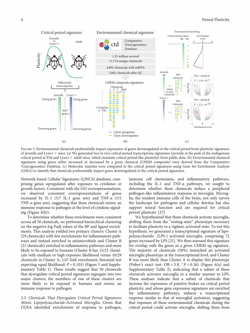
3.1. Molecular Matching of Critical Period andEnvironmental Chemical Signatures. We generated two criti-cal period signatures from transcriptomes of the primaryvisual cortex (V1) of juvenile wild-type mice during the peakof the critical period for visual cortex-mediated oculardominance plasticity at postnatal day (P) 26 [23] or adultLynx1-/- mice that have open-ended critical period plasticity
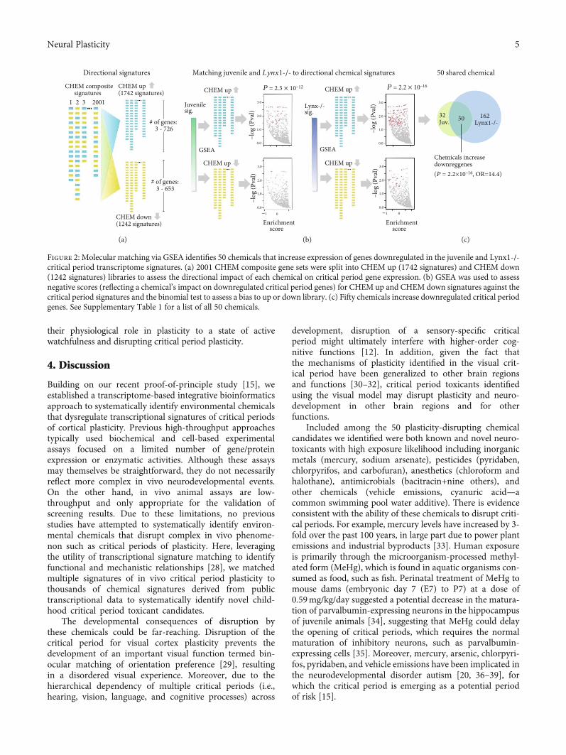
throughout life [24] in comparison with adult wild type,revealing differential expression of 9657 genes (signaturesderived from GSE89757 [16]) (Figure 1(a)). To determinethe impact of environmental chemicals on juvenile andLynx1-/- plasticity signatures, we used GSEA [25] to com-pute molecular matches of chemical gene expression signa-tures derived from the Comparative ToxicogenomicsDatabase (CTD) to critical period signatures. Using 2001composite chemical signatures (i.e., genes either increasedor decreased by a given chemical, referred to as CHEMcomposite) (Figure 1(b)), we found that chemicals were morelikely to impact the expression of genes that were downregu-lated in juvenile and Lynx1-/- critical period signatures,rather than genes that were upregulated (binomial tests:P = 1:8 × 10⁻⁴ and P < 2:2 × 10-16) (Figure 1(c)). Becauseenvironmental chemicals preferentially impact genesdownregulated in the critical period signatures, we usedGSEA to compute molecular matches between the direc-tional chemical signatures (CHEM up: sets of genesincreased by 1742 chemicals; CHEM down: sets of genesdecreased by 1242 chemicals) and assessed only negativeGSEA scores (reflecting a chemical’s impact on downregulatedcritical period genes) to find that chemicals tended to prefer-entially increase, as opposed to decrease, the expression ofgenes downregulated in both juvenile and Lynx1-/- signatures(binomial tests: P = 2:3 × 10-12 and P < 2:2 × 10-16)(Figures 2(a) and 2(b)). We focused our subsequent analysison 50 chemicals (of a total of 1742) that increased genes whoseexpression was downregulated in both of the critical periodsignatures, which was a significant overlap (Fisher’s exact test:P = 2:2 × 10-16, OR = 14:4) (Figure 2(b)). Genes downregu-lated in the critical period signatures are putative “brakes”on developmental brain plasticity, suggesting that these 50chemicals may disrupt neurodevelopment by prematurelyexpressing plasticity-dampening molecules.
3.2. Chemicals That Dysregulate Critical Period SignaturesConverge on Pathogen Response Inflammatory Pathways.The 50 chemicals shown by GSEA to impact both juvenileand Lynx1-/- signatures were diverse and included pesticides(e.g., pyridaben), antimicrobials (e.g., bacitracin), metals(e.g., mercury), anesthetics (e.g., halothane), and other com-pounds or mixtures (e.g., vehicle emissions) (SupplementaryTable 1). To gain insight into biological effects that might beshared by these diverse chemicals, we appliedchemogenomic enrichment analysis (CGEA) by calculatingoverrepresentation of biological pathways in each of the 50chemical signatures, relative to the remaining 1692 chemicalsignatures (see Figure 3 for the workflow). Using 5191 GeneOntology (GO) Biological Process (BP) gene sets, weidentified 33 BPs overrepresented in the 50 chemicalsignatures (at Padj < 0:05). CGEA enrichments of GO BPswere overwhelmingly associated to response to pathogen,immune cell chemotaxis, and inflammation (Figure 4(a)).
To understand the potential cytokine signaling by whichthese chemicals induce inflammatory responses, we com-puted overrepresentations for 96 ligand gene sets derivedfrom the Enrichr library (http://amp.pharm.mssm.edu/Enrichr/) [26] which includes the Library of Integrated
3Neural Plasticity
Network-based Cellular Signatures (LINCS) database, com-prising genes upregulated after exposure to cytokines orgrowth factors. Consistent with the GO overrepresentations,we observed consistent overrepresentations of genesincreased by IL-1 (5/7 IL-1 gene sets) and TNF-α (5/5TNF-α gene sets), suggesting that these chemicals mimic animmune response to pathogen at the level of cytokine signal-ing (Figure 4(b)).
To determine whether these enrichments were consistentacross all 50 chemicals, we performed hierarchical clusteringon the negative log Padj values of the BP and ligand enrich-ments. This analysis yielded two primary clusters: Cluster A(29 chemicals) with few enrichments for inflammatory path-ways and instead enriched in antimicrobials and Cluster B(21 chemicals) enriched in inflammatory pathways and morelikely to be exposed to humans (Cluster B has 15/21 chemi-cals with medium or high exposure likelihood versus 10/29chemicals in Cluster A; 2.07-fold enrichment; binomial testexpecting equal likelihood: P = 0:103) (Figure 5 and Supple-mentary Table 1). These results suggest that 50 chemicalsthat dysregulate critical period signatures segregate into twomajor clusters; the members of one of these clusters aremore likely to be exposed to humans and mimic animmune response to pathogen.
3.3. Chemicals That Dysregulate Critical Period SignaturesMimic Lipopolysaccharide-Activated Microglia. Given thatCGEA identified enrichments of response to pathogen,
immune cell chemotaxis, and inflammatory pathways,including the IL-1 and TNF-α pathways, we sought todetermine whether these chemicals induce a peripheralpathogen-like inflammatory response in microglia. Microg-lia, the resident immune cells of the brain, not only surveythe landscape for pathogens and cellular detritus but alsosupport neural function and are required for criticalperiod plasticity [27].
We hypothesized that these chemicals activate microglia,shifting them from the “resting-state” phenotype necessaryto facilitate plasticity to a vigilant, activated state. To test thishypothesis, we generated a transcriptional signature of lipo-polysaccharide- (LPS-) activated microglia, comprising 72genes increased by LPS [21]. We then assessed this signaturefor overlap with the genes in a given CHEM up signature.The majority of chemicals (58%) mimicked an activatedmicroglia phenotype at the transcriptional level, and ClusterB was more likely than Cluster A to display this phenotype(Fisher’s exact test: OR = 3:8, ∗P = 0:26) (Figure 6(a) andSupplementary Table 2), indicating that a subset of thesechemicals activates microglia in a similar manner to LPS.These analyses indicate that a subset of chemicals thatincrease the expression of putative brakes on critical periodplasticity, and whose gene expression signatures are enrichedfor inflammatory pathways, induces a transcriptionalresponse similar to that of microglial activation, suggestingthat exposure of these environmental chemicals during thecritical period could activate microglia, shifting them from
Critical period signatureJuvenile
Upreg genes Upreg genes
Downreg genes Downreg genes
orLynx1-/-
Adult
Differential
Juvenile Lynx1-/-
9657genes
transcriptome
Plas
ticity
(a)
Environmental chemical signature
Comparativectd Toxicogenomics
Database
1.25 million recored11,714 unique chemicals
4,892 chemicals with mRNA2,001 chemicals a�er QC
CHEM composite signaturesChemicals
1 2 3 2001
# of genes:3 - 750
Chem upregulatesChem downregulates
(b)
Transcriptional matching
Critical period chem.sig.
Upreg genes
Downreg genes
sig.Enrichment
score–1 0 +1
Juvenile2001 Dot = chemical
Dot = chemical
P = 1.8 × 10⁻⁴
P < 2.2 × 10⁻¹⁶
–1 0Enrichment score
+1
–log
(Pva
l)–l
og (P
val)
GSEAto each
chemicals
chemicals
2001chemicals
sig.
Lynx1-/-sig.
(c)
Figure 1: Environmental chemicals preferentially impact expression of genes downregulated in the critical period brain plasticity signaturesof juvenile and Lynx1-/- mice. (a) We generated two in vivo critical period transcriptome signatures (juvenile at the peak of the endogenouscritical period at P26 and Lynx1-/- adult mice, which maintain critical period-like plasticity) from public data. (b) Environmental chemicalsignatures using genes either increased or decreased by a given chemical (CHEM composite) were derived from the ComparativeToxicogenomics Database. (c) Molecular matches were computed to the critical period signatures using Gene Set Enrichment Analysis(GSEA) to identify that chemicals preferentially impact genes downregulated in the critical period signatures.
4 Neural Plasticity
their physiological role in plasticity to a state of activewatchfulness and disrupting critical period plasticity.
4. Discussion
Building on our recent proof-of-principle study [15], weestablished a transcriptome-based integrative bioinformaticsapproach to systematically identify environmental chemicalsthat dysregulate transcriptional signatures of critical periodsof cortical plasticity. Previous high-throughput approachestypically used biochemical and cell-based experimentalassays focused on a limited number of gene/proteinexpression or enzymatic activities. Although these assaysmay themselves be straightforward, they do not necessarilyreflect more complex in vivo neurodevelopmental events.On the other hand, in vivo animal assays are low-throughput and only appropriate for the validation ofscreening results. Due to these limitations, no previousstudies have attempted to systematically identify environ-mental chemicals that disrupt complex in vivo phenome-non such as critical periods of plasticity. Here, leveragingthe utility of transcriptional signature matching to identifyfunctional and mechanistic relationships [28], we matchedmultiple signatures of in vivo critical period plasticity tothousands of chemical signatures derived from publictranscriptional data to systematically identify novel child-hood critical period toxicant candidates.
The developmental consequences of disruption bythese chemicals could be far-reaching. Disruption of thecritical period for visual cortex plasticity prevents thedevelopment of an important visual function termed bin-ocular matching of orientation preference [29], resultingin a disordered visual experience. Moreover, due to thehierarchical dependency of multiple critical periods (i.e.,hearing, vision, language, and cognitive processes) across
development, disruption of a sensory-specific criticalperiod might ultimately interfere with higher-order cog-nitive functions [12]. In addition, given the fact thatthe mechanisms of plasticity identified in the visual crit-ical period have been generalized to other brain regionsand functions [30–32], critical period toxicants identifiedusing the visual model may disrupt plasticity and neuro-development in other brain regions and for otherfunctions.
Included among the 50 plasticity-disrupting chemicalcandidates we identified were both known and novel neuro-toxicants with high exposure likelihood including inorganicmetals (mercury, sodium arsenate), pesticides (pyridaben,chlorpyrifos, and carbofuran), anesthetics (chloroform andhalothane), antimicrobials (bacitracin+nine others), andother chemicals (vehicle emissions, cyanuric acid—acommon swimming pool water additive). There is evidenceconsistent with the ability of these chemicals to disrupt criti-cal periods. For example, mercury levels have increased by 3-fold over the past 100 years, in large part due to power plantemissions and industrial byproducts [33]. Human exposureis primarily through the microorganism-processed methyl-ated form (MeHg), which is found in aquatic organisms con-sumed as food, such as fish. Perinatal treatment of MeHg tomouse dams (embryonic day 7 (E7) to P7) at a dose of0.59mg/kg/day suggested a potential decrease in the matura-tion of parvalbumin-expressing neurons in the hippocampusof juvenile animals [34], suggesting that MeHg could delaythe opening of critical periods, which requires the normalmaturation of inhibitory neurons, such as parvalbumin-expressing cells [35]. Moreover, mercury, arsenic, chlorpyri-fos, pyridaben, and vehicle emissions have been implicated inthe neurodevelopmental disorder autism [20, 36–39], forwhich the critical period is emerging as a potential periodof risk [15].
Directional signatures 50 shared chemical
Chemicals increase downreggenes(P = 2.2×10⁻¹⁶, OR=14.4)
32Juv.
162Lynx1-/-50
GSEA
Juvenilesig.
CHEM up
CHEM up
–log
(Pva
l)–l
og (P
val)
–log
(Pva
l)–l
og (P
val)
P = 2.3 × 10⁻¹² P = 2.2 × 10⁻¹⁶CHEM up
CHEM up
GSEA
Lynx-/- sig.
Enrichmentscore
Enrichmentscore
–1 0 –1 0
Matching juvenile and Lynx1-/- to directional chemical signatures
3.0
2.0
1.0
0.0
3.0
2.0
1.0
0.0
3.0
2.0
1.0
0.0
3.0
2.0
1.0
0.0
CHEM composite signatures
20011 2 3
# of genes: 3 - 726
# of genes: 3 - 653
CHEM up (1742 signatures)
CHEM down (1242 signatures)
(a) (b) (c)
Figure 2: Molecular matching via GSEA identifies 50 chemicals that increase expression of genes downregulated in the juvenile and Lynx1-/-critical period transcriptome signatures. (a) 2001 CHEM composite gene sets were split into CHEM up (1742 signatures) and CHEM down(1242 signatures) libraries to assess the directional impact of each chemical on critical period gene expression. (b) GSEA was used to assessnegative scores (reflecting a chemical’s impact on downregulated critical period genes) for CHEM up and CHEM down signatures against thecritical period signatures and the binomial test to assess a bias to up or down library. (c) Fifty chemicals increase downregulated critical periodgenes. See Supplementary Table 1 for a list of all 50 chemicals.
5Neural Plasticity

A large portion of critical period-disrupting candidateswere antimicrobials (10 of 50) indicating that the down-stream pathways of antimicrobials may ultimately impact
brain development. Bacitracin is used in humans as an anti-biotic as well as in commercial farming practices to controlmicrobes and in the feed of swine, chickens, and other live-stock to promote growth [40]. Given the widespread admin-istration of antibiotics to livestock for human consumption,there is considerable concern about the impact of residualantibiotic in animal products and its impact on human health[41]. Moreover, there is a growing recognition of the impor-tance of the microbiome-immune-neural axis on health anddisease and antibiotics can profoundly disrupt healthymicrobiomes [42]. Bacitracin disrupts the microbiome andimpacts BDNF levels [43], a growth factor involved in theopening of the visual critical period [35].
Given the diversity of the 50 candidate plasticity disrup-tors, we applied a chemogenomic enrichment analysis(CGEA) approach to identify shared pathways among thesechemicals, which included response to pathogen, immunecell chemotaxis, and inflammatory pathways including IL-1and TNF-α cytokine signaling. This suggests that chemicalsthat disrupt critical period plasticity may be perceived asinvaders by the immune system, leading to induction of aninflammatory response. In the brain, this may involve activa-tion of microglia. Should this occur during the critical period,it might shift microglia away from their physiological role inexperience-dependent critical period plasticity [27] to a stateof active watchfulness in which they are not able to facilitateexperience-dependent brain development. Upon exposure totoxicants such as ozone and acetaminophen, peripheralimmune cells (e.g., macrophages) activate and induce aninflammatory response that includes cytokines such asTNF, mimicking the response to Gram-negative bacterialpathogens [44]. Given the role of TNF in activating microglia[45–47], soluble transport of TNF across the blood-brainbarrier [48] from peripherally stimulated immune cells couldactivate the stimulus. Future studies should assess the abilityof individual chemicals to activate microglia and disrupt crit-ical period plasticity.
This study was limited by the quality and breadth ofavailable chemical data, and a future work will benefit fromthe toxicology in the 21st Century (Tox21) program, anongoing effort to systematically profile the effect of tens ofthousands of chemicals on the expression of 1500 genes incell lines [49]. The chemical data used here were derivedfrom heterogeneous tissues in multiple animal and cellmodels, not specifically focused on neurons or the brain[50]. Hence, specificity for neuronal phenotypes could beimproved by extending current efforts to screen for damag-ing effects of toxicants in human cell lines [51] to neuronsderived from human-induced pluripotent stem cells (iPSCs).Finally, we limited ourselves to two models of critical periodplasticity; additional models, such as voluntary exercise-induced plasticity [52], may reveal additional insight regard-ing the mechanisms that can disrupt critical period plasticity.
In summary, we established an integrative bioinformaticsparadigm for generating rational hypotheses about theimpact of environmental chemicals on critical periods ofbrain plasticity, as well as their underlying mechanisms, withthe goal of identifying targets for therapeutic intervention.This approach could be generalized to other brain
Chemogenomic enrichment analysis(CGEA)
CHEM up (1742 chemical signatures)
# of genes: 3 - 726
Pathway gene sets
GO BP(5191 gene sets)
LINCS ligands(96 gene sets)
Pathway enrichments(GO BP or LINCS ligand gene sets)
GO BPor
LINCS ligands
123
N
Chemicals1 2 3 … 1742 Padj
Low
High
Overrep in 50 chem vs others
50 CP chems 1692 other chemsvs
vs
123
N
123
Netc until N
Top hits(Fig 4)
Clustering(Fig 5)
(a)
(b)
(c)
Figure 3: Chemogenomic enrichment analysis (CGEA) workflow.(a) Enrichments of 5191 Gene Ontology (GO) Biological Process(BP) and 96 Library of Integrated Network-based CellularSignatures (LINCS) ligand gene sets were calculated for 1742CHEM up signatures. (b) We calculated overrepresentation ofpathways in each of 50 chemical signatures that impact criticalperiod signatures, relative to the remaining 1692 chemicalsignatures. (c) Top overrepresentation hits were calculated(Figure 4), and hierarchical clustering was performed onenrichment Padj values (Figure 5).
6 Neural Plasticity

Gene Ontology Biological Process ORLeukocyte migration in inflammatory response (G0:0002523) 15.8
Defense response to fungus (G0:0050832) 17.1Sequestering of metal ion (G0:0051238) 14.2
Homotypic cell-cell adhesion (G0:0034109) 8.7Leukocyte aggregation (G0:0070486) 8.9
neg. reg. of endothelial cell apoptotic process (G0:2000352) 5.7Negative regulation of locomotion (G0:0040013) 4.4
Chronic inflammatory response (G0:0002544) 4.3reg. of adhesion-dependent cell spreading (G0:1900024) 17.6
Exocytosis (G0:0006887) 4.1Glial cell migration (G0:0008347) 6.0
pos. reg. of adhesion-dependent cell spreading (G0:1900026) 14.3
Negative regulation of cell migration (G0:0030336) 3.8Negative regulation of cell motility (G0:2000146) 3.8
Negative regulation of cellular component movemen1 (G0:0051271) 3. 7
Neutrophil migration (G0:1990266) 3.4Granulocyte migration (G0:0097530) 3.4
Neutrophil chemotaxis (G0:0030593) 3.4Coagulation (G0:0050817) 3.3
Granulocyte chemotaxis (G0:0071621) 3.3Platelet degranulation (G0:0002576) 3.4
Hemostasis (G0:0007599) 3.3Blood coagulation (G0:0007596) 3.3
neg. reg. of ext. apoptotic signaling by death domain (G0:1902042) 3. 7Positive regulation of Ras GTPase activity (G0:0032320) 10.9
Regulation of Ras GTPase activity (G0:0032318) 10.9 Positive regulation of vasoconstriction (G0:0045907) 4.3
Platelet activation (G0:0030168) 3.1Positive regulation of heterotypic cell-cell adhesion (G0:0034116) 3.2
Response to fungus (G0:0009620) 5.2�ymic T cell selection (G0:0045061) 18.3
Acute Inflammatory response (G0:0002526) 3.1Protein activation cascade (G0:0072376) 6.6
–Log10 (P-adj)0 1 2 3 4 5 6 7
(a)
Ligand OREPG-MCF7 6.7
PDGFBB-SKBR3 6.1 MSP-HS576T 6.4
HGF-MCF7 5.3 TNFA-MCF10A 4.4
IL1-MCF10A 4.3 MCSF-MDAMB231 4.1
SCF-SKBR3 3.9 GAS6-HS578T 3.7
GAS6-MDAMB231 3.7 INS-SKBR3 3.3
PDGFBB-BT20 3.9 GAS6-BT20 3.2
BTC-MDAMB231 3.2 IL 1-MDAMB231 3.1
BNGF-BT203.0 IL17-BT20 2.9
IFNG-MDAMB231 2.7 IL4-HS578T 2.6
IL1-SKBR3 2.5 IL1-HS578T 2.7
HRG-MDAMB231 2.8 TNFA-SKBR3 2.5
TNFA-HS578T 2.5 HGF-HS578T 2.4
IL6-SKBR3 2.4 GAS6-MCF7 2.9 IFNG-MCF7 2.6
IFNG-MCF1 OA 2.4IFNA-MCF7 2.7
IL1-BT20 2.4 BFGF-MCF7 2.2 MSP-SKBR3 2.1
HRG-BT20 2.3 BTC-SKBR3 2.0
–log10 (P-adj)0 1 2 3 4 5 6 7
(b)
Figure 4: Chemogenomic enrichment analysis of 50 chemicals that increase expression of genes downregulated in the critical periodsignatures reveals inflammatory, response to pathogen, and immune cell chemotaxis pathways. We computed gene set enrichments for theCHEM up library (1742 chemical signatures) across 5191 Gene Ontology (GO) Biological Process (BP) gene sets and 96 LINCS ligandgene sets to yield 9,042,722 and 167,232 enrichment P values, which were corrected for multiple testing using the Benjamini andHochberg approach. For each biological process or ligand, we calculated the overrepresentation of that gene set (if it was significant aftermultiple test correction) among the 50 chemicals identified as impacting both juvenile and Lynx1-/- critical period signatures, incomparison to the remaining 1692 chemicals, using a hypergeometric test (hypergea R package implementation). A pathway wasconsidered associated with a chemical if the enrichment Padj < 0:05, yielding (a) 33 GO BP gene sets and (b) 48 LINCS ligand gene sets.
Pict
ilisib
Fluc
onaz
ole
Clor
gylin
eCa
jani
nstil
bene
acid
Mon
obut
yryl
cyc
lic A
MP
Chlo
rpyr
ifos
Am
itrip
tylin
eCy
anur
ic ac
idIs
onia
zid
IMM
125
Adr
enoc
ortic
otro
pin
zinc
Nys
tatin
Nat
amyc
inQ
uini
dine
SB−4
3154
24-
Oxo
retin
oic a
cid
Pyrid
aben
Carb
amaz
epin
eD
oxyc
yclin
eA
brin
eD
emec
olci
neTr
ovafl
oxac
inCh
loro
pren
eM
ecam
ylam
ine
Pant
ogab
Ani
line
Asta
xant
hine
Baci
trac
inCo
rtic
oste
rone
Vin
blas
tine
Buth
ioni
ne su
lfoxi
min
eCh
loro
form
Vehi
cle e
miss
ions
Isop
rote
reno
lTr
imet
hylti
nPo
tass
ium
bro
mat
eQ
uartz
Dex
tran
sulfa
teLa
tex
Dic
lofe
nac
Hal
otha
neCI
104
4Te
trac
yclin
eD
obut
amin
eM
elam
ine
Carb
ofur
anCo
cain
eSo
dium
arse
nate
Asb
esto
s, se
rpen
tine
Mer
cury
Class ExposureHiMedLow
PesticideAntimicrobialMetalAnestheticOther
-Log(Padj)
151050
403020100
Response to pathogen, immune cell axis, inflammation pathways
Il-1 (5/7 sigs)TNF-a (5/5 sigs)
See Fig S1 for detailed clustering results
GO
BP
Liga
nds
Cluster A Cluster B
Figure 5: Clustering of chemical pathway enrichments identifies antimicrobial and inflammatory clusters. Hierarchical clustering (Ward Dmethod) on the negative log Padj values of Gene Ontology (GO) Biological Process (BP) and LINCS ligand enrichment analysis revealed twoclusters of chemicals. Cluster A (29 chemicals) contains few inflammatory pathway enrichments and 9 of the 10 antimicrobials in the set of 50chemicals examined, whereas Cluster B contains the majority of enrichments for response to pathogen, inflammation, immune cellchemotaxis, and IL-1/TNF-α. See Supplementary Figure 1 for detailed enrichment information.
7Neural Plasticity

phenotypes, allowing systematic assessment of the impact ofchemicals on a wide array of brain development phenotypes.
Data Availability
The data used to support the findings of this study areincluded within the article.
Conflicts of Interest
The authors declare no competing interests.
Authors’ Contributions
MRS, JTD, and HM designed the study; MRS, PY, and BRperformed the analyses; MRS, BK, JTD, and HM providedthe principal interpretations; MRS, JTD, and HM wrote themanuscript; and all authors approved the final manuscript.
Acknowledgments
This work was funded by a Traineeship, National Institute ofChild Health and Human Development-InterdisciplinaryTraining in Systems and Developmental Biology and BirthDefects Grant T32H-D0-75735 (to M.R.S.); the MindichChild Health and Development Institute Pilot Fund (toJ.T.D. and H.M.); the Knights Templar Eye Foundation (toH.M.); the March of Dimes (to H.M.); theWhitehall Founda-tion (to H.M.); the Harris Family Foundation (to J.T.D.); andthe National Institutes of Health Grants P30-ES-023515 (toJ.T.D. and H.M.), R01-DK-098242, U54-CA189201, andR56-AG058469 (to J.T.D.), and R01-EY-024918, R01-EY-026053, and R21 MH106919 (to H.M.).
Supplementary Materials
Supplementary Table 1: chemicals that commonly increaseputative brakes of both juvenile and Lynx1-KO tran-scriptome signatures. Supplementary Table 2: enrichmentstatistics for fifty chemicals that mimic the gene expressionphenotype induced by LPS-activated microglia (sorted byan ascending P value). Supplementary Figure 1: expandedresults of hierarchical clustering of GO BP and LINCS ligandCGEA for each of the 50 plasticity-disrupting candidate com-pounds (related to Figure 5). (Supplementary Materials)
References
[1] CAS, CAS REGISTRY - the gold standard for chemical sub-stance information, American Chemical Society, 2018.
[2] D. Markell, “An overview of TSCA, its history and key under-lying assumptions, and its place in environmental regulation,”Washington University Journal of Law & Policy, vol. 32,pp. 333–375, 2010.
[3] C. A. Boyle, S. Boulet, L. A. Schieve et al., “Trends in the prev-alence of developmental disabilities in US children, 1997–2008,” Pediatrics, vol. 127, no. 6, pp. 1034–1042, 2011.
[4] D. Rice and S. Barone Jr., “Critical periods of vulnerability forthe developing nervous system: evidence from humans andanimal models,” Environmental Health Perspectives, vol. 108,Suppl 3, pp. 511–533, 2000.
[5] S. E. Fox, P. Levitt, and C. A. Nelson III, “How the timing andquality of early experiences influence the development of brainarchitecture,” Child Development, vol. 81, no. 1, pp. 28–40,2010.
[6] J. S. Johnson and E. L. Newport, “Critical period effects in sec-ond language learning: the influence of maturational state onthe acquisition of English as a second language,” Cognitive Psy-chology, vol. 21, no. 1, pp. 60–99, 1989.
[7] T. L. Lewis and D. Maurer, “Multiple sensitive periods inhuman visual development: evidence from visually deprivedchildren,” Developmental Psychobiology, vol. 46, no. 3,pp. 163–183, 2005.
[8] C. A. Nelson, C. H. Zeanah, N. A. Fox, P. J. Marshall, A. T.Smyke, and D. Guthrie, “Cognitive recovery in sociallydeprived young children: the Bucharest Early InterventionProject,” Science, vol. 318, no. 5858, pp. 1937–1940, 2007.
[9] T. P. Nikolopoulos, G. M. O'Donoghue, and S. Archbold, “Ageat implantation: its importance in pediatric cochlear implanta-tion,” The Laryngoscope, vol. 109, no. 4, pp. 595–599, 1999.
[10] E. A. Schorr, N. A. Fox, V. vanWassenhove, and E. I. Knudsen,“Auditory-visual fusion in speech perception in children withcochlear implants,” Proceedings of the National Academy ofSciences of the United States of America, vol. 102, no. 51,pp. 18748–18750, 2005.
[11] J. J. LeBlanc and M. Fagiolini, “Autism: a “critical period” dis-order?,” Neural Plasticity, vol. 2011, Article ID 921680, 17pages, 2011.
[12] A. E. Takesian and T. K. Hensch, “Chapter 1 - balancing plas-ticity/stability across brain development,” in Progress in BrainResearch, M. N. M. M. Merzenich and M. V. V. Thomas, Eds.,pp. 3–34, Elsevier, 2013.
Pict
ilisib
Fluc
onaz
ole
Clor
gylin
eCa
jani
nstil
bene
acid
Mon
obut
yryl
cycl
ic A
MP
Chlo
rpyr
ifos
Am
itrip
tylin
eCy
anur
ic ac
idIs
onia
zid
IMM
125
Adr
enoc
ortic
otro
pin
zinc
Nys
tatin
Nat
amyc
inQ
uini
dine
SB−4
3154
24-
Oxo
retin
oic a
cid
Pyrid
aben
Carb
amaz
epin
eD
oxyc
yclin
eA
brin
eD
emec
olci
neTr
ovafl
oxac
inCh
loro
pren
eM
ecam
ylam
ine
Pant
ogab
Ani
line
Asta
xant
hine
Baci
trac
inCo
rtic
oste
rone
Vin
blas
tine
Buth
ioni
ne su
lfoxi
min
eCh
loro
form
Vehi
cle e
miss
ions
Isop
rote
reno
lTr
imet
hylti
nPo
tass
ium
bro
mat
eQ
uartz
Dex
tran
sulfa
teLa
tex
Dic
lofe
nac
Hal
otha
neCI
104
4Te
trac
yclin
eD
obut
amin
eM
elam
ine
Carb
ofur
anCo
cain
eSo
dium
arse
nate
Asb
esto
s, se
rpen
tine
Mer
cury
Cluster A Cluster B
-Log(Padj)
840
⁎
Figure 6: Fifty chemicals mimic the gene expression phenotype induced by LPS-activated microglia. We used Fisher’s exact test to calculatethe overlap of microglia genes increased by LPS activation to the genes in a given CHEM up signature. 58% of all chemicals were enriched (atPadj < 0:05), and Cluster B was more likely than Cluster A to display this phenotype (Fisher’s exact test: OR = 3:8, ∗P = 0:26). Chemicalsordered as in Figure 5.
8 Neural Plasticity

[13] E. A. Kelly, L. A. Opanashuk, and A. K. Majewska, “The effectsof postnatal exposure to low-dose bisphenol-A on activity-dependent plasticity in the mouse sensory cortex,” Frontiersin Neuroanatomy, vol. 8, 2014.
[14] A. E. Medina and A. S. Ramoa, “Early alcohol exposureimpairs ocular dominance plasticity throughout the criticalperiod,” Developmental Brain Research, vol. 157, no. 1,pp. 107–111, 2005.
[15] M. R. Smith, P. Yevoo, M. Sadahiro et al., “Integrative bioin-formatics identifies postnatal lead (Pb) exposure disruptsdevelopmental cortical plasticity,” Scientific Reports, vol. 8,no. 1, p. 16388, 2018.
[16] M. R. Smith, P. Burman, M. Sadahiro, B. A. Kidd, J. T. Dudley,and H. Morishita, “Integrative analysis of disease signaturesshows inflammation disrupts juvenile experience-dependentcortical plasticity,” eNeuro, vol. 3, no. 6, pp. ENEURO.0240–ENEU16.2016, 2016.
[17] G. K. Smyth, “Limma: linear models for microarray data,” inBioinformatics and Computational Biology Solutions Using Rand Bioconductor, pp. 397–420, Springer, New York, 2005.
[18] J. Lamb, E. D. Crawford, D. Peck et al., “The connectivity map:using gene-expression signatures to connect small molecules,genes, and disease,” Science, vol. 313, no. 5795, pp. 1929–1935, 2006.
[19] M. Bönn, Hypergea: hypergeometric tests, R Foundation, 2016.
[20] B. L. Pearson, J. M. Simon, E. S. McCoy, G. Salazar, G. Fragola,and M. J. Zylka, “Identification of chemicals that mimic tran-scriptional changes associated with autism, brain aging andneurodegeneration,” Nature Communications, vol. 7, no. 1,2016.
[21] M. L. Bennett, F. C. Bennett, S. A. Liddelow et al., “New toolsfor studying microglia in the mouse and human CNS,” Pro-ceedings of the National Academy of Sciences, vol. 113, no. 12,pp. E1738–E1746, 2016.
[22] Y. Benjamini and Y. Hochberg, “Controlling the false discov-ery rate: a practical and powerful approach to multiple test-ing,” Journal of the Royal Statistical Society. Series B(Methodological), vol. 57, no. 1, pp. 289–300, 1995.
[23] J. A. Gordon and M. P. Stryker, “Experience-dependent plas-ticity of binocular responses in the primary visual cortex ofthe mouse,” The Journal of Neuroscience, vol. 16, no. 10,pp. 3274–3286, 1996.
[24] H. Morishita, J. M. Miwa, N. Heintz, and T. K. Hensch,“Lynx1, a cholinergic brake, limits plasticity in adult visualcortex,” Science, vol. 330, no. 6008, pp. 1238–1240, 2010.
[25] A. Subramanian, P. Tamayo, V. K. Mootha et al., “Gene setenrichment analysis: a knowledge-based approach for inter-preting genome-wide expression profiles,” Proceedings of theNational Academy of Sciences, vol. 102, no. 43, pp. 15545–15550, 2005.
[26] E. Y. Chen, C. M. Tan, Y. Kou et al., “Enrichr: interactive andcollaborative HTML5 gene list enrichment analysis tool,”BMC Bioinformatics, vol. 14, no. 1, p. 128, 2013.
[27] G. O. Sipe, R. L. Lowery, M. È. Tremblay, E. A. Kelly, C. E.Lamantia, and A. K. Majewska, “Microglial P2Y12 is necessaryfor synaptic plasticity in mouse visual cortex,”Nature Commu-nications, vol. 7, no. 1, 2016.
[28] R. A. Hodos, B. A. Kidd, K. Shameer, B. P. Readhead, and J. T.Dudley, “In silico methods for drug repurposing and pharma-cology,” Wiley Interdisciplinary Reviews: Systems Biology andMedicine, vol. 8, no. 3, pp. 186–210, 2016.
[29] B.-S. Wang, R. Sarnaik, and J. Cang, “Critical period plasticitymatches binocular orientation preference in the visual cortex,”Neuron, vol. 65, no. 2, pp. 246–256, 2010.
[30] C. N. Levelt and M. Hübener, “Critical-period plasticity in thevisual cortex,” Annual Review of Neuroscience, vol. 35, no. 1,pp. 309–330, 2012.
[31] E. M. Nabel and H. Morishita, “Regulating critical periodplasticity: insight from the visual system to fear circuitryfor therapeutic interventions,” Frontiers in Psychiatry, vol. 4,2013.
[32] J. F. Werker and T. K. Hensch, “Critical periods in speech per-ception: new directions,” Annual Review of Psychology, vol. 66,no. 1, pp. 173–196, 2015.
[33] P. W. Davidson, G. J. Myers, and B. Weiss, “Mercury exposureand child development outcomes,” Pediatrics, vol. 113, 4Suppl, pp. 1023–1029, 2004.
[34] J. Umemori, F. Winkel, E. Castrén, and N. N. Karpova, “Dis-tinct effects of perinatal exposure to fluoxetine or methylmer-cury on parvalbumin and perineuronal nets, the markers ofcritical periods in brain development,” International Journalof Developmental Neuroscience, vol. 44, no. C, pp. 55–64, 2015.
[35] Z. J. Huang, A. Kirkwood, T. Pizzorusso et al., “BDNF regu-lates the maturation of inhibition and the critical period ofplasticity in mouse visual cortex,” Cell, vol. 98, no. 6,pp. 739–755, 1999.
[36] P. J. Landrigan, L. Lambertini, and L. S. Birnbaum, “A researchstrategy to discover the environmental causes of autism andneurodevelopmental disabilities,” Environmental Health Per-spectives, vol. 120, no. 7, pp. a258–a260, 2012.
[37] K. Thirtamara Rajamani, S. Doherty-Lyons, C. Bolden et al.,“Prenatal and early-life exposure to high-level diesel exhaustparticles leads to increased locomotor activity and repetitivebehaviors in mice,” Autism Research, vol. 6, no. 4, pp. 248–257, 2013.
[38] H. E. Volk, I. Hertz-Picciotto, L. Delwiche, F. Lurmann, andR. McConnell, “Residential proximity to freeways and autismin the CHARGE study,” Environmental Health Perspectives,vol. 119, no. 6, pp. 873–877, 2011.
[39] G. C. Windham, L. Zhang, R. Gunier, L. A. Croen, and J. K.Grether, “Autism spectrum disorders in relation to distribu-tion of hazardous air pollutants in the San Francisco BayArea,” Environmental Health Perspectives, vol. 114, no. 9,pp. 1438–1444, 2006.
[40] C. E. Dewey, B. D. Cox, B. E. Straw, E. J. Bush, and S. Hurd,“Use of antimicrobials in swine feeds in the United States,”Journal of Swine Health and Production, vol. 7, no. 1, pp. 19–25, 1999.
[41] M. D. Barton, “Antibiotic use in animal feed and its impact onhuman healt,” Nutrition Research Reviews, vol. 13, no. 2,pp. 279–299, 2000.
[42] T. C. Fung, C. A. Olson, and E. Y. Hsiao, “Interactions betweenthe microbiota, immune and nervous systems in health anddisease,” Nature Neuroscience, vol. 20, no. 2, pp. 145–155,2017.
[43] P. Bercik, E. Denou, J. Collins et al., “The intestinal microbiotaaffect central levels of brain-derived neurotropic factor andbehavior in mice,” Gastroenterology, vol. 141, no. 2, pp. 599–609.e3, 2011.
[44] D. L. Laskin and J. D. Laskin, “Role of macrophages andinflammatory mediators in chemically induced toxicity,” Tox-icology, vol. 160, no. 1-3, pp. 111–118, 2001.
9Neural Plasticity

[45] A. J. Bruce, W. Boling, M. S. Kindy et al., “Altered neuronaland microglial responses to excitotoxic and ischemic braininjury in mice lacking TNF receptors,” Nature Medicine,vol. 2, no. 7, pp. 788–794, 1996.
[46] R. Kuno, J. Wang, J. Kawanokuchi, H. Takeuchi, T. Mizuno,and A. Suzumura, “Autocrine activation of microglia by tumornecrosis factor-α,” Journal of Neuroimmunology, vol. 162,no. 1-2, pp. 89–96, 2005.
[47] K. Sriram, J. M. Matheson, S. A. Benkovic, D. B. Miller, M. I.Luster, and J. P. O'Callaghan, “Deficiency of TNF receptorssuppresses microglial activation and alters the susceptibilityof brain regions to MPTP-induced neurotoxicity: role ofTNF‐α1,” The FASEB Journal, vol. 20, no. 6, pp. 670–682,2006.
[48] W. A. Banks, A. J. Kastin, and R. D. Broadwell, “Passage ofcytokines across the blood-brain barrier,” Neuroimmunomo-dulation, vol. 2, no. 4, pp. 241–248, 1995.
[49] D. Mav, R. R. Shah, B. E. Howard et al., “A hybrid gene selec-tion approach to create the S1500+ targeted gene sets for use inhigh-throughput transcriptomics,” PLoS One, vol. 13, no. 2,article e0191105, 2018.
[50] A. P. Davis, C. J. Grondin, K. Lennon-Hopkins et al., “Thecomparative toxicogenomics database's 10th year anniversary:update 2015,” Nucleic Acids Research, vol. 43, no. D1,pp. D914–D920, 2014.
[51] F. Eduati, L. M. Mangravite, T. Wang et al., “Prediction ofhuman population responses to toxic compounds by a collab-orative competition,” Nature Biotechnology, vol. 33, no. 9,pp. 933–940, 2015.
[52] E. Kalogeraki, F. Greifzu, F. Haack, and S. Löwel, “Voluntaryphysical exercise promotes ocular dominance plasticity inadult mouse primary visual cortex,” Journal of Neuroscience,vol. 34, no. 46, pp. 15476–15481, 2014.
10 Neural Plasticity