synthetic biodegradable polymers as drug delivery systems for bone morphogenetic proteins
TRANSCRIPT

www.elsevier.com/locate/addr
Advanced Drug Delivery Revie
Synthetic biodegradable polymers as drug delivery systems for
bone morphogenetic proteins
N. Saitoa,T, N. Murakamib, J. Takahashib, H. Horiuchib, H. Otab, H. Katob,
T. Okadac, K. Nozakid, K. Takaokae
aDepartment of Physical Therapy, Shinshu University School of Health Sciences, 3-1-1 Asahi, Matsumoto, Nagano 390-8621, JapanbDepartment of Orthopaedic Surgery, Shinshu University School of Medicine, 3-1-1 Asahi, Matsumoto, Nagano 390-8621, Japan
cResearch Institute, Taki Chemical Co., Ltd., 64-1 Nishiwaki, Befucho, Kakogawa, Hyogo 675-0125, JapandApplied Pharmacology Laboratories, Institute for Drug Discovery Research, Yamanouchi Pharmaceutical Co., Ltd., 21 Miyukigaoka,
Tsukuba, Ibaraki 305-8585, JapaneDepartment of Orthopaedic Surgery, Osaka City University Graduate School of Medicine, 1-5-7 Asahimachi,
Abeno-ku, Osaka 545-0051, Japan
Received 8 April 2004; accepted 30 December 2004
Abstract
Bone morphogenetic proteins (BMP) induce bone formation in vivo, and clinical application in repair of bone fractures and
defects is expected. However, appropriate systems to deliver BMP for clinical use need to be developed. We synthesized a new
synthetic biodegradable polymer, poly-d,l-lactic acid-para-dioxanone-polyethylene glycol block copolymer (PLA-DX-PEG),
to serve as a biocompatible, biodegradable polymer for recombinant human (rh) BMP-2 delivery systems. In animal
experiments, new bone was efficiently formed and a large bone defect was repaired using PLA-DX-PEG/rhBMP-2 composites.
In addition, this new polymer could be used as an injectable delivery system for rhBMP-2. The rhBMP-2/PLA-DX-PEG
composites also could be combined with other materials such as hydroxyapatite or titanium. This new synthetic polymer might
be used for rhBMP-2 delivery in various clinical situations involving repair of bone, leading to great changes in orthopedic
treatment.
D 2005 Elsevier B.V. All rights reserved.
Keywords: Bone formation; Bone repair; Fracture; Bone defect; Recombinant human bone morphogenetic protein-2; Tissue engineering
0169-409X/$ - s
doi:10.1016/j.ad
T Correspondi
E-mail addr
ws 57 (2005) 1037–1048
ee front matter D 2005 Elsevier B.V. All rights reserved.
dr.2004.12.016
ng author. Tel./fax: +81 263 37 2409.
ess: [email protected] (N. Saito).

N. Saito et al. / Advanced Drug Delivery Reviews 57 (2005) 1037–10481038
Contents
1. Introduction . . . . . . . . . . . . . . . . . . . . . . . . . . . . . . . . . . . . . . . . . . . . . . . . . . . 1038
2. Bone morphogenetic proteins (BMP) and delivery systems . . . . . . . . . . . . . . . . . . . . . . . . . . . 1038
2.1. BMP . . . . . . . . . . . . . . . . . . . . . . . . . . . . . . . . . . . . . . . . . . . . . . . . . . . 1038
2.2. Delivery systems for BMP . . . . . . . . . . . . . . . . . . . . . . . . . . . . . . . . . . . . . . . . 1039
2.3. Synthetic polymers for BMP delivery system . . . . . . . . . . . . . . . . . . . . . . . . . . . . . . 1039
3. Development of new synthetic biodegradable polymers for rhBMP-2 delivery . . . . . . . . . . . . . . . . . 1040
3.1. Poly-d,l-lactic acid-polyethylene glycol block copolymer (PLA-PEG) . . . . . . . . . . . . . . . . . 1040
3.2. Poly-d,l-lactic acid-para-dioxanone-polyethylene glycol block copolymer (PLA-DX-PEG). . . . . . . 1040
4. Repair of bone tissues using rhBMP-2 and new synthetic polymers . . . . . . . . . . . . . . . . . . . . . . 1042
4.1. Repair of bone defect using composites of rhBMP-2 and synthetic polymers . . . . . . . . . . . . . . 1042
4.2. Injectable polymeric delivery systems for rhBMP-2 . . . . . . . . . . . . . . . . . . . . . . . . . . . 1043
4.3. Combination of the rhBMP-2/polymer composites with other materials . . . . . . . . . . . . . . . . . 1044
4.4. Development of a new artificial joint that restores a bone defect . . . . . . . . . . . . . . . . . . . . 1045
5. Conclusions . . . . . . . . . . . . . . . . . . . . . . . . . . . . . . . . . . . . . . . . . . . . . . . . . . . 1045
Acknowledgements . . . . . . . . . . . . . . . . . . . . . . . . . . . . . . . . . . . . . . . . . . . . . . . . . . 1046
References . . . . . . . . . . . . . . . . . . . . . . . . . . . . . . . . . . . . . . . . . . . . . . . . . . . . . . 1046
1. Introduction
The regeneration potential of human bone appears
to be limited, given that repair of large bone defects
such as those associated with comminuted fractures or
bone tumor resection usually remains unrepaired [1].
Such cases have been treated routinely with autogeneic
or allogeneic bone grafting. Major problems associated
with autogeneic grafting include limited anatomic
sources of donor bone and risk of morbidity from the
additional surgery for procurement of the graft. In
allogeneic bone grafting, major concerns are potential
risks of transmission of disease, immunologic reaction
of the host, poor osteogenic capacity of the trans-
planted bone, and high costs associated with a bone
banking system [2–4]. Current examination of alter-
natives to grafting techniques suggests three possible
new approaches to inducing new bone formation:
implantation of certain cytokines such as bone
morphogenetic proteins (BMP) in combination with
appropriate delivery systems at the target site [5–7];
transduction of genes encoding cytokines with osteo-
genic capacity into cells at repair sites [8,9]; and
transplantation of cultured osteogenic cells derived
from host bone marrow [10–13]. In our estimation, the
second approach represents the next major advance,
while the third requires considerable additional resour-
ces and time to procure and culture cells. The first
strategy appears to show the most practical promise for
the near future. Appropriate delivery systems are
essential to this technique. In this review, we outline
the development of new delivery systems for BMP and
preclinical animal experiments concerning bone tissue
regeneration that suggest clinical applications.
2. Bone morphogenetic proteins (BMP) and
delivery systems
2.1. BMP
BMP induce new bone formation by directing
mesenchymal stem cells toward chondroblastic and
osteoblastic differentiation, and causing them to pro-
liferate in vivo. BMP expression has been confirmed to
occur at the initial stage of the fracture healing process,
and to participate in a cascade regulating bone repair
processes. Also, new bone can be induced to form
heterotopically, such as when the BMP are implanted in
muscle in animal models using appropriate delivery
systems. These observations suggest that BMP could
be applied clinically to promotion of repair of bone.
BMP were first characterized in 1965 by Urist as a
biologically active molecule inducing new ectopic
bone formation from decalcified bone matrix in vivo
[14]. A cDNA encoding BMP was cloned by Wozney

N. Saito et al. / Advanced Drug Delivery Reviews 57 (2005) 1037–1048 1039
in 1988, and BMP were found to be a dimeric protein
with a molecular weight of about 32,000 [15–17]. An
important common feature of the BMPmolecules is the
position of cysteine residues in relation to the carboxyl
terminus. The positions of these seven cysteine
residues are the same as those in transforming growth
factor (TGF)-h, indicating that BMP molecules are
members of the TGF-h superfamily [18]. Today, the
BMP family consists of about 15 BMP [19].
BMP include bone formation during embryogene-
sis, growth, and adulthood. In fracture healing,
osteoprogenitor cells can respond to BMP and differ-
entiate into osteoblasts. BMP bind to their receptors on
progenitor cells, initiating signal transduction accord-
ing to the following sequence. BMP molecules bind to
a type IA or IB BMP receptor (BMPR-I) and to a type
II BMP receptor (BMPR-II) to form a heterotetramer.
These receptors are of the serine/threonine kinase type.
As a result of BMP binding, BMPR-II phosphorylate
the glycine/serine-rich domain of BMPR-I. BMPR-I
then phosphorylate the C-terminal domain of Smads 1,
5, and 8. [Smads is a term identifying homologues of
bMothers against decapentaplegicQ (Mad) and the
related genes, Sma.] Smad 6 blocks the phosphoryla-
tion cascade by binding to BMPR-I. Following
phosphorylation, Smads bind to Smad 4 and trans-
locate to the nucleus. On the other hand, when Smads
bind to Smad 6, the signal is terminated. Once inside
the osteoblast nucleus, Smads initiate and activate
Smad target gene transcription [20–22].
Among members of the BMP family, BMP-2, -4,
and -7 possess a strong ability to induce bone
formation. These BMP molecules have been synthe-
sized successfully by DNA recombination techniques;
the protein products (rBMP) have been shown to
possess the bone-inducing effect of BMP [23,24].
Thus, human-type BMP (rhBMP) have become
available for potential medical use. A number of
preclinical studies have assessed the efficacy of
rhBMP in healing of bone defects and acceleration
of fracture healing.
2.2. Delivery systems for BMP
New bone formation in vivo cannot be obtained
simply by injecting aqueous BMP solutions into the
area where bone is needed. Delivery systems that
retain BMP and release it slowly, as well as serving as
scaffolding for new bone formation, are essential. A
delivery system also must be biocompatible and
biodegradable; lack immunogenicity, toxicity, and
carcinogenicity; permit the biologic activity of BMP;
be easily handled; be sterilizble; and be inexpensive to
produce commercially. A large number of materials
that satisfy these conditions have been considered as
BMP delivery systems and tested in animals.
One of the first candidate materials was deminer-
alized bone matrix (DBM), from which BMP were
originally isolated [14,25]. Osteoconductive delivery
systems have included collagenous materials, such as
type I collagen (as sponges, gels, or fibrils) [19,26–
30], and type IV collagen [31,32]; inorganic ceramic
materials, such as hydroxyapatite (HA) (as a powder,
granules, or blocks) [33,34], tricalcium phosphate
(TCP) [35], glass ceramic, and other inorganic
materials; cartilage- or bone-derived materials, such
as coral, chitin, and bone mineral; and composites of
different types of these materials [20]. BMP have also
been used in combination with titanium and other
metal alloys [36].
Among these candidates, the most effective mate-
rial is type I collagen, which now is considered the
bgold standard.Q Type I collagen, a biologically
occurring polymer, is a major component of bone
and a suitable scaffold. In addition, collagen is
degraded and absorbed in vivo, allowing its disap-
pearance after new bone is formed. Since this collagen
was extracted from tendons and skin of pigs and
cattle, an atelocollagen was developed from these
sources largely eliminating antigenicity. In animal
tests, many excellent results have been obtained using
this collagen with BMP. This atelocollagen delivery
system also is used for clinical trials of BMP but some
antigenicity remains, posing a degree of risk of
immunologic reaction when used repetitively or in
large amounts. Furthermore, a potential risk exists for
transmission of infectious disease [37–39]. Finally,
biodegradability and other properties are difficult to
adjust. To avoid these problems, synthetic degradable
polymers have been examined as possible BMP
delivery systems.
2.3. Synthetic polymers for BMP delivery system
Synthetic biodegradable polymers pose no danger
of immunogenicity or possibility of disease trans-
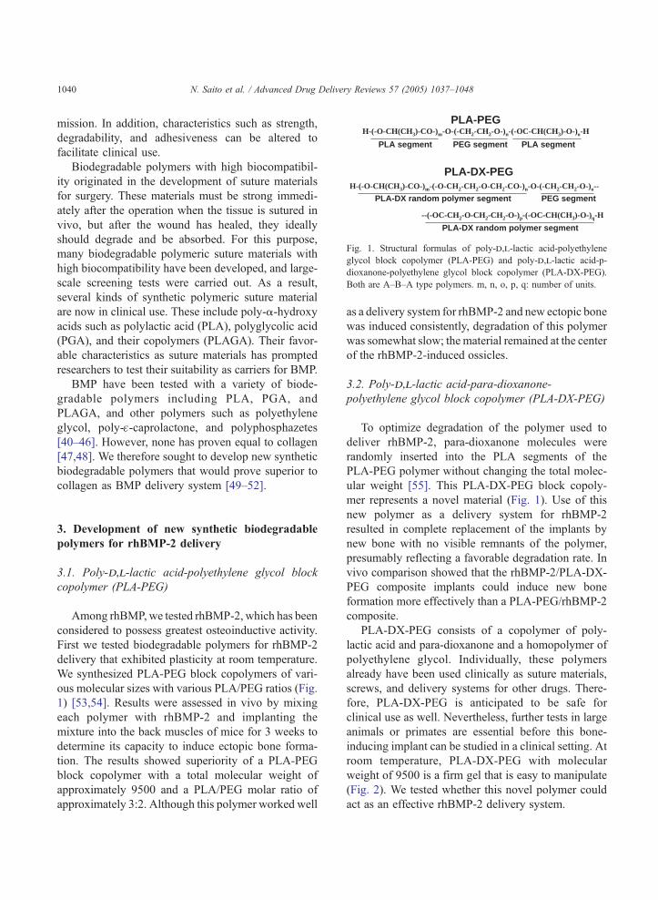
H-(-O-CH(CH3)-CO-)m-(-O-CH2-CH2-O-CH2-CO-)n-O-(-CH2-CH2-O-)o--
--(-OC-CH2-O-CH2-CH2-O-)p-(-OC-CH(CH3)-O-)q-H
PLA-DX random polymer segment PEG segment
PLA-PEGH-(-O-CH(CH3)-CO-)m-O-(-CH2-CH2-O-)n-(-OC-CH(CH3)-O-)o-H
PLA-DX random polymer segment
PLA-DX-PEG
PLA segment PEG segment PLA segment
Fig. 1. Structural formulas of poly-d,l-lactic acid-polyethylene
glycol block copolymer (PLA-PEG) and poly-d,l-lactic acid-p-
dioxanone-polyethylene glycol block copolymer (PLA-DX-PEG)
Both are A–B–A type polymers. m, n, o, p, q: number of units.
N. Saito et al. / Advanced Drug Delivery Reviews 57 (2005) 1037–10481040
mission. In addition, characteristics such as strength,
degradability, and adhesiveness can be altered to
facilitate clinical use.
Biodegradable polymers with high biocompatibil-
ity originated in the development of suture materials
for surgery. These materials must be strong immedi-
ately after the operation when the tissue is sutured in
vivo, but after the wound has healed, they ideally
should degrade and be absorbed. For this purpose,
many biodegradable polymeric suture materials with
high biocompatibility have been developed, and large-
scale screening tests were carried out. As a result,
several kinds of synthetic polymeric suture material
are now in clinical use. These include poly-a-hydroxy
acids such as polylactic acid (PLA), polyglycolic acid
(PGA), and their copolymers (PLAGA). Their favor-
able characteristics as suture materials has prompted
researchers to test their suitability as carriers for BMP.
BMP have been tested with a variety of biode-
gradable polymers including PLA, PGA, and
PLAGA, and other polymers such as polyethylene
glycol, poly-q-caprolactone, and polyphosphazetes
[40–46]. However, none has proven equal to collagen
[47,48]. We therefore sought to develop new synthetic
biodegradable polymers that would prove superior to
collagen as BMP delivery system [49–52].
3. Development of new synthetic biodegradable
polymers for rhBMP-2 delivery
3.1. Poly-d,l-lactic acid-polyethylene glycol block
copolymer (PLA-PEG)
Among rhBMP,we tested rhBMP-2, which has been
considered to possess greatest osteoinductive activity.
First we tested biodegradable polymers for rhBMP-2
delivery that exhibited plasticity at room temperature.
We synthesized PLA-PEG block copolymers of vari-
ous molecular sizes with various PLA/PEG ratios (Fig.
1) [53,54]. Results were assessed in vivo by mixing
each polymer with rhBMP-2 and implanting the
mixture into the back muscles of mice for 3 weeks to
determine its capacity to induce ectopic bone forma-
tion. The results showed superiority of a PLA-PEG
block copolymer with a total molecular weight of
approximately 9500 and a PLA/PEG molar ratio of
approximately 3:2. Although this polymer worked well
.
as a delivery system for rhBMP-2 and new ectopic bone
was induced consistently, degradation of this polymer
was somewhat slow; the material remained at the center
of the rhBMP-2-induced ossicles.
3.2. Poly-d,l-lactic acid-para-dioxanone-
polyethylene glycol block copolymer (PLA-DX-PEG)
To optimize degradation of the polymer used to
deliver rhBMP-2, para-dioxanone molecules were
randomly inserted into the PLA segments of the
PLA-PEG polymer without changing the total molec-
ular weight [55]. This PLA-DX-PEG block copoly-
mer represents a novel material (Fig. 1). Use of this
new polymer as a delivery system for rhBMP-2
resulted in complete replacement of the implants by
new bone with no visible remnants of the polymer,
presumably reflecting a favorable degradation rate. In
vivo comparison showed that the rhBMP-2/PLA-DX-
PEG composite implants could induce new bone
formation more effectively than a PLA-PEG/rhBMP-2
composite.
PLA-DX-PEG consists of a copolymer of poly-
lactic acid and para-dioxanone and a homopolymer of
polyethylene glycol. Individually, these polymers
already have been used clinically as suture materials,
screws, and delivery systems for other drugs. There-
fore, PLA-DX-PEG is anticipated to be safe for
clinical use as well. Nevertheless, further tests in large
animals or primates are essential before this bone-
inducing implant can be studied in a clinical setting. At
room temperature, PLA-DX-PEG with molecular
weight of 9500 is a firm gel that is easy to manipulate
(Fig. 2). We tested whether this novel polymer could
act as an effective rhBMP-2 delivery system.
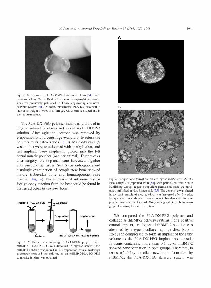
Fig. 2. Appearance of PLA-DX-PEG (reprinted from [51], with
permission from Marcel Dekker Inc.) requires copyright permission
since we previously published in Tissue engineering and novel
delivery systems [51]. At room temperature, PLA-DX-PEG with a
molecular weight of 9500 is a firm gel, which can be shaped and is
easy to manipulate.
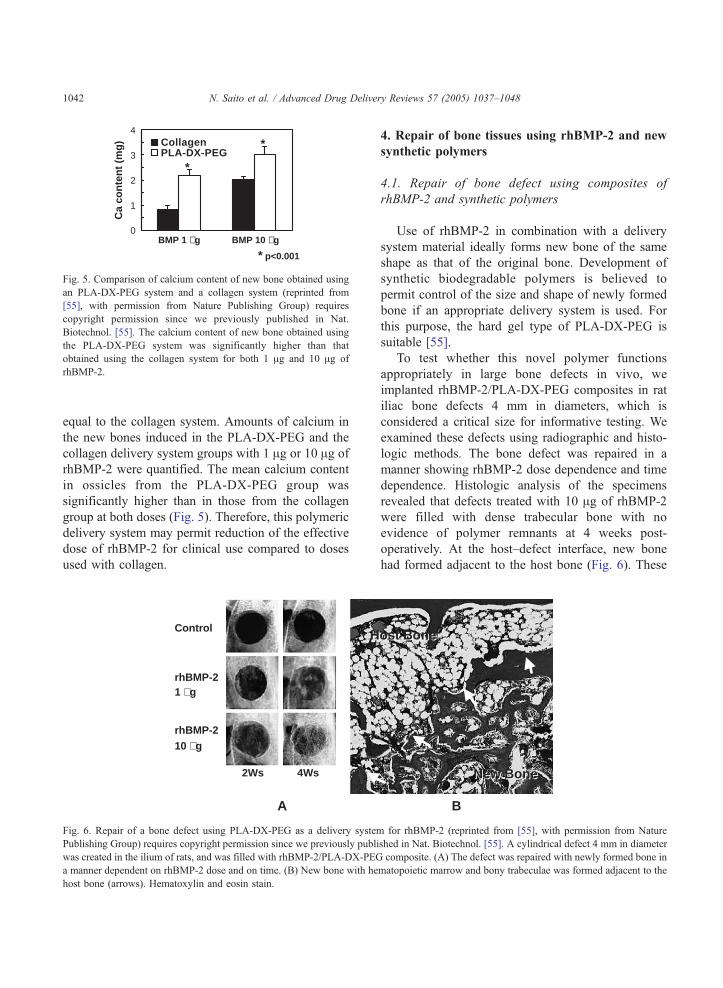
Fig. 4. Ectopic bone formation induced by the rhBMP-2/PLA-DX-
PEG composite (reprinted from [55], with permission from Nature
Publishing Group) requires copyright permission since we previ-
ously published in Nat. Biotechnol. [55]. The composite was placed
N. Saito et al. / Advanced Drug Delivery Reviews 57 (2005) 1037–1048 1041
The PLA-DX-PEG polymer mass was dissolved in
organic solvent (acetone) and mixed with rhBMP-2
solution. After agitation, acetone was removed by
evaporation with a centrifuge evaporator to return the
polymer to its native state (Fig. 3). Male ddy mice (5
weeks old) were anesthetized with diethyl ether, and
test implants were aseptically placed into the left
dorsal muscle pouches (one per animal). Three weeks
after surgery, the implants were harvested together
with surrounding tissues. Soft X-ray radiographs and
histologic examination of ectopic new bone showed
mature trabecular bone and hematopoietic bone
marrow (Fig. 4). No evidence of inflammatory or
foreign-body reaction from the host could be found in
tissues adjacent to the new bone.
Fig. 3. Methods for combining PLA-DX-PEG polymer with
rhBMP-2. PLA-DX-PEG was dissolved in organic solvent, and
rhBMP-2 solution was mixed in it. Evaporation with a centrifuge
evaporator removed the solvent, so an rhBMP-2/PLA-DX-PEG
composite implant was obtained.
in the back muscle of mouse, which was harvested after 3 weeks.
Ectopic new bone showed mature bone trabeculae with hemato-
poietic bone marrow. (A) Soft X-ray radiograph. (B) Photomicro-
graph. Hematoxylin and eosin stain.
We compared the PLA-DX-PEG polymer and
collagen as rhBMP-2 delivery systems. For a positive
control implant, an aliquot of rhBMP-2 solution was
absorbed by a type I collagen sponge disc, lyophi-
lized, and compressed to form an implant of the same
volume as the PLA-DX-PEG implant. As a result,
implants containing more than 0.5 Ag of rhBMP-2
showed bone formation in both groups. Therefore, in
terms of ability to elicit new bone formation by
rhBMP-2, the PLA-DX-PEG delivery system was
0
1
2
3
4C
a co
nte
nt
(mg
) CollagenPLA-DX-PEG
BMP 1 µg BMP 10 µg
* p<0.001
*
*
Fig. 5. Comparison of calcium content of new bone obtained using
an PLA-DX-PEG system and a collagen system (reprinted from
[55], with permission from Nature Publishing Group) requires
copyright permission since we previously published in Nat.
Biotechnol. [55]. The calcium content of new bone obtained using
the PLA-DX-PEG system was significantly higher than that
obtained using the collagen system for both 1 Ag and 10 Ag of
rhBMP-2.
N. Saito et al. / Advanced Drug Delivery Reviews 57 (2005) 1037–10481042
equal to the collagen system. Amounts of calcium in
the new bones induced in the PLA-DX-PEG and the
collagen delivery system groups with 1 Ag or 10 Ag of
rhBMP-2 were quantified. The mean calcium content
in ossicles from the PLA-DX-PEG group was
significantly higher than in those from the collagen
group at both doses (Fig. 5). Therefore, this polymeric
delivery system may permit reduction of the effective
dose of rhBMP-2 for clinical use compared to doses
used with collagen.
Control
rhBMP-2 1 µg
rhBMP-210 µg
2Ws 4Ws
HH
A
Fig. 6. Repair of a bone defect using PLA-DX-PEG as a delivery system
Publishing Group) requires copyright permission since we previously publi
was created in the ilium of rats, and was filled with rhBMP-2/PLA-DX-PEG
a manner dependent on rhBMP-2 dose and on time. (B) New bone with he
host bone (arrows). Hematoxylin and eosin stain.
4. Repair of bone tissues using rhBMP-2 and new
synthetic polymers
4.1. Repair of bone defect using composites of
rhBMP-2 and synthetic polymers
Use of rhBMP-2 in combination with a delivery
system material ideally forms new bone of the same
shape as that of the original bone. Development of
synthetic biodegradable polymers is believed to
permit control of the size and shape of newly formed
bone if an appropriate delivery system is used. For
this purpose, the hard gel type of PLA-DX-PEG is
suitable [55].
To test whether this novel polymer functions
appropriately in large bone defects in vivo, we
implanted rhBMP-2/PLA-DX-PEG composites in rat
iliac bone defects 4 mm in diameters, which is
considered a critical size for informative testing. We
examined these defects using radiographic and histo-
logic methods. The bone defect was repaired in a
manner showing rhBMP-2 dose dependence and time
dependence. Histologic analysis of the specimens
revealed that defects treated with 10 Ag of rhBMP-2
were filled with dense trabecular bone with no
evidence of polymer remnants at 4 weeks post-
operatively. At the host–defect interface, new bone
had formed adjacent to the host bone (Fig. 6). These
ost Boneost Bone
New BoneNew Bone
B
for rhBMP-2 (reprinted from [55], with permission from Nature
shed in Nat. Biotechnol. [55]. A cylindrical defect 4 mm in diameter
composite. (A) The defect was repaired with newly formed bone in
matopoietic marrow and bony trabeculae was formed adjacent to the

BMP/Polymer
Liquid37°C
Semisolid
A B
60°C
Fig. 7. Injectable polymeric delivery system for rhBMP-2 (reprinted
from [56], with permission from Elsevier) requires copyright
permission since we previously published in Bone [56]. (A) When
heated to 60 8C, the rhBMP-2/PLA-DX-PEG composite can be
injected percutaneously, avoiding need for surgical implantation.
Subsequently, implants become firm upon cooling to body temper-
ature, resulting in semisolid polymeric implants in vivo. (B) Soft X-
ray radiograph of new orthotopic bone formed by injection of the
rhBMP-2/PLA-DX-PEG composite in the muscle pouch on the
abraded surface of the murine femur 3 weeks after injection.
Fig. 8. Spinal fusion by injection of the rhBMP-2/PLA-DX-PEG
composite (reprinted from [58], with permission from Lippincot
Williams and Wilkins) requires copyright permission since we
previously published in J. Spinal Disord. [58]. PLA-DX-PEG with
rhBMP-2 was injected into the anterior longitudinal ligament of the
spine in dogs. New bone was formed on the anterior aspects o
vertebrae after 6 weeks (3D-CT, arrow).
N. Saito et al. / Advanced Drug Delivery Reviews 57 (2005) 1037–1048 1043
results suggest that rhBMP-2 in the PLA-DX-PEG
polymer delivery system should be suitable for
eliciting bone formation and healing in large bone
defects.
4.2. Injectable polymeric delivery systems for
rhBMP-2
Injectable delivery systems for rhBMP-2 could
provide a less invasive method for repair of bone
defects, avoiding extensive invasive surgery [56].
Clinical indications might include fresh fractures,
nonunion, or delayed union of bone causing serious
difficulty in fracture treatment, as well as large defects
often associated with bone tumor resection. As far as
we know, no such delivery system has been developed
or reported.
The new synthetic biodegradable PLA-DX-PEG
polymers feature an exquisite temperature-dependent
liquid–semisolid transition and work well as an
injectable rhBMP-2 delivery system. The thermo-
sensitive property of the rhBMP-2/PLA-DX-PEG
composite permits percutaneous injection after heat-
ing. Fluidity of the composite decreases as it cools
to body temperature, and the resultant semisolid
form provides a scaffold for bone formation as it
gradually releases rhBMP-2 into its immediate
surroundings.
The rhBMP-2 molecule is a heat-stable protein
[57]. For example, biologic activity of rhBMP-2 was
unchanged after heating to 60 8C for 30 min.
Considering the heat-stable character of rhBMP-2,
PLA-DX-PEG with molecular weight of 6400 could
be a suitable system for injectable delivery of rhBMP-
2. Together with rhBMP-2, this polymer heated to 60
8C could be injected as a liquid and then turn to a
semisolid form in vivo at 37 8C. The properties of thepolymer would allow retention of BMP for a period of
time sufficient to elicit new bone formation while
serving as a scaffold for further bone growth.
Eventually, it would be completely replaced by new
bone, avoiding surgery for removal since the polymer
is biodegradable (Fig. 7A). To further demonstrate the
efficacy of this polymer, 25 mg of PLA-DX-PEG
mixed with 10 Ag of rhBMP-2 was heated at 60 8C for
5 min and injected using a 14-gauge needle into
muscle overlying the surface of the murine femur.
Three weeks after injection, new bone was found at
the injection site, and was attached to the surface of
the femur (Fig. 7B). This new type of injectable
osteoinductive material should allow less invasive
surgery involving restoration or repair of bone.
We also tested this injection technique in spinal
fusion [58]. The rhBMP-2/PLA-DX-PEG composites
were injected into the anterior longitudinal ligaments
t
f

Fig. 9. Ectopic bone formation by hydroxyapatite (HA) with the
rhBMP-2/PLA-DX-PEG (reprinted from [54], with permission from
The Journal of Bone and Joint Surgery, Inc.) requires copyright
permission since we previously published in J. Bone Joint Surg.
Am. [54]. The rhBMP-2/PLA-DX-PEG composite was placed in
the pores the HA block, which was inserted in the back muscle of
mice. (A) Soft X-ray radiograph showed the new bone surrounding
HA at 3 weeks. (B) Histologic examination also showed new bone
within the pores of the HA.
Control
rhBMP-2120 µg
Ti
A
B
Fig. 10. Repair of a bone defect with a titanium syringe implant with
the rhBMP-2/PLA-DX-PEG composite (reprinted from [59], with
permission from 6 Wiley) requires copyright permission since we
previously published in J. Biomed. Mater. Res. [59]. A bone defec
of 1.5 cm was created in the humerus of rabbits, and three 5-mm
implants were placed in it. These were stabilized with an
intramedullary rod. (A) While the bone defect was not repaired in
control groups, the defect was restored in the 120 Ag rhBMP-2
group after 5 weeks. (B) Histologic examination showed that new
bone also had formed within the titanium mesh (Ti), and that new
bone had formed adjacent to the host bone. Hematoxylin and eosin
stain.
N. Saito et al. / Advanced Drug Delivery Reviews 57 (2005) 1037–10481044
of the canine spine. Six weeks later, new bone had
formed, bridging between the vertebrae anteriorly
(Fig. 8). If a pneumoscopic technique were used
jointly, anterior spinal fusion might be accomplished
by a less invasive approach.
4.3. Combination of the rhBMP-2/polymer composites
with other materials
The hydrophilic nature of the PLA-DX-PEG
polymer causes it to swell on contact with water.
This physical property provides an additional advant-
age for use of the polymer in combination with porous
materials. When a solid implant with pores filled with
the rhBMP-2/PLA-DX-PEG composite is implanted,
the composite will swell, extruding itself from the
pores to form a layer of composite.
To test this property, a combination of the rhBMP-
2/PLA-DX-PEG composite with porous hydroxyapa-
tite (HA) was used [54]. The rhBMP-2/PLA-DX-PEG
composite was placed in the pores of an HA block,
which then was inserted into the back muscle of mice.
Over 3 weeks, new bone had formed to surround the
HA. Histologic examination showed new bone
formation within the pores of the HA as well (Fig. 9).
Next, rhBMP-2 (120 Ag) was mixed with the
polymer (120 mg) and impregnated into titanium
fiber-mesh cylinders [59]. Three 5-mm cylinders were
placed end-to-end to fill a 15-mm defect created in the
humerus of adult rabbits and stabilized with an
intramedullary rod. In controls, the titanium fiber-
mesh cylinders contained the polymer but not rhBMP-
2. Six weeks after implantation, new bone had formed
on the surface of the implant and had bridged the
t

N. Saito et al. / Advanced Drug Delivery Reviews 57 (2005) 1037–1048 1045
defect. Defects treated with control implants were not
repaired (Fig. 10). These results provide strong
evidence that composite implants using rhBMP-2,
synthetic degradable polymers, and compatible mate-
rials provide enhanced regenerative potential for the
repair of a large bone defect. These techniques can
repair bones whose function requires great strength, as
a combination of rhBMP-2/PLA-DX-PEG composite
with HA or titanium represents a mechanically
durable osteoinductive material.
4.4. Development of a new artificial joint that restores
a bone defect
Total hip arthroplasty (THA) has become essen-
tially the standard procedure for treatment of various
hip lesions. However, one limitation of this operation
has been the eventual loosening of the prosthesis from
periprosthetic bone loss. At revision surgery, various
degrees of bone defect, both in the proximal femur
Control rhBMP-2PLA-DX-PEG
A B
Fig. 11. Repair of a periprosthetic bone defect using PLA-DX-PEG/
rhBMP-2 composite adherent to the prosthesis (reprinted from [60],
with permission from Elsevier) requires copyright permission since
we previously published in Biomaterials [60]. (A) Twelve weeks
after implantation, the implant with rhBMP-2/PLA-DX-PEG
showed new bone formation at the defect site. In the control group
without rhBMP-2, only a scant amount of new bone was seen at the
cut ends of the defects, which were not repaired. (B) By
microscopic examination of sections in the rhBMP-2 treatment
group, the new bone on the surface of implants showed normal
histology with hematopoietic marrow and bony trabeculae. New
bone formation also was observed within the pores of the titanium
mesh. Hematoxylin and eosin stain.
and the acetabulum, often are encountered; these
present challenges for sufficiently solid fixation of a
new prosthesis. Alternative approaches aimed at
overcoming this problem have included special design
of the revision prosthesis and allo- or autogeneic bone
grafting in combination with or without materials such
as hydroxyapatite. If such bone loss can be repaired
with use of rhBMP-2, revision surgery might be made
more effective.
To address the problem of loosening of the
prosthesis, we developed a new prosthesis combined
with rhBMP-2/PLA-DX-PEG composite [60]. We
tested efficacy of the rhBMP-2-containing prosthesis
in reconstructing a bone defect in a canine model
where the medial half of the proximal femur was
resected to create a defect that was repaired with
rhBMP-2/PLA-DX-PEG composite. Twelve weeks
after implantation, the original bone defects in the
rhBMP-2 treatment groups showed repair (Fig. 11).
Thus, this type of hybrid prosthesis may represent a
new modality for repair of bone defects or restora-
tion of lost bone mass encountered in revision
arthroplasty.
5. Conclusions
A new delivery system using PLA-DX-PEG
enabled creation of various osteoinductive materials
that could be used to heal fractures and repair large
bone defects. Importantly, this new rhBMP-2 delivery
system was developed using synthetic biodegradable
polymers, avoiding potential risks of disease trans-
mission or immunogenicity associated with use of
animal collagen or allogeneic bone grafts. Moreover,
this system avoids problems of autogenous bone
grafts such as limited supply of donor bone and the
need for additional surgery to harvest the bone, with
the risk of additional morbidity.
In summary, this new rhBMP-2 delivery system
represents an innovative potential therapy that is
safe, efficacious, and less invasive than current
approaches for repair of damaged bone. Further
work will be necessary to determine whether the
biocompatible and biodegradable properties exhibited
by the PLA-DX-PEG polymers in these studies are
replicated during the practical application of rhBMP-
2 in patient care.
N. Saito et al. / Advanced Drug Delivery Reviews 57 (2005) 1037–10481046
Acknowledgements
This work was supported by a Grant-in-Aid for
Scientific Research from the Ministry of Education,
Science, Sports and Culture, Japan, a Grant of Japan
Rheumatism Foundation, a Grant from Hip Joint
Foundation of Japan, a Grant from Japan Orthopae-
dics and Traumatology Foundation Inc., a Grant from
NOVARTIS Foundation (Japan) for the Promotion of
Science, and a Grant from TERUMO Life Science
Foundation.
References
[1] T.S. Einhorn, Enhancement of fracture-healing, J. Bone Jt.
Surg., Am. 77 (1995) 940–956.
[2] S.M. Doppelt, W.W. Tomford, A.B. Lucas, H.J. Mankin,
Operational and financial aspects of a hospital bone bank,
J. Bone Jt. Surg., Am. 63 (1981) 1472–1481.
[3] T.I. Malinin, O.V. Martinez, M.D. Brown, Banking of massive
osteoarticular and intercalary bone allografts: 12 years’
experience, Clin. Orthop. 197 (1985) 44–57.
[4] J.A. Memzek, S.P. Arnoczky, C.L. Swenson, Retroviral
transmission by the transplantation of connective-tissue
allografts, J. Bone Jt. Surg., Am. 76 (1995) 1034–1041.
[5] E. Canalis, Effect of growth factors on bone cell replication
and differentiation, Clin. Orthop. 193 (1985) 246–263.
[6] A.H. Reddi, Symbiosis of biotechnology and biomaterials:
application in tissue engineering of bone and cartilage, J. Cell.
Biochem. 56 (1994) 192–195.
[7] U. Ripamonti, A.H. Reddi, Tissue engineering, morpho-
genesis, and regeneration of the periodontal tissues by bone
morphogenetic proteins, Crit. Rev. Oral Biol. Med. 8 (1997)
154–163.
[8] J. Fang, Y.Y. Zhu, E. Smiley, J. Bonadio, J.P. Rouleau, S.A.
Goldstein, L.K. McCauley, B.L. Davidson, B.J. Roessler,
Stimulation of new bone formation by direct transfer of
osteogenic plasmid gene, Proc. Natl. Acad. Sci. U. S. A. 93
(1996) 5753–5758.
[9] J. Bonadio, E. Smiley, P. Patil, S. Goldstein, Localized,
direct plasmid gene delivery in vivo: prolonged therapy
results in reproducible tissue regeneration, Nat. Med. 5
(1999) 753–759.
[10] A.I. Caplan, Mesenchymal stem cells, J. Orthop. Res. 9 (1991)
641–650.
[11] S.E. Haynesworth, J. Goshima, V.M. Goldberg, A.I. Caplan,
Characterization of cells with osteogenic potential from human
marrow, Bone 13 (1992) 81–88.
[12] H. Ohgushi, Y. Dohi, T. Toshikawa, S. Tamai, S. Tabata, Y.
Suwa, In vitro bone formation by rat marrow cell
culture, J. Biomed. Mater. Res. 32 (1996) 341–348.
[13] S. Tamura, H. Kataoka, Y. Matsui, Y. Shionoya, K. Ohno, K.I.
Michi, K. Takahashi, A. Yamaguchi, The effects of trans-
plantation of osteoblastic cells with bone morphogenetic
protein (BMP)/carrier complex on bone repair, Bone 29
(2001) 169–175.
[14] M.R. Urist, Bone formation by autoinduction, Science 150
(1965) 893–899.
[15] J.M. Wozney, V. Rosen, A.J. Celeste, L.M. Mitsock, M.J.
Whitters, R.W. Kriz, R.M. Hewick, E.A. Wang, Novel
regulators of bone formation: molecular clones and activities,
Science 242 (1988) 1528–1534.
[16] K. Takaoka, H. Yoshikawa, J. Hashimoto, S. Miyamoto, K.
Masuhara, H. Nakahara, M. Matsui, K. Ono, Purification
and characterization of a bone-inducing protein from a
murine osteosarcoma (Dunn type), Clin. Orthop. 292 (1993)
122–129.
[17] K. Takaoka, H. Yoshikawa, J. Hashimoto, K. Masuhara, S.
Miyamoto, S. Suzuki, K. Ono, M. Matsui, S. Oikawa, N.
Tsuruoka, Y. Tawaragi, C. Inuzuka, T. Katayama, M.
Sugiyama, M. Tsujimoto, T. Nakanishi, H. Nakazatio, Gene
cloning and expression of a bone morphogenetic protein
derived from a murine osteosarcoma, Clin. Orthop. 294 (1994)
344–352.
[18] A.J. Celeste, J.A. Iannazzi, R.C. Taylor, R.M. Hewick, V.
Rosen, E.A. Wang, J.M. Wozney, Identification of trans-
forming growth factor h family members present in bone
inductive protein purified from bovine bone, Proc. Natl.
Acad. Sci. U. S. A. 87 (1990) 9843–9847.
[19] M. Geiger, R.H. Li, W. Friess, Collagen sponges for bone
regeneration with rhBMP-2, Adv. Drug Deliv. Rev. 55 (2003)
1613–1629.
[20] C.A. Kirker-Head, Potential applications and delivery strat-
egies for bone morphogenetic proteins, Adv. Drug Deliv. Rev.
43 (2000) 65–92.
[21] T. Sakou, Bone morphogenetic proteines: from basic studies to
clinical approaches, Bone 22 (1998) 591–603.
[22] J.M. Schmitt, K. Hwang, S.R. Winn, J.O. Hollinger, Bone
morphogenetic proteins: an update on basic biology and
clinical relevance, J. Orthop. Res. 17 (1999) 269–278.
[23] E.A. Wang, V. Rosen, Recombinant human bone morphoge-
netic protein induces bone formation, Proc. Natl. Acad. Sci.
U. S. A. 87 (1990) 2220–2224.
[24] J.M. Wozney, The bone morphogenetic protein family and
osteogenesis, Mol. Reprod. Dev. 32 (1992) 160–167.
[25] T.K. Sampath, J.E. Coughlin, R.M. Whetstone, D. Banach, C.
Corbett, R.J. Ridge, E. Ozkaynak, H. Oppermann, D.C. Rueger,
Bovine osteogenic protein is composed of dimers of OP-1 and
BMP2A, two members of the transforming growth factor beta
superfamily, J. Biol. Chem. 265 (1990) 13198–13250.
[26] K. Takaoka, M. Koezuka, H. Nakahara, Telopeptide-depleted
bovine skin collagen as a carrier for bone morphogenetic
protein, J. Orthop. Res. 9 (1991) 902–907.
[27] S.D. Cook, M.W. Wolfe, S.L. Salkeld, D.C. Rueger, Effect of
recombinant human osteogenic protein-1 on healing of
segmental defects in non-human primates, J. Bone Jt. Surg.,
Am. 77 (1995) 734–750.
[28] J.H. Schimandle, S.D. Boden, W.C. Hutton, Experimental
spinal fusion with recombinant human bone morphogenetic
protein-2, Spine 20 (1995) 1326–1337.
N. Saito et al. / Advanced Drug Delivery Reviews 57 (2005) 1037–1048 1047
[29] U. Ripamonti, M. Heliotis, D.C. Rueger, T.K. Sampath,
Induction of cementogenesis by recombinant human osteo-
genic protein-1 (hOP-1/BMP-7) in the baboon (Papio ursi-
nus), Arch. Oral Biol. 41 (1996) 121–126.
[30] H. Itoh, S. Ebara, M. Kamimura, Y. Tateiwa, T. Kinoshita, Y.
Yuzawa, K. Takaoka, Experimental spinal fusion with use of
recombinant human bone morphogenetic protein 2, Spine 24
(1999) 1402–1405.
[31] V.V. Viljanen, T.J. Gao, T.C. Lindholm, T.S. Lindholm, B.
Kommonen, Xenogeneic moose (Alces alces) bone morpho-
genetic protein (mBMP)-induced repair of critical-size skull
defects in sheep, Int. J. Oral Maxillofac. Surg. 25 (1996)
217–222.
[32] T.J. Gao, T.S. Lindholm, B. Kommonen, P. Ragni, A.
Paronzini, T.C. Lindholm, T. Jamsa, P. Jalovaara, Enhanced
healing of segmental tibial defects in sheep by a composite
bone substitute composed of tricalcium phosphate cylinder,
bone morphogenetic protein, and Type IV collagen, J.
Biomed. Mater. Res. 32 (1996) 505–512.
[33] E. Tsuruga, H. Takita, H. Itoh, Y. Wakisaka, Y. Kuboki, Pore
size of porous hydroxyapatite as the cell-substratum controls
BMP-induced osteogenesis, J. Biochem. 121 (1997) 317–324.
[34] J.A. Koempel, B.S. Patt, K. O’Grady, J. Wozney, D.M.
Toriumi, The effect of recombinant human bone morphoge-
netic protein-2 on the integration of porous hydroxyapatite
implants with bone, J. Biomed. Mater. Res. 41 (1998) 59–63.
[35] D.D. Lee, A. Tofighi, M. Aiolova, P. Chakravarthy, A.
Catalano, A. Majahad, D. Knaack, Alpha-BSM: a biomimetic
bone substitute and drug delivery vehicle, Clin. Orthop. 367
(1999) S396–S405.
[36] B.J. Cole, M.P. Bostrom, T.L. Pritchard, D.R. Sumner, E.
Tomin, J.M. Lane, A.J. Weiland, Use of bone morphogenetic
protein 2 on ectopic porous coated implants in the rat, Clin.
Orthop. 345 (1997) 219–228.
[37] F.H. Bach, J.A. Fishman, N. Daniels, J. Proimos, B. Anderson,
C.B. Carpenter, L. Forrow, S.C. Robson, H.V. Fineberg,
Uncertainty in xenotransplantation: individual benefit versus
collective risk, Nat. Med. 4 (1998) 141–144.
[38] D. Butler, M. Wadman, S. Lehrman, Q. Schiermeier, Last
chance to stop and think on risks of xenotransplants, Nature
391 (1998) 320–324.
[39] F. DeLustro, J. Dasch, J. Keefe, L. Ellingsworth, Immune
responses to allogeneic and xenogeneic implants of collagen
and collagen derivatives, Clin. Orthop. 260 (1990) 263–279.
[40] J.O. Hollinger, G.C. Battistone, Biodegradable bone repair
materials: synthetic polymers and ceramics, Clin. Orthop. 207
(1986) 290–305.
[41] R. Kenley, L. Marden, T. Turek, L. Jin, E. Ron, J.O. Hollinger,
Osseous regeneration in the rat calvarium using novel delivery
system for recombinant human bone morphogenetic protein-2
(rhBMP-2), J. Biomed. Mater. Res. 28 (1994) 1139–1147.
[42] S.C. Lee, M. Shea, M.A. Battle, K. Kozitza, E. Ron, T. Turek,
R.G. Schaub, W.C. Hayes, Healing of large segmental defects
in rat femurs is aided by rhBMP-2 in PLGA matrix, J. Biomed.
Mater. Res. 28 (1994) 1149–1156.
[43] M.C. Meikle, S. Papaioannou, T.J. Ratledge, P.M. Speight,
S.R. Watt-Smith, P.A. Hill, J.J. Reynolds, Effect of poly dl-
lactide-co-glycolide implants and xenogeneic bone matrix-
derived growth factors on calvarial bone repair in the rabbit,
Biomaterials 15 (1994) 513–521.
[44] H.S. Sandhu, L.E.A. Kanim, J.M. Kabo, J.M. Toth, E.N.
Zeegen, D. Liu, L.L. Seeger, E.G. Dawson, Evaluation of
rhBMP-2 with an OPLA carrier in a canine posterolateral
(transverse process) spinal fusion model, Spine 20 (1995)
2669–2682.
[45] M. Bostrom, J.M. Lane, E. Tomin, M. Browne, W. Berberian,
T. Turek, J. Smith, J. Wozeney, T. Schildhauer, Use of bone
morphogenetic protein-2 in the rabbit ulnar nonunion model,
Clin. Orthop. 327 (1996) 272–282.
[46] M. Mayer, J. Hollinger, E. Ron, J. Wozeney, Maxillary
alveolar cleft repair in dogs using recombinant human bone
morphogenetic protein-2 and polymer carrier, Plast. Reconstr.
Surg. 98 (1996) 247–259.
[47] O.M. Bostman, Absorbable implants for the fixation of
fracture, J. Bone Jt. Surg., Am. 73 (1991) 148–153.
[48] J.O. Hollinger, K. Leong, Poly(a-hydroxy acids): carriers
for bone morphogenetic proteins, Biomaterials 17 (1996)
187–194.
[49] K. Takaoka, N. Saito, S. Miyamoto, H. Yoshikawa, T. Okada,
in: Y. Ikada, T. Okano (Eds.), Bone-Inducing Implants: New
Synthetic Absorbable Poly-d,l-lactic Acid-Polyethylene Gly-
col Block Copolymers as BMP-Carriers, Tissue Engineering
for Therapeutic Use, vol. 3, Elsevier Science B.V., Amster-
dam, 1999, pp. 141–151.
[50] K. Takaoka, S. Miyamoto, N. Saito, T. Okada, in: D.L. Wise
(Ed.), New Synthetic Degradable Polymers as Carrier Materi-
als for BMP, Biomaterials Engineering and Devices: Human
Applications, vol. 1, The Humana Press, New Jersey, 2000,
pp. 239–249.
[51] N. Saito, H. Horiuchi, N. Murakami, J. Takahashi, T. Okada,
K. Nozaki, K. Takaoka, in: M.J. Yaszemski, D.J. Trantolo,
K.U. Lewandrowski, V. Hasirci, D.E. Altobelli, D.L. Wise
(Eds.), New Synthetic Biodegradable Polymers for Bone
Morphogenetic Protein Delivery Systems, Tissue Engineering
and Novel Delivery Systems, Marcel Dekker Inc., New York,
2004, pp. 475–482.
[52] N. Saito, K. Takaoka, New synthetic biodegradable polymers
as BMP carriers for bone tissue engineering, Biomaterials 24
(2003) 2287–2293.
[53] N. Saito, T. Okada, S. Toba, S. Miyamoto, K. Takaoka, New
synthetic absorbable polymers as BMP-carriers: plastic proper-
ties of poly-d,l-lactic acid-polyethylene glycol block copoly-
mers, J. Biomed. Mater. Res. 47 (1999) 104–110.
[54] N. Saito, T. Okada, H. Horiuchi, N. Murakami, J. Takahashi,
M. Nawata, H. Ota, S. Miyamoto, K. Nozaki, K. Takaoka,
Biodegradable poly-d,l-lactic acid-polyethylene glycol block
copolymers as a BMP delivery system for inducing bone,
J. Bone Jt. Surg., Am. 83 (Suppl. 1) (2001) 92–98.
[55] N. Saito, T. Okada, H. Horiuchi, N. Murakami, J. Takahashi,
M. Nawata, H. Ota, K. Nozaki, K. Takaoka, A biodegradable
polymer as a cytokine delivery system for inducing bone
formation, Nat. Biotechnol. 19 (2001) 332–335.
[56] N. Saito, T. Okada, H. Horiuchi, H. Ota, J. Takahashi, N.
Murakami, M. Nawata, S. Kojima, K. Nozaki, K. Takaoka,
N. Saito et al. / Advanced Drug Delivery Reviews 57 (2005) 1037–10481048
Local bone formation by injection of recombinant human bone
morphogenetic protein-2 contained in polymer carriers, Bone
32 (2003) 381–386.
[57] H. Izawa, Y. Hachiya, T. Kawai, K. Muramatsu, Y. Narita, N.
Ban, H. Yishizawa, The effect of heat-treated human bone
morphogenetic protein on clinical implantation, Clin. Orthop.
390 (2001) 252–258.
[58] J. Takahashi, N. Saito, S. Ebara, T. Kinoshita, H. Itoh, T.
Okada, K. Nozaki, K. Takaoka, Anterior thoracic spinal
fusion in dogs by injection of recombinant human bone
morphogenetic-2 and a synthetic polymer, J. Spinal Disord.
16 (2003) 137–143.
[59] N. Murakami, N. Saito, H. Horiuchi, T. Okada, K. Nozaki, K.
Takaoka, Repair of segmental defects in rabbit humeri with
titanium fiber mesh cylinders containing recombinant human
bone morphogenetic protein-2 (rhBMP-2) and a synthetic
polymer, J. Biomed. Mater. Res. 62 (2002) 169–174.
[60] N. Murakami, N. Saito, J. Takahashi, H. Ota, H. Horiuchi, M.
Nawata, T. Okada, K. Nozaki, K. Takaoka, Repair of a
proximal femoral bone defect in dogs using a porous surfaced
prosthesis in combination with recombinant BMP-2 and a
synthetic polymer carrier, Biomaterials 24 (2003) 2153–2159.