synthesis, structural characterization and antimicrobial
TRANSCRIPT
Accepted Manuscript
Synthesis, structural characterization and antimicrobial activity of silver(I) com-plexes with 1-benzyl-1H-tetrazoles
Tina P. Andrejević, Andrea M. Nikolic, Biljana Đ. Glišić, Hubert Wadepohl,Sandra Vojnovic, Mario Zlatović, Miloš Petković, Jasmina Nikodinovic-Runic,Igor M. Opsenica, Miloš I. Djuran
PII: S0277-5387(18)30457-1DOI: https://doi.org/10.1016/j.poly.2018.08.001Reference: POLY 13323
To appear in: Polyhedron
Received Date: 26 June 2018Revised Date: 1 August 2018Accepted Date: 2 August 2018
Please cite this article as: T.P. Andrejević, A.M. Nikolic, B.D. Glišić, H. Wadepohl, S. Vojnovic, M. Zlatović, M.Petković, J. Nikodinovic-Runic, I.M. Opsenica, M.I. Djuran, Synthesis, structural characterization and antimicrobialactivity of silver(I) complexes with 1-benzyl-1H-tetrazoles, Polyhedron (2018), doi: https://doi.org/10.1016/j.poly.2018.08.001
This is a PDF file of an unedited manuscript that has been accepted for publication. As a service to our customerswe are providing this early version of the manuscript. The manuscript will undergo copyediting, typesetting, andreview of the resulting proof before it is published in its final form. Please note that during the production processerrors may be discovered which could affect the content, and all legal disclaimers that apply to the journal pertain.
1
Ms. No. POLY-D-18-00610 (revised version)
Synthesis, structural characterization and antimicrobial activity of
silver(I) complexes with 1-benzyl-1H-tetrazoles
Tina P. Andrejevića,#, Andrea M. Nikolicb,#, Biljana Đ. Glišića,*, Hubert Wadepohlc, Sandra
Vojnovicd, Mario Zlatovićb, Miloš Petkoviće, Jasmina Nikodinovic-Runicd, Igor M. Opsenicab,*,
Miloš I. Djuranf
aUniversity of Kragujevac, Faculty of Science, Department of Chemistry, R. Domanovića 12,
34000 Kragujevac, Serbia
bUniversity of Belgrade-Faculty of Chemistry, Studentski trg 16, 11158 Belgrade, Serbia
cAnorganisch-Chemisches Institut, University of Heidelberg, Im Neuenheimer Feld 270, 69120
Heidelberg, Germany
dInstitute of Molecular Genetics and Genetic Engineering, University of Belgrade, Vojvode Stepe
444a, 11000 Belgrade, Serbia
eDepartment of Organic Chemistry, Faculty of Pharmacy, University of Belgrade, 11000
Belgrade, Serbia
fSerbian Academy of Sciences and Arts, Knez Mihailova 35, 11000 Belgrade, Serbia
#T.P.A. and A.M.N. contributed equally.
Corresponding authors. Tel.: +381 34 300 251; fax: +381 34 335 040 (B.Đ. Glišić); Tel.: +381
11 33 36 684 (I.M. Opsenica).
E-mail address: [email protected] (B.Đ. Glišić); [email protected] (I.M. Opsenica).
2
Abstract
Herein, we report the synthesis and structural characteristics of three tetrazole-containing
compounds, 1-benzyl-1H-tetrazole (bntz), 1-benzyl-1H-tetrazol-5-amine (bntza) and 1-(4-
methoxybenzyl)-1H-tetrazol-5-amine (mbntza) and the corresponding silver(I) complexes of the
general formula [Ag(NO3-O)(L-N4)2]n, L = bntz (1), bntza (2) and mbntza (3). Silver(I)
complexes 1 – 3 and 1-benzyl-1H-tetrazoles have been studied in detail by NMR, IR and UV-Vis
spectroscopic methods and the structures of 1 and 2 have been determined by single-crystal X-
ray diffraction analysis. The results of these analyses revealed a monodentate coordination of the
ligands to Ag(I) ion via the N4 tetrazole nitrogen. The antimicrobial potential of silver(I)
complexes 1 – 3 was evaluated against the broad panel of Gram-positive and Gram-negative
bacteria and fungi, displaying their remarkable inhibiting activity with MIC (minimal inhibitory
concentration) values in the range 2 – 8 and 0.16 – 1.25 μg/mL (3.8 – 16.3 and 0.31 – 2.15 μM),
respectively. On the other hand, 1-benzyl-1H-tetrazoles used for the synthesis of the silver(I)
complexes were not active against the investigated strains, suggesting that the activity of the
complexes originates from the Ag(I) ion exclusively. Moreover, silver(I) complexes 1 – 3 have
good therapeutic potential, which can be deduced from their moderate cytotoxicity on the human
fibroblast cell line MRC5, with IC50 values falling in the range 30 – 60 μg/mL (57.7 – 103.4
μM).
Keywords: 1-Benzyl-1H-tetrazoles, Silver(I) complexes, Structural characterization,
Antimicrobial activity, Cytotoxicity
3
1. Introduction
Silver(I) complexes exhibit a wide range of applications in the medicinal and analytical
chemistry, catalysis and the polymer design [1,2]. The use of silver(I) complexes in medicine is
mainly related to their well known antibacterial and antifungal properties which are attributed to
the presence of Ag(I) ion [3-6]. This metal ion is highly toxic to microorganisms, well tolerated
by mammalians and does not present a serious toxic risk to human body [7,8]. Nevertheless,
silver poisoning can occur among people chronically exposed to this metal [5]. Thus, argyria, the
permanent discoloration of the skin, has been observed in persons that have ingested both
metallic silver and silver(I) compounds in small doses during a long time period [5]. Although
most scientists believe that argyria is the most serious health effect caused by the silver, there are
also a few reports claiming that silver can cause brain damage, seizure or a persistent vegetative
state [5].
In comparison to the simple silver(I) salts, the complexes of this metal ion represent an
alternative formulation of silver with tunable antimicrobial properties. Many silver(I) complexes
have shown superior antimicrobial activity in comparison with the silver(I) salts [1,9], as well as
lower in vitro cytotoxicity on the healthy human lung fibroblast cell line MRC5 and in vivo
embryotoxicity [10]. One of the key factors determining the antimicrobial potential of silver(I)
complexes is the type of donor atom coordinated to Ag(I) ion and, consequently, the rate of the
ligand replacement rather than the complexes’ solubility, charge and degree of polymerization
[11]. For instance, silver(I) complexes with nitrogen donors have shown an effective and wide-
spectrum antimicrobial activity [11-13]. The effectiveness of this type of silver(I) complexes was
connected with the presence of a weak AgN bond, which can be easily cleaved in their
4
interaction with different biomolecules, such as thiol-containing proteins and nucleic acids, the
process that is supposed to be a prerequisite for their antimicrobial action [6,11-13].
Aromatic nitrogen-containing heterocyclic compounds (N-heterocycles) are one of
the most important class of the nitrogen donor ligands utilized in the synthesis of
biologically active silver(I) complexes [6]. These compounds represent structural
moieties of many natural products and biologically important molecules, as well as
pharmacologically used agents [14]. So far, different N-heterocycles such as imidazole
and its substituted derivatives [7,15-18], tetrazole [19], pyridine and its derivatives [20-
24], quinolines [25], diazines and benzodiazines [9,10,26], bipyridines [27], terpyridines
[28] and phenantrolines [29-36] have been used for the synthesis of mono- and
polynuclear silver(I) complexes, which have shown a remarkable activity against a broad
panel of bacterial and fungal strains which can lead to many infections.
Even though tetrazoles, five-membered aromatic heterocycles which contain four
nitrogen atoms within one ring, are not a part of any natural product, they have attracted
considerable attention as structural components of energetic materials [37], bioactive compounds
[38], nanomaterials [39] and as ligands for complexation to different metal ions [40-42]. Besides
the unsubstituted tetrazole, there is a growing interest in the investigation of compounds
containing N-linked substituents attached to the ring carbon of tetrazole, especially for 5-amino-
1H-tetrazole and its derivatives [40]. 5-Amino-1H-tetrazole motif is interesting, because it is
nitrogen-rich compound which possesses five potential binding sites (one amino and four
tetrazole nitrogen atoms) [43].
Herein, we have synthesized 1-benzyl-1H-tetrazole (bntz), 1-benzyl-1H-tetrazol-5-amine
(bntza) and 1-(4-methoxybenzyl)-1H-tetrazol-5-amine (mbntza), all containing 1-benzyl-1H-
5
tetrazole motif [44] and used them as ligands for the synthesis of silver(I) complexes of the
general formula [Ag(NO3-O)(L-N4)2]n, L = bntz (1), bntza (2) and mbntza (3) (Scheme 1). The
1-benzyl-1H-tetrazoles and the corresponding silver(I) complexes were evaluated for in vitro
antimicrobial and antiproliferative activities against the normal human lung fibroblast cell line to
properly address their therapeutic potential.
2. Experimental
2.1. Materials and methods
Unless stated otherwise, all solvents and reagents were obtained from commercial
sources and used without further purification. Elemental microanalyses of the synthesized 1-
benzyl-1H-tetrazoles and silver(I) complexes for carbon, hydrogen and nitrogen were performed
by the Microanalytical Laboratory, Faculty of Chemistry, University of Belgrade. The IR spectra
were recorded as KBr pellets on a Perkin Elmer Spectrum 100 spectrometer over the
wavenumber range of 4000 – 450 cm-1. The NMR spectra of the 1-benzyl-1H-tetrazoles and
silver(I) complexes were recorded at 25 C on a Bruker Avance III Ultrashield (1H at 500 MHz,
13C at 125 MHz) and Bruker Avance III 400 MHz spectrometer (1H at 400 MHz, 13C at 101
MHz), respectively. In order to investigate the solution stability of silver(I) complexes, the 1H
NMR spectra were recorded immediately after their dissolution in DMSO-d6, as well as after 48
h standing in the dark at room temperature. The UV-Vis spectra were recorded on a Shimadzu
spectrophotometer in DMSO/H2O (1 : 9, v/v). Compounds bntza and mbntza were analyzed by
high resolution tandem mass spectrometry using LTQ Orbitrap XL (Thermo Fisher Scientific
Inc., USA) mass spectrometer. The sample was dissolved in CH3CN and it was injected directly.
Ionization was done in positive mode on heated electrospray ionization (HESI) probe. HESI
6
parameters were: spray voltage 4.7 kV, vaporizer temperature 60 °C, sheath and auxiliary gas
flow 24 and 10 (arbitrary units), respectively, capillary voltage 49 V, capillary temperature 275
°C, tube lens voltage 80 V, resolution (at m/z 400): 30000.
2.2. Synthesis of 1-benzyl-1H-tetrazoles
The tetrazole-containing compounds, 1-benzyl-1H-tetrazole (bntz), 1-benzyl-1H-tetrazol-
5-amine (bntza) and 1-(4-methoxybenzyl)-1H-tetrazol-5-amine (mbntza), were synthesized by
previously described methods [45,46]. These compounds were pure based on elemental
microanalysis and NMR spectroscopy.
2.2.1. 1-Benzyl-1H-tetrazole (bntz)
1-Benzyl-1H-tetrazole (bntz) was synthesized according to the procedure described by
Satoh and Marcopulos as a yellow crystalline solid (see Supplementary data) [45]. Yield: 40%
(598.0 mg). Mp 52 – 55 °C. IR (KBr, ν, cm-1): 3113, 3065 (ν(Car–H)), ~3000 (ν(C–H)), 1670
(ν(Car=N)), 1495 (ν(N–Car=N)), 1465 (δ(CH2)), 1386 (ν(N=N)), 1243 (ν(C–N)), 1162 (ν(N–N)),
710 (γ(Car–H)). 1H NMR (500 Hz, CDCl3): δ 5.60 (s, 2H, C6H), 7.29 – 7.32 (m, 2H, C8H and
C12H), 7.38 – 7.42 (m, 3H, C9H – C11H), 8.57 ppm (s, 1H, C5H). 13C{1H} NMR (125 Hz,
CDCl3): δ 52.09 (C6), 128.23 (C8 and C12), 129.29 (C10), 129.33 (C9 and C11), 132.78 (C7),
142.38 ppm (C5). UV-Vis (DMSO/H2O, λmax, nm): 257.0 (ε = 3.6.102 M-1cm-1).
2.2.2. 1-Benzyl-1H-tetrazol-5-amine (bntza)
The amino-containing bntza compound was synthesized according to the previously
reported method as a colorless solid (see Supplementary data) [46]. Yield: 59% (939.0 mg).
7
Mp 184 – 186 °C. IR (KBr, ν, cm-1): 3338 (νas(NH2)), 3153 (νs(NH2)), 3064 (ν(Car–H)), ~3000
(ν(C–H)), 1642 (ν(Car=N)), 1485 (ν(N–Car=N)), 1430 (δ(CH2)), 1336 (ν(N=N)), 1269 (ν(C–N)),
1125 (ν(N–N)), 723 (γ(Car–H)). 1H NMR (500 Hz, DMSO-d6): δ 5.35 (s, 2H, C6H), 6.82 (s, 2H,
NH2), 7.20 – 7.24 (m, 2H, C8H and C12H), 7.28 – 7.32 (m, 1H, C10H), 7.33 – 7.38 ppm (m, 2H,
C9H and C11H). 13C{1H} NMR (125 Hz, DMSO-d6): δ 47.53 (C6), 127.52 (C8 and C12),
127.93 (C10), 128.70 (C9 and C11), 135.41 (C7), 155.52 ppm (C5). UV-Vis (DMSO/H2O, λmax,
nm): 238.0 (ε = 2.4.103 M-1cm-1). HRMS (HESI/Orbitrap) m/z [M + H]+ calc for C8H10N5:
176.09362; found 176.09299.
2.2.3. 1-(4-Methoxybenzyl)-1H-tetrazol-5-amine (mbntza)
Following the procedure described for bntza [46], compound mbntza was obtained from
4-(methoxyphenyl)methanamine as a colorless crystalline solid. Yield: 62% (185.0 mg). Mp
183 – 185 °C. IR (KBr, ν, cm-1): 3332 (νas(NH2)), 3162 (νs(NH2)), 3025, 2961 (ν(Car–H)), 2840,
2742 (ν(C–H)), 1660 (ν(Car=N)), 1513 (ν(N–Car=N)), 1458 (δ(CH3)), 1436 (δ(CH2)), 1307
(ν(N=N)), 1281 (ν(C–N)), 1253 (ν(C–O)), 1182 (ν(N–N)), 792, 677 (γ(Car–H)). 1H NMR (500
Hz, DMSO-d6): δ 3.72 (s, 3H, OCH3), 5.26 (s, 2H, C6H), 6.79 (s, 2H, NH2), 6.91 (d, J = 9.0 Hz,
2H, C9H and C11H), 7.21 ppm (d, J = 9.0 Hz, 2H, C8H and C12H). 13C{1H} NMR (125 Hz,
DMSO-d6): δ 47.11 (C6), 55.13 (OCH3), 114.08 (C9 and C11), 127.31 (C7), 129.21 (C8 and
C12), 155.00 (C5), 159.01 ppm (C10). UV-Vis (DMSO/H2O, λmax, nm): 238.0 (ε = 3.5.103
M-1cm-1), 273.0 (ε = 1.4.103 M-1cm-1). HRMS (HESI/Orbitrap) m/z [M + Na]+ calc for
C9H11N5ONa: 228.08613; found 228.08524.
8
2.3. Synthesis of silver(I) complexes 1 – 3
Silver(I) complexes with 1-benzyl-1H-tetrazoles, [Ag(NO3-O)(bntz-N4)2]n (1), [Ag(NO3-
O)(bntza-N4)2]n (2) and [Ag(NO3-O)(mbntza-N4)2]n (3), were synthesized in accordance to the
previously described method for the preparation of the silver(I) complexes with
diazanaphthalenes [10]. The purity of the complexes was confirmed by elemental microanalysis
and NMR spectroscopy.
The solution of 1.0 mmol of the corresponding 1-benzyl-1H-tetrazole (160.2 mg of bntz
for 1, 175.2 mg of bntza for 2 and 205.2 mg of mbntza for 3) in 5.0 mL of ethanol was added
slowly under stirring to the solution containing an equimolar amount of AgNO3 (169.9 mg) in
10.0 mL of ethanol. The reaction mixture was stirred for 3 – 4 h on a magnetic stirrer, in a sealed
round-bottom flask, protected from light at room temperature. Complexes 1 and 3 were obtained
from the mother ethanol solution after its cooling in the refrigerator for several days. The crystals
of complex 2 were obtained after the crude product, resulting from the reaction of AgNO3 and
bntza, was dissolved in 10 mL of acetonitrile. Yield (calculated on the basis of the N-
heterocyclic ligand): 152.0 mg (62%) for 1, 189.9 mg (73%) for 2 and 191.5 mg (66%) for 3.
Anal. calcd. for 1 (C16H16AgN9O3; MW = 490.25): C, 39.20; H, 3.29; N, 25.71. Found:
C, 38.87; H, 3.22; N, 26.04%. IR (KBr, ν, cm-1): 3113 (ν(Car–H)), ~3000 (ν(C–H)), 1654
(ν(Car=N)), 1494 (ν(N–Car=N)), 1384, 1354 (νas(NO3)), 1244 (ν(C–N)), 1164 (ν(N–N)), 711
(γ(Car–H)). 1H NMR (400 Hz, DMSO-d6): δ 5.72 (s, 2H, C6H), 7.30 – 7.44 (m, 5H, C8H –
C12H), 9.53 ppm (s, 1H, C5H). 13C{1H} NMR (101 Hz, DMSO-d6): δ 51.26 (C6), 128.62 (C8
and C12), 128.94 (C10), 129.37 (C9 and C11), 135.31 (C7), 144.51 ppm (C5). UV-Vis
(DMSO/H2O, λmax, nm): 257.0 (ε = 5.3.102 M-1cm-1).
9
Anal. calcd. for 2 (C16H18AgN11O3; MW = 520.28): C, 36.94; H, 3.49; N, 29.62. Found:
C, 36.65; H, 3.62; N, 29.88%. IR (KBr, ν, cm-1): 3322 (νas(NH2)), 3203 (νs(NH2)), ~3000 (ν(Car–
H) and ν(C–H)), 1647 (ν(Car=N)), 1483 (ν(N–Car=N)), 1440 (δ(CH2)), 1384, 1364 (νas(NO3)),
1334 (ν(N=N)), 1204 (ν(C–N)), 1144 (ν(N–N)), 708 (γ(Car–H)). 1H NMR (400 Hz, DMSO-d6):
δ 5.38 (s, 2H, C6H), 6.90 (s, 2H, NH2), 7.24 (d, J = 6.9 Hz, 2H, C8H and C12H), 7.33 (d, J = 7.1
Hz, 1H, C10H), 7.38 ppm (t, J = 7.1 Hz, 2H, C9H and C11H). 13C{1H} NMR (101 Hz, DMSO-
d6): δ 48.12 (C6), 128.02 (C8 and C12), 128.44 (C10), 129.19 (C9 and C11), 135.79 (C7),
155.96 ppm (C5). UV-Vis (DMSO/H2O, λmax, nm): 239.0 (ε = 2.1.103 M-1cm-1).
Anal. calcd. for 3 (C18H22AgN11O5; MW = 580.31): C, 37.25; H, 3.82; N, 26.55. Found:
C, 36.60; H, 3.63; N, 26.19%. IR (KBr, ν, cm-1): 3337 (νas(NH2)), 3160 (νs(NH2)), ~3000 (ν(Car–
H) and ν(C–H)), 1656 (ν(Car=N)), 1514 (ν(N–Car=N)), 1478 (δ(CH3)), 1441 (δ(CH2)), 1384,
1306 (νas(NO3)), 1253 (ν(C–O)), 1181 (ν(N–N)), 792 (γ(Car–H)). 1H NMR (400 Hz, DMSO-d6):
δ 3.73 (s, 3H, OCH3), 5.28 (s, 2H, C6H), 6.86 (s, 2H, NH2), 6.92 (d, J = 8.6 Hz, 2H, C9H and
C11H), 7.22 ppm (d, J = 8.6 Hz, 2H, C8H and C12H). 13C{1H} NMR (101 Hz, DMSO-d6): δ
47.64 (C6), 55.60 (OCH3), 114.56 (C9 and C11), 127.70 (C7), 129.70 (C8 and C12), 155.73
(C5), 159.49 ppm (C10). UV-Vis (DMSO/H2O, λmax, nm): 238.0 (ε = 3.4.103 M-1cm-1), 273.0 (ε
= 1.4.103 M-1cm-1).
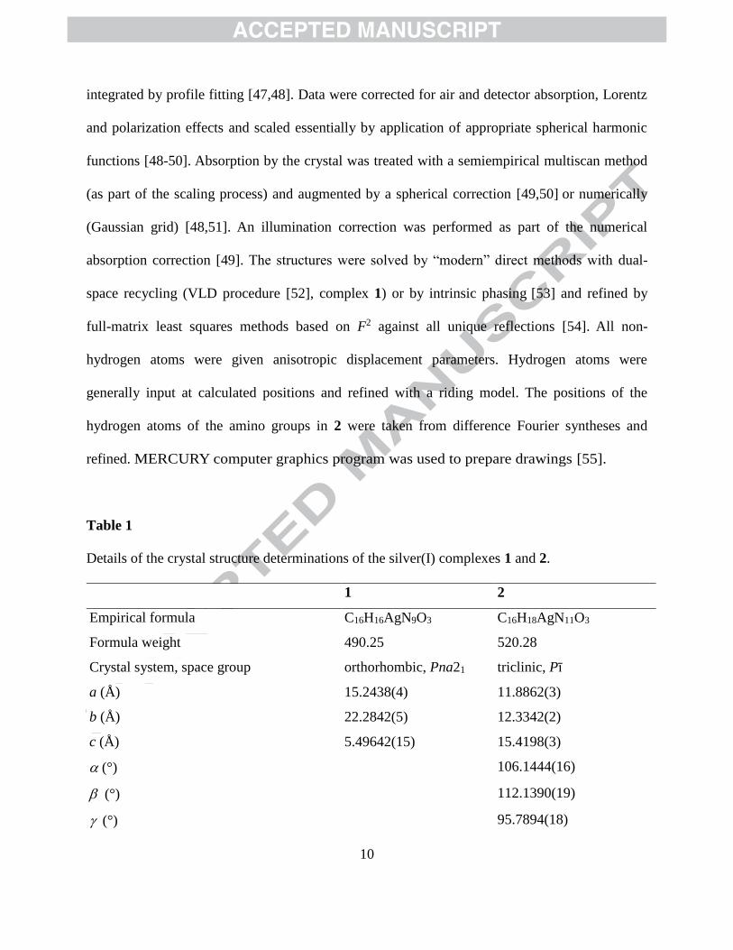
2.4. Crystallographic data collection and refinement of the structures
Crystal data and details of the structure determinations are compiled in Table 1. Full
shells of intensity data were collected at low temperature with an Agilent Technologies
Supernova-E CCD diffractometer (Mo- or Cu-K radiation, microfocus X-ray tube, multilayer
mirror optics). Detector frames (typically -, occasionally -scans, scan width 0.4...1°) were
10
integrated by profile fitting [47,48]. Data were corrected for air and detector absorption, Lorentz
and polarization effects and scaled essentially by application of appropriate spherical harmonic
functions [48-50]. Absorption by the crystal was treated with a semiempirical multiscan method
(as part of the scaling process) and augmented by a spherical correction [49,50] or numerically
(Gaussian grid) [48,51]. An illumination correction was performed as part of the numerical
absorption correction [49]. The structures were solved by “modern” direct methods with dual-
space recycling (VLD procedure [52], complex 1) or by intrinsic phasing [53] and refined by
full-matrix least squares methods based on F2 against all unique reflections [54]. All non-
hydrogen atoms were given anisotropic displacement parameters. Hydrogen atoms were
generally input at calculated positions and refined with a riding model. The positions of the
hydrogen atoms of the amino groups in 2 were taken from difference Fourier syntheses and
refined. MERCURY computer graphics program was used to prepare drawings [55].
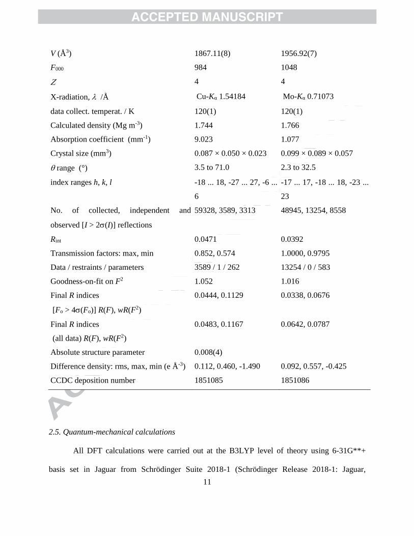
Table 1
Details of the crystal structure determinations of the silver(I) complexes 1 and 2.
1 2
Empirical formula C16H16AgN9O3 C16H18AgN11O3
Formula weight 490.25 520.28
Crystal system, space group orthorhombic, Pna21 triclinic, Pī
a (Å) 15.2438(4) 11.8862(3)
b (Å) 22.2842(5) 12.3342(2)
c (Å) 5.49642(15) 15.4198(3)
(°) 106.1444(16)
(°) 112.1390(19)
(°) 95.7894(18)
11
V (Å3) 1867.11(8) 1956.92(7)
F000 984 1048
4 4
X-radiation, /Å Cu-Kα 1.54184 Mo-Kα 0.71073
data collect. temperat. / K 120(1) 120(1)
Calculated density (Mg m-3) 1.744 1.766
Absorption coefficient (mm-1) 9.023 1.077
Crystal size (mm3) 0.087 × 0.050 × 0.023 0.099 × 0.089 × 0.057
range (°) 3.5 to 71.0 2.3 to 32.5
index ranges h, k, l -18 ... 18, -27 ... 27, -6 ...
6
-17 ... 17, -18 ... 18, -23 ...
23
No. of collected, independent and
observed [I > 2(I)] reflections
59328, 3589, 3313 48945, 13254, 8558
Rint 0.0471 0.0392
Transmission factors: max, min 0.852, 0.574 1.0000, 0.9795
Data / restraints / parameters 3589 / 1 / 262 13254 / 0 / 583
Goodness-on-fit on F2 1.052 1.016
Final R indices
[Fo > 4(Fo)] R(F), wR(F2)
0.0444, 0.1129 0.0338, 0.0676
Final R indices
(all data) R(F), wR(F2)
0.0483, 0.1167 0.0642, 0.0787
Absolute structure parameter 0.008(4)
Difference density: rms, max, min (e Å-3) 0.112, 0.460, -1.490 0.092, 0.557, -0.425
CCDC deposition number 1851085 1851086
2.5. Quantum-mechanical calculations
All DFT calculations were carried out at the B3LYP level of theory using 6-31G**+
basis set in Jaguar from Schrödinger Suite 2018-1 (Schrödinger Release 2018-1: Jaguar,
12
Schrödinger, LLC, New York, NY, 2018.). The B3LYP method is known to give good
descriptions of transition metal complexes [56]. All structures were prepared in Schrödinger
Suite and optimized in Jaguar using same aforementioned parameters prior to calculation.
2.6. Antimicrobial studies
MIC concentrations of 1 – 3 and 1-benzyl-1H-tetrazole ligands were determined
according to the standard broth microdilution assays, recommended by the National Committee
for Clinical Laboratory Standards (M07-A8) for bacteria and Standards of European Committee
on Antimicrobial Susceptibility Testing (v 7.3.1: Method for the determination of broth dilution
minimum inhibitory concentrations of antifungal agents for yeasts) for Candida spp. The tested
compounds were dissolved in DMSO at 50 mg/mL. The highest concentration used was 500
µg/mL. Bacterial test organisms included: Staphylococcus aureus ATCC 25923, Listeria
monocytogenes NCTC 11994, Micrococcus luteus ATCC 379, Pseudomonas aeruginosa PAO1
NCTC 10322, and Candida strains: C. albicans ATCC 10231, C. glabrata ATCC 2001, C.
krusei ATCC 6258, C. parapsilosis ATCC 22019. The inoculums were 105 colony forming units,
cfu/mL, for bacteria and 104 cfu/mL for Candida spp. The MIC value was recorded as the lowest
concentration that inhibited the growth after 24 h at 37 °C.
2.7. In vitro cytotoxicity assay
Cell viability was tested by the 3-(4,5-dimethylthiazol-2-yl)-2,5-diphenyltetrazolium
bromide (MTT) assay [57]. The assay was carried out using human lung fibroblasts (MRC5)
after 48 h of cell incubation in the media, containing compounds at concentrations ranging from
0.2 – 200 µg/mL. The MRC5 cell line was maintained in the RPMI-1640 medium, supplemented
13
with 100 μg/mL streptomycin, 100 U/mL penicillin and 10% (v/v) fetal bovine serum (FBS) (all
from Sigma, Munich, Germany) as a monolayer (1 x 10⁴ cells per well) and grown in humidified
atmosphere of 95% air and 5% CO2 at 37 °C. The extent of MTT reduction was measured
spectrophotometrically at 540 nm using a Tecan Infinite 200 Pro multiplate reader (Tecan Group
Ltd., Männedorf, Switzerland), and the cell survival was expressed as percentage of the control
(untreated cells). The percentage viability values were plotted against the log of concentration
and a sigmoidal dose response curve was calculated by non-linear regression analysis, using the
Graphpad Prism software, version 5.0 for Windows (Graphpad Software, CA, USA).
Cytotoxicity is expressed as the concentration of the compound inhibiting growth by 50% (IC50).
3. Results and discussion
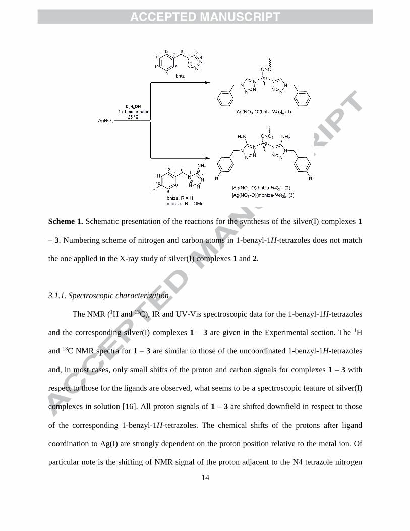
3.1. Synthesis and structural characterization of 1-benzyl-1H-tetrazoles and the silver(I)
complexes 1 – 3
Three tetrazole-containing compounds, 1-benzyl-1H-tetrazole (bntz), 1-benzyl-1H-
tetrazol-5-amine (bntza) and 1-(4-methoxybenzyl)-1H-tetrazol-5-amine (mbntza), were
synthesized by the previously reported methods (Scheme S1) [45,46]. They further reacted with
AgNO3 in 1 : 1 mole ratio in ethanol at room temperature to yield [Ag(NO3-O)(bntz-N4)2]n (1),
[Ag(NO3-O)(bntza-N4)2]n (2) and [Ag(NO3-O)(mbntza-N4)2]n (3) complexes (Scheme 1). In
complexes 1 – 3, the corresponding tetrazole ligand is monodentatedly coordinated to Ag(I) ion
via the N4 nitrogen atom.
14
Scheme 1. Schematic presentation of the reactions for the synthesis of the silver(I) complexes 1
– 3. Numbering scheme of nitrogen and carbon atoms in 1-benzyl-1H-tetrazoles does not match
the one applied in the X-ray study of silver(I) complexes 1 and 2.
3.1.1. Spectroscopic characterization
The NMR (1H and 13C), IR and UV-Vis spectroscopic data for the 1-benzyl-1H-tetrazoles
and the corresponding silver(I) complexes 1 – 3 are given in the Experimental section. The 1H
and 13C NMR spectra for 1 – 3 are similar to those of the uncoordinated 1-benzyl-1H-tetrazoles
and, in most cases, only small shifts of the proton and carbon signals for complexes 1 – 3 with
respect to those for the ligands are observed, what seems to be a spectroscopic feature of silver(I)
complexes in solution [16]. All proton signals of 1 – 3 are shifted downfield in respect to those
of the corresponding 1-benzyl-1H-tetrazoles. The chemical shifts of the protons after ligand
coordination to Ag(I) are strongly dependent on the proton position relative to the metal ion. Of
particular note is the shifting of NMR signal of the proton adjacent to the N4 tetrazole nitrogen
15
binding center, i.e. C5H proton, in the spectrum of 1. Thus, C5H in the spectrum of
uncoordinated bntz gives a singlet at δ 8.57 ppm, and it was shifted downfield at 9.53 ppm (Δδ =
0.96 ppm) after its coordination to the Ag(I) ion. For the complexes 1 – 3, 1H NMR spectra
remained unmodified over 48 h, indicating their stability in solution during that time. More
specifically, no decomposition of the complexes to the free ligands and no coordination of
DMSO to Ag(I) were observed.
From the 13C spectra of the complex 1, it can be concluded that bntz is coordinated to the
Ag(I) ion through the N4 tetrazole nitrogen, considering the large shift (Δδ = 2.13 ppm) for the
N4 adjacent carbon, C5, compared to the uncoordinated bntz. On the other hand, only a small
shift of ~1 ppm is observed for the carbon atoms of bntza and mbntza after their coordination to
Ag(I).
The IR spectra of the complexes 1 – 3, in comparison to those of 1-benzyl-1H-tetrazoles,
display certain differences which may give an insight in the type of bonds and their structures. A
band attributed to the nitrate asymmetric stretching vibrations in the IR spectra of 1 – 3 is split
into two bands with relatively small separation, being an indication of nitrate coordination in
these complexes [58]. The splitting of the nitrate asymmetric stretching vibrations in the IR
spectra of 1 – 3 is in accordance with that observed for the polynuclear [Ag(NO3)(qz)]n complex
(qz is quinazoline) which contains nitrate as a bridging ligand between two Ag(I) ions [9]. The
IR spectra of complexes 2 and 3 also show the expected bands at approximately 3300 and 3200
cm-1, which are due to the asymmetric and symmetric stretching vibrations of the amino group,
respectively [59].
The UV-Vis spectra of silver(I) complexes 1 – 3, recorded in DMSO/H2O, resemble
those of the corresponding 1-benzyl-1H-tetrazole ligands. In the complexes 1 and 2, the
16
corresponding absorbance peaks at ~ 257.0 and 239.0 nm, respectively, are caused by the
characteristic π → π* transitions in the ligand [60,61]. The absorbance peak for 2 shows slight
red shift compared to that for the free ligand (λ = 238.0 nm). The UV-vis spectra of mbntza and
the corresponding complex 3 have two maximum absorbance peaks at 238.0 and 273.0 nm,
which can be assigned to π → π* transition of the phenyl ring and n → π* intraligand transition
of its methoxy group, respectively [62].
3.1.2. Description of the single crystal structures
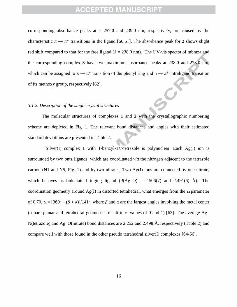
The molecular structures of complexes 1 and 2 with the crystallographic numbering
scheme are depicted in Fig. 1. The relevant bond distances and angles with their estimated
standard deviations are presented in Table 2.
Silver(I) complex 1 with 1-benzyl-1H-tetrazole is polynuclear. Each Ag(I) ion is
surrounded by two bntz ligands, which are coordinated via the nitrogen adjacent to the tetrazole
carbon (N1 and N5, Fig. 1) and by two nitrates. Two Ag(I) ions are connected by one nitrate,
which behaves as bidentate bridging ligand (d(Ag–O) = 2.506(7) and 2.491(6) Å). The
coordination geometry around Ag(I) in distorted tetrahedral, what emerges from the τ4 parameter
of 0.70, τ4 = [360o – (β + α)]/141o, where β and α are the largest angles involving the metal center
(square-planar and tetrahedral geometries result in τ4 values of 0 and 1) [63]. The average Ag–
N(tetrazole) and Ag–O(nitrate) bond distances are 2.252 and 2.498 Å, respectively (Table 2) and
compare well with those found in the other pseudo tetrahedral silver(I) complexes [64-66].
17
Fig. 1. Molecular structures of silver(I) complexes 1 and 2. Displacement ellipsoids are drawn at
50% probability level and H atoms are represented by spheres of arbitrary size.
Similar to 1, silver(I) complex 2 contains two monodentatedly coordinated 1-
benzyl-1H-tetrazol-5-amine via the nitrogen adjacent to the tetrazole carbon (N1/N51 and
N6/N56, Fig. 1). The coordinated tetrazole nitrogens of the two bntza ligands are almost
equidistant from the Ag(I) ion (Table 2) and are in agreement with the other silver(I)
complexes with aromatic N-heterocycles [10]. One of the nitrate oxygens acts as acceptor
18
in bifurcated hydrogen bonding with the amino groups of both tetrazolamino ligands on
the same silver(I) ion (Table 3). An additional comparatively weak interaction of two
nitrate oxygens with the silver(I) ions leads to polymeric Ag-(NO3)-Ag-(NO3)- chains
(Table 2). Interestingly, an ordered superstructure with a dinuclear [Ag(NO3)(bntza)2]2
repetition unit is formed, with the only significant difference between the two very similar
mononuclear subunits being the rotational conformations of the benzyl substituents.
Table 2
Selected bond distances (Å) and valence angles (o) in silver(I) complexes 1 and 2.
1 2
Ag—O1 2.506(7) Ag1—N1 2.1155(16)
Ag—O2i 2.491(6) Ag1—N6 2.1100(17)
Ag—N1 2.241(5) Ag51—N51 2.1202(17)
Ag—N5 2.264(5) Ag51—N56 2.1145(17)
O1—N9 1.259(11) Ag1-O81 2.6859(16)
O2—N9 1.241(11) Ag1-O92 2.8882(15)
O3—N9 1.255(7) Ag51-O82iii 2.8644(15)
Ag51-O91 2.6579(16)
O1—Ag—O2i 83.80(15) N1—Ag1—N6 175.67(7)
N1—Ag—O1 112.9(2) N2—N1—Ag1 121.14(3)
N1—Ag—O2i 101.7(2) C1—N1—Ag1 131.99(14)
N1—Ag—N5 148.56(18) N7—N6—Ag1 122.22(13)
N5—Ag—O1 95.6(2) C9—N6—Ag1 130.58(14)
N5—Ag—O2i 93.9(2) N51—Ag51—N56 174.66(7)
N9—O1—Ag 100.0(5) N52—N51—Ag51 122.78(13)
N9—O2—Agii 103.9(4) C51—N51—Ag51 130.32(14)
N2—N1—Ag 123.4(4) N57—N56—Ag51 120.84(13)
C1—N1—Ag 129.8(4) C59—N56—Ag51 132.34(14)
N6—N5—Ag 122.7(4)
C9—N5—Ag 130.4(5)
Symmetry code: (i) x, y, z-1; (ii) x, y, z+1; (iii) x, 1+y, z
19
Table 3
Hydrogen bond parameters for silver(I) complex 2.
D—H···A D—H (Å) H···A (Å) D···A (Å) D—H···A (º)
N5—H5a···O92 0.88(2) 1.97(2) 2.839(2) 170(2)
N10—H10B···O83i 0.82(3) 2.06(3) 2.866(3) 169(3)
N55—H55A···O82ii 0.81(2) 2.04(3) 2.847(2) 171(2)
N60—H60B···O93iii 0.80(3) 2.08(3) 2.862(3) 164(3)
Symmetry codes: (i) −x+2, −y+1, −z+2; (ii) x, y+1, z; (iii) –x+2, −y+2, −z+2
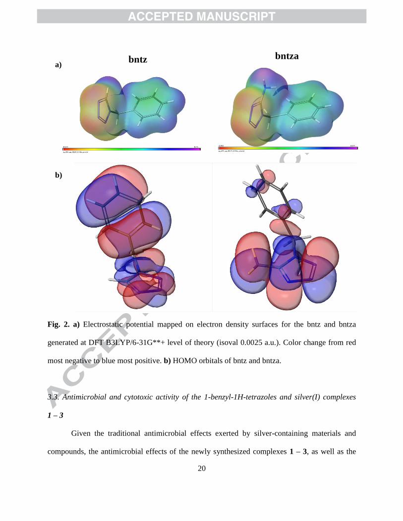
3.2.Computational studies
In order to gain a better understanding of the coordination mode of the synthesized 1-
benzyl-1H-tetrazoles toward the Ag(I) ion, the structures of bntz and bntza were optimized and
their molecular electrostatic potential (MEP) mapped on electron density surface generated at
B3LYP/6-31G**+ level of theory was examined (Fig. 2a). The MEP is a useful property for
analyzing and predicting the sites or regions of a molecule to which an approaching electrophile
(metal ion) is initially attracted [67,68]. The regions of negative potential are colored in red,
while the blue regions have positive potential. From Fig. 2a, one could see that in bntz and bntza
molecules, the negative electrostatic region is localized on the N3 and N4 tetrazole nitrogens,
indicating that both of these donors are almost equally accessible to metal ion. However, the
results of calculations suggest that the electron density in the HOMO (Highest Occupied
Molecular Orbital) is mainly distributed over the tetrazole ring, and some slight preference for
the coordinating nitrogen atom N4 could be found (Fig. 2b). These theoretical findings are in line
with the X-ray results for the complexes 1 and 2, all together supporting the selective formation
of silver(I) complexes in which the corresponding tetrazole-containing ligand is coordinated via
the nitrogen which is adjacent to the tetrazole carbon atom.
20
Fig. 2. a) Electrostatic potential mapped on electron density surfaces for the bntz and bntza
generated at DFT B3LYP/6-31G**+ level of theory (isoval 0.0025 a.u.). Color change from red
most negative to blue most positive. b) HOMO orbitals of bntz and bntza.
3.3. Antimicrobial and cytotoxic activity of the 1-benzyl-1H-tetrazoles and silver(I) complexes
1 – 3
Given the traditional antimicrobial effects exerted by silver-containing materials and
compounds, the antimicrobial effects of the newly synthesized complexes 1 – 3, as well as the
bntz
b)
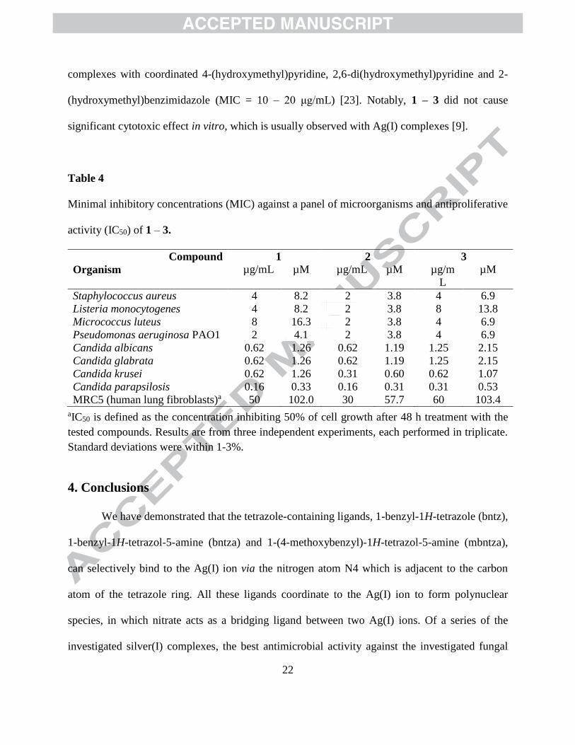
a) bntza
21
corresponding ligands have been evaluated against the panel of bacteria and Candida strains
(Table 4). While 1-benzyl-1H-tetrazoles used for the synthesis of the silver(I) complexes were
not active against the investigated strains at concentrations higher than 500 µg/mL (data not
shown), 1 – 3 showed growth inhibiting activity against all tested strains, suggesting that the
activity of the complexes originates from the presence of Ag(I) ions. The MIC values of the
silver(I) complexes were between 2 – 8 µg/mL (3.8 – 16.3 µM) against bacterial strains, while
they were able to inhibit Candida, both albicans and non-albicans strains at 0.16 – 1.25 µg/mL
(0.31 – 2.15 µM) MIC concentration values (Table 4). Three complexes exhibited comparable
activity, with 2 being slightly more active across the microbial panel. From these data, no
difference has been observed between Gram-positive and Gram-negative strains, while anti-
Candida activity of these complexes was more prominent, with C. parapsilosis being the most
sensitive strain. More importantly, 1 – 3 were moderately cytotoxic against human lung
fibroblast cell line, with the best selectivity index being 312 (for 1 and C. parapsilosis) giving
reason for further evaluations of these complexes for therapeutic applications. These results are
of special importance having in mind, that emerging non-albicans Candida species, such as C.
parapsilosis, C. glabrata, C. tropicalis and C. krusei, are increasingly recognized as causative
agents of infections ranging from superficial to life-threatening disseminated bloodstream and
deep-tissue infections [69].
Good antimicrobial activity of 1 – 3 was comparable or slightly better to that observed
for silver(I) complexes with aromatic nitrogen-containing heterocycles such as pyridazine,
pyrimidine, pyrazine, quinoxaline, phenazine, quinazoline and phthalazine [9,10,26]. Unlike these
complexes, 1 – 3 showed higher activity against selection of Candida strains. Improved activity
against C. albicans in comparison to the antibacterial activity has also been observed for silver(I)
22
complexes with coordinated 4-(hydroxymethyl)pyridine, 2,6-di(hydroxymethyl)pyridine and 2-
(hydroxymethyl)benzimidazole (MIC = 10 – 20 μg/mL) [23]. Notably, 1 – 3 did not cause
significant cytotoxic effect in vitro, which is usually observed with Ag(I) complexes [9].
Table 4
Minimal inhibitory concentrations (MIC) against a panel of microorganisms and antiproliferative
activity (IC50) of 1 – 3.
Compound 1 2 3
Organism µg/mL µM µg/mL µM µg/m
L
µM
Staphylococcus aureus 4 8.2 2 3.8 4 6.9
Listeria monocytogenes 4 8.2 2 3.8 8 13.8
Micrococcus luteus 8 16.3 2 3.8 4 6.9
Pseudomonas aeruginosa PAO1 2 4.1 2 3.8 4 6.9
Candida albicans 0.62 1.26 0.62 1.19 1.25 2.15
Candida glabrata 0.62 1.26 0.62 1.19 1.25 2.15
Candida krusei 0.62 1.26 0.31 0.60 0.62 1.07
Candida parapsilosis 0.16 0.33 0.16 0.31 0.31 0.53
MRC5 (human lung fibroblasts)a 50 102.0 30 57.7 60 103.4
aIC50 is defined as the concentration inhibiting 50% of cell growth after 48 h treatment with the
tested compounds. Results are from three independent experiments, each performed in triplicate.
Standard deviations were within 1-3%.
4. Conclusions
We have demonstrated that the tetrazole-containing ligands, 1-benzyl-1H-tetrazole (bntz),
1-benzyl-1H-tetrazol-5-amine (bntza) and 1-(4-methoxybenzyl)-1H-tetrazol-5-amine (mbntza),
can selectively bind to the Ag(I) ion via the nitrogen atom N4 which is adjacent to the carbon
atom of the tetrazole ring. All these ligands coordinate to the Ag(I) ion to form polynuclear
species, in which nitrate acts as a bridging ligand between two Ag(I) ions. Of a series of the
investigated silver(I) complexes, the best antimicrobial activity against the investigated fungal
23
and bacterial strains is observed for the silver(I) complex [Ag(NO3-O)(bntza-N4)2]n, with
tetrazole ligand containing the amino group and unsubstituted benzyl ring. At the same time, this
complex is the most cytotoxic; nevertheless, its selectivity indices are, in some cases, higher than
180. These findings serve as a basis for further design of tetrazole-containing compounds, which
could be used as ligands for complexation of Ag(I) ion, what can be of importance for the
development of novel silver-based antimicrobials.
Acknowledgements
This research has been financially supported by the Ministry of Education, Science and
Technological Development of the Republic of Serbia, under Grants No. 172008, 172036 and
173048, the [email protected] SCOPES Institutional Partnership (Project No.
IZ74Z0_160515) and the Serbian Academy of Sciences and Arts (Project No. F128). The authors
acknowledge the support of the FP7 RegPot project FCUB ERA GA No. 256716. The EC does
not share responsibility for the content of the article.
Appendix A. Supplementary data
Supplementary Material associated with this article includes the synthetic procedures for
the preparation of 1-benzyl-1H-tetrazoles and Scheme S1. CCDC 1851085 and 1851086
contains the supplementary crystallographic data for this paper. These data can be obtained free
of charge from The Cambridge Crystallographic Data Centre via
https://www.ccdc.cam.ac.uk/data_request/cif.
24
References
[1] F. Marchetti, J. Palmucci, C. Pettinari, R. Pettinari, S. Scuri, I. Grappasonni, M.
Cocchioni, M. Amati, F. Lelj, A. Crispini, Inorg. Chem. 55 (2016) 5453-5466.
[2] A.N. Khlobystov, A.J. Blake, N.R. Champness, D.A. Lemenovskii, A.G. Majouga, N.V.
Zyk, M. Schröder, Coord. Chem. Rev. 222 (2001) 155-192.
[3] K.M. Fromm, Nat. Chem. 3 (2011) 178.
[4] K.M. Fromm, Appl. Organomet. Chem. 27 (2013) 683-687.
[5] S. Eckhardt, P.S. Brunetto, J. Gagnon, M. Priebe, B. Giese, K.M. Fromm, Chem. Rev.
113 (2013) 4708-4754.
[6] S. Medici, M. Peana, G. Crisponi, V.M. Nurchi, J.I. Lachowicz, M. Remelli, M.A.
Zoroddu, Coord. Chem. Rev. 327-328 (2016) 349-359.
[7] P. Kleyi, R.S. Walmsley, M.A. Fernandes, N. Torto, Z.R. Tshentu, Polyhedron 41 (2012)
25-29.
[8] A.B.G. Lansdown, Crit. Rev. Toxicol. 37 (2007) 237-250.
[9] N.D. Savić, B.Đ. Glišić, H. Wadepohl, A. Pavic, L. Senerovic, J. Nikodinovic-Runic,
M.I. Djuran, MedChemComm 7 (2016) 282-291.
[10] B.Đ. Glišić, L. Senerovic, P. Comba, H. Wadepohl, A. Veselinovic, D.R. Milivojevic,
M.I. Djuran, J. Nikodinovic-Runic, J. Inorg. Biochem. 155 (2016) 115-128.
[11] I. Tsyba, B. Bun-kit Mui, R. Bau, R. Noguchi, K. Nomiya, Inorg. Chem. 42 (2003) 8028-
8032.
[12] K. Nomiya, S. Takahashi, R. Noguchi, S. Nemoto, T. Takayama, M. Oda, Inorg. Chem.
39 (2000) 3301-3311.
[13] K. Nomiya, K. Tsuda, T. Sudoh, M. Oda, J. Inorg. Biochem. 68 (1997) 39-44.
25
[14] J.A. Joule, M. Keith, Heterocyclic Chemistry, Blackwell Science, Oxford, 2000.
[15] M. McCann, R. Curran, M. Ben-Shoshan, V. McKee, M. Devereux, K. Kavanagh, A.
Kellett, Polyhedron 56 (2013) 180-188.
[16] U. Kalinowska-Lis, A. Felczak, L. Chęcińska, K. Zawadzka, E. Patyna, K. Lisowska, J.
Ochocki, Dalton Trans. 44 (2015) 8178-8189.
[17] U. Kalinowska-Lis, A. Felczak, L. Chęcińska, M. Małecka, K. Lisowska, J. Ochocki,
New J. Chem. 40 (2016) 694-704.
[18] R. Rowan, T. Tallon, A.M. Sheahan, R. Curran, M. McCann, K. Kavanagh, M.
Devereux, V. McKee, Polyhedron 25 (2006) 1771-1778.
[19] K. Nomiya, R. Noguchi, M. Oda, Inorg. Chim. Acta 298 (2000) 24-32.
[20] S.H. Alisir, B. Sariboga, Y. Topcu, S.-Y. Yang, J. Inorg. Organomet. Polym. 23 (2013)
1061-1067.
[21] Z. Vargová, M. Almáši, D. Hudecová, D. Titková, I. Rostášová, V. Zeleňák, K.
Györyová, J. Coord. Chem. 67 (2014) 1002-1021.
[22] S.H. Alisir, S. Demir, B. Sariboga, O. Buyukgungor, J. Coord. Chem. 68 (2015) 155-168.
[23] U. Kalinowska-Lis, A. Felczak, L. Chęcińska, K. Lisowska, J. Ochocki, J. Organomet.
Chem. 749 (2014) 394-399.
[24] U. Kalinowska-Lis, A. Felczak, L. Chęcińska, I. Szabłowska-Gadomska, E. Patyna, M.
Małecki, K. Lisowska, J. Ochocki, Molecules 21 (2016) 87-100.
[25] A.A.A. Massoud, V. Langer, Y.M. Gohar, M.A.M. Abu-Youssef, J. Jänis, G. Lindberg,
K. Hansson, L. Öhrström, Inorg. Chem. 52 (2013) 4046-4060.
[26] N.D. Savić, D.R. Milivojevic, B.Đ. Glišić, T. Ilic-Tomic, J. Veselinovic, A. Pavic, B.
Vasiljevic, J. Nikodinovic-Runic, M.I. Djuran, RSC Adv. 6 (2016) 13193-13206.
26
[27] S. Aslam, A.A. Isab, M.A. Alotaibi, M. Saleem, M. Monim-ul-Mehboob, S. Ahmad, I.
Georgieva, N. Trendafilova, Polyhedron 115 (2016) 212-218.
[28] M.A. Fik, A. Gorczynski, M. Kubicki, Z. Hnatejko, A. Fedoruk-Wyszomirska, E.
Wyszko, M. Giel-Pietraszuk, V. Patroniak, Eur. J. Med. Chem. 86 (2014) 456-468.
[29] M. McCann, M. Geraghty, M. Devereux, D. O’Shea, J. Mason, L. O’Sullivan, Met.
Based Drugs 7 (2000) 185-193.
[30] B. Coyle, K. Kavanagh, M. McCann, M. Devereux, M. Geraghty, BioMetals 16 (2003)
321-329.
[31] B. Coyle, P. Kinsella, M. McCann, M. Devereux, R. O’Connor, K. Kavanagh, Toxicol. In
Vitro 18 (2004) 63-70.
[32] M. McCann, B. Coyle, S. McKay, P. McCormack, K. Kavanagh, M. Devereux, V.
McKee, P. Kinsella, R. O’Connor, M. Clynes, BioMetals 17 (2004) 635-645.
[33] A. Eshwika, B. Coyle, M. Devereux, M. McCann, K. Kavanagh, BioMetals 17 (2004)
415-422.
[34] C. Deegan, B. Coyle, M. McCann, M. Devereux, D.A. Egan, Chem.-Biol. Interact. 164
(2006) 115-125.
[35] L. Thornton, V. Dixit, L.O.N. Assad, T.P. Ribeiro, D.D. Queiroz, A. Kellett, A. Casey, J.
Colleran, M.D. Pereira, G. Rochford, M. McCann, D. O’Shea, R. Dempsey, S. McClean,
A.F.-A. Kia, M. Walsh, B. Creaven, O. Howe, M. Devereux, J. Inorg. Biochem. 159
(2016) 120-132.
[36] N.D. Savić, S. Vojnovic, B.Đ. Glišić, A. Crochet, A. Pavic, G.V. Janjić, M. Pekmezović,
I.M. Opsenica, K.M. Fromm, J. Nikodinovic-Runic, M.I. Djuran, submitted
[37] V.A. Ostrovskii, E.A. Popova, R.E. Trifonov, Adv. Heterocycl. Chem. 123 (2017) 1-62.
27
[38] C.-X. Wei, M. Bian, G.-H. Gong, Molecules 20 (2015) 5528-5553.
[39] S.V. Voitekhovich, V. Lesnyak, N. Gaponik, A. Eychmüller, Small 11 (2015) 5728-5739.
[40] V.A. Ostrovskii, G.I. Koldobskii, R.E. Trifonov, Comprehensive Heterocyclic Chemistry
III: Tetrazoles, Elsevier Ltd., 2008.
[41] G. Aromí, L. A. Barrios, O. Roubeau, P. Gamez, Coord. Chem. Rev. 255 (2011) 485-
546.
[42] M.A. Malik, M.Y. Wani, S.A. Al-Thabaiti, R.A. Shiekh, J. Incl. Phenom. Macrocycl.
Chem. 78 (2014) 15-37.
[43] D.J.St. Jean Jr., C. Fotsch, J. Med. Chem. 55 (2012) 6002-6020.
[44] A. Mahmooda, I.U. Khan, R.L. Longo, A. Irfan, S.A. Shahzad, C. R. Chim. 18 (2015)
422-429.
[45] Y.H. Joo, J.M. Shreeve, Org. Lett. 10 (2008) 4665-4667.
[46] Y. Satoh, N. Marcopulos, Tetrahedron Lett. 36 (1995) 1759-1762.
[47] K. Kabsch, in: M.G. Rossmann, E. Arnold (Eds.), International Tables for
Crystallography Vol. F, Ch. 11.3, Kluwer Academic Publishers, Dordrecht, 2001.
[48] CrysAlisPro, Agilent Technologies UK Ltd., Oxford, UK, 2011-2014, and Rigaku
Oxford Diffraction, Rigaku Polska Sp.z o.o., Wrocław, Poland, 2015-2018.
[49] SCALE3 ABSPACK, CrysAlisPro, Agilent Technologies UK Ltd., Oxford, UK, 2011-
2014, and Rigaku Oxford Diffraction, Rigaku Polska Sp.z o.o., Wrocław, Poland, 2015-
2018.
[50] R.H. Blessing, Acta Crystallogr. Sect. A: Found. Crystallogr. 51 (1995) 33-38.
[51] W.R. Busing, H.A. Levy, Acta Cryst. 10 (1957) 180-182.
28
[52] (a) M.C. Burla, R. Caliandro, B. Carrozzini, G.L. Cascarano, C. Cuocci, C. Giacovazzo,
M. Mallamo, A. Mazzone, G. Polidori, SIR2014, CNR IC, Bari, Italy, 2014; (b) M.C.
Burla, R. Caliandro, B. Carrozzini, G.L. Cascarano, C. Cuocci, C. Giacovazzo, M.
Mallamo, A. Mazzone, G. Polidori, J. Appl. Crystallogr. 48 (2015) 306-309.
[53] (a) G.M. Sheldrick, SHELXT, University of Göttingen and Bruker AXS GmbH,
Karlsruhe, Germany, 2012-2018; (b) M. Ruf, B.C. Noll, Application Note SC-XRD 503,
Bruker AXS GmbH, Karlsruhe, Germany, 2014; (c) G.M. Sheldrick, Acta Crystallogr.
Sect. A: Found. Crystallogr. 71 (2015) 3-8.
[54] (a) G.M. Sheldrick, SHELXL–20xx, University of Göttingen and Bruker AXS GmbH,
Karlsruhe, Germany 2012-2018; (b) G.M. Sheldrick, Acta Crystallogr. Sect. A: Found.
Crystallogr. 64 (2008) 112-122; (c) G.M. Sheldrick, Acta Crystallogr. Sect. C: Cryst.
Struct. Commun. 71 (2015) 3-8.
[55] I.J. Bruno, J.C. Cole, P.R. Edgington, M. Kessler, C.F. Macrae, P. McCabe, J. Pearson,
R. Taylor, Acta Crystallogr. Sect. B: Struct. Sci. 58 (2002) 389-397.
[56] S. Niu, M.B. Hall, Chem. Rev. 100 (2000) 353-406.
[57] M.B. Hansen, S.E. Nielsen, K. Berg, Re-examination and further development of a
precise and rapid dye method for measuring cell growth/cell kill, J. Immunol. Methods
119 (1989) 203-210.
[58] A.S. Potapov, E.A. Nudnova, A.I. Khlebnikov, V.D. Ogorodnikov, T.V. Petrenko, Inorg.
Chem. Commun. 53 (2015) 72-75.
[59] S. Chattopadhyay, P. Chakraborty, M.G.B. Drew, A. Ghosh, Inorg. Chim. Acta 362
(2009) 502-508.
29
[60] J.-A. Zhang, M. Pan, J.-Y. Zhang, H.-K. Zhang, Z.-J. Fan, B.-S. Kang, C.-Y. Su,
Polyhedron 28 (2009) 145-149.
[61] Y. Jiang, C.-F. Zhu, Z. Zheng, J.-B. He, Y. Wang, Inorg. Chim. Acta 451 (2016) 143-
147.
[62] S.M. Tailor, U.H. Patel, J. Coord. Chem. 68 (2015) 2192-2207.
[63] L. Yang, D.R. Powell, R.P. Houser, Dalton Trans. (2007) 955-964.
[64] C. Pettinari, F. Marchetti, G. Lupidi, L. Quassinti, M. Bramucci, D. Petrelli, L.A. Vitali,
M.F.C.G. da Silva, L.M.D.R.S. Martins, P. Smoleński, A.J. Pombeiro, Inorg. Chem. 50
(2011) 11173-11183.
[65] D.L. Reger, E.A. Foley, M.D. Smith, Inorg. Chem. 49 (2010) 234-242.
[66] D.L. Reger, R.P. Watson, J.R. Gardinier, M.D. Smith, Inorg. Chem. 43 (2004) 6609-
6619.
[67] F.J. Luque, J.M. López, M. Orozco, Theor. Chem. Acc. 103 (2000) 343-345.
[68] R.V. Pinjari, S.P. Gejji, J. Phys. Chem. A 112 (2008) 12679-12686.
[69] S.G. Whaley, E.L. Berkow, J.M. Rybak, A.T. Nishimoto, K.S. Barker, P.D. Rogers,
Front. Microbiol. 7 (2017) 2173.

30
Table and Figure Captions
Table 1
Details of the crystal structure determinations of the silver(I) complexes 1 and 2.
Table 2
Selected bond distances (Å) and valence angles (o) in silver(I) complexes 1 and 2.
Table 3
Hydrogen bond parameters for silver(I) complex 2.
Table 4
Minimal inhibitory concentrations (MIC) against a panel of microorganisms and antiproliferative
activity (IC50) of 1 – 3.
Scheme 1. Schematic presentation of the reactions for the synthesis of the silver(I) complexes 1
– 3. Numbering scheme of nitrogen and carbon atoms in 1-benzyl-1H-tetrazoles does not match
the one applied in the X-ray study of silver(I) complexes 1 and 2.
Fig. 1. Molecular structures of silver(I) complexes 1 and 2. Displacement ellipsoids are drawn at
50% probability level and H atoms are represented by spheres of arbitrary size.
Fig. 2. a) Electrostatic potential mapped on electron density surfaces for the bntz and bntza
generated at DFT B3LYP/6-31G**+ level of theory (isoval 0.0025 a.u.). Color change from red
most negative to blue most positive. b) HOMO orbitals of bntz and bntza.
31
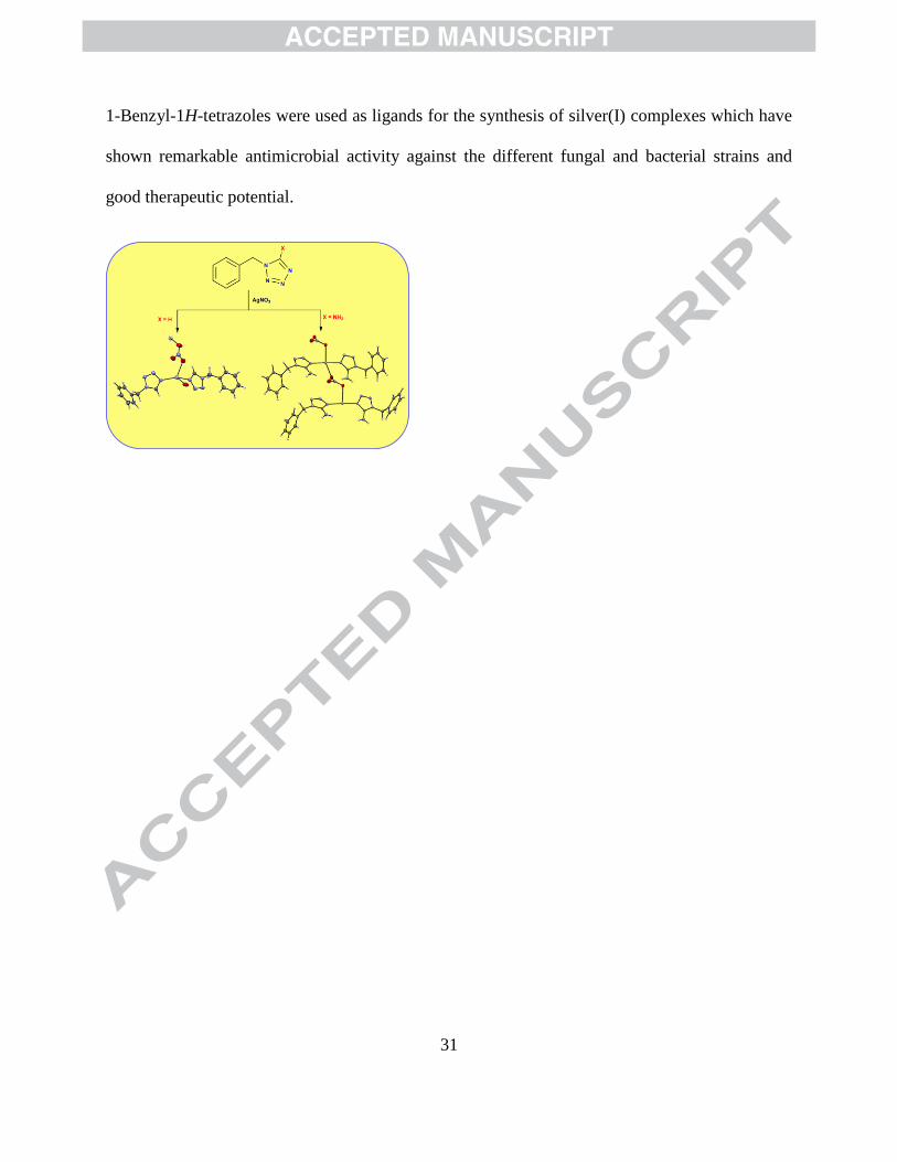
1-Benzyl-1H-tetrazoles were used as ligands for the synthesis of silver(I) complexes which have
shown remarkable antimicrobial activity against the different fungal and bacterial strains and
good therapeutic potential.

32