synthesis of silver nanoparticles using silybum...
TRANSCRIPT
International Journal of Nanoscience and NanotechnologyVolume 9, No.4, December 2013, Pages 221-226
Synthesis of Silver Nanoparticles
Using Silybum Marianum
Seed Extract
R. Mohammadinejad , Sh. Pour seyedi , A. Baghizadeh , Sh. Ranjbar, G. A. Mansoori°
Department of Bioengine ering, University of Illinois at Chicago, Chicago , USA
(*) Corresponding author: [email protected] (Received:20 Oct. 2013 and Accepted: 19 Nov. 2013)
Abstract:
Green synthesis of nanoJXlrlicles, as fundamental building blocks of nanotechnology, has recently attracted considerable attention. Silver nanoparticles have unique physiochemical, biological and environmentalproperties which make them useful in a wide range of applications. In the present paper we report our research results on biosynthesis <?,{silver nanoparticlesfrom silverprecursor using milk-thistle plant (Silybum marianum) seed extract. The resulting synthesi=ed Ag nanoparticles (AgNPs) were characteri=ed with UVvisible spectroscopy. X-ray diffraction (XRD) and transmission electron microscopy (FEM). Our measurementsshowed that S. marianum seed extract can mediate facile and eco-friendly biosynthesis of colloidal sphericalsilver nanoparticles (AgNPs) of si=e range 1-25 nm. The colloidal AgNPs were formed at room temperatureand were observed to be highly stable even after 6 months. Keywords: milk-thistle plant , Silver nano[Xlrticles, Silybum marianum, stable colloids, UV-vis spectroscopy,XRD, TEM
1. INTRODUCTION
Advancement of nanotechnology is based on the availability of well-defined and well-characterized molecules, macromolecules, nanostructures, and supramolecules as its fundamental molecular building blocks. These are small particles of 1-100 nm with high specific surfac-e area. Nanostructure materials show unique physical, chemical, biological and environmental properties, including catalytic activity, optical, electronic and magnetic properties, which have increased their applications in research, industry, agriculture, environment and medicine (Khataee and Mansoori 2011; Mansoori
2005; Mansoori et al. 2007, 2008, 2012). Facile green silver nanoparticles (AgNPs) are blossoming field of research and have high potential as commercialized nanomaterials (Chaloupka et al. 2010; Vahabi et al. 2011; Mansoori 2013). As an effective antimicrobial agent, facile green AgNPs have the potential for large-scale applications in the formulation of dental resin composites (Kassaee et al. 2008), bone cement (Alt et al. 2004), water and air filters (Jain and Pradeep 2005; Sharma et al. 2009), clothing and textiles, medical devices and implants ( de Mel et al. 2012), cosmetics (K.okura et al. 2010) and packaging (Azeredo 2009). Besides their antimicrobial properties, silver nanoparticles
221
222
and silver nanocomposites or nanohybrids have other interesting characteristics which will further enable them to be used in catalysts, biosensors, conductive inks, electronic devices and solar cells (Tsuji et al. 2012; Wijnhoven et al. 2009). They can be produced economically and in large / industrial scale (Vahabi et al. 2011; Mansoori 2013). Several techniques to manufacture AgNPs are proposed. Generally, AgNPs are prepared by a variety of biological, chemical and physical methods, but majority of these techniques are either expensive and/or environmentally hazardous. In addition the synthesized nanoparticles by most methods may be unstable and tend to agglomerate quickly and become useless unless capping agents are applied for their stabilization (Sintubin et al. 2012). Chemicals used for synthesis and stabilization of nanoparticles could be also toxic. The need for clean and reliable synthetic protocols for nanomaterials synthesis leads to the developing interest in benign / green biological approaches (Vahabi et al. 2011; Zaki et al. 2011; Mansoori 2013). In recent years many live bio-organisms such as bacteria, fungi, algae, plants and extracts or metabolites from them have been mediated for synthesis of AgNPs. The reduction of Ag+ to Ag0 occurs by combinations of biomolecules such as proteins, polysaccharides, and flavonoids (Park et al. 2011; Vahabi et al. 2011; Mansoori 2013). Certain biological synthesis of metal and their alloy nanoparticles is
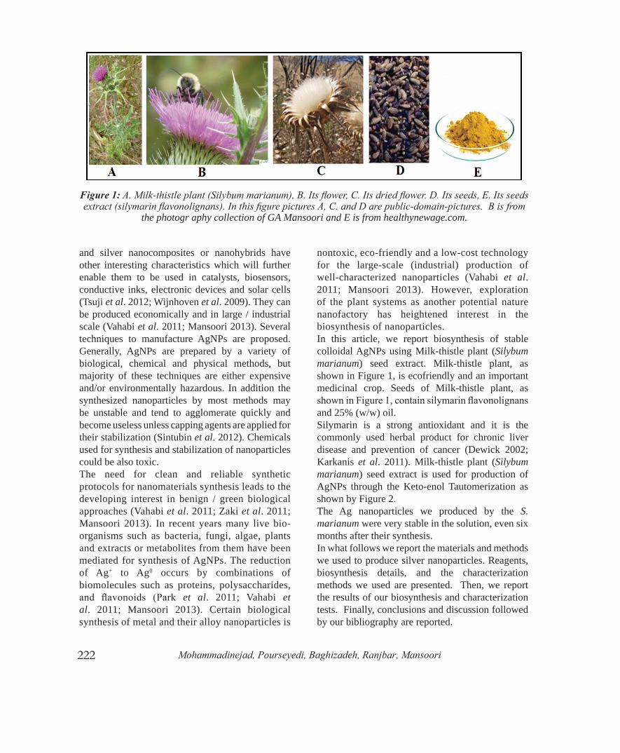
nontoxic, eco-friendly and a low-cost technology for the large-scale (industrial) production of well-characterized nanoparticles (Vahabi et al. 2011; Mansoori 2013). However, exploration of the plant systems as another potential nature nanofactory has heightened interest in the biosynthesis of nanoparticles. In this article, we report biosynthesis of stable colloidal AgNPs using Milk-thistle plant (Silybum marianum) seed extract. Milk-thistle plant, as shown in Figure 1, is ecofriendly and an important medicinal crop. Seeds of Milk-thistle plant, as showninFigure1,containsilymarinflavonolignansand 25% (w/w) oil.Silymarin is a strong antioxidant and it is the commonly used herbal product for chronic liver disease and prevention of cancer (Dewick 2002; Karkanis et al. 2011). Milk-thistle plant (Silybum marianum) seed extract is used for production of AgNPs through the Keto-enol Tautomerization as shown by Figure 2.The Ag nanoparticles we produced by the S. marianum were very stable in the solution, even six months after their synthesis. In what follows we report the materials and methods we used to produce silver nanoparticles. Reagents, biosynthesis details, and the characterization methods we used are presented. Then, we report the results of our biosynthesis and characterization tests. Finally, conclusions and discussion followed by our bibliography are reported.
Figure1: A. Milk-thistle plant (Silybum marianum), B. Its flower, C. Its dried flower. D. Its seeds, E. Its seeds extract (silymarin flavonolignans). In this figure pictures A, C. and D are public-domain-pictures. B is from
the photogr aphy collection of GA Mansoori and E is from healthynewage.com.
Mohammadinejad, Pourseyedi, Baghizadeh, Ranjbar, Mansoori

223
2. MATERIALS AND METHODS
2.1. Reagents
Silver nitrate (AgNO3) was purchased from Merck, Germany. Seeds of S. marianum were obtained from, Pakanbazr, Isfahan, Iran.
2.2. Biosynthesis of AgNPs
For seed extract preparation 5 g dry seeds of S. marianum were washed several times withdeionized (DI) water to remove dust. Seeds wereadded to 100 mL boiling DI water. After boiling for20 min, 3 mL of seed extract was added to 47 mLof 10-3 M AgNO3 solution for AgNPs synthesis atroom temperature.
2.3. Characterization of AgNPs
2.3.1. UV–vis spectroscopyThe biosynthesis of AgNPs was monitored periodically using a UV–vis spectrophotometer (Cary 50, Australia) at different times at room temperature. These measurements operated at a resolution of 1 nm and wavelength range between 300 and 600 nm.
2.3.2. X-ray diffractionThe formation and quality of compounds were gained by XRD technique. For this purpose, biosynthesized AgNPs colloid was centrifuged (at 18,000 rpm; 25◦C) for 20min’s,washedwithDI water and re-centrifuged in four cycles. Then purifiedAgNPsweredriedand subjected toXRD
experiment. AgNPs were then coated on silicon wafer and X-ray diffraction was performed on an X-ray diffractometer (X’Pert Pro MPD) operated at40 kV and 40 mA. The scanning was done in theregionof2θfrom20◦to80◦.
2.3.3. Transmission electron microscopyTransmission Electron Microscopy (TEM) was performed on Philips CM-10 model (HT 100KV) for determining the morphology of AgNPs. The sample was sonicated for 15 min. A drop of this solution was loaded on carbon-coated copper grids, and allowed to evaporate.
3. RESULTS AND DISCUSSION
3.1. UV–vis absorbance studies
Reduction of the Ag+ to Ag0 during exposure to the S. marianum seed extract was followed by colorchange of the solution from colorless to yellow.These color changes aroused out of the excitationof surface plasmon vibrations with the AgNPs(Mulvaney, 1996). The UV–vis spectra producedare shown in Figure 3.It is observed that the maximum absorbance of Agnanoparticles occurs at 425 nm, indicating that AgNPs were produced. It was also observed that reduction ofsilver ions into nanoparticles started after 3 hours ofreaction and completed after almost 24 hours. Figure4 shows the UV–vis absorption spectra of silversynthesized nanoparticles after storage for 6 monthsto test the stability of the AgNPs.
Figure2: Silymarin flavonolignans keto-enol tautomerization used to produce silver nanoparticles.
InternationalJournalofNanoscienceandNanotechnology

224
Figure3: UV–vis spectra showing absorption recorded as a function of 10−3 M aqueous solution of silver nitrate with S. marianum seed extract as a function of time. (a) Color of S. marianum seed
extract before adding silver nitrate (b) Color of S. marianum seed extract after adding silver
nitrate at 24 h.
As it can be seen, the absorption peaks of the AgNPs shift only slightly,without a significant change inthe color. This indicates that the as-prepared AgNPs are stable over a long period (Liu et al. 2009).
3.2. XRD analysis
The formations of the nano-crystalline Ag particles werefurtherconfirmedbytheXRDanalysisdepictedin Figure 5.
Figure4: UV–vis spectra of biosynthesized silver nanoparticles by S. marianum seed extract after
six months.
Figure5: XRD pattern of silver nanoparticles synthesized by treating AgNO3 solution with
Silybum marianum seed extract.
Intensepeakswereobservedat2θvaluesof38.098o, 44.154o, 64.674o, and 77.544o, corresponding to (111), (200), (220) and (311) Bragg’s reflectionbased on the face-centered-cubic (fcc) crystal structure of AgNPs. The broadening of Bragg’s peaks indicates the formation of nanoparticles. The XRD pattern thus clearly shows that the AgNPs formed by the reduction of Ag+ ions by S. marianum seed extract are crystalline in nature. No additional peak appeared in XRD pattern, indicating a high purity of biosynthesized AgNPs.
3.3. TEM analysis
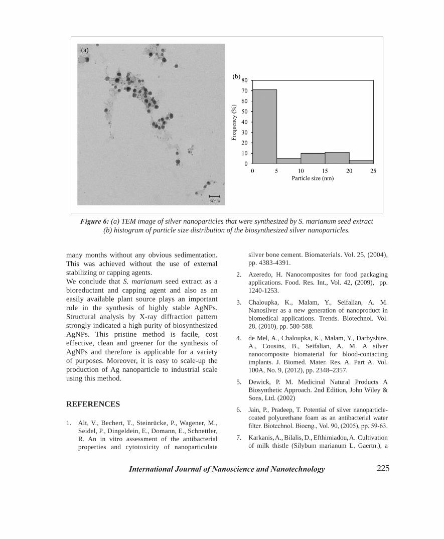
Transmission electron microscopy (TEM) was used to determine the morphology (size and shape) of nanoparticles. The TEM images of the prepared AgNPs at 50 nm scales are shown in the Figure 6a.TEM images show that they have spherical shape. Particle size distribution histogram determined from TEM is shown in Figure 6b. Ag nanoparticles range in size from 1 to 25nm.
4. CONCLUSION
Silybum marianum seed extract was successfully used for the single-pot biosynthesis of spherical AgNPs in ambient conditions with the size range from 1 to 25 nm, as inferred from the TEM imaging. UV analysis indicated stability of the AgNPs for
Mohammadinejad, Pourseyedi, Baghizadeh, Ranjbar, Mansoori
225
many months without any obvious sedimentation. This was achieved without the use of external stabilizing or capping agents. We conclude that S. marianum seed extract as a bioreductant and capping agent and also as an easily available plant source plays an important role in the synthesis of highly stable AgNPs. Structural analysis by X-ray diffraction pattern strongly indicated a high purity of biosynthesized AgNPs. This pristine method is facile, cost effective, clean and greener for the synthesis of AgNPs and therefore is applicable for a variety of purposes. Moreover, it is easy to scale-up the production of Ag nanoparticle to industrial scale using this method.
REFERENCES
1. Alt, V., Bechert, T., Steinrücke, P., Wagener, M.,Seidel, P., Dingeldein, E., Domann, E., Schnettler,R. An in vitro assessment of the antibacterialproperties and cytotoxicity of nanoparticulate
silver bone cement. Biomaterials. Vol. 25, (2004), pp. 4383-4391.
2. Azeredo, H. Nanocomposites for food packagingapplications. Food. Res. Int., Vol. 42, (2009), pp.1240-1253.
3. Chaloupka, K., Malam, Y., Seifalian, A. M.Nanosilver as a new generation of nanoproduct inbiomedical applications. Trends. Biotechnol. Vol.28, (2010), pp. 580-588.
4. de Mel, A., Chaloupka, K., Malam, Y., Darbyshire,A., Cousins, B., Seifalian, A. M. A silvernanocomposite biomaterial for blood-contactingimplants. J. Biomed. Mater. Res. A. Part A. Vol.100A, No. 9, (2012), pp. 2348–2357.
5. Dewick, P. M. Medicinal Natural Products ABiosynthetic Approach. 2nd Edition, John Wiley &Sons, Ltd. (2002)
6. Jain, P., Pradeep, T. Potential of silver nanoparticle-coated polyurethane foam as an antibacterial waterfilter.Biotechnol.Bioeng.,Vol.90,(2005),pp.59-63.
7. Karkanis, A., Bilalis, D., Efthimiadou, A. Cultivation of milk thistle (Silybum marianum L. Gaertn.), a
Figure6: (a) TEM image of silver nanoparticles that were synthesized by S. marianum seed extract (b) histogram of particle size distribution of the biosynthesized silver nanoparticles.
InternationalJournalofNanoscienceandNanotechnology
226
medicinal weed. Ind. Crop. Prod., Vol. 34, (2011), pp. 825-830.
8. Kassaee, M., Akhavan, A., Sheikh, N., Sodagar, A.. Antibacterial effects of a new dental acrylic resincontaining silver nanoparticles. J. Appl. Polym. Sci.,Vol. 110, (2008), pp. 1699-1703.
9. Khataee, A. and Mansoori, G.A. Nanostructuredtitanium Dioxide Materials. Word Scientific,Hackensack, USA (2011).
10. Kokura, S., Handa, O., Takagi, T., Ishikawa, T.,Naito, Y., Yoshikawa, T. Silver nanoparticles as asafe preservative for use in cosmetics. Nanomed-Nanotechnol., Vol. 6, (2010), pp. 570-574.
11. Liu, Y., Chen, S., Zhong, L., Wu, G. Preparationof high-stable silver nanoparticle dispersion byusing sodium alginate as a stabilizer under gammaradiation. Radiat. Phys. Chem., Vol. 78, (2009), pp.251-255.
12. Mansoori, G. A. Principles of nanotechnology:molecular-based study of condensed matter in smallsystems.WordScientific,Hackensack,USA(2005).
13. Mansoori,G.A.,George,T.F.,Assoufid,L.,Zhang,G. Molecular building blocks for nanotechnology:From diamondoids to nanoscale materials andapplications. Springer. New York, NY (2007).
14. Mansoori, G. A. Synthesis of Nanoparticles byFungi, U.S. Patent “US 8,394,421 B2”, (2013).
15. Mansoori G. A., Bastami T. R., Ahmadpour,A., Eshaghi, Z. . Environmental applicationof nanotechnology. Annual Review of NanoResearch, Vol. 2, Chapter 10, Cao, G. and Brinker,C.J. (Editors).WordScientific,Hackensack,USA(2008).
16. Mansoori G.A., De Araujo PLB, De Araujo ESDiamondoid molecules: with applications inbiomedicine, materials science, nanotechnology andpetroleum science. Word Scientific, Hackensack,USA (2012).
17. Mulvaney, P. Surface plasmon spectroscopy ofnanosized metal particles. Langmuir., Vol. 12,(1996), pp. 788-800.
18. Park, Y., Hong, Y., Weyers, A., Kim, Y., Linhardt,R. Polysaccharides and phytochemicals: a naturalreservoir for the green synthesis of gold and silvernanoparticles. IET Nanobiotechnol., Vol. 5, (2011),pp. 69-78.
19. Sharma, V. K., Yngard, R. A., Lin, Y. . Silvernanoparticles: green synthesis and their antimicrobial activities. Adv. Colloid Interfac., Vol. 145, (2009),pp. 83-96.
20. Sintubin, L., Verstraete, W., Boon, N. . Biologicallyproduced nanosilver: Current state and futureperspectives. Biotechnol. Bioeng., Vol. 109, No. 10,(2012), pp. 2422–2436.
21. Tsuji, M., Gomi, S., Maeda, Y., Matsunaga, M.,Hikino, S., Uto, K., Tsuji, T., Kawazumi, H. . RapidTransformation from Spherical Nanoparticles,Nanorods, Cubes, or Bipyramids to TriangularPrisms of Silver with PVP, Citrate, and H2O2.Langmuir., Vol. 28, (2012), pp. 8845-8861.
22. Vahabi, K., Mansoori, G.A. and Karimi, S. .Biosynthesis of Silver Nanoparticles by FungusTrichoderma Reesei (A Route for Large-ScaleProduction of AgNPs). Insciences J., Vol. 1, No. 1,(2011), pp. 65-79; doi:10.5640/insc.010165 ISSN1664-171X.
23. Wijnhoven, S.W.P., Peijnenburg, W.J.G.M., Herberts, C.A., Hagens, W.I., Oomen, A.G., Heugens, E.H.W.,Roszek, B., Bisschops, J., Gosens, I., Van De Meent,D. Nano-silver-a review of available data andknowledge gaps in human and environmental riskassessment. Nanotoxicology., Vol. 3, (2009), pp.109-138.
24. Zaki, S., El Kady, M., Abd-El-Haleem, D.Biosynthesis and structural characterization of silvernanoparticles from bacterial isolates. Mater. Res.Bull., Vol. 46, (2011), pp. 1571-1576.
Mohammadinejad, Pourseyedi, Baghizadeh, Ranjbar, Mansoori