synthesis of metallic nanoparticles from beta vulgaris ... · a single-pot green chemistry approach...
TRANSCRIPT
ORIGINAL PAPER
Synthesis of metallic nanoparticles from Beta vulgaris usinga single-pot green chemistry approach and their environmentalengineering application
Carol Zethu Ngwenya1 • Karabo Seteno Obed Ntwampe1 • Nothemba Silwana1
Received: 1 July 2016 / Accepted: 26 October 2016 / Published online: 3 November 2016
� Springer International Publishing Switzerland 2016
Abstract In this study, a method for the green synthesis of
nanoparticles using Beta vulgaris (B. vulgaris) extracts was
developed. B. vulgaris was solely used in a synthetic,
chemical-free, redox reaction, as a source of the precursor
metal, reducing and capping agent. Metals accumulated in
B. vulgaris were reduced to nanosize by betanin, an
anthocyanin present in the B. vulgaris which has reducing
and stabilising capabilities. Nanoparticles were synthesised
in a solution containing 1% (v/v) B. vulgaris juice extract,
at 80 �C and a pH of 8. The nanoparticles were charac-
terised using TEM, SEM–EDS, UV–Vis, FTIR, and
PXRD. It was determined that a hydroxyl group from the
betanin releases an active H? proton, which subsequently
reduces metal ions. Results showed that spherical-shaped
Ca-based crystal nanoparticles of approximately 5 nm were
successfully synthesised using the developed method.
Supplementing the Ca-based nanoparticles in biosurfactant
producing cultures of Bacillus licheniformis STK 01
improved the emulsification index of kerosene from 20 to
50%—signifying the usability of the nanoparticles in
environmental engineering applications in which
hydrophobic compounds are targeted for bioremediation.
Keywords Beta vulgaris � Betanin � Environmental
engineering � Nanobiotechnology � Nanoparticles
Introduction
Nanoparticles, due to their ideal particle size and unique
properties, have been of great research interest for appli-
cations in the field of biotechnology. The focus has been on
the active integration of microbial biotechnology and
nanotechnology [23]. Current trends also focus on the
application of nanoparticles to improve biochemical pro-
cesses. For example, iron (Fe) nanoparticles increased
glycolipid biosurfactant production by 80% using Nacar-
diopsis sp. MSA13A [13] with the produced biosurfactant
being used as green stabilisers for the biological synthesis
of nanoparticles [14]. This indicates the need for the syn-
thesis of suitable nanoparticles for biochemical processes
and environmental engineering applications.
Nanoparticles are highly reactive, and their character-
istics depend on their particle size, shape, and composition.
Nanoparticles are synthesised by different physical and
chemical methods. They can either be synthesised using a
top–down approach which involves the breaking down of a
large particle into nanosize, or a bottom–up approach,
whereby atoms are assembled to molecular structures that
fall within the nanorange [23]. The most commonly used
method is the chemical reduction method that involves
reducing metal salts to metal atoms by using a reducing
agent. The currently employed chemical and physical
processes for nanoparticle synthesis are labour-intensive,
use high radiation, temperature, and pressure, as well as
analytical grade chemicals as reducing and stabilising
agents. The residue and/or waste from such processes can
become detrimental to environmental health and living
organisms when disposed-off into the environment. Thus,
the synthesis of nanoparticles using the green chemistry
approach is therefore recommended. Research has focused
on the development of clean, non-toxic and
& Karabo Seteno Obed Ntwampe
1 Bioresource Engineering Research Group (BioERG),
Department of Biotechnology, Cape Peninsula University of
Technology, P.O. Box 652, Cape Town 8000, South Africa
123
Nanotechnol. Environ. Eng. (2016) 1:11
DOI 10.1007/s41204-016-0012-5

environmentally benign production processes for
nanoparticle synthesis. There have been many reported
attempts at synthesising nanoparticles in supposedly envi-
ronmentally friendly, non-toxic processes that promote the
green chemistry approach [9, 17]. For the green chemistry
approach, biological/plant extracts are used to replace
hazardous chemicals, mostly as reducing and stabilising
agents.
In a study by Gan et al. [9], palm oil mill effluent was
used as a reducing agent for the synthesis of gold
nanoparticles from a chloroauric acid solution (HAuCl4) as
a metal precursor. However, this approach still produces
toxic chemical waste from the source of the metal pre-
cursor. Similarly, Le et al. [17] produced silica nanoparti-
cles from Vietnamese rice husks as a source of the metal
precursor but used analytical grade chemicals as reducing
and stabilising agents. Additionally, numerous studies have
reported on the green synthesis of silver nanoparticles
particularly as a noble metal with vast applications
[4, 7, 8, 10, 26]. In these studies, various plant extracts
have shown potential to facilitate the bioreduction of the
silver-based metal precursor to silver nanoparticles [1].
This approach has been widely used for the green synthesis
of silver nanoparticles. However, the major setback of
these methods is that most reported using analytical grade
AgNO3 as a precursor. Upon bioreduction of Ag2? to Ag–
NPs, the NO32- is most likely to end-up in wastewater.
This may result in pollution of freshwater sources by
causing eutrophication and acidification [25]. Alterna-
tively, agrowaste material such as Beta vulgaris, which is
known to contain various metals [2] including reducing
agents [27], can be used for the synthesis of nanoparticles.
This approach to green synthesis of nanoparticles can add
to the current methods being developed for the synthesis of
nanoparticles intended for environmental engineering
applications.
As previously mentioned that B. vulgaris has been
shown to be a rich source of metals and valuable com-
pounds such as betanin with high metal ion reducing
capabilities [22], the present study was undertaken to
develop a single-pot method for the green synthesis of
nanoparticles from B. vulgaris.
Experimental methods and materials
Preparation of Beta vulgaris solutions
for nanoparticle synthesis
B. vulgaris extract solution was prepared from B. vulgaris
agrowaste obtained from a fruit- and vegetable-processing
facility located in Cape Town, Western Cape, South Africa.
The facility is located within close proximity of the Cape
Peninsula University of Technology, District Six campus.
The waste was milled and oven-dried at 70 �C for 72 h,
and thereafter, it was pulverised to a size less than
0.30 mm. A mass of 10 g of the pulverised B. vulgaris was
added to 1 L of sterile distilled water (sdH2O). Addition-
ally, a juice extract was obtained by juicing freshly peeled
B. vulgaris tubers in a blender (Russell HobbsTM, Eng-
land), and the removal of B. vulgaris debri was achieved by
filtering all the solutions including juice extracts through a
125-mm Whatman� filter paper.
Preparation of an Aloe vera solution to enhance
nanoparticle synthesis
Aloe vera (A. vera) was obtained within the premises of the
Cape Peninsula University of Technology, District Six
campus. A. vera contains bioactive phytochemicals that can
facilitate nanoparticle formation. The A. vera leaf was cut
into small fractions, and 20 g of the sliced A. vera was
added to 200 mL of sdH2O. An A. vera extract solution
was prepared by boiling at 100 �C for 60 min. The solution
was filtered through a 125-mm Whatman� filter paper to
remove debri prior to usage.
Preparation of CaCO3 solution from egg shells
for pH adjustment
Eggshells were obtained from a campus restaurant of the
Cape Peninsula University of Technology, District Six
campus. The eggshells were washed in running tap water
and oven-dried at 70 �C for 24 h and milled to produce a
fine powder. A mass of 50 g of powdered eggshells was
added to 100 mL of sdH2O and autoclaved at 121 �C for
15 min and filtered through a 125-mm Whatman� paper.
The eggshell solution was used to adjust the pH of some
solutions used in this study.
Process conditions assessed for the synthesis
of nanoparticles
Experimental runs (n = 13, in triplicate—see Table 1)
were used to optimise pH, juice extract/solution concen-
tration, and temperature for the synthesis of nanoparticles.
The pH was tested at acidic (low/high acidity) and alkaline
(low/high alkalinity) conditions. The pH adjustment was
accomplished by either using 1 M NaOH and/or CaCO3
extracted from eggshells (for the alkaline study), while 1 M
HCl was utilised for the studies conducted under acidic
conditions. The temperature used was either at room tem-
perature (RT) or at 80 �C [22]. The presence and synthesis
of nanoparticles was initially determined by a colorimetric
technique [12], as the interaction of plant extracts with
metal ions in solution alters the properties of the extract,
11 Page 2 of 13 Nanotechnol. Environ. Eng. (2016) 1:11
123
resulting in a colour change. Subsequently, favourable
process conditions were identified.
The effect of A. vera on nanoparticle synthesis
The effect of A. vera in the synthesis of nanoparticles was
evaluated (experimental run 9, Table 1), with the A. vera
solution being added to the B. vulgaris extract solution at a
ratio of 1:1. The CaCO3 solution from eggshell extract
solution was used to adjust the pH of the media.
Nanoparticle synthesis from B. vulgaris solutions
and juice extract
A comparative study between the B. vulgaris agrowaste
extract solutions and the juice extract solution was evalu-
ated. Different concentrations of juice extract were evalu-
ated (see Table 1, experimental runs 10–13). The reaction
was carried out at optimised pH and temperature, and the
presence of nanoparticles was assessed based on the colour
change in the solutions and reaction time.
Purification and characterisation of nanoparticles
Nanoparticle purification was done according to a method
developed by Kowalczyk et al. [15], which is based on
centrifugation. Briefly, samples were centrifuged at
1400 rpm for 10 min at a temperature of 4 �C, followed by
pellet re-suspension in deionised water to remove B. vul-
garis debri and extracellular polymeric substances (EPS).
The supernatant was discarded, and the pellet was dried at
60 �C for 24 h. The dried pellet was then used for char-
acterisation using microscopy and spectrophotometric
techniques. The IR spectra of the nanoparticles were
measured using a PerkinElmer FTIR spectrometer with a
scan range of 7800–4000 cm-1 while a UV–Vis spec-
trophotometer was used to determine the absorption spectra
in the range of 200–800 nm.
Furthermore, powder X-ray diffraction was used to
determine the diffraction pattern and crystalline properties
of the particles using a D2 Phaser BRUKER diffractometer
with Cu-Ka (1.54184 A). The size and shape of the
nanoparticles was determined using a FEI/Tecnai T20
TEM operated at 200 kV, with further confirmation being
done using a Zeiss/Leo 1450 SEM–EDS, which also aided
with the elemental analysis. Additionally, ImageJ software
was utilised to analyse 2D TEM images, by initially setting
the scale as enlisted on the images, to further quantify the
particle size distribution of the nanoparticles, using the
analysis tool within the software [16].
Environmental engineering application:
biosurfactant activity by emulsifying kerosene
A volume of 5 mL of kerosene was added to 5 mL of the
cell-free supernatant, recovered at 11 000 g for 10 min,
containing the biosurfactant produced by Bacillus licheni-
formis STK 01 using B. vulgaris media supplemented with
nanoparticles in a graduated 15-mL centrifuge tube. The
mixture was mixed using a vortex for 5 min and left to
stand for 24 h. The emulsification index was calculated
using Eq. 1 (in triplicate, with average emulsification index
reported):
E24 ¼ Total volume of the emulsion
Total volume of the aqueous solution þ emulsion� 100
ð1Þ
Table 1 Process parameters for
determining suitable conditions
for the synthesis of
nanoparticles
Sample ID Temperature pH B. vulgaris solution/juice BV:AV ratio pH adjusting solution
1 RT 2 10 mL extract solution 1:0 1 M HCl
2 RT 6 10 mL extract solution 1:0 1 M HCl
3 RT 8 10 mL extract solution 1:0 1 M NaOH
4 RT 10 10 mL extract solution 1:0 1 M NaOH
5 80 �C 2 10 mL extract solution 1:0 1 M HCl
6 80 �C 6 10 mL extract solution 1:0 1 M HCl
7 80 �C 8 10 mL extract solution 1:0 1 M NaOH
8 80 �C 10 10 mL extract solution 1:0 1 M NaOH
9 80 �C 8 10 mL extract solution 1:1 CaCO3/EgS
10 80 �C 8 100% (v/v) juice extract 1:0 1 M NaOH
11 80 �C 8 10% (v/v) juice extract 1:0 1 M NaOH
12 80 �C 8 1% (v/v) juice extract 1:0 1 M NaOH
13 80 �C 8 0.1% (v/v) juice extract 1:0 1 M NaOH
RT room temperature, BV B. vulgaris, AV A. vera, EgS eggshell
All experiments were performed in triplicate
Nanotechnol. Environ. Eng. (2016) 1:11 Page 3 of 13 11
123

Results and discussion
Process parameters for nanoparticle synthesis
To develop a suitable method, different parameters were
assessed and optimised. The first part of the study focussed
on the determination of the optimum pH and temperature
for nanoparticle synthesis. The B. vulgaris solution was
used to effectively determine a suitable pH (high/low
acidity and high/low alkalinity). Summarised results for the
experimental runs which were used to monitor and confirm
the colour change including reaction time are as shown in
Table 2—some of which culminated in suitably sized
nanoparticles.
During the formation of nanoparticles, a series of col-
our changes was observed as a result of the betanin which
was degraded under different conditions, thus facilitating
the reduction of metals to nanosize. As shown in Table 2,
there was minimal significant colour change observed in
runs 1, 2, 5, and 6 under low pH. This indicated that a
chemical reaction was non-existent. The pH normally
determines the stability of the betanin, which was previ-
ously reported to be stable at a pH range of 3 to 7 [6]. At
this pH range, betanin loses its ability to bind and reduce
metal ions which effectively reduces the synthesis of
nanoparticles and thus the yield. For runs 3, 4, and 7 with
an alkaline pH, a chemical reaction was observed by a
significant colour change in the samples, with no reaction
being observed for run 8 at pH 10. In runs 3 and 7, with
low alkalinity, the colour changed from purple red to a
dark brown colour. The colour change was an indication
of betanin degradation, resulting in the formation of a
brown colour as previously determined by De Azeredo
et al. [6]. At alkaline pH, betanin contains more nega-
tively charged functional groups to efficiently bind and
reduce cations.
Additionally, the brown colour is normally observed in
the green synthesis of nanoparticles to detect the biore-
duction of metals using plant extracts [20, 30]. The slight
colour change observed in run 4 can be attributed to the
slight degradation of betanin within the samples. Different
colour changes for nanoparticle synthesis facilitated by
betanin from B. vulgaris at different pH were noted in a
study conducted by Parameshwaran et al. [22]. This reaf-
firmed that pH affects the stability of the betanin catalysis
which promotes subsequent bioreduction of the metal ions.
Similarly, elevated temperature promotes betanin degra-
dation and subsequent formation of nanoparticles. Experi-
mental runs 5–8 were performed at ambient temperature
under natural light to promote the photocatalytic degrada-
tion of the betanin. Minimal colour change was observed in
runs 5, 6, and 8. However, a slow colorimetric reaction was
observed for run 7 samples, for which the colour changed
from purple red to dark brown after 72 h. This suggested
the slow photocatalytic degradation of betanin, as the sta-
bility of betanin can also be impaired by natural light [6].
The degradation of betanin in run 7 samples was also
enhanced by the alkalinity of the medium compared with
samples in runs 5 and 6. Samples for runs 9 and 10 were
both heated to a temperature of 80 �C, which resulted in
the gradual colour change from purple red to a dark brown
and bright yellow colour, respectively. At elevated tem-
peratures, betanin degradation mechanisms vary. It can
either be degraded by isomeration, decarboxylation, or
cleavage [24]. During the formation of nanoparticles, ele-
vated temperatures enhance the synthesis process by
increasing the nucleation rate [6]; thus, elevated tempera-
tures were observed to increase the reaction rates compared
with samples in experiments in which ambient tempera-
tures were used.
To confirm nanoparticle production, TEM was used to
characterise the size of the particles (Fig. 1). Furthermore,
Table 2 Summarised
comparative analysis of process
parameters and observations for
nanoparticle synthesis
Sample ID pH Temp Supplement Colour change Reaction time
1 2 80 �C – – –
2 6 80 �C – – –
3 8 80 �C – Purple red to dark brown 90 min
4 10 80 �C – Purple red to dark red 90 min
5 2 RT – – –
6 6 RT – – –
7 8 RT – Purple red to brown red 72 h
8 10 RT – – –
9 8 80 �C A. vera and eggshell extract Purple red to dark brown 90 min
10 8 80 �C – Reddish/pink bright yellow 90 min
RT room temperature
All experiments were performed in triplicate
11 Page 4 of 13 Nanotechnol. Environ. Eng. (2016) 1:11
123
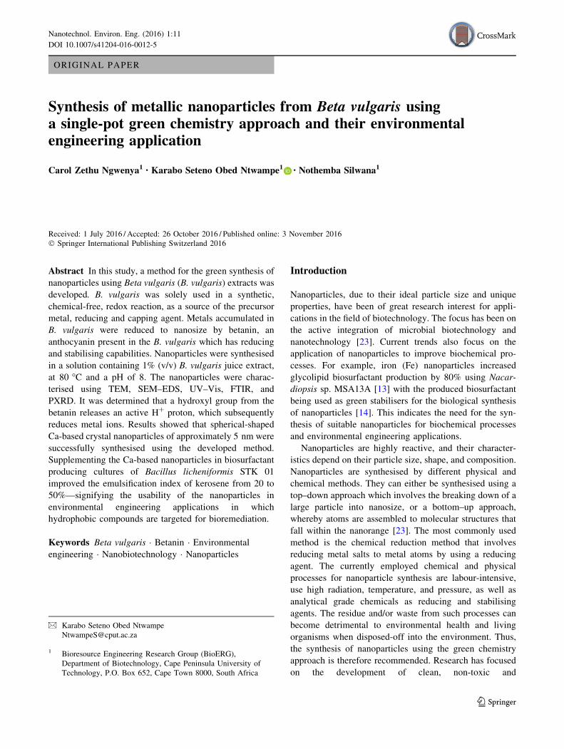
SEM–EDS was also used to confirm the shape and to
determine particle composition by performing elemental
analysis (Fig. 7). Electron microscope analysis was only
carried out on samples that showed a positive chemical
reaction. A freshly prepared and untreated B. vulgaris juice
extract was used in control experiments. For TEM analysis,
Fig. 1 TEM images of the
synthesised nanoparticles
Nanotechnol. Environ. Eng. (2016) 1:11 Page 5 of 13 11
123

copper grids were coated with a few drops of the samples
by using a Pasteur pipette subsequent to drying under a
lamp. The grids were also used for SEM–EDS analysis.
The images obtained from the TEM were examined and
confirmed the synthesis of nanoparticles. The control
experiments showed irregular structures of 0.2 lm
(Fig. 1a). SEM images for control samples revealed what
seemed to be biological debri, i.e. conglomerated gelati-
nous biomass (pectin-like structures) and/or EPS (Fig. 2a–
d). EPS, constituting polysaccharides including proteins, is
known to aggregate in hyperhaline solutions. Similarly,
‘‘low methoxyl pectins form gelatinous structures in the
presence of Ca2? ions at low pH (3–4.5), whereas high
methoxyl pectins rapidly form thermally irreversible gels
in the presence of sufficient sucrose at low pH’’ [28]—pH
and constituents prevalent in untreated B. vulgaris juice
extracts. These structures are clearly different from those
obtained in treated samples, i.e. Fig. 1d. Furthermore,
samples for experimental run 3 indicated the formation of
well-defined spherical-shaped particles of approximately
46.55 nm. These particles were encapped within a clear
capping agent demonstrating betanin’s reducing and
Fig. 2 SEM images of distinguishable biological debri in control experiments (untreated B. vulgaris juice extract)
11 Page 6 of 13 Nanotechnol. Environ. Eng. (2016) 1:11
123

capping capabilities (Fig. 1b), with some larger ([50 nm
to\100 nm) irregular nanoparticles (Fig. 1c, d), using a
1% (v/v) solution from dried B. vulgaris.
TEM and SEM–EDS analyses confirmed spherical-
shaped structures of the particle in treated extracts, with
elemental analysis, suggesting Ca as the major constituent.
Samples in experimental run 4 indicated an irregular-
shaped crystal-like structure which falls within the nano-
size range (Fig. 1c). No metal constituents were detected
from the SEM–EDS spectrum for the irregular-shaped non-
crystal-like structures found in samples for run 7 performed
under ambient temperature, suggesting EPS conglomera-
tion. Al and Si were also detected, presumably as a result of
embedded Al and Si assumed to be from the soil. On
comparing TEM images in samples for run 7, to those of
run 3 (Fig. 1d and b, respectively), the dark shade of the
particles was as a result of diffracted light by the crystalline
structure of the particles. The light structures observed in
TEM images of run 7 samples (Fig. 1d) showed that the
particles were large and perhaps irregular in shape.
Moreover, spherical crystal particles are formed at high
temperatures compared with room temperatures [19], as
observed in run 7, which was processed at room tempera-
ture (Table 1). The successful parameters used for samples
in run 3 were used for further development of the method.
The only limitation of the methods used for the first part of
this study was the low yields of the metallic nanoparticles
obtained.
Improvement of nanoparticle yields using A. vera
In an attempt to improve yields and optimise the in vitro
reaction, biological agents such as A. vera can be used as
supplements. Equivalent quantities of the B. vulgaris
solution and the A. vera were added and incubated at 80 �Cand pH 8 for samples in run 9. For a sustainable greener
method, CaCO3 from eggshell extracts was also used to
adjust the pH and as a supplementary source of Ca2?.
Eggshells have been reported to be a rich source of min-
eralised CaCO3, comprising 94% of the eggshells [21].
The colour change and reaction rate of these samples
were similar to those initially observed for run 3. There-
fore, A. vera had an insignificant impact on the nanopar-
ticle synthesis, although the colour change was similar to
that observed in run 3. However, TEM images (Fig. 3)
revealed a decrease in nanoparticle size and an increase in
particle yields for samples supplemented with A. vera. This
was assumed to be aided by the bioactive phytochemicals
in A. vera. The particles synthesised in run 9 were spherical
with a size of less than 10 nm. Moreover, A. vera exhibited
capping properties, with the nanoparticles synthesised
being closely aggregated and encapsulated within the A.
vera capping agent. The yield and the particle size distri-
bution of the nanoparticles in the samples supplemented
with A. vera demonstrated an improvement in comparison
with those without A. vera.
B. vulgaris juice concentration optimisation
for nanoparticle synthesis
Previous studies have shown that the quantity of the plant
extracts required during green synthesis of nanoparticles
plays a significant role in nanoparticle formation
[9, 20, 22]. In this study, B. vulgaris extract solutions were
compared with different concentrations of fresh B. vulgaris
juice, i.e. with experiments (Table 1, experimental runs
Fig. 3 TEM image of the
sample supplemented with A.
vera
Nanotechnol. Environ. Eng. (2016) 1:11 Page 7 of 13 11
123
10–13). Experimental run 3 (Table 2) samples, containing
B. vulgaris extract solution, were also used for comparative
analyses. The fresh juice extract was used within 48 h of
extraction to prevent the solution becoming viscous as a
result of moisture loss. B. vulgaris juice extracts contain-
ing, 10% (v/v), 1% (v/v), and 0.5% (v/v) (Table 1) were
used. These samples were processed with parameters used
successfully in experimental run 3 for nanoparticle syn-
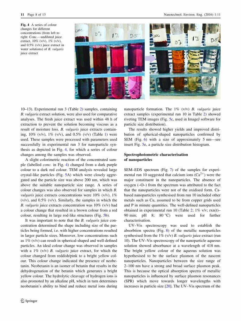
thesis as depicted in Fig. 4, for which a series of colour
changes among the samples was observed.
A slight colorimetric reaction of the concentrated sam-
ple (labelled conc. in Fig. 4) changed from a dark purple
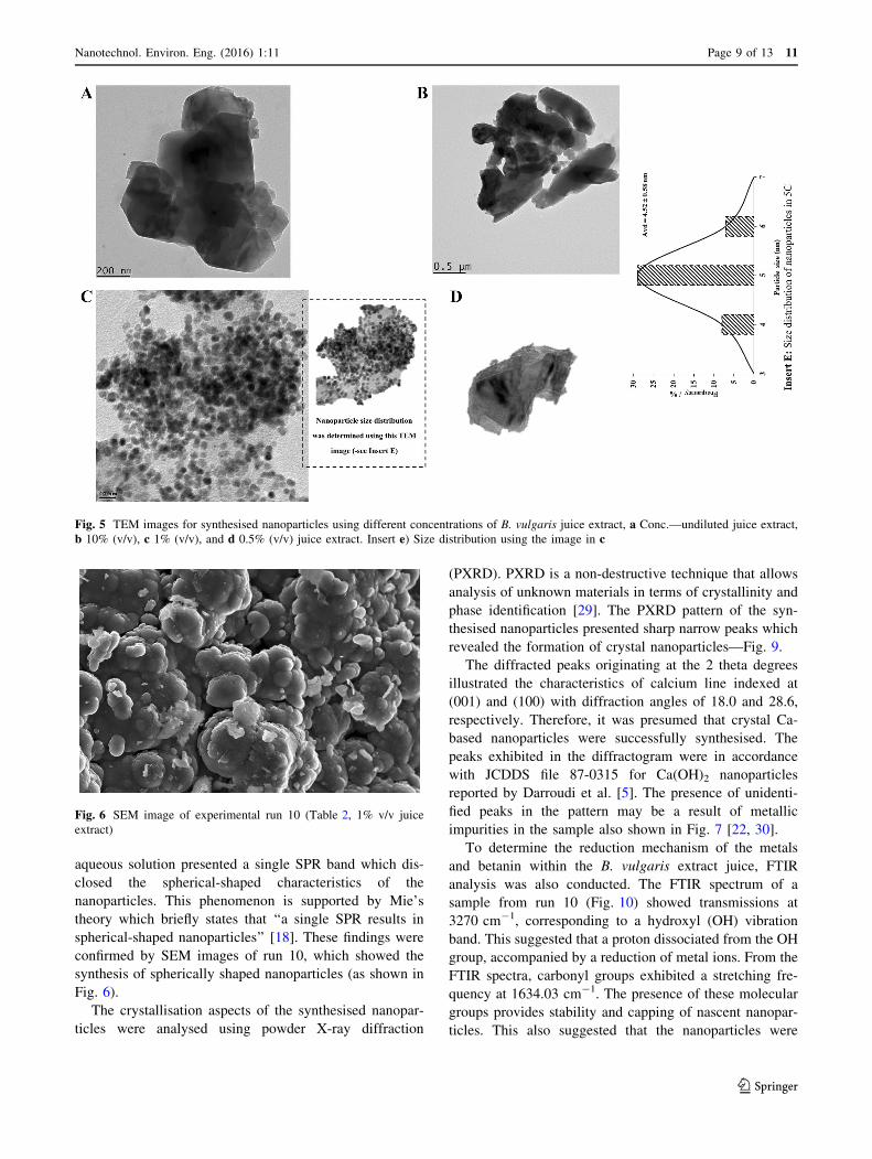
colour to a dark red colour. TEM analysis revealed large
crystal-like particles (Fig. 5A) which were closely aggre-
gated and the particle size was above 200 nm, which was
above the suitable nanoparticle size range. A series of
colour changes was also observed for samples in which B.
vulgaris juice extracts concentrations were 10% (v/v), 1%
(v/v), and 0.5% (v/v). Similarly, the samples in which the
B. vulgaris juice extracts concentration was 10% (v/v) had
a colour change that resulted in a brown colour from a red
colour, resulting in large rod-like structures (Fig. 5b).
It was important to note that the B. vulgaris juice con-
centration determined the shape including size of the par-
ticles being formed, i.e. with higher concentrations resulted
in larger particle sizes. Moreover, low concentrations such
as 1% (v/v) can result in spherical-shaped and well-defined
particles. An ideal colour change was observed in samples
with a 1% (v/v) B. vulgaris juice extract, for which the
colour changed from reddish/pink to a bright yellow col-
our. This colour change indicated the presence of neobe-
tanin. Neobetanin is an isomer of betanin that results in the
dehydrogenation of the betanin which generates a bright
yellow colour. The hydrolytic cleavage of hydrogen ions is
also promoted by an alkaline pH, which in turn determines
neobetanin’s ability to bind and reduce metal ions during
nanoparticle formation. The 1% (v/v) B. vulgaris juice
extract samples (experimental run 10 in Table 2) showed
riveting TEM images (Fig. 5c, used in ImageJ software for
particle size distribution).
The results showed higher yields and improved distri-
bution of spherical-shaped nanoparticles confirmed by
SEM (Fig. 6) with a size of approximately 5 nm—see
insert Fig. 5e, a particle size distribution histogram.
Spectrophotometric characterisation
of nanoparticles
SEM–EDS spectrum (Fig. 7) of the samples for experi-
mental run 10 suggested that calcium ions (Ca2?) were the
major constituent in the nanoparticles. The absence of
oxygen (–O–) from the spectrum was attributed to the fact
that the nanoparticles were not of the oxidised form. Ca-
based nanoparticles synthesised from run 10 included other
metals such as Cu, assumed to be from copper grids used
and P in minute quantities. The well-defined nanoparticles
obtained in experimental run 10 (Table 2; 1% v/v; rxn(t)–
90 min; pH 8; 80 �C) were used for further
characterisation.
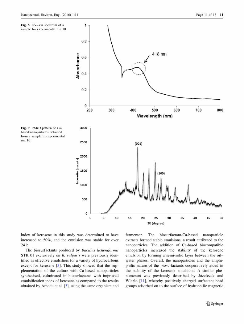
UV–Vis spectroscopy was used to establish the
absorbtion spectra (Fig. 8) of the metallic nanoparticles
synthesised from the 1% (v/v) B. vulgaris juice extract (run
10). The UV–Vis spectroscopy of the nanoparticle aqueous
solution showed absorbance at a wavelength of 418 nm.
The bright yellow colour of the aqueous solution was
hypothesised to be the surface plasmon of the nascent
nanoparticles. Nanoparticles between the size range of
2–100 nm have a strong and broad surface plasmon peak.
This is because the optical absorption spectra of metallic
nanoparticles is influenced by surface plasmon resonances
(SPR) which move towards longer wavelengths with
increases in particle size [20]. The UV–Vis spectrum of the
Fig. 4 A series of colour
changes for different
concentrations (from left to
right: Conc.—undiluted juice
extract, 10% (v/v), 1% (v/v),
and 0.5% (v/v) juice extract in
water solutions) of B. vulgaris
juice extract
11 Page 8 of 13 Nanotechnol. Environ. Eng. (2016) 1:11
123
aqueous solution presented a single SPR band which dis-
closed the spherical-shaped characteristics of the
nanoparticles. This phenomenon is supported by Mie’s
theory which briefly states that ‘‘a single SPR results in
spherical-shaped nanoparticles’’ [18]. These findings were
confirmed by SEM images of run 10, which showed the
synthesis of spherically shaped nanoparticles (as shown in
Fig. 6).
The crystallisation aspects of the synthesised nanopar-
ticles were analysed using powder X-ray diffraction
(PXRD). PXRD is a non-destructive technique that allows
analysis of unknown materials in terms of crystallinity and
phase identification [29]. The PXRD pattern of the syn-
thesised nanoparticles presented sharp narrow peaks which
revealed the formation of crystal nanoparticles—Fig. 9.
The diffracted peaks originating at the 2 theta degrees
illustrated the characteristics of calcium line indexed at
(001) and (100) with diffraction angles of 18.0 and 28.6,
respectively. Therefore, it was presumed that crystal Ca-
based nanoparticles were successfully synthesised. The
peaks exhibited in the diffractogram were in accordance
with JCDDS file 87-0315 for Ca(OH)2 nanoparticles
reported by Darroudi et al. [5]. The presence of unidenti-
fied peaks in the pattern may be a result of metallic
impurities in the sample also shown in Fig. 7 [22, 30].
To determine the reduction mechanism of the metals
and betanin within the B. vulgaris extract juice, FTIR
analysis was also conducted. The FTIR spectrum of a
sample from run 10 (Fig. 10) showed transmissions at
3270 cm-1, corresponding to a hydroxyl (OH) vibration
band. This suggested that a proton dissociated from the OH
group, accompanied by a reduction of metal ions. From the
FTIR spectra, carbonyl groups exhibited a stretching fre-
quency at 1634.03 cm-1. The presence of these molecular
groups provides stability and capping of nascent nanopar-
ticles. This also suggested that the nanoparticles were
Fig. 5 TEM images for synthesised nanoparticles using different concentrations of B. vulgaris juice extract, a Conc.—undiluted juice extract,
b 10% (v/v), c 1% (v/v), and d 0.5% (v/v) juice extract. Insert e) Size distribution using the image in c
Fig. 6 SEM image of experimental run 10 (Table 2, 1% v/v juice
extract)
Nanotechnol. Environ. Eng. (2016) 1:11 Page 9 of 13 11
123

capped by a polymer as shown in Fig. 1B for experimental
run 3. The general decrease in the bands observed between
571.87 and 406.09 cm-1 indicated metallic nanoparticles
synthesis facilitated by betanin. At this point, the functional
groups of betanin interacted with the metals found in B.
vulgaris juice for the formation of nanoparticles. A similar
trend was observed in the FTIR spectrum for betanin, i.e.
for the analysis of the surface structure of Ag nanoparticles
[22]. This suggested that there was a reaction between free
metal ions and betanin found in B. vulgaris for the for-
mation of nanoparticles, a hypothesis used to elucidate the
mechanism for the synthesis of the Ca-based nanoparticles.
Overall, the synthesised nanoparticles can be used in
enhancing the bioavailability of hydrophobic recalcitrant
persistent organic pollutants in bioremediation studies—
Sect. 3.5 demonstrates the application of the produced
nanoparticles reported in this study.
Nanoparticles from Beta vulgaris: an environmental
engineering application
The nanoparticles synthesised were used in a system uti-
lised to produce biosurfactants in fed-batch fermentations.
Subsequently, kerosene was emulsified using the biosur-
factants produced by Bacillus licheniformis STK 01 in B.
vulgaris media supplemented with the Ca-based nanopar-
ticles. Table 3 shows the emulsification index of kerosene
by Bacillus licheniformis STK 01-Ca-based nanoparticle
extracts; results which were compared with those obtained
in a previous study by Amodu et al. [3]. The emulsification
Fig. 7 SEM–EDS spectrum of
a sample in experimental run 10
11 Page 10 of 13 Nanotechnol. Environ. Eng. (2016) 1:11
123
index of kerosene in this study was determined to have
increased to 50%, and the emulsion was stable for over
24 h.
The biosurfactants produced by Bacillus licheniformis
STK 01 exclusively on B. vulgaris were previously iden-
tified as effective emulsifiers for a variety of hydrocarbons
except for kerosene [3]. This study showed that the sup-
plementation of the culture with Ca-based nanoparticles
synthesised, culminated in biosurfactants with improved
emulsification index of kerosene as compared to the results
obtained by Amodu et al. [3], using the same organism and
fermentor. The biosurfactant-Ca-based nanoparticle
extracts formed stable emulsions, a result attributed to the
nanoparticles. The addition of Ca-based biocompatible
nanoparticles increased the stability of the kerosene
emulsion by forming a semi-solid layer between the oil–
water phases. Overall, the nanoparticles and the amphi-
philic nature of the biosurfactants cooperatively aided in
the stability of the kerosene emulsions. A similar phe-
nomenon was previously described by Jozefczak and
Wlazło [11], whereby positively charged surfactant head
groups adsorbed on to the surface of hydrophilic magnetic
Fig. 8 UV–Vis spectrum of a
sample for experimental run 10
Fig. 9 PXRD pattern of Ca-
based nanoparticles obtained
from a sample in experimental
run 10
Nanotechnol. Environ. Eng. (2016) 1:11 Page 11 of 13 11
123

nanoparticles, exposing hydrophobic tails, which aided the
stabilisation of emulsions studied.
Conclusion
A single-pot green chemistry method for the synthesis of
Ca-based nanoparticles using B. vulgaris extracts was
developed. It can be concluded that elevated temperatures
and alkaline pH conditions influence the bioreduction of
metal ions accumulated in B. vulgaris, which were con-
verted into nanoparticles. Electron microscopes and spec-
trophotometric analysis revealed the successful synthesis of
spherical-shaped Ca-based crystal nanoparticles of
approximately 5 nm following bioreduction by betanin.
Furthermore, the supplementation of the synthesised
nanoparticles in a fed-batch biosurfactant production sys-
tem culminated in improved emulsification activity of the
biosurfactant produced as demonstrated by the improved
emulsification of kerosene. Overall, nanoparticles from
Beta vulgaris can be applied in environmental engineering
applications such as the bioremediation of hydrophobic
contaminants in polluted environments.
Acknowledgements The authors would like to acknowledge the
funding from the Cape Peninsula University of Technology (CPUT)
University Research Fund (URF RK 16) and National Research
Foundation (NRF).
References
1. Ahmed S, Ahmad M, Swami B, Ikram S (2016) A review on
plants extract mediated synthesis of silver nanoparticles for
antimicrobial applications: a green expertise. IJAR 7(1):17–28
2. Amodu OS, Ntwampe SK, Ojumu TV (2014) Emulsification of
hydrocarbons by biosurfactant: exclusive use of agrowaste.
BioResources 9(2):3508–3525
3. Amodu OS, Ntwampe SKO, Ojumu TV (2014) Optimization of
biosurfactant production by Bacillus licheniformis STK 01 grown
exclusively on Beta vulgaris waste using response surface
methodology. BioResources 9(3):5045–5065
4. Basu S, Maji P, Ganguly J (2015) Rapid green synthesis of silver
nanoparticles by aqueous extract of seeds of Nyctanthes arbor-
tristis. J Appl Nanosci 6(1):1–5. doi:10.1007/s13204-015-0407-9
5. Darroudi M, Bagherpour M, Hosseini HA, Ebrahimi M (2016)
Biopolymer-assisted green synthesis and characterization of cal-
cium hydroxide nanoparticles. Ceram Int 3(42):3816–3819.
doi:10.1016/j.ceramint.2015.11.045
6. De Azeredo HMC, Pereira AC, De Souza ACR, Gouveia ST,
Mendes KCB (2009) Study on efficiency of betacyanin extraction
from red beetroots. Int J Food Sci Technol 44(12):2464–2469.
doi:10.1111/j.1365-2621.2009.02037.x
7. Dhand V, Soumya L, Bharadwaj S, Chakra S, Bhatt D, Sreedhar
B (2016) Green synthesis of silver nanoparticles using Coffea
arabica seed extract and its antibacterial activity. Mat Sci Eng C
Biol S 58:36–43. doi:10.1016/j.msec.2015.08.018
8. Edison T, Lee Y, Sethuraman M (2016) Green synthesis of silver
nanoparticles using Terminalia cuneata and its catalytic action in
reduction of direct yellow-12 dye. Spectrochim Acta A
161:122–129. doi:10.1016/j.saa.2016.02.044
9. Gan PP, Ng SH, Huang Y, Li SFY (2012) Green synthesis of gold
nanoparticles using palm oil mill effluent (POME): a low-cost
and eco-friendly viable approach. Biores Technol 113:132–135.
doi:10.1016/j.biortech.2012.01.015
Fig. 10 FTIR spectrum of a
sample from experimental run
10
Table 3 Emulsification of
kerosene by Bacillus
licheniformis STK 01/Ca-based
nanoparticle extracts
References E24
Amodu et al. [2, 3] 20%
This study 50%
11 Page 12 of 13 Nanotechnol. Environ. Eng. (2016) 1:11
123

10. Ghaedi M, Yousefinejad M, Safarpoor M, Khafri H, Purkait M
(2015) Rosmarinus officinalis leaf extract mediated green syn-
thesis of silver nanoparticles and investigation of its antimicrobial
properties. Ind Eng Chem Res 31:167–172. doi:10.1016/j.jiec.
2015.06.020
11. Jozefczak R, Wlazło R (2015) Ultrasonic studies of emulsion
stability in the presence of magnetic nanoparticles. Adv Condens
Matter Phys. doi:10.1155/2015/398219
12. Kalimuthu K, Suresh Babu R, Venkataraman D, Bilal M, Gur-
unathan S (2008) Biosynthesis of silver nanocrystals by Bacillus
licheniformis. Colloids Surf B Biointerfaces 65(1):150–153.
doi:10.1016/j.colsurfb.2008.02.018
13. Kiran GS, Nishanth LA, Priyadharshini S, Anitha K, Selvin J
(2014) Effect of Fe nanoparticle on growth and glycolipid bio-
surfactant production under solid state culture by marine No-
cardiopsis sp. MSA13A. BMC Biotechnol 14(1):1–10. doi:10.
1186/1472-6750-14-48
14. Kiran GS, Selvin J, Manilal A, Sujith S (2011) Biosurfactants as
green stabilizers for the biological synthesis of nanoparticles. Crit
Rev Biotechnol 31(4):354–364. doi:10.3109/07388551.2010.
539971
15. Kowalczyk B, Lagzi I, Grzybowski BA (2011) Nanoseparations:
strategies for size and/or shape-selective purification of
nanoparticles. Curr Opin Colloid Interface Sci 16(2):135–148.
doi:10.1016/j.cocis.2011.01.004
16. Kumar S, Sharma JK (2016) Stable phase CdS nanoparticles for
optoelectronics: a study on surface morphology, structural and
optical characterization. Mater Sci Poland 34(2):368–373. doi:10.
1515/msp-2016-0033
17. Le VH, Thuc CNH, Thuc HH (2013) Synthesis of silica
nanoparticles from Vietnamese rice husk by sol–gel method.
Nanoscale Res Lett 8(1):58. doi:10.1186/1556-276X-8-58
18. Link S, El-Sayed MA (2000) Shape and size dependence of
radiative, non-radiative and photothermal properties of gold
nanocrystals. Annu Rev Phys Chem 19(3):409–453. doi:10.1080/
01442350050034180
19. Makarov VV, Makarova SS, Love AJ, Sinitsyna OV, Dudnik AO,
Yaminsky IV, Taliansky ME, Kalinina NO (2014) Biosynthesis
of stable iron oxide nanoparticles in aqueous extracts of Hordeum
vulgare and Rumex acetosa plants. Langmuir 30:5982–5988.
doi:10.1021/la5011924
20. Mubayi A, Chatterji S, Rai PM, Watal G (2012) Evidence based
green synthesis of nanoparticles. Adv Mater Lett 3(6):519–525.
doi:10.5185/amlett.2012.icnano.353
21. Murakami FS, Rodrigues PO, De Campos CMT, Silva MAS
(2007) Physicochemical study of CaCO3 from egg shells. Cienc
Technol Aliment 27(3):658–662. doi:10.1590/S0101-
20612007000300035
22. Parameshwaran R, Kalaiselvam S, Jayavel R (2013) Green syn-
thesis of silver nanoparticles using B. vulgaris: role of process
conditions on size distribution and surface structure. Mater Chem
Phys 140(1):135–147. doi:10.1016/j.matchemphys.2013.03.012
23. Płaza GA, Chojniak J, Banat IM (2014) Biosurfactant mediated
biosynthesis of selected metallic nanoparticles. Int J Mol Sci
15(8):13720–13737. doi:10.3390/ijms150813720
24. Reshmi SK, Aravindhan KM, Suganyadavi P (2012) The effect of
light, temperature, pH on stability of betacyanin pigments in
Basella alba fruit. Asian J Pharm Clin Res 4(3):107–110
25. Rodrıguez Arredondo M, Kuntke P, Jeremiasse A, Sleutels T,
Buisman C, ter Heijne A (2015) Bioelectrochemical systems for
nitrogen removal and recovery from wastewater. Environ Sci
Water Res Technol 1(1):22–33. doi:10.1039/C4EW00066H
26. Sadeghi B, Gholamhoseinpoor F (2015) A study on the stability
and green synthesis of silver nanoparticles using Ziziphora
tenuior (Zt) extract at room temperature. Spectrochim Acta Mol
Biomol Spectrosc 134:310–315. doi:10.1016/j.saa.2014.06.046
27. Singh B, Hathan BS (2014) Chemical composition, functional
properties and processing of beetroot—a review. IJSER
5(1):679–684
28. Srivastava P, Malviya R (2011) Sources of pectin, extraction and
its applications in pharmaceutical industry—an overview. Indian
J Nat Prod Resour 2(1):10–18
29. Taglieri G, Mondelli C, Daniele V, Pusceddu E, Trapanati A
(2013) Synthesis and X-ray diffraction analysis of calcium
hydroxide nanoparticles in aqueous suspension. AMPC
3(1A):108–112. doi:10.4236/ampc.2013.31A013
30. Usman MS, Ibrahim NA, Shameli K, Zainuddin N, Yunus
WMZW (2012) Copper nanoparticles mediated by chitosan:
synthesis and characterization via chemical methods. Molecules
17(12):14928–14936. doi:10.3390/molecules171214928
Nanotechnol. Environ. Eng. (2016) 1:11 Page 13 of 13 11
123