synthesis and characterization of single crystal pbo ... · synthesis and characterization of...
TRANSCRIPT

CERAMICSINTERNATIONAL
Available online at www.sciencedirect.com
http://dx.doi.org/0272-8842/& 20
nCorrespondinE-mail addre
raminyousefi@ia
Please cite thi(2014), http://d
Ceramics International ] (]]]]) ]]]–]]]www.elsevier.com/locate/ceramint
Synthesis and characterization of single crystal PbO nanoparticlesin a gelatin medium
Ramin Yousefia,n, Ali Khorsand Zakb, Farid Jamali-Sheinic, Nay Ming Huangd,Wan Jefrey Basirune,f, M. Sookhakiand
aDepartment of Physics, Masjed-Soleiman Branch, Islamic Azad University (I.A.U), Masjed-Soleiman, IranbNanotechnology Laboratory, Esfarayen University, North Khorasan, Iran
cDepartment of Physics, Ahwaz Branch, Islamic Azad University, Ahwaz, IrandLow Dimensional Materials Research Center, Department of Physics, Faculty of Science, University of Malaya, 50603 Kuala Lumpur, Malaysia
eDepartment of Chemistry, University of Malaya, 50603 Kuala Lumpur, MalaysiafNanotechnology & Catalysis Research Centre (NanoCat), Institute of Postgraduate Studies, University of Malaya, 50603 Kuala Lumpur, Malaysia
Received 12 March 2014; received in revised form 30 March 2014; accepted 30 March 2014
Abstract
Single-crystal, lead-oxide nanoparticles (PbO-NPs) were synthesized via the sol–gel method in a gelatin medium. Long-chain gelatincompounds were utilized to terminate the growth of the PbO-NPs and to stabilize them. The gel that was obtained was calcined at 500, 550, and600 1C for 1 h. The synthesized PbO-NPs were characterized using an X-ray diffraction (XRD) analysis, a thermogravimetric analysis (TGA),scanning electron microscopy (SEM), energy-dispersive X-ray spectroscopy (EDX), and high-magnification and high-resolution transmissionelectron microscopy (TEM and HRTEM). The XRD patterns indicated a phase transformation from tetragonal to orthorhombic as the calcinationtemperature increased. The SEM and TEM images showed that the nanoparticles started to form at a temperature range of 550–600 1C. Inaddition, the TEM results showed that the average particle size of the nanoparticles was �45 nm. The HRTEM images revealed single-crystalPbO-NPs with an orthorhombic phase. The optical properties of the PbO-NPs were studied using a UV–visible (UV–vis) spectrophotometer. TheUV–vis absorption spectra of the PbO-NPs indicated absorption peaks in the visible region, which were attributed to the band gap of the PbO-NPs with an orthorhombic phase.& 2014 Elsevier Ltd and Techna Group S.r.l. All rights reserved.
Keywords: A. Sol–gel processes; B. Nanocomposites; C. Optical properties; PbO nanoparticles; Calcination temperature
1. Introduction
Over past few decades, the understanding of semiconductornanostructures has allowed significant contributions to the devel-opment of nanotechnology. These new developments and applica-tions of semiconductor nanostructures have prompted both thescientific and industrial communities to focus on this class ofmaterials. The huge surface-to-volume ratio of semiconductornanostructures results in a strong sensitivity of the excitons tosurface states, as well as defects caused by their reduced size.
10.1016/j.ceramint.2014.03.18014 Elsevier Ltd and Techna Group S.r.l. All rights reserved.
g author. Tel.: þ98 9166224993; fax: þ98 6813330093.sses: [email protected],umis.ac.ir (R. Yousefi).
s article as: R. Yousefi, et al., Synthesis and characterization of six.doi.org/10.1016/j.ceramint.2014.03.180
Therefore, introducing a simple route to grow these nanostructuresand investigate the electrical and optical properties of nanodimen-sional semiconductors is extremely important in order to under-stand in detail how the electronic states are modified by these typesof effects in a nearly 0D structure.Lead oxide, which is one of these semiconductor nanostructures,
has important applications in storage batteries, the glass industry,and pigments [1]. To date, various forms of lead oxide and theirnanostructure compositions have been reported, including nano-plates, nanostars [1], nanorods [2], nanopowders [3], nanosheetsand nanotubes [4]. In addition, different oxidation states of leadresulting from different experimental conditions have been reported.It is well known that the lead-to-oxygen ratio determines the bandgap and, hence, the color, e.g., PbO is yellow, Pb3O4 is red,
ngle crystal PbO nanoparticles in a gelatin medium, Ceramics International

Fig. 1. TGA–DTA curves of initial resin from 20 1C to 800 1C.
R. Yousefi et al. / Ceramics International ] (]]]]) ]]]–]]]2
and Pb12O19 is brown. However, there has been greater focuson PbO nanostructures than on other forms of lead oxidesbecause of the photoconductive properties of these nanostructures.
PbO is an indirect band gap semiconductor with tetragonaland orthorhombic phases. The tetragonal and orthorhombicphases of PbO have band gaps of 1.9–2.2 eV and 2.6 eV,respectively [5]. Recently, several new routes have been usedto synthesize PbO nanostructures, such as calcination [6],sonochemical methods [7], gel combustion [8], anodic oxida-tion [9], hydrothermal method [10], and thermal decomposi-tion [11]. In general, the preparation of nanoparticles is acomplicated process, and a wide variety of different variablesmay affect the properties of the final product. Some importantvariables have distinct effects on the properties of the finalproduct, whereas others may have only minor effects. Whenpreparing nanoparticles such as PbO nanoparticles (NPs), it isvery important to obtain a narrow size distribution for the finalproduct and to be able to control the morphology of the NPs.These objectives can be achieved by using a suitable poly-merization agent in the sol–gel process.
In this work, a simple sol–gel route was created to preparePbO-NPs in a gelatin medium for the first time. Gelatin wasused as the polymerization agent, and it served as a terminatorfor growing the PbO-NPs because it expands during thecalcination process, making it difficult for the particles tocome together. The size, morphology, and crystallinity of theresulting PbO-NPs were investigated. This method can be usedto prepare PbO-NPs in large quantities. Thus, it is suitable forlarge-scale production.
2. Experimental
Seven grams of lead nitrate were dissolved in a minimumvolume of distilled water, and the solution was stirred for30 min. Ten grams of gelatin were dissolved in 75 ml ofdistilled water and stirred for 30 min at 60 1C to achieve aclear gelatin solution. The lead nitrate solution was added to thegelatin solution, and the container was moved to a water bath ata fixed temperature of 90 1C. Stirring was continued to obtain amilky colored resin. The resin became hard after the temperatureof the container was reduced to room temperature. The finalproduct was calcined at different temperatures (500, 550, and600 1C) in air for 2 h to obtain PbO-NPs with a yellow color.
To determine the best calcination temperature, the gel wascharacterized by thermogravimetric analysis (TGA, Q600).X-ray diffraction (XRD) (Philips, X'Pert, CuKa) was used toevaluate the phase characteristics of the samples, and a HitachiH-7100 electron microscope was used to examine the shapesand sizes of the nanoparticles. In addition, the nanostructuresthat were obtained were characterized using a high-resolutiontransmission electron microscope (HRTEM, JEOL JEM-2100F,operated at 200 kV). Elemental analyses of the products wereconducted using an energy-dispersive X-ray spectroscope(EDX), which was attached to a field emission scanning electronmicroscope (FESEM, Quanta 200F). The optical properties ofthe PbO-NPs were characterized at room temperature using aUV–vis spectrometer (PerkinElmer, Inc.).
Please cite this article as: R. Yousefi, et al., Synthesis and characterization of si(2014), http://dx.doi.org/10.1016/j.ceramint.2014.03.180
3. Results and discussion
The thermogravimetric analysis and derivative analysis (TGA/DTA) curves of the PbO-NPs synthesized by the sol–gel methodin gelatin are presented in Fig. 1. The heating process was startedat about 20 1C, and the temperature was increased to 800 1C at atemperature rate change of 10 1C/min. The first weight loss of19.1% (bends of Ed1 and Ed2) occurred between 20 1C and280 1C, and it was attributed to the loss of adsorbed water andcrystal water because the gradual dehydration of lead hydroxideoccurs in this temperature range. The second stage (37.8% weightloss) occurred at 280–480 1C, and it was attributed to thedecomposition of chemically bound groups, corresponding tothe bends of Ed3 and Ed4. In this case, it represented thedecomposition of gelatin molecules to form other organiccompound(s). The third mass loss of 8.5% (Ed4) occurred inthe range of 480–500 1C, and it was associated with the furtheroxidation of the lead components. An additional mass loss of27.8% (Ed5) occurred between 500 1C and 575 1C, and it wasassociated with the final oxidation of the lead components. Thetotal mass loss of the sample was 93.2%. No weight loss between575 1C and 800 1C was detected on the TGA curve, whichindicated that the stable PbO-NPs product had been formed.Fig. 2 shows the XRD patterns of the synthesized lead oxide
at 500 1C, 550 1C, and 600 1C. Four different phases of leadoxide can be indexed in the patterns. To allow clearerobservations, a magnified image of the patterns between 261and 351 is displayed on the left side of the figure. The resultsshow that, for the sample calcined at 500 1C, the tetragonalphase of PbO (Ref. code: 00-001-0796) was the most crystallite-like structure, whereas, for the samples prepared at highertemperatures, the majority of the structures had the form oforthorhombic PbO (Ref. code: 00-005-0570). This changing ofthe PbO phase from tetragonal to orthorhombic above 550 1Cwas in good agreement with the literature [5]. Some additionalpeaks were also observed, which were attributed to the presenceof monoclinic Pb2O3 (Ref. code: 00-023-0331) and tetragonalPb3O4 (Ref. code: 00-041-1493), both of which are indexed inthe pattern. However, the intensity of these diffraction peakswas negligible compared to the PbO peaks.
ngle crystal PbO nanoparticles in a gelatin medium, Ceramics International

Fig. 2. XRD patterns of nanostructures obtained at calcination temperatures of 500 1C (blue), 550 1C (red), and 600 1C (green). (For interpretation of the referencesto color in this figure legend, the reader is referred to the web version of this article.)
Fig. 3. SEM images and EDX spectra of nanostructures formed at calcination temperatures of (a) 500 1C, (b) 550 1C, and (c) 600 1C.
R. Yousefi et al. / Ceramics International ] (]]]]) ]]]–]]] 3
Please cite this article as: R. Yousefi, et al., Synthesis and characterization of single crystal PbO nanoparticles in a gelatin medium, Ceramics International(2014), http://dx.doi.org/10.1016/j.ceramint.2014.03.180
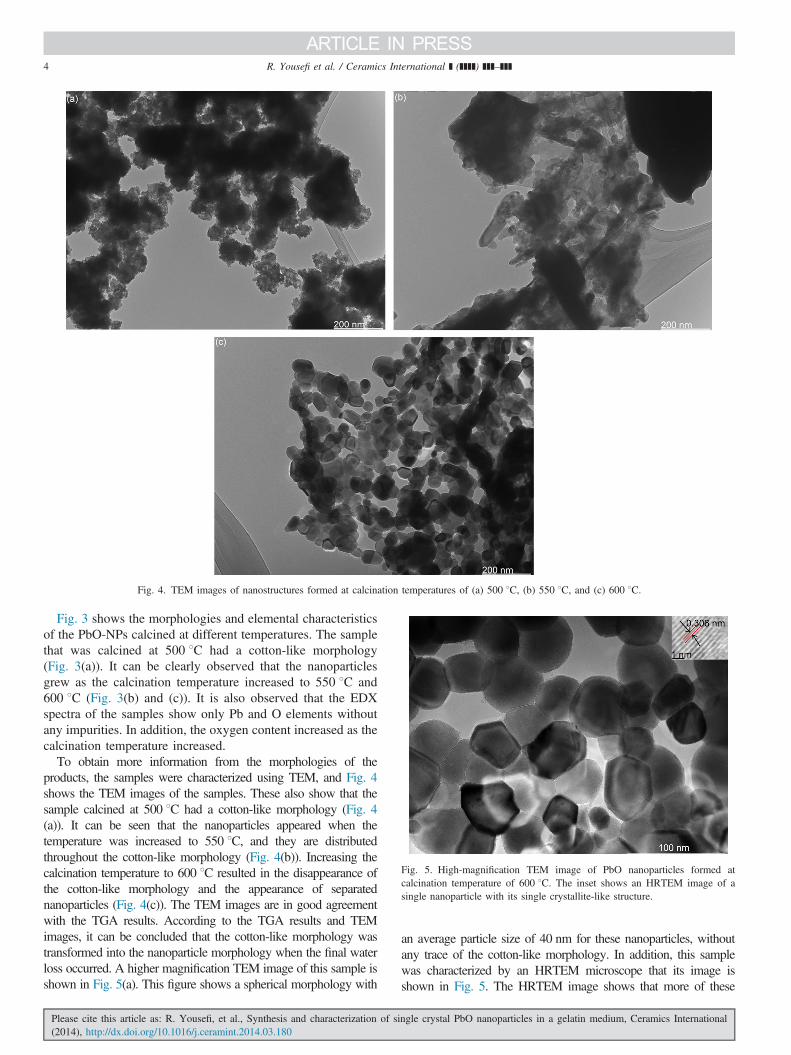
Fig. 4. TEM images of nanostructures formed at calcination temperatures of (a) 500 1C, (b) 550 1C, and (c) 600 1C.
Fig. 5. High-magnification TEM image of PbO nanoparticles formed atcalcination temperature of 600 1C. The inset shows an HRTEM image of asingle nanoparticle with its single crystallite-like structure.
R. Yousefi et al. / Ceramics International ] (]]]]) ]]]–]]]4
Fig. 3 shows the morphologies and elemental characteristicsof the PbO-NPs calcined at different temperatures. The samplethat was calcined at 500 1C had a cotton-like morphology(Fig. 3(a)). It can be clearly observed that the nanoparticlesgrew as the calcination temperature increased to 550 1C and600 1C (Fig. 3(b) and (c)). It is also observed that the EDXspectra of the samples show only Pb and O elements withoutany impurities. In addition, the oxygen content increased as thecalcination temperature increased.
To obtain more information from the morphologies of theproducts, the samples were characterized using TEM, and Fig. 4shows the TEM images of the samples. These also show that thesample calcined at 500 1C had a cotton-like morphology (Fig. 4(a)). It can be seen that the nanoparticles appeared when thetemperature was increased to 550 1C, and they are distributedthroughout the cotton-like morphology (Fig. 4(b)). Increasing thecalcination temperature to 600 1C resulted in the disappearance ofthe cotton-like morphology and the appearance of separatednanoparticles (Fig. 4(c)). The TEM images are in good agreementwith the TGA results. According to the TGA results and TEMimages, it can be concluded that the cotton-like morphology wastransformed into the nanoparticle morphology when the final waterloss occurred. A higher magnification TEM image of this sample isshown in Fig. 5(a). This figure shows a spherical morphology with
Please cite this article as: R. Yousefi, et al., Synthesis and characterization of si(2014), http://dx.doi.org/10.1016/j.ceramint.2014.03.180
an average particle size of 40 nm for these nanoparticles, withoutany trace of the cotton-like morphology. In addition, this samplewas characterized by an HRTEM microscope that its image isshown in Fig. 5. The HRTEM image shows that more of these
ngle crystal PbO nanoparticles in a gelatin medium, Ceramics International

Fig. 6. (a) UV–vis absorbance spectra of nanoparticles synthesized at 550 1C and 600 1C; (b) band gaps of nanoparticles synthesized at 550 1C and 600 1C usingthe Kubelka–Munk method.
R. Yousefi et al. / Ceramics International ] (]]]]) ]]]–]]] 5
nanoparticles are orthorhombic phase PbO, because the HRTEMimage shows that the length of the lattice space is 0.306 nm, whichis close to the corresponding value of 0.306 nm of the (100) plane,which indicates that the crystals were stacked along the (100) planeof the orthorhombic phase PbO. In addition, no defects areobserved between the lattice spaces. Therefore, the NPs that wereobtained were single crystals with a high crystalline quality.
Fig. 6(a) shows the UV–vis absorption spectra of the PbONPs prepared at calcination temperatures of 550 1C and600 1C. The spectra show a characteristic absorption peak ofPbO at a wavelength of approximately 420 nm for bothsamples. Moreover, the indirect band gap energies of PbOcan be estimated from a plot of (αhυ)1/2 versus the photoenergy(hυ) according to the Kubelka–Munk model, where α is theabsorption coefficient [12]. Fig. 6(b) shows this result. It canbe seen that the band gap of the nanoparticles was approxi-mately 2.75 eV, which is in close agreement with the valuesreported by another researcher for orthorhombic phase PbO[13].
4. Conclusions
Single crystal PbO-NPs were synthesized using a simple sol–gelmethod at different calcination temperatures. The XRD patternsindicated a phase transformation from tetragonal to orthorhombicwhen the calcination temperature was increased from 500 1C to600 1C. A microscopy study of the products showed that the NPsstarted to form at temperatures above 550 1C, and their formationwas complete at 600 1C. In addition, the TEM images showed anarrow distribution of the NPs. The HRTEM results showed thatthe NPs were single crystals and had a high crystalline quality. TheUV–vis results showed that the band gap of the NPs was in thevisible region. This simple and effective method can be used togrow PbO NPs at larger scales, making them available for use inphotoconductive applications.
Acknowledgements
R. Yousefi and F. Jamali-Sheini gratefully acknowledge theIslamic Azad University (I.A.U), Masjed-Soleiman and Ahwaz
Please cite this article as: R. Yousefi, et al., Synthesis and characterization of si(2014), http://dx.doi.org/10.1016/j.ceramint.2014.03.180
Branches, respectively, for their support of this research work.In addition, N.M. Huang acknowledges a High ImpactResearch Grant from the Ministry of Higher Education ofMalaysia (UM.C/P/HIR/MOHE/SC/21).
References
[1] K.C. Chen, C.W. Wang, Y.I. Lee, H.G. Liu, Nanoflakes and nanostars ofβ-PbO formed at the air/water interface, Colloids Surf. A 373 (2010)124–129.
[2] S. Ghasemi, M.F. Mousavi, M. Shamsipur, H. Karami, Sonochemical-assisted synthesis of nano-structured lead dioxide, Ultrason. Sonochem.15 (2008) 448–455.
[3] M.M. Kashani-Motlagh, M. Karami Mahmoudabad, Synthesis andcharacterization of lead oxide nano-powders by sol–gel method, J. Sol–Gel Sci. Technol. 59 (2011) 106–110.
[4] L. Shi, Y. Xu, Q. Li, Controlled growth of lead oxide nanosheets, scrollednanotubes, and nanorods, Cryst. Growth Des. 8 (2008) 3521–3525.
[5] J.C. Schottmiller, Photoconductivity in tetragonal and orthorhombic leadmonoxide layers, J. Appl. Phys. 37 (1966) 3505.
[6] L. Li, X. Zhu, D. Yang, L. Gao, J. Liu, R. Vasant Kumar, J. Yang,Preparation and characterization of nano-structured lead oxide from spentlead acid battery paste, J. Hazard. Mater. 203–204 (2011) 274–282.
[7] M.J. Soltanian Fard, F. Rastaghi, N. Ghanbari, Sonochemical synthesis ofnew nano-two-dimensional lead (II) coordination polymer: as precursorfor preparation of PbO nano-structure, J. Mol. Struct. 1032 (2013)133–137.
[8] M.K. Mahmoudabad, M.M. Kashani-Motlagh, Synthesis and character-ization of PbO nanostructure and NiO doped with PbO throughcombustion of citrate/nitrate gel, Inorg. Chem. Commun. 24 (2012)32–39.
[9] D.P. Singh, O.N. Srivastava, Synthesis of micron-sized hexagonal andflowerlike nanostructures of lead oxide (PbO2) by anodic oxidation oflead, Nano–Micro Lett. 3 (2011) 255–259.
[10] B. Jia, L. Gao, Synthesis and characterization of single crystalline PbOnanorods via a facile hydrothermal method, Mater. Chem. Phys. 100(2006) 351–354.
[11] F. Behnoudnia, H. Dehghani, Synthesis and characterization of novelthree-dimensional-cauliflower-like nanostructure of lead (II) oxalate andits thermal decomposition for preparation of PbO, Inorg. Chem. Com-mun. 24 (2012) 32–39.
[12] A. Khorsand Zak, R. Yousefi, W.H. Abd Majid, M.R. Muhamad, Facilesynthesis and X-ray peak broadening studies of Zn1�xMgxO, Ceram. Int.38 (2012) 2059–2064.
[13] C.A. Cattley, A. Stavrinadis, R. Beal, J. Moghal, A.G. Cook, P.S. Grant,J.M. Smith, H. Assender, A.A.R. Watt, Colloidal synthesis of lead oxidenanocrystals for photovoltaics, Chem. Commun. 46 (2010) 2802–2804.
ngle crystal PbO nanoparticles in a gelatin medium, Ceramics International