synthesis and bioevaluation of iodine-131 directly labeled...
TRANSCRIPT
Research ArticleSynthesis and Bioevaluation of Iodine-131Directly Labeled Cyclic RGD-PEGylated Gold Nanorods forTumor-Targeted Imaging
Yingying Zhang12 Yongxue Zhang12 Lianglan Yin12 Xiaotian Xia12 Fan Hu12
QingYao Liu12 Chunxia Qin12 and Xiaoli Lan12
1Department of Nuclear Medicine Union Hospital Tongji Medical CollegeHuazhong University of Science and Technology Wuhan 430022 China2Hubei Key Laboratory of Molecular Imaging Union Hospital Tongji Medical CollegeHuazhong University of Science and Technology Wuhan 430022 China
Correspondence should be addressed to Chunxia Qin qin chunxiahusteducn and Xiaoli Lan lxl730724hotmailcom
Received 6 September 2017 Accepted 4 December 2017 Published 24 December 2017
Academic Editor Yuebing Wang
Copyright copy 2017 Yingying Zhang et alThis is an open access article distributed under the Creative CommonsAttribution Licensewhich permits unrestricted use distribution and reproduction in any medium provided the original work is properly cited
IntroductionRadiolabeled gold nanoparticles play an important role in biomedical applicationThe aim of this study was to prepareiodine-131 (131I)-labeled gold nanorods (GNRs) conjugated with cyclic RGD and evaluate its biological characteristics for targetedimaging of integrin 120572v1205733-expressing tumors Methods HS-PEG(5000)-COOH molecules were applied to replace CTAB coveringthe surface of bare GNRs for better biocompatibility and c(RGDfK) peptides were conjugated onto the carboxyl terminal of GNR-PEG-COOH via EDCNHS coupling reactionsThe nanoconjugate was characterized and 131I was directly tagged on the surface ofGNRs via AuI bonds for SPECTCT imagingWe preliminarily studied the characteristics of the probe and its feasibility for tumor-targeting SPECTCT imaging ResultsThe [131I]GNR-PEG-cRGD probe was prepared in a simple and rapidmanner and was stablein both PBS and fetal bovine serum It targeted selectively and could be taken up by tumor cells mainly via integrin 120572v1205733-receptor-mediated endocytosis In vivo imaging biodistribution and autoradiography results showed evident tumor uptake in integrin 120572v1205733-expressing tumors ConclusionsThese promising results showed that this smart nanoprobe can be used for angiogenesis-targetedSPECTCT imaging Furthermore the nanoprobe possesses a remarkable capacity for highly efficient photothermal conversion inthe near-infrared region suggesting its potential as a multifunctional theranostic agent
1 Introduction
Ideal physicochemical properties a high binding affinity forselected molecules with thiol terminal groups and remark-able photoacoustic features provide gold nanoparticles(GNPs) with significant capabilities for biomedical applica-tions [1 2] Different forms of gold nanostructures such asGNPs [3] gold nanorods (GNRs) [4] gold nanocages [5]gold nanospheres [6] and gold nanoshells [7] have beeninvestigated for molecular imaging and therapy As a repre-sentative GNP GNRs have attracted considerable attention inrecent years because of their small size ease of preparationand bioconjugation strong absorption and scattering prop-erties and well-characterized biocompatibility [8] The longsurface plasmon resonance (LSPR) of GNRs can be finely
tuned by the aspect ratio [9] which gives rise tomany excitingpossibilities for biosensing optical imaging photothermaltherapy and drug delivery With a proper aspect ratio theLSPR of GNRs can be located in the near-infrared region(NIR 650ndash900 nm) that is particularly suitable for in vivoimaging and photothermal therapy [9ndash11]
The high reaction activity of the surface in the crystalstructure of GNRs allows multiple functionalizations includ-ing target ligands (eg peptides [12] folic acid [13] and anti-bodies [14]) and imaging agents (eg fluorescent radionu-clide and contrast reagents) [15 16] Cetyltrimethylammoni-um bromide (CTAB) a kind of cationic surfactant stabilizeris essential for the synthesis of GNRs However CTAB mole-cules exhibit strong cytotoxicity that can induce cell apoptosisand autophagy by damaging mitochondria and generating
HindawiContrast Media amp Molecular ImagingVolume 2017 Article ID 6081724 10 pageshttpsdoiorg10115520176081724
2 Contrast Media amp Molecular Imaging
intracellular reactive oxygen species [17] Fortunately CTABcan be replaced or conjugated with many functional groups[2] Introducing polyethylene glycol (PEG) to the surfaceof nanoparticles achieves better biocompatibility and lowercytotoxicity by decreasing the opsonization effect and mini-mizing nonspecific uptake by the reticuloendothelial systemin vivo for a longer blood circulation time [8] In additionit has been shown that halide ions chemisorb onto the goldsurface with Au-X and its binding strength varies as I gt Br gtCl [18] Iodine-131 (131I 11990512 = 801 days) a radionuclide withgamma emission of 364 keV and beta emission of 0608 keVprovides imaging feasibility and a beta-emitting therapeuticeffect which makes it the optimal choice for application as atheranostic agent [19]
Extensive angiogenesis exists in solid tumors which canbe used as a diagnostic and therapeutic target Integrin 120572v1205733is a cell adhesion molecule overexpressed on most tumorcells for regulation of angiogenesis and plays important rolesin various stages such as malignant transformation tumorgrowth progression invasion and metastasis [20] An Arg-Gly-Asp- (RGD-) based strategy to target integrin 120572v1205733 isone of the most promising and best studied in oncologicalresearch [20] especially cyclic RGD (cRGD) peptides whichhave higher affinity selectivity and stability than linear pep-tides [16] Furthermore hypervasculature a defective vascu-lar architecture poor lymphatic drainage or recovery systemand greatly increased production of a number of permeabilitymediators facilitate nanosized particle extravasation from theblood pool which can be retained in solid tumors known asthe enhanced permeability and retention (EPR) effect [21]Therefore cRGD-conjugated nanodrugs can accumulate intumor tissues actively through target molecules and passivelybecause of the EPR effect resulting in increased curativeefficacy and reduced side effects [22 23]
Based on the above theoretical fundamentals we syn-thesized a smart multifunctional nanoprobe 131I-labeledcRGD-conjugated PEG-modified GNRs and evaluated thefeasibility of the nanoprobe for tumor-targeted imaging byin vitro cell experiments and in vivo tumor-bearing mouseimaging
2 Materials and Methods
21 Chemicals and Materials The chemicals and materi-als included a gold colloid solution (GNR-PEG) (XirsquoanRuixi Biological Tech Co Ltd Xirsquoan China) N-(3-dimethylaminopropyl)-N1015840-ethylcarbodiimide hydrochloride(EDCsdotHCl) (Aladdin Bio-Chem Tech Co Ltd ShanghaiChina) N-hydroxysuccinimide (NHS) (Sinopharm Chem-ical Reagent Co Ltd Shanghai China) cyclo (Arg-Gly-Asp-d-Phe-Lys) [c(RGDfK)] (GL Biochem Ltd ShanghaiChina) sodium iodide-131 ([131I]NaI) (Atom High TechBeijing China) and instant thin-layer chromatography-silicagel (iTLC-SG) (Agilent Tech Santa Clara CA USA)
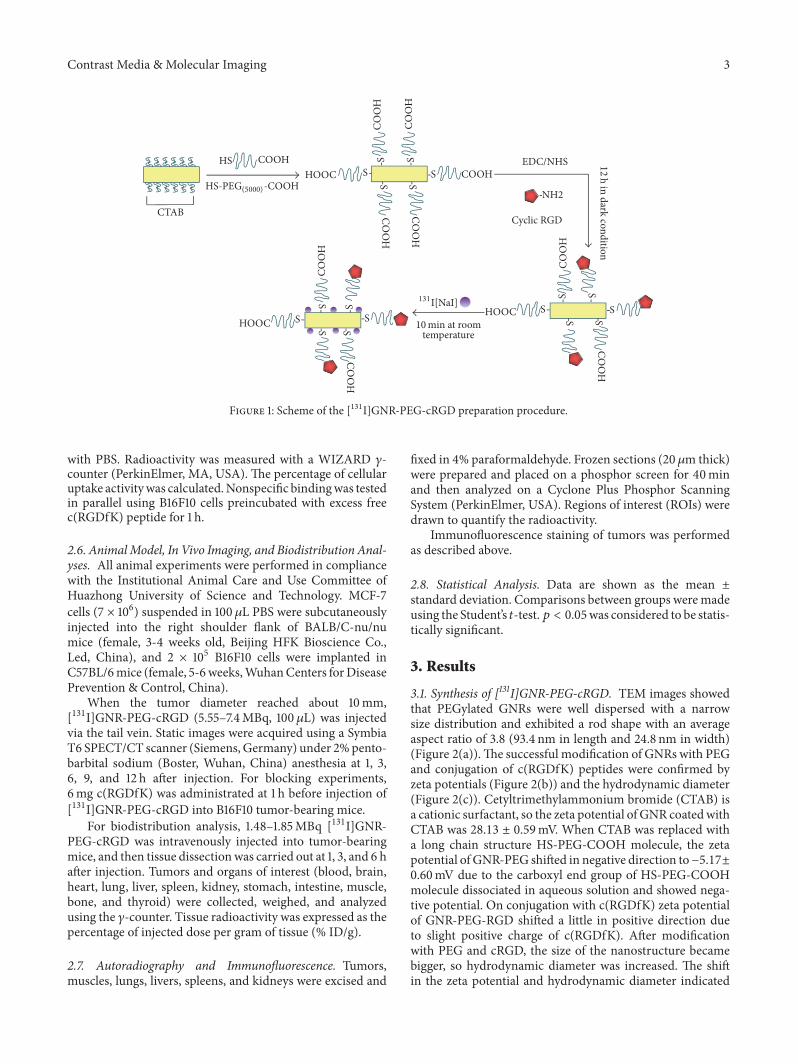
22 Synthesis of [131I]GNR-PEG-cRGD A scheme of the[131I]GNR-PEG-cRGD preparation procedure is shown inFigure 1
First EDCsdotHCl (588 times 10minus2mmol) and NHS (588 times10minus2mmol) were individually dissolved in 500 120583L ultrapuredeionized (DI) water added to 1mL of a GNR-PEG solution(059mg AumL) and allowed to react overnight in the darkat room temperature Subsequently 500120583L c(RGDfK) pep-tide (589 times 10minus3mmol) was added to the reaction mixturefollowed by stirring for 12 h in the dark at room temperatureSonication was conducted discontinuously during the reac-tion process to avoid formation of precipitates or aggregatesThe final product was purified by centrifugation (8000 rpmfor 15min at 4∘C) redispersed in 1mL DI water and stored at4∘C in the dark
Radiolabeling was performed before use Briefly[131I]NaI (1111MBqmL) was added to the GNR-PEG-cRGDsolution (118 120583gmL) and allowed to react for 15min at roomtemperature followed by centrifugation (8000 rpm for 15minat 4∘C) and then redispersed in 1mL phosphate-bufferedsaline (PBS)
23 Characterization of GNRs The morphology and sizeof GNRs were characterized by transmission electronmicroscopy (TEM) Optical absorption spectra were mea-sured on a UV-Vis-NIR spectrophotometer (722S JinghuaInstrument Shanghai China) The hydrodynamic diameterand zeta potential were measured by ZetaPALS zeta poten-tial analyzer (Brookhaven Instrument Corp Holtsville NYUSA) The in vitro stability of [131I]GNR-PEG-cRGD in PBSand fetal bovine serum (FBS) was determined by mixing01mL [131I]GNR-PEG-cRGD with an equal volume of PBSFBS and incubating at 37∘C for 48 h Radiochemical stabilitywas monitored by iTLC-SG with a 09 sodium chloridesolution as the solvent on a radioactive chromatographyscanner (Zhongcheng Hefei China) at 6 12 24 and 48 h
24 Cell Culture and Analysis of Integrin 120572v1205733 ExpressionIntegrin 120572v1205733-positive B16F10 mouse malignant melanomacells and integrin 120572v1205733-negative MCF-7 human breast can-cer cells (American Type Culture Collection ManassasVA USA) were cultured in Dulbeccorsquos modified Eaglersquosmedium (Gibco Carlsbad CA USA) containing 10 (vv)FBS (Gibco) and 1 antibiotics (100Uml penicillin and100Uml streptomycin Beyotime Shanghai China) at 37∘Cwith 5 CO2 The expression of integrin 120572v1205733 was confirmedby immunofluorescence with a primary anti-integrin 120572v1205733antibody (1 100 Bioss Beijing China) and Cy3-conjugatedgoat anti-rabbit secondary antibody (1 50 Aspen WuhanChina) as described previously [24] Anti-rabbit IgG (CellSignaling Technology Inc USA) instead of the primaryantibody was used as the control
25 In Vitro Cell Binding Assay B16F10 and MCF-7 cellswere seeded in 24-well plates at a density of 2 times 105 cellsper well incubated at 37∘C overnight and then treated with08mL [131I]GNR-PEG-cRGD (0074MBqwell) at 37∘C for30 60 120 and 240min The medium was then removedand the cells were collected and washed twice with PBS Thecell pellet was lysed with 1N NaOH and then washed twice
Contrast Media amp Molecular Imaging 3
S COOHSCO
OH
SCO
OH
S
S
S
COO
H
COO
H
COO
H
S
COO
H
HOOCHS COOH EDCNHS
NH2
Cyclic RGDS
COO
H
S
S
CTAB
SSS
SCO
OH
temperature
S
SHOOCSSHOOC
HS-PEG(5000)-COOH
10min at room
12h in dark condition
131I[NaI]
Figure 1 Scheme of the [131I]GNR-PEG-cRGD preparation procedure
with PBS Radioactivity was measured with a WIZARD 120574-counter (PerkinElmer MA USA) The percentage of cellularuptake activitywas calculatedNonspecific bindingwas testedin parallel using B16F10 cells preincubated with excess freec(RGDfK) peptide for 1 h
26 AnimalModel In Vivo Imaging and Biodistribution Anal-yses All animal experiments were performed in compliancewith the Institutional Animal Care and Use Committee ofHuazhong University of Science and Technology MCF-7cells (7 times 106) suspended in 100120583L PBS were subcutaneouslyinjected into the right shoulder flank of BALBC-nunumice (female 3-4 weeks old Beijing HFK Bioscience CoLed China) and 2 times 105 B16F10 cells were implanted inC57BL6mice (female 5-6weeksWuhanCenters forDiseasePrevention amp Control China)
When the tumor diameter reached about 10mm[131I]GNR-PEG-cRGD (555ndash74MBq 100 120583L) was injectedvia the tail vein Static images were acquired using a SymbiaT6 SPECTCT scanner (Siemens Germany) under 2pento-barbital sodium (Boster Wuhan China) anesthesia at 1 36 9 and 12 h after injection For blocking experiments6mg c(RGDfK) was administrated at 1 h before injection of[131I]GNR-PEG-cRGD into B16F10 tumor-bearing mice
For biodistribution analysis 148ndash185MBq [131I]GNR-PEG-cRGD was intravenously injected into tumor-bearingmice and then tissue dissectionwas carried out at 1 3 and 6 hafter injection Tumors and organs of interest (blood brainheart lung liver spleen kidney stomach intestine musclebone and thyroid) were collected weighed and analyzedusing the 120574-counter Tissue radioactivity was expressed as thepercentage of injected dose per gram of tissue ( IDg)
27 Autoradiography and Immunofluorescence Tumorsmuscles lungs livers spleens and kidneys were excised and
fixed in 4 paraformaldehyde Frozen sections (20120583m thick)were prepared and placed on a phosphor screen for 40minand then analyzed on a Cyclone Plus Phosphor ScanningSystem (PerkinElmer USA) Regions of interest (ROIs) weredrawn to quantify the radioactivity
Immunofluorescence staining of tumors was performedas described above
28 Statistical Analysis Data are shown as the mean plusmnstandard deviation Comparisons between groups weremadeusing the Studentrsquos 119905-test119901 lt 005was considered to be statis-tically significant
3 Results
31 Synthesis of [131I]GNR-PEG-cRGD TEM images showedthat PEGylated GNRs were well dispersed with a narrowsize distribution and exhibited a rod shape with an averageaspect ratio of 38 (934 nm in length and 248 nm in width)(Figure 2(a))The successful modification of GNRs with PEGand conjugation of c(RGDfK) peptides were confirmed byzeta potentials (Figure 2(b)) and the hydrodynamic diameter(Figure 2(c)) Cetyltrimethylammonium bromide (CTAB) isa cationic surfactant so the zeta potential of GNR coatedwithCTAB was 2813 plusmn 059mV When CTAB was replaced witha long chain structure HS-PEG-COOH molecule the zetapotential of GNR-PEG shifted in negative direction tominus517plusmn060mV due to the carboxyl end group of HS-PEG-COOHmolecule dissociated in aqueous solution and showed nega-tive potential On conjugation with c(RGDfK) zeta potentialof GNR-PEG-RGD shifted a little in positive direction dueto slight positive charge of c(RGDfK) After modificationwith PEG and cRGD the size of the nanostructure becamebigger so hydrodynamic diameter was increased The shiftin the zeta potential and hydrodynamic diameter indicated
4 Contrast Media amp Molecular Imaging
50nm
(a)
GNR-CTABGNR-PEGGNR-PEG-cRGD
minus10
0
10
20
30
40
Zeta
pot
entia
l (m
V)
(b)
GNR-CTABGNR-PEGGNR-PEG-cRGD
0
50
100
150
200
250
Hyd
rody
nam
ic d
iam
eter
(nm
)
(c)
GNR-CTABGNR-PEGGNR-PEG-cRGD
00
02
04
06
08
10
Abso
rban
ce in
tens
ity
600 800 1000 1200400Wavelength (nm)
(d)
PBSSerum
80
85
90
95
100
Radi
oche
mic
al p
urity
()
6 12 24 480Time (h)
(e)
Figure 2 Characterization of GNR-PEG-cRGD (a) TEM of GNR-PEG Zeta potentials (b) hydrodynamic diameter (c) and UV-vis spectra(d) of GNR-CTAB GNR-PEG and GNR-PEG-cRGD (e) In vitro stability of [131I]GNR-PEG-cRGD in PBS and FBS at 6 12 24 and 48 hafter labeling
Contrast Media amp Molecular Imaging 5
Cy3 DAPI Merge
(a)
B16F10MCF-7
0
10
20
30
40
50
Cell
upt
ake r
atio
()
200 3000 100Time (min)
(b)
Figure 3 (a) Immunofluorescence staining of integrin 120572v1205733 in B16F10 (upper row) and MCF-7 (lower row) cells The nucleus werecounterstained with DAPI The red fluorescence intensity is proportional to the expression level of integrin 120572v1205733 (times200) (b) Results of cellbinding assays at various time points
successful conjugation of PEG and c(RGDfK) peptides UV-vis absorbance spectra showed no obvious change after PEGmodification and cRGD conjugation with a maximum UV-vis absorption peak at around 780 nm (Figure 2(d))
For radiolabeling of GNRs the radiochemical yield of[131I]GNR-PEG-cRGD was 6454 plusmn 381 (119899 = 4) and theradiochemical purity was 9817 plusmn 086 (119899 = 4) after cen-trifugation [131I]GNR-PEG-cRGD had favorable stability invitro (Figure 2(e)) with radiochemical purities of 9779 plusmn050 in PBS and 9559 plusmn 073 in FBS at 48 h after labeling
32 Integrin 120572v1205733 Expression and Cell Binding Immunofluo-rescence demonstrated that the integrin 120572v1205733 expression level
in B16F10 cells was significantly higher than that in MCF-7 cells (Figure 3(a)) Therefore B16F10 cells were used asthe experimental group while MCF-7 cells were used as thenegative control
As shown in Figure 3(b) [131I]GNR-PEG-cRGD exhib-ited specific binding because the cell binging ratio of[131I]GNR-PEG-cRGD in B16F10 cells increased as timeelapsed and reached a peak (3820 plusmn 148) at 120min whilethe accumulation of [131I]GNR-PEG-cRGD in MCF-7 cellswas much lower than that in B16F10 cells (119901 lt 005) with noobvious change over timeThe specificity was also confirmedby receptor blocking experiments with an uptake ratio of2061 plusmn 115 at 120min
6 Contrast Media amp Molecular Imaging
41
312 h9 h6 h3 h1 h(a)
41
312 h9 h6 h3 h1 h(b)
41
312 h9 h6 h3 h1 h(c)
Figure 4 Representative whole body SPECTCT images of B16F10 (a) blocked B16F10 (b) and MCF-7 (c) tumor-bearing mice at 1 3 6 9and 12 h after intravenous injection of [131I]GNR-PEG-cRGD Arrows indicate tumor sites
33 In Vivo Analyses SPECTCT images (Figure 4) and bio-distribution analyses (Table 1) showed evident specific tumoruptake [131I]GNR-PEG-cRGD had accumulated in B16F10tumors quickly and effectively at 1 h after injection Remark-ably the tumor uptake increased gradually over time andreached the peak value at about 6 h and tumors were clearlyvisualized at 12 h after injection However nanoprobes inMCF-7 tumors were almost undetectable at all time pointsIn blocking experiments B16F10 tumor uptake was clearlyreduced Biodistribution results revealed that B16F10 tumoruptake of [131I]GNR-PEG-cRGD was gradually increased to509 plusmn 068 IDg (119899 = 4) at 6 h after injection which wassignificantly higher compared with MCF-7 tumors (159 plusmn039 IDg 119899 = 4 119901 lt 005) and the blocked group (221 plusmn052 IDg 119899 = 4119901 lt 005) Tumormuscle ratioswere 999plusmn298 (B16F10) 367plusmn092 (MCF-7) and 387plusmn093 (blocked)
at 6 h Liver spleen and lungs had remarkable radioactivityuptake The kidneys showed low uptake of about 2 IDg
34 Autoradiography and Immunofluorescence Autoradiog-raphy also revealed abundant radioactivity accumulation inB16F10 tumors and little radioactivity accumulation in MCF-7 tumors (ROIs 373plusmn075 versus 127plusmn047 119899 = 4119901 lt 005)which demonstrated specific targeting of the nanoprobeHigh radioactivity had accumulated in samples of the lungsliver and spleen while radioactivity distribution in muscleswas sparse (Figure 5(a))These ex vivo results were consistentwith in vivo analyses
Immunofluorescence staining of integrin 120572v1205733 in B16F10sections revealed intense fluorescence but little fluorescencein MCF-7 sections (Figure 5(b)) which validated abundant
Contrast Media amp Molecular Imaging 7
Table 1 Biodistribution of [131I]GNR-PEG-cRGD in C57BL6 mice with B16F10 tumors and athymic mice with MCF-7 tumors at 1 3 and6 h after intravenous administration (119899 = 4)
Tissue B16F10 B16F10 blocking MCF-71 h 3 h 6 h 6 h 1 h 3 h 6 h
Blood 353 plusmn 131 453 plusmn 158 319 plusmn 035 397 plusmn 125 314 plusmn 210 31 plusmn 031 244 plusmn 028Brain 020 plusmn 009 018 plusmn 003 017 plusmn 002 015 plusmn 009 018 plusmn 009 015 plusmn 003 010 plusmn 002Heart 114 plusmn 044 145 plusmn 027 085 plusmn 019 078 plusmn 017 045 plusmn 016 093 plusmn 017 047 plusmn 017Lung 1925 plusmn 459 1174 plusmn 197 707 plusmn 025 693 plusmn 024 2056 plusmn 924 1741 plusmn 104 331 plusmn 187Liver 3205 plusmn 537 2883 plusmn 717 2651 plusmn 493 2759 plusmn 501 3018 plusmn 487 3151 plusmn 571 2760 plusmn 334Spleen 1128 plusmn 063 986 plusmn 105 555 plusmn 069 632 plusmn 101 1599 plusmn 174 857 plusmn 070 620 plusmn 118Kidney 234 plusmn 075 260 plusmn 104 229 plusmn 021 301 plusmn 091 229 plusmn 151 200 plusmn 036 150 plusmn 037Stomach 359 plusmn 181 612 plusmn 228 343 plusmn 044 402 plusmn 059 349 plusmn 269 544 plusmn 275 470 plusmn 137Intestine 152 plusmn 055 160 plusmn 057 123 plusmn 048 104 plusmn 029 115 plusmn 077 135 plusmn 025 089 plusmn 047Muscle 047 plusmn 019 059 plusmn 008 051 plusmn 008 057 plusmn 012 054 plusmn 022 058 plusmn 023 042 plusmn 007Bone 259 plusmn 078 329 plusmn 147 169 plusmn 013 170 plusmn 021 202 plusmn 085 272 plusmn 068 126 plusmn 071Thyroid 288 plusmn 037 225 plusmn 109 293 plusmn 075 301 plusmn 092 326 plusmn 215 293 plusmn 026 359 plusmn 268Tumor 357 plusmn 125 402 plusmn 145 509 plusmn 068 221 plusmn 052 131 plusmn 088 175 plusmn 026 159 plusmn 039Uptake ratio
Tumorblood 101 plusmn 011 080 plusmn 029 158 plusmn 013 056 plusmn 022 052 plusmn 007 057 plusmn 004 063 plusmn 019Tumormuscle 706 plusmn 253 689 plusmn 237 999 plusmn 298 387 plusmn 093 217 plusmn 106 326 plusmn 098 367 plusmn 092
integrin 120572v1205733 expression in B16F10 tumors but low expres-sion in MCF-7 tumors
4 Discussion
An 131I-labeled cRGD-conjugated PEG-modifiedGNRprobewas synthesized successfully Compared with a reportedspherical gold nanoprobe with single surface plasma reso-nance in visible light [12] which limits certain applicationsin the medical field the probe we designed and synthesizedpossesses distinct advantages in the field of optical biologicalapplications Excellent surface plasma resonances in theNIR region confer GNRs with minimal light absorption byhemoglobin and water maximal penetration [25] and highlyefficient photothermal conversion [9] making them particu-larly attractive for optoacoustic imaging [13] and photother-mal therapy [26]
There has been increasing use of radioisotopes to labelnanoparticles such as positron emitters copper-64 [26] gal-lium-67 [27] and zirconium-89 [28] and single photonemitters Technetiom-99m [3] and radioiodine (eg iodine-125 [29] iodine-123 and iodine-131 [30]) Inmost conditionsa bifunctional chelator such as DOTA or HYNIC must beconjugated to the nanoparticle for radiolabeling Thereforemodification of nanoparticles is required before labelingIt has been shown that iodide ions have high affinity andstrong binding to the surface of GNRs [8 29 31 32] Iodideions absorb preferentially onto facets of GNRs and form thestrongest bonds with Au which probably leads to the forma-tion of a surface of AuI [18] Such simple chemistry betweeniodine and GNRs allows straightforward and efficient label-ing of radioiodine to GNRs without iodination reagents oriodine-accepting functional groups such as a phenol residue[29] Iodine-125 has been reported to directly label GNRs in
a simple and rapid manner [8 29] Here we first report thein vitro and in vivo behaviors of directly 131I-labeledGNRs bysimply mixing a GNR colloid with [131I]NaI at room temper-ature for a short time This reaction takes place fairly rapidlyand completely which agreeswell with the previous reports ofhigh affinity and strong binding of iodide ions to the surfaceof GNRs [18] The labeling method is characterized by sim-plicity a short reaction time mild reaction conditions andhigh yield Another advantage is that purification is simpleby centrifugation to remove free [131I]Iodide ions Further-more this nanoprobe has excellent stability with radiochem-istry purity greater than 95 after incubation in PBS or FBSfor 48 h 131I is widely used in clinics and easy to obtain Theproperties of 120573-emitters are used for radiotherapy Passingthrough tissue the ejected 120573-particles (ie electrons) interactwith atoms mainly in water molecules and lose their energyleading to the generation of excited and ionized atoms andfree radicals that are responsible for DNA damage in cellsby inducing single-strand breaks in DNA [33] making 131Ithe optimal choice for application as a theranostic agent [19]Our ultimate goal is to use this probe for both imaging andtherapy so 131I was chosen
The significant difference in cellular uptake of [131I]GNR-PEG-cRGD by B16F10 and MCF-7 cells confirmed receptor-specific internalization of the probes suggesting that theprobes were taken up by tumor cells via receptor-mediatedendocytosis However [131I]GNR-PEG-cRGD accumulationin MCF-7 cells and blocked B16F10 cells was up to about20 because of passive uptake which may be explained bythe following three reasons First the nanometric size ofthe probe is known to be taken up by all mammalian celltypes [34] Second serum proteins in the cell culture maybe absorbed onto the surface of nanoparticles or targetedc(RGDfK) peptides which can be taken up by cells and can
8 Contrast Media amp Molecular Imaging
Kidn
ey
Sple
en
Live
r
Lung
Mus
cle
Tum
or(a)
Cy3 DAPI Merge
(b)
Figure 5 (a) Autoradiography of tumor tissue and organs B16F10 tumor-bearing mice (upper row) and MCF-7 tumor-bearing mice (lowerrow) (b) Immunofluorescence staining of integrin 120572v1205733 in B16F10 (upper row) and MCF-7 (lower row) tumors (times200)
mediate uptake of nanoparticles into cells nonspecifically viathemechanismof receptor-mediated endocytosis [34]Thirdtumor cell membranes have a predominant negative chargewhereas the zeta potential of [131I]GNR-PEG-cRGD waspositive which can bind efficiently to the surface of tumorcells by electrostatic attraction and then promote nonspecificuptake via clathrin-mediated endocytosis [35]
SPECTCT imaging results revealed high [131I]GNR-PEG-cRGD accumulation in B16F10 tumors the tumorsof blocked B16F10 mice and MCF-7 mice are also visiblealthough they are not very clear In biodistribution studytumor uptake values were 509 plusmn 068 221 plusmn 052 and 159 plusmn039 IDg at 6 h pi in B16F10 blocked B16F10 and MCF-7mice with tumormuscle ratio of 999plusmn 298 387plusmn 093 and367 plusmn 092 respectively These results indicate that low EPReffects existed in B16F10MCF-7 bearing mouse due to thecharacteristics of nanoparticles and solid tumor and traceraccumulation in B16F10 can be explained by both integrin120572v1205733-specific endocytosis and EPR effect in which activetargeting plays a vital role
Theoretically due to the large size of these nanoparticlesusually optimal uptake would be observed at late time points
Actually our in vitro cell binding results were 2852 plusmn 1003602 plusmn 120 382 plusmn 148 and 3911 plusmn 180 at 30 60 120and 240min and nearly reached a platform at 120min our invivo imaging also revealed highest tumor uptake present at6 h and decreased overtime In addition significant thyroiduptake is also observed which increased overtime in ourstudies thyroid uptake was also observed in reported 125I-Labeled Gold Nanorods papers as expected [8 29] Onepossible explanation of short blood circulation (dropped at6 h pi) and rapid thyroid accumulation (eg 1 h pi) was invivo deiodination from nanoparticles Our in vitro stabilityresultsmay not reflect real condition because iTLCmight notbe able to identify the disassociated[131I] that might also beabsorbed by protein(s) in blood We will use FPLC to furtherassess the serum and in vivo stability of [131I]GNR-PEG-cRGD and late time points will be included in our therapystudy
Compared with other radiotherapeutic systems such asthose based on Lu-177-gold nanoparticles-RGD one studycompared 177Lu-labeled monomeric dimeric and multi-meric RGD peptides for the therapy of tumors expressinga(n)b(3) integrins 177Lu-AuNP-c(RGDfK)Cwas demonstrated
Contrast Media amp Molecular Imaging 9
as the best one for targeted radionuclide therapy of tumorsexpressing a(n)b(3) integrins with highest tumor uptake of642 plusmn 071 IDg at 6 h [36] our [131I]GNR-PEG-cRGDsystem has similar in vivo stability (509 plusmn 068 IDg at 6 h)Another study reports that the mean tumor residence timesof 177Lu-AuNP-RGD were 616 plusmn 58 h [37] And we will getthe data in our therapy study
Although CTAB was replaced with HS-PEG(5000)-COOHfor better stability and biocompatibility significant uptakewas still observed in the liver and spleen because of abundantmacrophages in the reticuloendothelial system of the liverand spleen and the colloidal nature of the probe which havebeen well documented in previous reports of radiolabelednanoparticles [3 4] Efforts have been made to understandand minimize uptake by the liver and spleen as much aspossible One study reported that targeting ligands on the sur-face of nanoparticlesmight even be detrimental because theirexposure can accelerate nanoparticle opsonization and bloodclearance by the immune system resulting in high uptakein the liver and spleen [35] Morales-Avila et al [3] studiedthe biodistribution of GNPs using various administrationmethods Their results showed that intravenous administra-tion resulted in higher liver and spleen accumulation thanintraperitoneal administration because intravenous admin-istration leads to opsonization followed by substantial uptakeby macrophages located in the liver and spleen Our resultsrevealed that [131I]GNR-PEG-cRGD had accumulated in theliver and spleen at an early time point and gradually declinedover time the reasonmay be due to the radiolabeled nanosys-temaccumulation by reticuloendothelial system (RES)meta-bolized by the hepatobiliary system it is also possible that theactivity eliminated by the hepatobiliary system correspondsto the free iodide and not to the radiolabeled nanosystem
However the detailed metabolism mechanism in vivois still not understood The size of a nanoparticle may beanother influencing factor It has been reported that GNPs ofless than 5-6 nm in size can be removed from the body viathe kidney which can minimize nonspecific accumulation byRES [29] In addition the final metabolic pattern of largersized nanoparticles is associated with the shape and surfacechemistry [38] Our results also demonstrated that the invivo environment is far more complex than in vitro modelsystems
In summary a stable and tumor-specific SPECT imagingnanoparticle probe was successfully prepared in this studyThe probe can specifically target integrin 120572v1205733-expressingtumor cells both in vitro and in vivo mainly by receptor-mediated endocytosis Importantly the radiolabelingmethodis simple and fast with a high yield and high stability Thesepromising results demonstrate that our [131I]GNR-PEG-cRGD probe can be used as an angiogenesis-targeted SPECTimaging probe Currently more detailed studies to improvethe in vivo fate of the [131I]GNR-PEG-cRGD probe and theuse of this multifunctional probe as a theranostic agent areongoing
5 Conclusion
In this study a smart nanoprobe [131I]GNR-PEG-cRGD wassuccessfully developed and it showed specific binding ability
with integrin 120572v1205733 indicating its potential as a multifunc-tional theranostic agent for tumors
Conflicts of Interest
The authors declare that there are no conflicts of interestregarding the publication of this paper
Acknowledgments
This study was supported by the National Natural ScienceFoundation of China (no 81401444) and the IndependentInnovationResearch Fund ofHuazhongUniversity of Science(no 2014QN038)
References
[1] E C Dreaden A M Alkilany X Huang C J Murphy andM A El-Sayed ldquoThe golden age gold nanoparticles for bio-medicinerdquo Chemical Society Reviews vol 41 no 7 pp 2740ndash2779 2012
[2] S Same A Aghanejad S A Nakhjavani J Barar and Y OmidildquoRadiolabeled theranostics Magnetic and gold nanoparticlesrdquoBioImpacts vol 6 no 3 pp 169ndash181 2016
[3] E Morales-Avila G Ferro-Flores B E Ocampo-Garcıa et alldquoMultimeric system of 99119898Tc-labeled gold nanoparticles conju-gated to c[RGDfK(C)] formolecular imaging of tumor120572(v)120573(3)expressionrdquo Bioconjugate Chemistry vol 22 no 5 pp 913ndash9222011
[4] Y Xiao H Hong V Z Matson et al ldquoGold nanorods conju-gated with doxorubicin and cRGD for combined anti-cancerdrug delivery and PET imagingrdquoTheranostics vol 2 no 8 pp757ndash768 2012
[5] L Au D Zheng F Zhou Z-Y Li X Li and Y Xia ldquoA quanti-tative study on the photothermal effect of immuno gold nano-cages targeted to breast cancer cellsrdquoACSNano vol 2 no 8 pp1645ndash1652 2008
[6] M Tian W Lu R Zhang et al ldquoTumor uptake of hollow goldnanospheres after intravenous and intra-arterial injectionPETCT study in a rabbit VX2 liver cancer modelrdquo MolecularImaging and Biology vol 15 no 5 pp 614ndash624 2013
[7] H Xie Z J Wang A Bao B Goins andW T Phillips ldquoIn vivoPET imaging and biodistribution of radiolabeled gold nano-shells in rats with tumor xenograftsrdquo International Journal ofPharmaceutics vol 395 no 1-2 pp 324ndash330 2010
[8] X Shao A Agarwal J R Rajian N A Kotov and X WangldquoSynthesis and bioevaluation of 125I-labeled gold nanorodsrdquoNanotechnology vol 22 no 13 Article ID 135102 2011
[9] C-H Chou C-D Chen and C R C Wang ldquoHighly effi-cient wavelength-tunable gold nanoparticle based optother-mal nanoconvertorsrdquo The Journal of Physical Chemistry B vol109 no 22 pp 11135ndash11138 2005
[10] C Wang C Bao S Liang et al ldquoRGD-conjugated silica-coatedgold nanorods on the surface of carbon nanotubes for targetedphotoacoustic imaging of gastric cancerrdquo Nanoscale ResearchLetters vol 9 no 1 article 264 2014
[11] Z Heidari M Salouti and R Sariri ldquoBreast cancer photother-mal therapy based on gold nanorods targeted by covalently-coupled bombesin peptiderdquo Nanotechnology vol 26 no 19Article ID 195101 2015
10 Contrast Media amp Molecular Imaging
[12] N Su YDangG Liang andG Liu ldquoIodine-125-labeled cRGD-gold nanoparticles as tumor-targeted radiosensitizer and imag-ing agentrdquo Nanoscale Research Letters vol 10 article 160 2015
[13] J Zhong L Wen S Yang L Xiang Q Chen and D XingldquoImaging-guided high-efficient photoacoustic tumor therapywith targeting gold nanorodsrdquo Nanomedicine NanotechnologyBiology and Medicine vol 11 no 6 pp 1499ndash1509 2015
[14] P P Joshi S J Yoon W G Hardin S Emelianov and K VSokolov ldquoConjugation of antibodies to gold nanorods throughFc portion synthesis and molecular specific imagingrdquo Biocon-jugate Chemistry vol 24 no 6 pp 878ndash888 2013
[15] L Vigderman B P Khanal and E R Zubarev ldquoFunctional goldnanorods synthesis self-assembly and sensing applicationsrdquoAdvanced Materials vol 24 no 36 pp 4811ndash4841 2012
[16] R Haubner F Bruchertseifer M Bock H Kessler M Schwai-ger and H-J Wester ldquoSynthesis and biological evaluation ofa 99mTc-labelled cyclic RGD peptide for imaging the 120572v1205733expressionrdquo Nuklearmedizin vol 43 no 1 pp 26ndash32 2004
[17] J Wan J-H Wang T Liu Z Xie X-F Yu and W Li ldquoSurfacechemistry but not aspect ratio mediates the biological toxicityof gold nanorods in vitro and in vivordquo Scientific Reports vol 5Article ID 11398 2015
[18] S Singh R Pasricha U M Bhatta P V Satyam M Sastryand B L V Prasad ldquoEffect of halogen addition to monolayerprotected gold nanoparticlesrdquo Journal of Materials Chemistryvol 17 no 16 pp 1614ndash1619 2007
[19] S V Gudkov N Y Shilyagina V A Vodeneev and A VZvyagin ldquoTargeted radionuclide therapy of human tumorsrdquoInternational Journal of Molecular Sciences vol 17 no 1 article33 2015
[20] M A Dechantsreiter E Planker BMatha et al ldquoN-methylatedcyclic RGD peptides as highly active and selective 1205721199071205733 integrinantagonistsrdquo Journal of Medicinal Chemistry vol 42 no 16 pp3033ndash3040 1999
[21] H Maeda J Wu T Sawa Y Matsumura and K Hori ldquoTumorvascular permeability and the EPR effect in macromoleculartherapeutics a reviewrdquo Journal of Controlled Release vol 65 no1-2 pp 271ndash284 2000
[22] V Torchilin ldquoTumor delivery of macromolecular drugs basedon the EPR effectrdquoAdvanced Drug Delivery Reviews vol 63 no3 pp 131ndash135 2011
[23] J Fang H Nakamura and H Maeda ldquoThe EPR effect uniquefeatures of tumor blood vessels for drug delivery factorsinvolved and limitations and augmentation of the effectrdquoAdvancedDrugDelivery Reviews vol 63 no 3 pp 136ndash151 2011
[24] C Qin X Lan J He et al ldquoAn in vitro and in vivo evaluation ofa reporter geneprobe system hERL18F-FESrdquo PLoS ONE vol8 no 4 Article ID e61911 2013
[25] R Weissleder ldquoA clearer vision for in vivo imagingrdquo NatureBiotechnology vol 19 no 4 pp 316-317 2001
[26] X Sun X Huang X Yan et al ldquoChelator-free 64Cu-integratedgold nanomaterials for positron emission tomography imagingguided photothermal cancer therapyrdquo ACS Nano vol 8 no 8pp 8438ndash8446 2014
[27] C Tsoukalas G Laurent G Jimenez Sanchez et al ldquoInitial invitro and in vivo assessment of AuDTDTPA-RGD nanopar-ticles for Gd-MRI and 68Ga-PET dual modality imagingrdquoEJNMMI Physics vol 2 no 1 article A89 2015
[28] L Karmani V Bouchat C Bouzin et al ldquo 89Zr-labeled anti-endoglin antibody-targeted gold nanoparticles for imagingcancer implications for future cancer therapyrdquo Nanomedicinevol 9 no 13 pp 1923ndash1937 2014
[29] Y-H Kim J Jeon S H Hong et al ldquoTumor targeting andimaging using cyclic RGD-PEGylated gold nanoparticle probeswith directly conjugated iodine-125rdquo Small vol 7 no 14 pp2052ndash2060 2011
[30] H-W Kao Y-Y Lin C-C Chen et al ldquoEvaluation of EGFR-targeted radioimmuno-gold-nanoparticles as a theranosticagent in a tumor animal modelrdquo Bioorganic amp MedicinalChemistry Letters vol 23 no 11 pp 3180ndash3185 2013
[31] D K Smith N R Miller and B A Korgel ldquoIodide in CTABprevents gold nanorod formationrdquo Langmuir vol 25 no 16 pp9518ndash9524 2009
[32] W Cheng S Dong and E Wang ldquoIodine-induced gold-nano-particle fusionfragmentationaggregation and iodine-linkednanostructured assemblies on a glass substraterdquo AngewandteChemie International Edition vol 42 no 4 pp 449ndash452 2003
[33] H Hong Y Zhang J Sun andW Cai ldquoMolecular imaging andtherapy of cancer with radiolabeled nanoparticlesrdquoNano Todayvol 4 no 5 pp 399ndash413 2009
[34] B D Chithrani A A Ghazani andW CW Chan ldquoDetermin-ing the size and shape dependence of gold nanoparticle uptakeinto mammalian cellsrdquo Nano Letters vol 6 no 4 pp 662ndash6682006
[35] O Harush-Frenkel E Rozentur S Benita and Y AltschulerldquoSurface charge of nanoparticles determines their endocyticand transcytotic pathway in polarized MDCK cellsrdquo Biomacro-molecules vol 9 no 2 pp 435ndash443 2008
[36] M Luna-Gutierrez G Ferro-Flores B Ocampo-Garcıa et al ldquo177Lu-labeled monomeric dimeric and multimeric RGD pep-tides for the therapy of tumors expressing 120572(])120573(3) integrinsrdquoJournal of Labelled Compounds and Radiopharmaceuticals vol55 no 4 pp 140ndash148 2012
[37] A Vilchis-Juarez G Ferro-Flores C Santos-Cuevas et alldquoMolecular targeting radiotherapy with Cyclo-RGDfK(C) pep-tides conjugated to 177Lu-labeled gold nanoparticles in tumor-bearingmicerdquo Journal of Biomedical Nanotechnology vol 10 no3 pp 393ndash404 2014
[38] X Huang X Peng Y Wang et al ldquoA reexamination of activeand passive tumor targeting by using rod-shaped gold nano-crystals and covalently conjugated peptide ligandsrdquo ACS Nanovol 4 no 10 pp 5887ndash5896 2010
Submit your manuscripts athttpswwwhindawicom
Stem CellsInternational
Hindawi Publishing Corporationhttpwwwhindawicom Volume 2014
Hindawi Publishing Corporationhttpwwwhindawicom Volume 2014
MEDIATORSINFLAMMATION
of
Hindawi Publishing Corporationhttpwwwhindawicom Volume 2014
Behavioural Neurology
EndocrinologyInternational Journal of
Hindawi Publishing Corporationhttpwwwhindawicom Volume 2014
Hindawi Publishing Corporationhttpwwwhindawicom Volume 2014
Disease Markers
Hindawi Publishing Corporationhttpwwwhindawicom Volume 2014
BioMed Research International
OncologyJournal of
Hindawi Publishing Corporationhttpwwwhindawicom Volume 2014
Hindawi Publishing Corporationhttpwwwhindawicom Volume 2014
Oxidative Medicine and Cellular Longevity
Hindawi Publishing Corporationhttpwwwhindawicom Volume 2014
PPAR Research
The Scientific World JournalHindawi Publishing Corporation httpwwwhindawicom Volume 2014
Immunology ResearchHindawi Publishing Corporationhttpwwwhindawicom Volume 2014
Journal of
ObesityJournal of
Hindawi Publishing Corporationhttpwwwhindawicom Volume 2014
Hindawi Publishing Corporationhttpwwwhindawicom Volume 2014
Computational and Mathematical Methods in Medicine
OphthalmologyJournal of
Hindawi Publishing Corporationhttpwwwhindawicom Volume 2014
Diabetes ResearchJournal of
Hindawi Publishing Corporationhttpwwwhindawicom Volume 2014
Hindawi Publishing Corporationhttpwwwhindawicom Volume 2014
Research and TreatmentAIDS
Hindawi Publishing Corporationhttpwwwhindawicom Volume 2014
Gastroenterology Research and Practice
Hindawi Publishing Corporationhttpwwwhindawicom Volume 2014
Parkinsonrsquos Disease
Evidence-Based Complementary and Alternative Medicine
Volume 2014Hindawi Publishing Corporationhttpwwwhindawicom
2 Contrast Media amp Molecular Imaging
intracellular reactive oxygen species [17] Fortunately CTABcan be replaced or conjugated with many functional groups[2] Introducing polyethylene glycol (PEG) to the surfaceof nanoparticles achieves better biocompatibility and lowercytotoxicity by decreasing the opsonization effect and mini-mizing nonspecific uptake by the reticuloendothelial systemin vivo for a longer blood circulation time [8] In additionit has been shown that halide ions chemisorb onto the goldsurface with Au-X and its binding strength varies as I gt Br gtCl [18] Iodine-131 (131I 11990512 = 801 days) a radionuclide withgamma emission of 364 keV and beta emission of 0608 keVprovides imaging feasibility and a beta-emitting therapeuticeffect which makes it the optimal choice for application as atheranostic agent [19]
Extensive angiogenesis exists in solid tumors which canbe used as a diagnostic and therapeutic target Integrin 120572v1205733is a cell adhesion molecule overexpressed on most tumorcells for regulation of angiogenesis and plays important rolesin various stages such as malignant transformation tumorgrowth progression invasion and metastasis [20] An Arg-Gly-Asp- (RGD-) based strategy to target integrin 120572v1205733 isone of the most promising and best studied in oncologicalresearch [20] especially cyclic RGD (cRGD) peptides whichhave higher affinity selectivity and stability than linear pep-tides [16] Furthermore hypervasculature a defective vascu-lar architecture poor lymphatic drainage or recovery systemand greatly increased production of a number of permeabilitymediators facilitate nanosized particle extravasation from theblood pool which can be retained in solid tumors known asthe enhanced permeability and retention (EPR) effect [21]Therefore cRGD-conjugated nanodrugs can accumulate intumor tissues actively through target molecules and passivelybecause of the EPR effect resulting in increased curativeefficacy and reduced side effects [22 23]
Based on the above theoretical fundamentals we syn-thesized a smart multifunctional nanoprobe 131I-labeledcRGD-conjugated PEG-modified GNRs and evaluated thefeasibility of the nanoprobe for tumor-targeted imaging byin vitro cell experiments and in vivo tumor-bearing mouseimaging
2 Materials and Methods
21 Chemicals and Materials The chemicals and materi-als included a gold colloid solution (GNR-PEG) (XirsquoanRuixi Biological Tech Co Ltd Xirsquoan China) N-(3-dimethylaminopropyl)-N1015840-ethylcarbodiimide hydrochloride(EDCsdotHCl) (Aladdin Bio-Chem Tech Co Ltd ShanghaiChina) N-hydroxysuccinimide (NHS) (Sinopharm Chem-ical Reagent Co Ltd Shanghai China) cyclo (Arg-Gly-Asp-d-Phe-Lys) [c(RGDfK)] (GL Biochem Ltd ShanghaiChina) sodium iodide-131 ([131I]NaI) (Atom High TechBeijing China) and instant thin-layer chromatography-silicagel (iTLC-SG) (Agilent Tech Santa Clara CA USA)
22 Synthesis of [131I]GNR-PEG-cRGD A scheme of the[131I]GNR-PEG-cRGD preparation procedure is shown inFigure 1
First EDCsdotHCl (588 times 10minus2mmol) and NHS (588 times10minus2mmol) were individually dissolved in 500 120583L ultrapuredeionized (DI) water added to 1mL of a GNR-PEG solution(059mg AumL) and allowed to react overnight in the darkat room temperature Subsequently 500120583L c(RGDfK) pep-tide (589 times 10minus3mmol) was added to the reaction mixturefollowed by stirring for 12 h in the dark at room temperatureSonication was conducted discontinuously during the reac-tion process to avoid formation of precipitates or aggregatesThe final product was purified by centrifugation (8000 rpmfor 15min at 4∘C) redispersed in 1mL DI water and stored at4∘C in the dark
Radiolabeling was performed before use Briefly[131I]NaI (1111MBqmL) was added to the GNR-PEG-cRGDsolution (118 120583gmL) and allowed to react for 15min at roomtemperature followed by centrifugation (8000 rpm for 15minat 4∘C) and then redispersed in 1mL phosphate-bufferedsaline (PBS)
23 Characterization of GNRs The morphology and sizeof GNRs were characterized by transmission electronmicroscopy (TEM) Optical absorption spectra were mea-sured on a UV-Vis-NIR spectrophotometer (722S JinghuaInstrument Shanghai China) The hydrodynamic diameterand zeta potential were measured by ZetaPALS zeta poten-tial analyzer (Brookhaven Instrument Corp Holtsville NYUSA) The in vitro stability of [131I]GNR-PEG-cRGD in PBSand fetal bovine serum (FBS) was determined by mixing01mL [131I]GNR-PEG-cRGD with an equal volume of PBSFBS and incubating at 37∘C for 48 h Radiochemical stabilitywas monitored by iTLC-SG with a 09 sodium chloridesolution as the solvent on a radioactive chromatographyscanner (Zhongcheng Hefei China) at 6 12 24 and 48 h
24 Cell Culture and Analysis of Integrin 120572v1205733 ExpressionIntegrin 120572v1205733-positive B16F10 mouse malignant melanomacells and integrin 120572v1205733-negative MCF-7 human breast can-cer cells (American Type Culture Collection ManassasVA USA) were cultured in Dulbeccorsquos modified Eaglersquosmedium (Gibco Carlsbad CA USA) containing 10 (vv)FBS (Gibco) and 1 antibiotics (100Uml penicillin and100Uml streptomycin Beyotime Shanghai China) at 37∘Cwith 5 CO2 The expression of integrin 120572v1205733 was confirmedby immunofluorescence with a primary anti-integrin 120572v1205733antibody (1 100 Bioss Beijing China) and Cy3-conjugatedgoat anti-rabbit secondary antibody (1 50 Aspen WuhanChina) as described previously [24] Anti-rabbit IgG (CellSignaling Technology Inc USA) instead of the primaryantibody was used as the control
25 In Vitro Cell Binding Assay B16F10 and MCF-7 cellswere seeded in 24-well plates at a density of 2 times 105 cellsper well incubated at 37∘C overnight and then treated with08mL [131I]GNR-PEG-cRGD (0074MBqwell) at 37∘C for30 60 120 and 240min The medium was then removedand the cells were collected and washed twice with PBS Thecell pellet was lysed with 1N NaOH and then washed twice
Contrast Media amp Molecular Imaging 3
S COOHSCO
OH
SCO
OH
S
S
S
COO
H
COO
H
COO
H
S
COO
H
HOOCHS COOH EDCNHS
NH2
Cyclic RGDS
COO
H
S
S
CTAB
SSS
SCO
OH
temperature
S
SHOOCSSHOOC
HS-PEG(5000)-COOH
10min at room
12h in dark condition
131I[NaI]
Figure 1 Scheme of the [131I]GNR-PEG-cRGD preparation procedure
with PBS Radioactivity was measured with a WIZARD 120574-counter (PerkinElmer MA USA) The percentage of cellularuptake activitywas calculatedNonspecific bindingwas testedin parallel using B16F10 cells preincubated with excess freec(RGDfK) peptide for 1 h
26 AnimalModel In Vivo Imaging and Biodistribution Anal-yses All animal experiments were performed in compliancewith the Institutional Animal Care and Use Committee ofHuazhong University of Science and Technology MCF-7cells (7 times 106) suspended in 100120583L PBS were subcutaneouslyinjected into the right shoulder flank of BALBC-nunumice (female 3-4 weeks old Beijing HFK Bioscience CoLed China) and 2 times 105 B16F10 cells were implanted inC57BL6mice (female 5-6weeksWuhanCenters forDiseasePrevention amp Control China)
When the tumor diameter reached about 10mm[131I]GNR-PEG-cRGD (555ndash74MBq 100 120583L) was injectedvia the tail vein Static images were acquired using a SymbiaT6 SPECTCT scanner (Siemens Germany) under 2pento-barbital sodium (Boster Wuhan China) anesthesia at 1 36 9 and 12 h after injection For blocking experiments6mg c(RGDfK) was administrated at 1 h before injection of[131I]GNR-PEG-cRGD into B16F10 tumor-bearing mice
For biodistribution analysis 148ndash185MBq [131I]GNR-PEG-cRGD was intravenously injected into tumor-bearingmice and then tissue dissectionwas carried out at 1 3 and 6 hafter injection Tumors and organs of interest (blood brainheart lung liver spleen kidney stomach intestine musclebone and thyroid) were collected weighed and analyzedusing the 120574-counter Tissue radioactivity was expressed as thepercentage of injected dose per gram of tissue ( IDg)
27 Autoradiography and Immunofluorescence Tumorsmuscles lungs livers spleens and kidneys were excised and
fixed in 4 paraformaldehyde Frozen sections (20120583m thick)were prepared and placed on a phosphor screen for 40minand then analyzed on a Cyclone Plus Phosphor ScanningSystem (PerkinElmer USA) Regions of interest (ROIs) weredrawn to quantify the radioactivity
Immunofluorescence staining of tumors was performedas described above
28 Statistical Analysis Data are shown as the mean plusmnstandard deviation Comparisons between groups weremadeusing the Studentrsquos 119905-test119901 lt 005was considered to be statis-tically significant
3 Results
31 Synthesis of [131I]GNR-PEG-cRGD TEM images showedthat PEGylated GNRs were well dispersed with a narrowsize distribution and exhibited a rod shape with an averageaspect ratio of 38 (934 nm in length and 248 nm in width)(Figure 2(a))The successful modification of GNRs with PEGand conjugation of c(RGDfK) peptides were confirmed byzeta potentials (Figure 2(b)) and the hydrodynamic diameter(Figure 2(c)) Cetyltrimethylammonium bromide (CTAB) isa cationic surfactant so the zeta potential of GNR coatedwithCTAB was 2813 plusmn 059mV When CTAB was replaced witha long chain structure HS-PEG-COOH molecule the zetapotential of GNR-PEG shifted in negative direction tominus517plusmn060mV due to the carboxyl end group of HS-PEG-COOHmolecule dissociated in aqueous solution and showed nega-tive potential On conjugation with c(RGDfK) zeta potentialof GNR-PEG-RGD shifted a little in positive direction dueto slight positive charge of c(RGDfK) After modificationwith PEG and cRGD the size of the nanostructure becamebigger so hydrodynamic diameter was increased The shiftin the zeta potential and hydrodynamic diameter indicated
4 Contrast Media amp Molecular Imaging
50nm
(a)
GNR-CTABGNR-PEGGNR-PEG-cRGD
minus10
0
10
20
30
40
Zeta
pot
entia
l (m
V)
(b)
GNR-CTABGNR-PEGGNR-PEG-cRGD
0
50
100
150
200
250
Hyd
rody
nam
ic d
iam
eter
(nm
)
(c)
GNR-CTABGNR-PEGGNR-PEG-cRGD
00
02
04
06
08
10
Abso
rban
ce in
tens
ity
600 800 1000 1200400Wavelength (nm)
(d)
PBSSerum
80
85
90
95
100
Radi
oche
mic
al p
urity
()
6 12 24 480Time (h)
(e)
Figure 2 Characterization of GNR-PEG-cRGD (a) TEM of GNR-PEG Zeta potentials (b) hydrodynamic diameter (c) and UV-vis spectra(d) of GNR-CTAB GNR-PEG and GNR-PEG-cRGD (e) In vitro stability of [131I]GNR-PEG-cRGD in PBS and FBS at 6 12 24 and 48 hafter labeling
Contrast Media amp Molecular Imaging 5
Cy3 DAPI Merge
(a)
B16F10MCF-7
0
10
20
30
40
50
Cell
upt
ake r
atio
()
200 3000 100Time (min)
(b)
Figure 3 (a) Immunofluorescence staining of integrin 120572v1205733 in B16F10 (upper row) and MCF-7 (lower row) cells The nucleus werecounterstained with DAPI The red fluorescence intensity is proportional to the expression level of integrin 120572v1205733 (times200) (b) Results of cellbinding assays at various time points
successful conjugation of PEG and c(RGDfK) peptides UV-vis absorbance spectra showed no obvious change after PEGmodification and cRGD conjugation with a maximum UV-vis absorption peak at around 780 nm (Figure 2(d))
For radiolabeling of GNRs the radiochemical yield of[131I]GNR-PEG-cRGD was 6454 plusmn 381 (119899 = 4) and theradiochemical purity was 9817 plusmn 086 (119899 = 4) after cen-trifugation [131I]GNR-PEG-cRGD had favorable stability invitro (Figure 2(e)) with radiochemical purities of 9779 plusmn050 in PBS and 9559 plusmn 073 in FBS at 48 h after labeling
32 Integrin 120572v1205733 Expression and Cell Binding Immunofluo-rescence demonstrated that the integrin 120572v1205733 expression level
in B16F10 cells was significantly higher than that in MCF-7 cells (Figure 3(a)) Therefore B16F10 cells were used asthe experimental group while MCF-7 cells were used as thenegative control
As shown in Figure 3(b) [131I]GNR-PEG-cRGD exhib-ited specific binding because the cell binging ratio of[131I]GNR-PEG-cRGD in B16F10 cells increased as timeelapsed and reached a peak (3820 plusmn 148) at 120min whilethe accumulation of [131I]GNR-PEG-cRGD in MCF-7 cellswas much lower than that in B16F10 cells (119901 lt 005) with noobvious change over timeThe specificity was also confirmedby receptor blocking experiments with an uptake ratio of2061 plusmn 115 at 120min
6 Contrast Media amp Molecular Imaging
41
312 h9 h6 h3 h1 h(a)
41
312 h9 h6 h3 h1 h(b)
41
312 h9 h6 h3 h1 h(c)
Figure 4 Representative whole body SPECTCT images of B16F10 (a) blocked B16F10 (b) and MCF-7 (c) tumor-bearing mice at 1 3 6 9and 12 h after intravenous injection of [131I]GNR-PEG-cRGD Arrows indicate tumor sites
33 In Vivo Analyses SPECTCT images (Figure 4) and bio-distribution analyses (Table 1) showed evident specific tumoruptake [131I]GNR-PEG-cRGD had accumulated in B16F10tumors quickly and effectively at 1 h after injection Remark-ably the tumor uptake increased gradually over time andreached the peak value at about 6 h and tumors were clearlyvisualized at 12 h after injection However nanoprobes inMCF-7 tumors were almost undetectable at all time pointsIn blocking experiments B16F10 tumor uptake was clearlyreduced Biodistribution results revealed that B16F10 tumoruptake of [131I]GNR-PEG-cRGD was gradually increased to509 plusmn 068 IDg (119899 = 4) at 6 h after injection which wassignificantly higher compared with MCF-7 tumors (159 plusmn039 IDg 119899 = 4 119901 lt 005) and the blocked group (221 plusmn052 IDg 119899 = 4119901 lt 005) Tumormuscle ratioswere 999plusmn298 (B16F10) 367plusmn092 (MCF-7) and 387plusmn093 (blocked)
at 6 h Liver spleen and lungs had remarkable radioactivityuptake The kidneys showed low uptake of about 2 IDg
34 Autoradiography and Immunofluorescence Autoradiog-raphy also revealed abundant radioactivity accumulation inB16F10 tumors and little radioactivity accumulation in MCF-7 tumors (ROIs 373plusmn075 versus 127plusmn047 119899 = 4119901 lt 005)which demonstrated specific targeting of the nanoprobeHigh radioactivity had accumulated in samples of the lungsliver and spleen while radioactivity distribution in muscleswas sparse (Figure 5(a))These ex vivo results were consistentwith in vivo analyses
Immunofluorescence staining of integrin 120572v1205733 in B16F10sections revealed intense fluorescence but little fluorescencein MCF-7 sections (Figure 5(b)) which validated abundant
Contrast Media amp Molecular Imaging 7
Table 1 Biodistribution of [131I]GNR-PEG-cRGD in C57BL6 mice with B16F10 tumors and athymic mice with MCF-7 tumors at 1 3 and6 h after intravenous administration (119899 = 4)
Tissue B16F10 B16F10 blocking MCF-71 h 3 h 6 h 6 h 1 h 3 h 6 h
Blood 353 plusmn 131 453 plusmn 158 319 plusmn 035 397 plusmn 125 314 plusmn 210 31 plusmn 031 244 plusmn 028Brain 020 plusmn 009 018 plusmn 003 017 plusmn 002 015 plusmn 009 018 plusmn 009 015 plusmn 003 010 plusmn 002Heart 114 plusmn 044 145 plusmn 027 085 plusmn 019 078 plusmn 017 045 plusmn 016 093 plusmn 017 047 plusmn 017Lung 1925 plusmn 459 1174 plusmn 197 707 plusmn 025 693 plusmn 024 2056 plusmn 924 1741 plusmn 104 331 plusmn 187Liver 3205 plusmn 537 2883 plusmn 717 2651 plusmn 493 2759 plusmn 501 3018 plusmn 487 3151 plusmn 571 2760 plusmn 334Spleen 1128 plusmn 063 986 plusmn 105 555 plusmn 069 632 plusmn 101 1599 plusmn 174 857 plusmn 070 620 plusmn 118Kidney 234 plusmn 075 260 plusmn 104 229 plusmn 021 301 plusmn 091 229 plusmn 151 200 plusmn 036 150 plusmn 037Stomach 359 plusmn 181 612 plusmn 228 343 plusmn 044 402 plusmn 059 349 plusmn 269 544 plusmn 275 470 plusmn 137Intestine 152 plusmn 055 160 plusmn 057 123 plusmn 048 104 plusmn 029 115 plusmn 077 135 plusmn 025 089 plusmn 047Muscle 047 plusmn 019 059 plusmn 008 051 plusmn 008 057 plusmn 012 054 plusmn 022 058 plusmn 023 042 plusmn 007Bone 259 plusmn 078 329 plusmn 147 169 plusmn 013 170 plusmn 021 202 plusmn 085 272 plusmn 068 126 plusmn 071Thyroid 288 plusmn 037 225 plusmn 109 293 plusmn 075 301 plusmn 092 326 plusmn 215 293 plusmn 026 359 plusmn 268Tumor 357 plusmn 125 402 plusmn 145 509 plusmn 068 221 plusmn 052 131 plusmn 088 175 plusmn 026 159 plusmn 039Uptake ratio
Tumorblood 101 plusmn 011 080 plusmn 029 158 plusmn 013 056 plusmn 022 052 plusmn 007 057 plusmn 004 063 plusmn 019Tumormuscle 706 plusmn 253 689 plusmn 237 999 plusmn 298 387 plusmn 093 217 plusmn 106 326 plusmn 098 367 plusmn 092
integrin 120572v1205733 expression in B16F10 tumors but low expres-sion in MCF-7 tumors
4 Discussion
An 131I-labeled cRGD-conjugated PEG-modifiedGNRprobewas synthesized successfully Compared with a reportedspherical gold nanoprobe with single surface plasma reso-nance in visible light [12] which limits certain applicationsin the medical field the probe we designed and synthesizedpossesses distinct advantages in the field of optical biologicalapplications Excellent surface plasma resonances in theNIR region confer GNRs with minimal light absorption byhemoglobin and water maximal penetration [25] and highlyefficient photothermal conversion [9] making them particu-larly attractive for optoacoustic imaging [13] and photother-mal therapy [26]
There has been increasing use of radioisotopes to labelnanoparticles such as positron emitters copper-64 [26] gal-lium-67 [27] and zirconium-89 [28] and single photonemitters Technetiom-99m [3] and radioiodine (eg iodine-125 [29] iodine-123 and iodine-131 [30]) Inmost conditionsa bifunctional chelator such as DOTA or HYNIC must beconjugated to the nanoparticle for radiolabeling Thereforemodification of nanoparticles is required before labelingIt has been shown that iodide ions have high affinity andstrong binding to the surface of GNRs [8 29 31 32] Iodideions absorb preferentially onto facets of GNRs and form thestrongest bonds with Au which probably leads to the forma-tion of a surface of AuI [18] Such simple chemistry betweeniodine and GNRs allows straightforward and efficient label-ing of radioiodine to GNRs without iodination reagents oriodine-accepting functional groups such as a phenol residue[29] Iodine-125 has been reported to directly label GNRs in
a simple and rapid manner [8 29] Here we first report thein vitro and in vivo behaviors of directly 131I-labeledGNRs bysimply mixing a GNR colloid with [131I]NaI at room temper-ature for a short time This reaction takes place fairly rapidlyand completely which agreeswell with the previous reports ofhigh affinity and strong binding of iodide ions to the surfaceof GNRs [18] The labeling method is characterized by sim-plicity a short reaction time mild reaction conditions andhigh yield Another advantage is that purification is simpleby centrifugation to remove free [131I]Iodide ions Further-more this nanoprobe has excellent stability with radiochem-istry purity greater than 95 after incubation in PBS or FBSfor 48 h 131I is widely used in clinics and easy to obtain Theproperties of 120573-emitters are used for radiotherapy Passingthrough tissue the ejected 120573-particles (ie electrons) interactwith atoms mainly in water molecules and lose their energyleading to the generation of excited and ionized atoms andfree radicals that are responsible for DNA damage in cellsby inducing single-strand breaks in DNA [33] making 131Ithe optimal choice for application as a theranostic agent [19]Our ultimate goal is to use this probe for both imaging andtherapy so 131I was chosen
The significant difference in cellular uptake of [131I]GNR-PEG-cRGD by B16F10 and MCF-7 cells confirmed receptor-specific internalization of the probes suggesting that theprobes were taken up by tumor cells via receptor-mediatedendocytosis However [131I]GNR-PEG-cRGD accumulationin MCF-7 cells and blocked B16F10 cells was up to about20 because of passive uptake which may be explained bythe following three reasons First the nanometric size ofthe probe is known to be taken up by all mammalian celltypes [34] Second serum proteins in the cell culture maybe absorbed onto the surface of nanoparticles or targetedc(RGDfK) peptides which can be taken up by cells and can
8 Contrast Media amp Molecular Imaging
Kidn
ey
Sple
en
Live
r
Lung
Mus
cle
Tum
or(a)
Cy3 DAPI Merge
(b)
Figure 5 (a) Autoradiography of tumor tissue and organs B16F10 tumor-bearing mice (upper row) and MCF-7 tumor-bearing mice (lowerrow) (b) Immunofluorescence staining of integrin 120572v1205733 in B16F10 (upper row) and MCF-7 (lower row) tumors (times200)
mediate uptake of nanoparticles into cells nonspecifically viathemechanismof receptor-mediated endocytosis [34]Thirdtumor cell membranes have a predominant negative chargewhereas the zeta potential of [131I]GNR-PEG-cRGD waspositive which can bind efficiently to the surface of tumorcells by electrostatic attraction and then promote nonspecificuptake via clathrin-mediated endocytosis [35]
SPECTCT imaging results revealed high [131I]GNR-PEG-cRGD accumulation in B16F10 tumors the tumorsof blocked B16F10 mice and MCF-7 mice are also visiblealthough they are not very clear In biodistribution studytumor uptake values were 509 plusmn 068 221 plusmn 052 and 159 plusmn039 IDg at 6 h pi in B16F10 blocked B16F10 and MCF-7mice with tumormuscle ratio of 999plusmn 298 387plusmn 093 and367 plusmn 092 respectively These results indicate that low EPReffects existed in B16F10MCF-7 bearing mouse due to thecharacteristics of nanoparticles and solid tumor and traceraccumulation in B16F10 can be explained by both integrin120572v1205733-specific endocytosis and EPR effect in which activetargeting plays a vital role
Theoretically due to the large size of these nanoparticlesusually optimal uptake would be observed at late time points
Actually our in vitro cell binding results were 2852 plusmn 1003602 plusmn 120 382 plusmn 148 and 3911 plusmn 180 at 30 60 120and 240min and nearly reached a platform at 120min our invivo imaging also revealed highest tumor uptake present at6 h and decreased overtime In addition significant thyroiduptake is also observed which increased overtime in ourstudies thyroid uptake was also observed in reported 125I-Labeled Gold Nanorods papers as expected [8 29] Onepossible explanation of short blood circulation (dropped at6 h pi) and rapid thyroid accumulation (eg 1 h pi) was invivo deiodination from nanoparticles Our in vitro stabilityresultsmay not reflect real condition because iTLCmight notbe able to identify the disassociated[131I] that might also beabsorbed by protein(s) in blood We will use FPLC to furtherassess the serum and in vivo stability of [131I]GNR-PEG-cRGD and late time points will be included in our therapystudy
Compared with other radiotherapeutic systems such asthose based on Lu-177-gold nanoparticles-RGD one studycompared 177Lu-labeled monomeric dimeric and multi-meric RGD peptides for the therapy of tumors expressinga(n)b(3) integrins 177Lu-AuNP-c(RGDfK)Cwas demonstrated
Contrast Media amp Molecular Imaging 9
as the best one for targeted radionuclide therapy of tumorsexpressing a(n)b(3) integrins with highest tumor uptake of642 plusmn 071 IDg at 6 h [36] our [131I]GNR-PEG-cRGDsystem has similar in vivo stability (509 plusmn 068 IDg at 6 h)Another study reports that the mean tumor residence timesof 177Lu-AuNP-RGD were 616 plusmn 58 h [37] And we will getthe data in our therapy study
Although CTAB was replaced with HS-PEG(5000)-COOHfor better stability and biocompatibility significant uptakewas still observed in the liver and spleen because of abundantmacrophages in the reticuloendothelial system of the liverand spleen and the colloidal nature of the probe which havebeen well documented in previous reports of radiolabelednanoparticles [3 4] Efforts have been made to understandand minimize uptake by the liver and spleen as much aspossible One study reported that targeting ligands on the sur-face of nanoparticlesmight even be detrimental because theirexposure can accelerate nanoparticle opsonization and bloodclearance by the immune system resulting in high uptakein the liver and spleen [35] Morales-Avila et al [3] studiedthe biodistribution of GNPs using various administrationmethods Their results showed that intravenous administra-tion resulted in higher liver and spleen accumulation thanintraperitoneal administration because intravenous admin-istration leads to opsonization followed by substantial uptakeby macrophages located in the liver and spleen Our resultsrevealed that [131I]GNR-PEG-cRGD had accumulated in theliver and spleen at an early time point and gradually declinedover time the reasonmay be due to the radiolabeled nanosys-temaccumulation by reticuloendothelial system (RES)meta-bolized by the hepatobiliary system it is also possible that theactivity eliminated by the hepatobiliary system correspondsto the free iodide and not to the radiolabeled nanosystem
However the detailed metabolism mechanism in vivois still not understood The size of a nanoparticle may beanother influencing factor It has been reported that GNPs ofless than 5-6 nm in size can be removed from the body viathe kidney which can minimize nonspecific accumulation byRES [29] In addition the final metabolic pattern of largersized nanoparticles is associated with the shape and surfacechemistry [38] Our results also demonstrated that the invivo environment is far more complex than in vitro modelsystems
In summary a stable and tumor-specific SPECT imagingnanoparticle probe was successfully prepared in this studyThe probe can specifically target integrin 120572v1205733-expressingtumor cells both in vitro and in vivo mainly by receptor-mediated endocytosis Importantly the radiolabelingmethodis simple and fast with a high yield and high stability Thesepromising results demonstrate that our [131I]GNR-PEG-cRGD probe can be used as an angiogenesis-targeted SPECTimaging probe Currently more detailed studies to improvethe in vivo fate of the [131I]GNR-PEG-cRGD probe and theuse of this multifunctional probe as a theranostic agent areongoing
5 Conclusion
In this study a smart nanoprobe [131I]GNR-PEG-cRGD wassuccessfully developed and it showed specific binding ability
with integrin 120572v1205733 indicating its potential as a multifunc-tional theranostic agent for tumors
Conflicts of Interest
The authors declare that there are no conflicts of interestregarding the publication of this paper
Acknowledgments
This study was supported by the National Natural ScienceFoundation of China (no 81401444) and the IndependentInnovationResearch Fund ofHuazhongUniversity of Science(no 2014QN038)
References
[1] E C Dreaden A M Alkilany X Huang C J Murphy andM A El-Sayed ldquoThe golden age gold nanoparticles for bio-medicinerdquo Chemical Society Reviews vol 41 no 7 pp 2740ndash2779 2012
[2] S Same A Aghanejad S A Nakhjavani J Barar and Y OmidildquoRadiolabeled theranostics Magnetic and gold nanoparticlesrdquoBioImpacts vol 6 no 3 pp 169ndash181 2016
[3] E Morales-Avila G Ferro-Flores B E Ocampo-Garcıa et alldquoMultimeric system of 99119898Tc-labeled gold nanoparticles conju-gated to c[RGDfK(C)] formolecular imaging of tumor120572(v)120573(3)expressionrdquo Bioconjugate Chemistry vol 22 no 5 pp 913ndash9222011
[4] Y Xiao H Hong V Z Matson et al ldquoGold nanorods conju-gated with doxorubicin and cRGD for combined anti-cancerdrug delivery and PET imagingrdquoTheranostics vol 2 no 8 pp757ndash768 2012
[5] L Au D Zheng F Zhou Z-Y Li X Li and Y Xia ldquoA quanti-tative study on the photothermal effect of immuno gold nano-cages targeted to breast cancer cellsrdquoACSNano vol 2 no 8 pp1645ndash1652 2008
[6] M Tian W Lu R Zhang et al ldquoTumor uptake of hollow goldnanospheres after intravenous and intra-arterial injectionPETCT study in a rabbit VX2 liver cancer modelrdquo MolecularImaging and Biology vol 15 no 5 pp 614ndash624 2013
[7] H Xie Z J Wang A Bao B Goins andW T Phillips ldquoIn vivoPET imaging and biodistribution of radiolabeled gold nano-shells in rats with tumor xenograftsrdquo International Journal ofPharmaceutics vol 395 no 1-2 pp 324ndash330 2010
[8] X Shao A Agarwal J R Rajian N A Kotov and X WangldquoSynthesis and bioevaluation of 125I-labeled gold nanorodsrdquoNanotechnology vol 22 no 13 Article ID 135102 2011
[9] C-H Chou C-D Chen and C R C Wang ldquoHighly effi-cient wavelength-tunable gold nanoparticle based optother-mal nanoconvertorsrdquo The Journal of Physical Chemistry B vol109 no 22 pp 11135ndash11138 2005
[10] C Wang C Bao S Liang et al ldquoRGD-conjugated silica-coatedgold nanorods on the surface of carbon nanotubes for targetedphotoacoustic imaging of gastric cancerrdquo Nanoscale ResearchLetters vol 9 no 1 article 264 2014
[11] Z Heidari M Salouti and R Sariri ldquoBreast cancer photother-mal therapy based on gold nanorods targeted by covalently-coupled bombesin peptiderdquo Nanotechnology vol 26 no 19Article ID 195101 2015
10 Contrast Media amp Molecular Imaging
[12] N Su YDangG Liang andG Liu ldquoIodine-125-labeled cRGD-gold nanoparticles as tumor-targeted radiosensitizer and imag-ing agentrdquo Nanoscale Research Letters vol 10 article 160 2015
[13] J Zhong L Wen S Yang L Xiang Q Chen and D XingldquoImaging-guided high-efficient photoacoustic tumor therapywith targeting gold nanorodsrdquo Nanomedicine NanotechnologyBiology and Medicine vol 11 no 6 pp 1499ndash1509 2015
[14] P P Joshi S J Yoon W G Hardin S Emelianov and K VSokolov ldquoConjugation of antibodies to gold nanorods throughFc portion synthesis and molecular specific imagingrdquo Biocon-jugate Chemistry vol 24 no 6 pp 878ndash888 2013
[15] L Vigderman B P Khanal and E R Zubarev ldquoFunctional goldnanorods synthesis self-assembly and sensing applicationsrdquoAdvanced Materials vol 24 no 36 pp 4811ndash4841 2012
[16] R Haubner F Bruchertseifer M Bock H Kessler M Schwai-ger and H-J Wester ldquoSynthesis and biological evaluation ofa 99mTc-labelled cyclic RGD peptide for imaging the 120572v1205733expressionrdquo Nuklearmedizin vol 43 no 1 pp 26ndash32 2004
[17] J Wan J-H Wang T Liu Z Xie X-F Yu and W Li ldquoSurfacechemistry but not aspect ratio mediates the biological toxicityof gold nanorods in vitro and in vivordquo Scientific Reports vol 5Article ID 11398 2015
[18] S Singh R Pasricha U M Bhatta P V Satyam M Sastryand B L V Prasad ldquoEffect of halogen addition to monolayerprotected gold nanoparticlesrdquo Journal of Materials Chemistryvol 17 no 16 pp 1614ndash1619 2007
[19] S V Gudkov N Y Shilyagina V A Vodeneev and A VZvyagin ldquoTargeted radionuclide therapy of human tumorsrdquoInternational Journal of Molecular Sciences vol 17 no 1 article33 2015
[20] M A Dechantsreiter E Planker BMatha et al ldquoN-methylatedcyclic RGD peptides as highly active and selective 1205721199071205733 integrinantagonistsrdquo Journal of Medicinal Chemistry vol 42 no 16 pp3033ndash3040 1999
[21] H Maeda J Wu T Sawa Y Matsumura and K Hori ldquoTumorvascular permeability and the EPR effect in macromoleculartherapeutics a reviewrdquo Journal of Controlled Release vol 65 no1-2 pp 271ndash284 2000
[22] V Torchilin ldquoTumor delivery of macromolecular drugs basedon the EPR effectrdquoAdvanced Drug Delivery Reviews vol 63 no3 pp 131ndash135 2011
[23] J Fang H Nakamura and H Maeda ldquoThe EPR effect uniquefeatures of tumor blood vessels for drug delivery factorsinvolved and limitations and augmentation of the effectrdquoAdvancedDrugDelivery Reviews vol 63 no 3 pp 136ndash151 2011
[24] C Qin X Lan J He et al ldquoAn in vitro and in vivo evaluation ofa reporter geneprobe system hERL18F-FESrdquo PLoS ONE vol8 no 4 Article ID e61911 2013
[25] R Weissleder ldquoA clearer vision for in vivo imagingrdquo NatureBiotechnology vol 19 no 4 pp 316-317 2001
[26] X Sun X Huang X Yan et al ldquoChelator-free 64Cu-integratedgold nanomaterials for positron emission tomography imagingguided photothermal cancer therapyrdquo ACS Nano vol 8 no 8pp 8438ndash8446 2014
[27] C Tsoukalas G Laurent G Jimenez Sanchez et al ldquoInitial invitro and in vivo assessment of AuDTDTPA-RGD nanopar-ticles for Gd-MRI and 68Ga-PET dual modality imagingrdquoEJNMMI Physics vol 2 no 1 article A89 2015
[28] L Karmani V Bouchat C Bouzin et al ldquo 89Zr-labeled anti-endoglin antibody-targeted gold nanoparticles for imagingcancer implications for future cancer therapyrdquo Nanomedicinevol 9 no 13 pp 1923ndash1937 2014
[29] Y-H Kim J Jeon S H Hong et al ldquoTumor targeting andimaging using cyclic RGD-PEGylated gold nanoparticle probeswith directly conjugated iodine-125rdquo Small vol 7 no 14 pp2052ndash2060 2011
[30] H-W Kao Y-Y Lin C-C Chen et al ldquoEvaluation of EGFR-targeted radioimmuno-gold-nanoparticles as a theranosticagent in a tumor animal modelrdquo Bioorganic amp MedicinalChemistry Letters vol 23 no 11 pp 3180ndash3185 2013
[31] D K Smith N R Miller and B A Korgel ldquoIodide in CTABprevents gold nanorod formationrdquo Langmuir vol 25 no 16 pp9518ndash9524 2009
[32] W Cheng S Dong and E Wang ldquoIodine-induced gold-nano-particle fusionfragmentationaggregation and iodine-linkednanostructured assemblies on a glass substraterdquo AngewandteChemie International Edition vol 42 no 4 pp 449ndash452 2003
[33] H Hong Y Zhang J Sun andW Cai ldquoMolecular imaging andtherapy of cancer with radiolabeled nanoparticlesrdquoNano Todayvol 4 no 5 pp 399ndash413 2009
[34] B D Chithrani A A Ghazani andW CW Chan ldquoDetermin-ing the size and shape dependence of gold nanoparticle uptakeinto mammalian cellsrdquo Nano Letters vol 6 no 4 pp 662ndash6682006
[35] O Harush-Frenkel E Rozentur S Benita and Y AltschulerldquoSurface charge of nanoparticles determines their endocyticand transcytotic pathway in polarized MDCK cellsrdquo Biomacro-molecules vol 9 no 2 pp 435ndash443 2008
[36] M Luna-Gutierrez G Ferro-Flores B Ocampo-Garcıa et al ldquo177Lu-labeled monomeric dimeric and multimeric RGD pep-tides for the therapy of tumors expressing 120572(])120573(3) integrinsrdquoJournal of Labelled Compounds and Radiopharmaceuticals vol55 no 4 pp 140ndash148 2012
[37] A Vilchis-Juarez G Ferro-Flores C Santos-Cuevas et alldquoMolecular targeting radiotherapy with Cyclo-RGDfK(C) pep-tides conjugated to 177Lu-labeled gold nanoparticles in tumor-bearingmicerdquo Journal of Biomedical Nanotechnology vol 10 no3 pp 393ndash404 2014
[38] X Huang X Peng Y Wang et al ldquoA reexamination of activeand passive tumor targeting by using rod-shaped gold nano-crystals and covalently conjugated peptide ligandsrdquo ACS Nanovol 4 no 10 pp 5887ndash5896 2010
Submit your manuscripts athttpswwwhindawicom
Stem CellsInternational
Hindawi Publishing Corporationhttpwwwhindawicom Volume 2014
Hindawi Publishing Corporationhttpwwwhindawicom Volume 2014
MEDIATORSINFLAMMATION
of
Hindawi Publishing Corporationhttpwwwhindawicom Volume 2014
Behavioural Neurology
EndocrinologyInternational Journal of
Hindawi Publishing Corporationhttpwwwhindawicom Volume 2014
Hindawi Publishing Corporationhttpwwwhindawicom Volume 2014
Disease Markers
Hindawi Publishing Corporationhttpwwwhindawicom Volume 2014
BioMed Research International
OncologyJournal of
Hindawi Publishing Corporationhttpwwwhindawicom Volume 2014
Hindawi Publishing Corporationhttpwwwhindawicom Volume 2014
Oxidative Medicine and Cellular Longevity
Hindawi Publishing Corporationhttpwwwhindawicom Volume 2014
PPAR Research
The Scientific World JournalHindawi Publishing Corporation httpwwwhindawicom Volume 2014
Immunology ResearchHindawi Publishing Corporationhttpwwwhindawicom Volume 2014
Journal of
ObesityJournal of
Hindawi Publishing Corporationhttpwwwhindawicom Volume 2014
Hindawi Publishing Corporationhttpwwwhindawicom Volume 2014
Computational and Mathematical Methods in Medicine
OphthalmologyJournal of
Hindawi Publishing Corporationhttpwwwhindawicom Volume 2014
Diabetes ResearchJournal of
Hindawi Publishing Corporationhttpwwwhindawicom Volume 2014
Hindawi Publishing Corporationhttpwwwhindawicom Volume 2014
Research and TreatmentAIDS
Hindawi Publishing Corporationhttpwwwhindawicom Volume 2014
Gastroenterology Research and Practice
Hindawi Publishing Corporationhttpwwwhindawicom Volume 2014
Parkinsonrsquos Disease
Evidence-Based Complementary and Alternative Medicine
Volume 2014Hindawi Publishing Corporationhttpwwwhindawicom
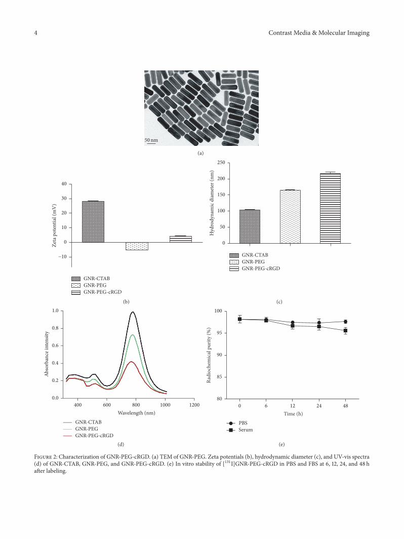
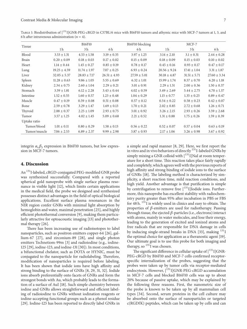
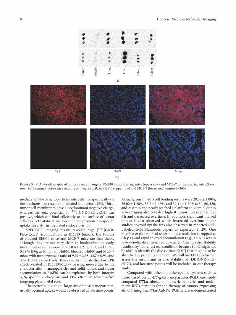