synthesis and anticancer activity of gold porphyrin linked
TRANSCRIPT

HAL Id: hal-03015360https://hal.archives-ouvertes.fr/hal-03015360
Submitted on 19 Nov 2020
HAL is a multi-disciplinary open accessarchive for the deposit and dissemination of sci-entific research documents, whether they are pub-lished or not. The documents may come fromteaching and research institutions in France orabroad, or from public or private research centers.
L’archive ouverte pluridisciplinaire HAL, estdestinée au dépôt et à la diffusion de documentsscientifiques de niveau recherche, publiés ou non,émanant des établissements d’enseignement et derecherche français ou étrangers, des laboratoirespublics ou privés.
Distributed under a Creative Commons Attribution| 4.0 International License
Synthesis and Anticancer Activity of Gold PorphyrinLinked to Malonate Diamine Platinum Complexes
Isabelle Toubia, Christophe Nguyen, Stéphane Diring, Lamiaa Ali, LudivineLarue, Rabab Aoun, Céline Frochot, Magali Gary-Bobo, Marwan Kobeissi,
Fabrice Odobel
To cite this version:Isabelle Toubia, Christophe Nguyen, Stéphane Diring, Lamiaa Ali, Ludivine Larue, et al..Synthesis and Anticancer Activity of Gold Porphyrin Linked to Malonate Diamine PlatinumComplexes. Inorganic Chemistry, American Chemical Society, 2019, 58 (18), pp.12395-12406.�10.1021/acs.inorgchem.9b01981�. �hal-03015360�
1
Synthesis and anticancer activity of gold porphyrin linked to malonate
diamine platinum complexes
Isabelle Toubia,ad# Christophe Nguyen,b# Stéphane Diring,a Lamiaa M. A. Ali,b Ludivine Laruec,
Rabab Aoun,d Céline Frochot,*c Magali Gary-Bobo,*b Marwan Kobeissi,*d Fabrice Odobel*a
aCEISAM, Chimie Et Interdisciplinarité, Synthèse, Analyse, Modélisation, CNRS, UMR CNRS
6230, UFR des Sciences et des Techniques ; 2, rue de la Houssinière - BP 92208; 44322 NANTES
Cedex 3 (France). E-mail:[email protected]
bIBMM, Institut des Biomolécules Max Mousseron, UMR 5247 CNRS, UM-Faculté de
Pharmacie, 15 Avenue Charles 9 Flahault, 34093, Montpellier cedex 05 (France). E-mail :
cLRGP, Laboratoire Réactions et Génie des Procédés, UMR 7274 CNRS-Université de Lorraine,
1 rue Grandville, 54000 Nancy, France. E-mail : [email protected]
dLaboratoireRammalRammal, Equipe de Synthèse Organique Appliquée SOA, Université
Libanaise, Faculté des Sciences 5, Nabatieh, Liban. E-mail: [email protected]
#These authors contributed equally to this study.
Synopsis
Novel anticancer agents containing a gold porphyrin appended to a platinum complex were
synthesized and characterized. Interestingly, the heterobimetallic conjugates showed
enhanced cytotoxic activity due to cooperative effects from the two metals in addition to
selectivity to cancer cells.
2
Abstract
Recently, Gold(III) porphyrins have gained great interest as anticancer drugs not only for stability of
gold(III), but also for the functionalization of porphyrin to allow bridging with another metal such as
platinum(II). We report here, for the first time, the synthesis of three new bi-metal compounds
containing gold(III) porphyrin conjugated to a platinum diamine moiety through malonate bridging to
obtain enhanced cytotoxicity from both metals combined to the phototoxicity of the porphyrin. The
three complexes differ by type of diamine ligands around platinum(II) which are ammonia (NH3),
cyclohexane diamine (CyDA) and pyridine methyl amine (Py). The synthesis was carried out using
complexation of activated malonic acid derivatives with aqua diamino-platinum(II) complexes and the
products were characterized by IR, NMR, mass spectra and by elementary analysis. Cytotoxic activity
of the conjugates was screened in both healthy and cancer cell lines, human fibroblast cells (FS-68)
and human breast cancer cells (MCF-7), and was compared to the corresponding platinum(II)
complexes. The cyclohexyldiamine (CyDA) derivative exhibited the most cytotoxic effect among the
series. The results showed that gold(III)/Pt(II)conjugates are more potent by 2 to 5.6-fold than the
corresponding platinum complexes. Moreover, the dyad AuP-PtCyDA is 18% more potent and also
more selective towards cancer cells than the parent gold porphyrin substituted with malonic acid. On
the other hand, the IC50 of dyad AuP-PtCyDA is 43% lower than that of AuTPP, but more selective
towards healthy cells. Singlet oxygen measurements indicated that gold(III) porphyrin derivatives are
poor oxygen sensitizers and cell death occurred potentially due to generation of others reactive oxygen
species (ROS) upon reductive quenching of the gold porphyrin excited state. In addition, the increase
in cancer cell death obtained after light irradiation is totally absent in healthy cells, demonstrating the
specificity of this PDT treatment on cancer cells. Our findings imply that the incorporation of two
different cytotoxic metals in the same molecule represents a remarkable cytotoxic effect compared to
traditional homometallic Pt(II) drugs.
Introduction
Cancer still remains one the major cause of mortality in the world in spite of the many
progresses made in medical treatments for several decades. Conventional cancer treatment includes
surgery, chemotherapy and radiotherapy. Platinum based compounds, such as Cisplatin, are widely
used as anticancer therapeutic agents, but they have dose-limiting side effects and some cancer lines
have become resistant to these drugs.1
3
Photodynamic therapy (PDT) is an approved clinical treatment in oncology. It has many
advantages such as low-cost, less invasiveness and minimal side-effects than conventional
chemotherapy.2 PDT requires a photosensitizer, such as a porphyrin derivatives, which are known to
better concentrate within the tumor tissue.3 Currently, many porphyrin based photosensitizers are
developed used in the clinic.2c, 34 The Photofrin was the first photosensitiser approved by the Food and
Drug Administration (FDA) in 1993 for bladder cancer therapy. Now, it is used for several cancers. Then,
a second generation of photoactive compounds was produced and developed for clinical use such as
Foscan, Metvix, Laserphyrin, Redaporphin. In addition, several compounds such as Tookad,
Radachlorin are under clinical trials for cancer treatment. Light excitation of photosensitizers produces
reactive oxygen species (ROS) in tumor and ultimately leads to cancer cell death, a method that confers
higher selectivity to cancer cells than Cisplatin-based chemotherapy treatments.
The combination of a porphyrin or a phthalocyanine with an appended platinum,5 gold,6 or
ruthenium,7 complex has been attempted to design innovative anticancer drugs. Interesting synergetic
anticancer properties are obtained thanks to the dark chemostatic effect of the metal complex
amplified by the PDT effect generated upon light excitation of the porphyrin or the phthalocyanine.5a,
b Recently, Che and co-workers reported the great pverotential of cationic gold(III) porphyrins as
cytotoxic agent towards various cancer cell lines.8 This discovery has stimulated many studies with gold
porphyrins to develop anticancer drugs.9 Interestingly, while Cisplatin anticancer drugs bind to DNA
and induce cell death by binding to purine nucleic bases such as guanine and adenine,1a gold porphyrins
target mitochondria and particularly the heat shock protein. 10As a consequence, the combination of
Cisplatin derivative with gold porphyrin could lead to synergistic effect owing to different modes of
action.
The most general mechanism of action of PDT is the generation of singlet oxygen by energy
transfer from the photosensitizer excited triplet state to the triplet ground state of oxygen (classified
as type II mechanism in PDT). It is well-accepted that photosensitizers with triplet excited states are
more favorable than singlet ones to sensitize oxygen, because oxygen has a triplet ground state
therefore energy transfer process from excited states of similar multiplicity are more efficient.11
Gold porphyrins are known to undergo intersystem crossing with unity quantum yield owing
to the heavy atom effect induced by the presence of gold.12 As a result, itis reasonable to anticipate
that gold porphyrins could be promising candidates for PDT, such as it was shown for palladium
porphyrins.11d, eHowever, to the best of our knowledge, there is so far no single study of the phototoxic
activity of gold porphyrin derivatives.
In this work, we have investigated the combination of Cisplatin derivatives covalently
connected to a gold porphyrin in order to take advantage, on one hand, of the intrinsic cytotoxicity of
the platinum complex added to that of the gold porphyrin and, on the other hand, of the potentially
4
high phototoxicity of gold porphyrin upon light excitation (Chart 1). In addition, the affinity of
porphyrin for tumor cells could also enhance selectivity and concentration of Cisplatin into tumor and
thus increase the anticancer activity. However, this affinity is not sufficient legitimating that targeting
strategies are currently explored to enhance the selectivity towards cancer cells.13 Overall, the gold
porphyrin platinum complex conjugates could have superior anticancer activity than individual
compounds.
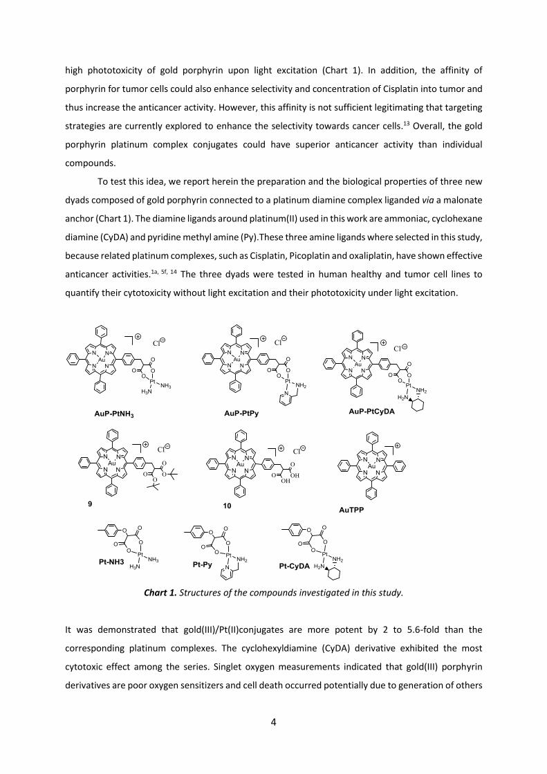
To test this idea, we report herein the preparation and the biological properties of three new
dyads composed of gold porphyrin connected to a platinum diamine complex liganded via a malonate
anchor (Chart 1). The diamine ligands around platinum(II) used in this work are ammoniac, cyclohexane
diamine (CyDA) and pyridine methyl amine (Py).These three amine ligands where selected in this study,
because related platinum complexes, such as Cisplatin, Picoplatin and oxaliplatin, have shown effective
anticancer activities.1a, 5f, 14 The three dyads were tested in human healthy and tumor cell lines to
quantify their cytotoxicity without light excitation and their phototoxicity under light excitation.
Chart 1. Structures of the compounds investigated in this study.
It was demonstrated that gold(III)/Pt(II)conjugates are more potent by 2 to 5.6-fold than the
corresponding platinum complexes. The cyclohexyldiamine (CyDA) derivative exhibited the most
cytotoxic effect among the series. Singlet oxygen measurements indicated that gold(III) porphyrin
derivatives are poor oxygen sensitizers and cell death occurred potentially due to generation of others

5
reactive oxygen species (ROS) upon reductive quenching of the gold porphyrin excited state. Overall
this study indicates that the incorporation of two different cytotoxic metals in the same molecule
represents a remarkable cytotoxic effect compared to traditional homometallic Pt(II) drugs and that
an increased cancer cell death was obtained after light irradiation only in cancer cells.
Synthesis of the compounds
The synthesis of the reference platinum compounds (Pt-NH3, Pt-Py and PtCyDA) is illustrated in
Scheme 1. First, p-cresol 1 was O-alkylated with bromoditertbutyl malonate 2 according to a
Williamson substitution reaction affording 3 in 36% yield. We suspect that this lower yield stems from
a second O-alkylation of compound 3, concomitant with its bromination and further nucleophilic
substitution by p-cresol as already reported on a similar compound.15 The tertbutyl esters of the
malonate were subsequently hydrolyzed with trifluoroacetic acid (TFA) to give compound 4. Finally,
the dicarboxylato platinum complexes were obtained in 92% yield according to Dhara’s methodology,
which consists in activating the carboxylic acid groups by deprotonation using sodium hydroxide before
being reacted with the platinum amino precursor containing two weakly bound aqua ligands.14a, 16
Scheme 1. Synthetic route for the preparation of the reference platinum complexes. Reagents and
conditions: a) THF, NaOH, RT, 24h, 36% ; b) TFA, 70oC, 1h, 100% ; c) EtOH, NaOH ; d) EtOH/Water (5/5),
diamine(dinitro)platinum(II), RT, 48h, 92%.
6
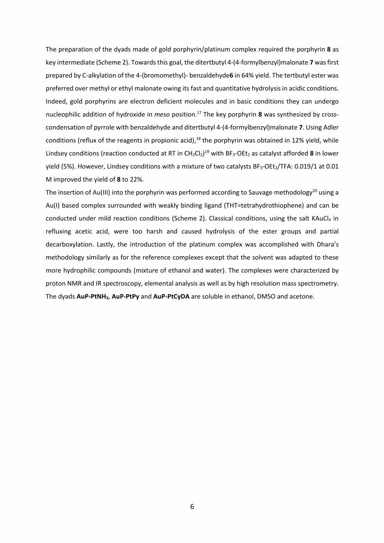
The preparation of the dyads made of gold porphyrin/platinum complex required the porphyrin 8 as
key intermediate (Scheme 2). Towards this goal, the ditertbutyl 4-(4-formylbenzyl)malonate 7 was first
prepared by C-alkylation of the 4-(bromomethyl)- benzaldehyde6 in 64% yield. The tertbutyl ester was
preferred over methyl or ethyl malonate owing its fast and quantitative hydrolysis in acidic conditions.
Indeed, gold porphyrins are electron deficient molecules and in basic conditions they can undergo
nucleophilic addition of hydroxide in meso position.17 The key porphyrin 8 was synthesized by cross-
condensation of pyrrole with benzaldehyde and ditertbutyl 4-(4-formylbenzyl)malonate 7. Using Adler
conditions (reflux of the reagents in propionic acid),18 the porphyrin was obtained in 12% yield, while
Lindsey conditions (reaction conducted at RT in CH2Cl2)19 with BF3-OEt2 as catalyst afforded 8 in lower
yield (5%). However, Lindsey conditions with a mixture of two catalysts BF3-OEt2/TFA: 0.019/1 at 0.01
M improved the yield of 8 to 22%.
The insertion of Au(III) into the porphyrin was performed according to Sauvage methodology20 using a
Au(I) based complex surrounded with weakly binding ligand (THT=tetrahydrothiophene) and can be
conducted under mild reaction conditions (Scheme 2). Classical conditions, using the salt KAuCl4 in
refluxing acetic acid, were too harsh and caused hydrolysis of the ester groups and partial
decarboxylation. Lastly, the introduction of the platinum complex was accomplished with Dhara’s
methodology similarly as for the reference complexes except that the solvent was adapted to these
more hydrophilic compounds (mixture of ethanol and water). The complexes were characterized by
proton NMR and IR spectroscopy, elemental analysis as well as by high resolution mass spectrometry.
The dyads AuP-PtNH3, AuP-PtPy and AuP-PtCyDA are soluble in ethanol, DMSO and acetone.
7
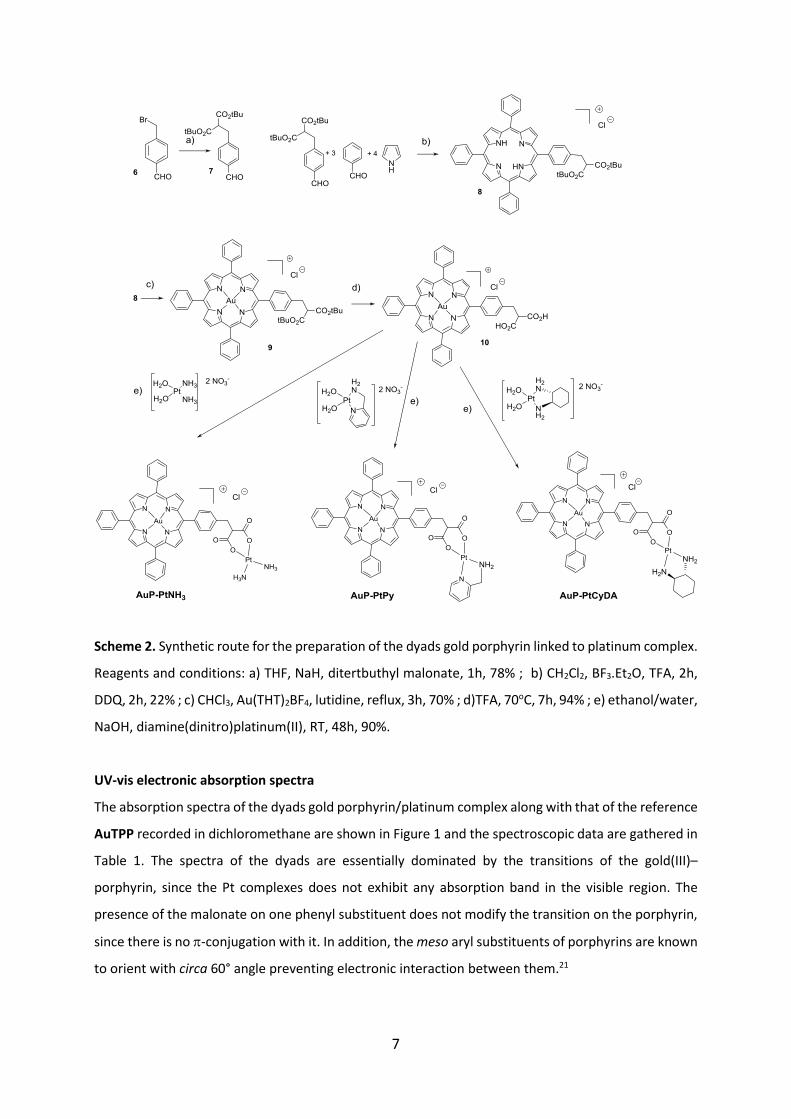
Scheme 2. Synthetic route for the preparation of the dyads gold porphyrin linked to platinum complex.
Reagents and conditions: a) THF, NaH, ditertbuthyl malonate, 1h, 78% ; b) CH2Cl2, BF3.Et2O, TFA, 2h,
DDQ, 2h, 22% ; c) CHCl3, Au(THT)2BF4, lutidine, reflux, 3h, 70% ; d)TFA, 70oC, 7h, 94% ; e) ethanol/water,
NaOH, diamine(dinitro)platinum(II), RT, 48h, 90%.
UV-vis electronic absorption spectra
The absorption spectra of the dyads gold porphyrin/platinum complex along with that of the reference
AuTPP recorded in dichloromethane are shown in Figure 1 and the spectroscopic data are gathered in
Table 1. The spectra of the dyads are essentially dominated by the transitions of the gold(III)–
porphyrin, since the Pt complexes does not exhibit any absorption band in the visible region. The
presence of the malonate on one phenyl substituent does not modify the transition on the porphyrin,
since there is no -conjugation with it. In addition, the meso aryl substituents of porphyrins are known
to orient with circa 60° angle preventing electronic interaction between them.21
8
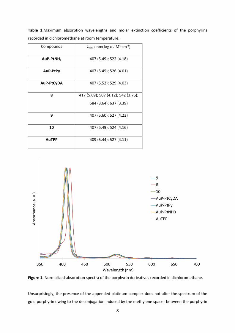
Table 1.Maximum absorption wavelengths and molar extinction coefficients of the porphyrins
recorded in dichloromethane at room temperature.
Compounds abs / nm(log / M-1cm-1)
AuP-PtNH3 407 (5.49); 522 (4.18)
AuP-PtPy 407 (5.45); 526 (4.01)
AuP-PtCyDA 407 (5.52); 529 (4.03)
8 417 (5.69); 507 (4.12); 542 (3.76);
584 (3.64); 637 (3.39)
9 407 (5.60); 527 (4.23)
10 407 (5.49); 524 (4.16)
AuTPP 409 (5.44); 527 (4.11)
Figure 1. Normalized absorption spectra of the porphyrin derivatives recorded in dichloromethane.
Unsurprisingly, the presence of the appended platinum complex does not alter the spectrum of the
gold porphyrin owing to the deconjugation induced by the methylene spacer between the porphyrin

9
and the Pt complex. The absorption bands of the gold(III)–porphyrin are characterized by a
hypsochromic shift of both the Soret and the Q-bands relative to those in the free base porphyrin
(Figure 1). These spectral features are explained by AuIII-N bonding interactions, consequently leading
to stabilization of the HOMO levels, and an increase in the HOMO–LUMO gap with respect to that of
the free base.22
Singlet oxygen measurements
Most of the porphyrins used in PDT operate according to a type II mechanism, that is the production
of singlet oxygen by energy transfer from the photosensitizer excited state to oxygen present in the
biological tissues.2a, b The singlet oxygen quantum yield production was therefore measured to assess
the potential of these gold porphyrin systems to act as sensitizers for PDT. The measurements were
recorded in methanol and the production of singlet oxygen was assayed by recording the band at 1270
nm of singlet oxygen emission after gold porphyrin systems illumination. Unfortunately, none of the
gold porphyrin and dyads prepared in this work produced detectable luminescence signal setting, thus
a singlet oxygen quantum yield was below 1%. This unexpected result probably stems from the short
lived triplet excited state of gold(III) porphyrins (≈ 1.5 ns), which decay to ground state before the
energy transfer to surrounding oxygen could take place.12, 23
Biological activity
In order to determine the therapeutic potential of these new compounds, we have first studied the
cytotoxicity induced in human breast cancer cells (MCF-7) and in healthy human fibroblasts (FS-68).
The cells were incubated in darkness during 72 h with increasing concentrations of each compound
(from 0.01 to 100 µM). The concentrations of the drugs which led to 50% cell mortality (IC50) were
determined. As shown in Figure 2, obtained cytotoxicity data in both cell lines showed the classical
sigmoidal dose-response curves when plotted as a logarithmic function of concentration (µM). Figure
2a,b illustrates the dose dependent cytotoxic effect of the reference platinum complexes Pt-NH3, Pt-
Py and PtCyDA which are in the range of those reported with similar compounds and the ranking is
consistent with previous studies5f, 24: Pt-NH3<Pt-Py<PtCyDA. Overall, the Pt complex with the ligand
CyDA is the most cytotoxic of the series with a lowerIC50 value than Cisplatin (IC50(Cisplatin)≈ 5 M).25
Importantly, the cell death is very low in healthy fibroblasts, IC50 is too high to be calculate precisely
(≥100 µM) (Figure 2c). This result highlights the targeting of cancer cells by these compounds.

10
Figure 2.Cytotoxic study of Pt-NH3, Pt-Py and PtCyDA. (a)Human breast cancer cells (MCF-7) and (b)
healthy fibroblasts cells (FS-68) were incubated 72 h with increasing concentrations of Pt-NH3, Pt-Py
and PtCyDA and maintained in darkness. Values are means ± standard deviation of 3 experiments. (c)
IC50 values are reported for each cell line.
Then, the cytotoxic activity of gold porphyrins AuTPP, 9 and 10 was investigated in both human
breast cancer cells (Figure 3a) and in healthy fibroblasts cells (Figure 3b). The results first show that
overall, the gold porphyrins are more potent than the reference platinum complexes. Although the
difference of cytotoxicity in cancer cells between Pt-CyDA and 10 is small, but on healthy fibroblasts
this difference become very high; close to 100 fold (Table 3c). In addition, the introduction of the
malonate substituent on the porphyrin decreases its cytotoxic activity, since the simple AuTPP has the
lowest IC50. This result is consistent with the report of Che and co-workers, showing that any
substituent pattern on AuTPP results in a weakening of its cytotoxicity.8a Important features for the
anticancer activity of gold(III) porphyrins are the size of the macrocycle, which must fit in the cavity of
the target receptor (certainly heat shock protein)10 and the lipophilicity of the porphyrin because its
biological properties might be related to the fact it is a planar aromatic organic cation.26 Thus, complex
9 with bulky hydrophobic malonate substituent displayed the least activity, which might be attributed
to traffic entrance into cells. A long floppy alkyl chain might have been more favorable to tether the
platinum complex and to maintain the cytotoxic activity of gold(III) porphyrin. Finally, we note that for

11
gold porphyrins AuTPP and 10, the cytotoxicity is lower on healthy cells than cancerous ones, a
significant advantage to decrease side effects during the treatment.
Figure 3. Cytotoxic study of AuTPP, 9 and 10. (a)Human breast cancer cells (MCF-7) and (b) healthy
fibroblasts cells (FS-68) were incubated 72 h with increasing concentrations of AuTPP, 9 and 10and
maintained in darkness. Values are means ± standard deviation of 3 experiments. (c) IC50 values are
reported for each cell line.
Then, the photodynamic therapy efficiency of the gold porphyrins was studied in order to determine
the possibility of a dual therapy that could increase, after light excitation, the cell death already due to
the cytotoxicity of the compound (Figure 4). Cancer cells were thus incubated for 24 h with AuTPP, 9
and 10at a concentration of 0.5 µM and then irradiated or not with the mercury lamp of a fluorescent
microscope (20 min, exc = 390 – 420 nm, 39 J.cm-2). The cell death quantification performed 2 days
after illumination demonstrates a decrease in living cells treated and submitted to photoexcitation. In
fact, light excitation induces a supplementary cell death in addition to their intrinsic cytotoxicity and
the most significant effect was obtained by the photoexcitation of AuTPP for which the illumination
induced 18% of cell death to add to the 70% of cell death obtained without light. However, the
additional cell mortality generated upon light excitation remains modest, indicating that gold
porphyrins have weak, but not null, cytotoxic as compared for other sensitizers, such as free base
porphyrins.13a, 27 As mentioned above, the low phototoxicity of gold porphyrins is certainly explained

12
by the very weak singlet oxygen generation quantum yield of these compounds, since porphyrins
usually display higher ones.27a, 28
Figure 4.Phototoxicity of AuTPP, 9 and 10incubated at 0.5 µM with MCF-7 cells during 24 h and
irradiated 20 min at exc = 390 – 420 nm (39 J.cm-2).Values are means ± standard deviation of 3
experiments.
Finally, the toxicity (Figure 5) and phototoxicity (Figure 6) of the three dyads were tested. On cancer
and healthy cells (Figure 5a,b,c), all of them display IC50 values at the micromolar level, which are lower
than those of the reference platinum complexes, but remain in the same range, let alone a bit better,
as that of the reference gold porphyrin 10. Again, the most active dyad is AuP-PtCyDA, where the Pt is
liganded with cyclohexane diamine. The cytotoxicity effect of the bimetal porphyrin conjugates AuP-
PtNH3, AuP-PtPy and AuP-PtCyDA was increased by 5.6, 4.7 and 2 fold enhancement, respectively,
compared to their corresponding Pt-complexes. These results would suggest that porphyrins granted
the transport of the complex inside the cancer cell for more favorable cytotoxicity. As expected, the
CyDA complex showed the highest effect.

13
Figure 5. Cytotoxic study of dyads AuP-PtNH3, AuP-PtPy and AuP-PtCyDA. (a)Human breast cancer
cells (MCF-7) and (b) healthy fibroblasts cells (FS-68) were incubated 72 h with increasing
concentrations of dyads AuP-PtNH3, AuP-PtPy and AuP-PtCyDAand maintained in darkness. Values
are means ± standard deviation of 3 experiments. (c) IC50 values are reported for each cell line.
Similarly as for the reference gold porphyrins (AuTPP, 9 and 10), light excitation increases cell
mortality, but to a low extent (Figure 6). Similarly as for the reference gold porphyrins (AuTPP, 9 and
10), light excitation increases cell mortality, but to a low extent (Figure 6). In addition, AuP-PtCyDA
does not exhibit PDT efficiency in these conditions.”

14
Figure 6.Phototoxicityofdyads AuP-PtNH3,AuP-PtPyandAuP-PtCyDAincubated at 0.5 µM with MCF-7
cells during 24 h and irradiated 20 min at exc = 390 – 420 nm (39 J.cm-2). Values are means ± standard
deviation of 3 experiments.
To confirm that the phototoxicity induced by photoexcitation was due to PDT effect, we studied the
reactive oxygen species (ROS) production in MCF-7 cells human cells treated with the compounds Pt-
CyDA (as negative control), AuTPP and AuP-PtPy with or without laser irradiation. Cells were first
incubated with DCFH2-DA (2’,7’-dichlorodihydrofluorescein diacetate), which is a non-fluorescent
molecule. In the presence of ROS produced within the cell, this molecule is oxidized into the
fluorescent 2’,7’-dichlorofluorescein (DCF) and its green luminescence can be detected at = 450 nm
using fluorescent microscope. The results, reported in Figure 7, showed that the light induced an
increase in green fluorescence inside the cells incubated with AuTPP and AuP-PtPy traducing thus a
ROS production and suggesting a cell death induced by PDT. The singlet oxygen measurements have
shown that all these porphyrin derivatives are poor oxygen photosensitizers, therefore it is likely that
the ROS production is induced by generating cytotoxic radicals via electron transfer from excited AuP+
to surrounding organic molecules such as glutathione, a ubiquitous antioxidant in cells, upon reductive
quenching of AuP+ excited state, which is a strong oxidant.12 Accordingly, PDT effect can arise by a type
I mechanism, which passes via the formation of oxidized species, inducing thus an oxidative stress by
oxidizing GSH to glutathione disulfide. Photosensitizers operating upon a type I mechanism is
particularly interesting in hypoxic environments found in many solid tumours.
Figure 7.ROS production in MCF-7 cells incubated with Pt-CyDA, AuP-PtCyDA and AuTPP at 0.5 µM
during 24 h and irradiated 20 min at exc = 390 – 420 nm (39 J.cm-2). Green luminescence shows the
generation of ROS detected at = 450 nm.

15
Finally, we decided to study the PDT effect in healthy fibroblasts in the same conditions as those used
for cancer cells (Figure 8). For this, FS-68 cells were incubated 24 h with 0.5 µM of compounds and
were submitted to light excitation at exc = 390 – 420 nm, 39 J.cm-2. The quantification of living cells 2
days after the PDT experiment showed a total absence of phototoxic effect. This result highlights the
specific effect of PDT on cancer cells and suggests that there could be no photoxicity on healthy cells
around the tumor.
Figure 8. Phototoxicity of gold porphyrins 10, 9, AuTPP and dyads AuP-PtNH3,AuP-PtPy,AuP-
PtCyDAincubated at 0.5 µM with healthy fibroblasts (FS-68) during 24 h and irradiated 20 min at exc =
390 – 420 nm (39 J.cm-2). Values are means ± standard deviation of 3 experiments.
Conclusion
For the first time, we report three new dyads consisting of two appended cytotoxic moieties such as a
gold(III) porphyrin and a platinum complex and their biological properties towards cancer cells were
investigated in the dark (chemostatic effect) and upon light excitation (PDT effect) and compared to
their corresponding individual platinum(II) complexes. To evaluate the decrease in side effects that
could occur with such compounds, the effect on healthy cells was also studied. The main results of this
work are: i) the diamine ligand around the Pt centre has a high impact on the cytotoxicity of the
resulting complex and cyclohexane diamine is the best ligand; ii) the gold porphyrins are poor singlet
16
oxygen photosensitizers and thereby exhibit low phototoxicity with the present systems, but
improvement can be obtained with modification of the porphyrin core, such as fluorination. The
presence of fluorine atoms on meso phenyl substituents could enhance both the oxidizing power29 and
the lifetime of the triplet excited state. Indeed, it is postulated that gold porphyrins display ligand to
metal charge transfer transitions, which are responsible of the fast decay of the triplet excited state.23
However, the phototoxicity is not null and most probably result by a type I mechanism; iii) the
introduction of a malonate group of one phenyl substituent of AuTPPs lightly decreases cyto- and
photoxicity the gold porphyrin; iv) the dyad AuP-PtCyDA displays interesting cytotoxic activity towards
cancer cells, for which the mechanism of action would be interesting to be investigated.The enhanced
cytotoxicity indicates that porphyrins allow the delivery of the complex into cancer cells. The complex
might have dual biological functioning between Au(III) and Pt(II) arms. The former as reported would
interact with mitochondria and the latter would target DNA; both mechanisms cause cancer cell death.
Importantly, all compounds present a close or clearly higher cytotoxic activity in cancer cells than
healthy cells and the PDT effect, even slight, induced around 15 or 20% more cancer cell death for
AuTPP, AuP-PtNH3 and AuP-PtPy in conditions totally harmless for healthy cells (0% cell death).
Further biological analysis would clarify the anticancer activity if it is based on specific metal or both.
Additional analysis of the intracellular metal contents such as ICP-analysis to monitor the uptake of the
whole stable bimetal conjugate by cancer cells would clarify the delivery mechanism. On the other
hand, the incorporate mode of action exhibited by the two metals may be even amplified using an
alternative photosensitizer. These studies are in due course in our laboratory.
Acknowledgments
We thank Campus France (Eiffel grant), Pays de la Loire (LUMOMAT project), CNRS-L and the Lebanese
University for the financial support of this project.
Experimental part
Synthesis of the compounds
Generalities
17
1H and 13C NMR spectra were recorded on an AVANCE 300 UltraShield BRUKER and AVANCE 400
BRUKER. Chemical shifts for 1H and 13C NMR spectra are referenced relative to residual proton in the
deuterated solvent (CDCl3 = 7.26 ppm for 1H, DMSO-d8 = 2.50 ppm for 1H) or to an internal reference
(TMS, = 0 ppm for both 1H). NMR spectra were recorded at room temperature, chemical shifts are
written in ppm and coupling constants in Hz. High-resolution mass (HR-MS) spectra were obtained by
electrospray ionization coupled with high resolution ion trap orbitrap (LTQ-Orbitrap, ThermoFisher
Scientific),working in ion-positive or ion-negative mode.
Chemicals were purchased from Sigma-Aldrich or Alfa Aesar and used as received. Thin-layer
chromatography (TLC) was performed on silica sheets precoated with Merck 5735 Kieselgel 60F254.
Column chromatography was carried out with Merck 5735 Kieselgel 60F (0.040-0.063 mm
mesh).Bromo ditertbutyl malonate 2, 4-(bromomethyl)-benzaldehyde6and platinum precursors
Pt(OH2)2(NH3)2-(NO3)2, Pt(OH2)2(Py)-(NO3)2 and Pt(OH2)2(CyDA)-(NO3)2were synthesized according to
literature.
Ditertbutylmalonate p-cresol (3). A total of 1 g (9.24 mmol) of p-cresol 1 and 2.7 g (9.24 mmol) of
bromoditertbutyl malonate2were dissolved under argon in 20 ml of distilled THF. 0.36 g (9 mmol) of
pulverized NaOH was added, and the mixture was stirred for 24 h at room temperature. The organic
solution was diluted with water and extracted with dichloromethane. The combined organic phases
were washed with water and dried over Na2SO4.The solvent was evaporated and the crude product
was purified by column chromatography (SiO2; CH2Cl2/petroleum ether 5/5). Yield: (1.08 g, 36%).NMR
(1H, CDCl3, 300 MHz) δ (ppm): 7.07-7.04 (d, 2H, J = 8.63 Hz), 6.86-6.84 (d, 2H, J = 8.63 Hz), 4.92 (s, 1H),
2.26 (s, 3H), 1.48 (s, 18H). HRMS (ES+) [M+] m/z 345.1672 found, 345.1678 calc.
Compound (4). 206 mg (0.63 mmol) of 3 was dissolved in 1 ml TFA and heated at 70°C for one hour.
The solvent was evaporated and the product was dried under vacuum. Yield: (134 mg, 100%). IR (ATR):
1706 cm-1 (C=O).NMR (1H, CDCl3, 300 MHz) δ (ppm): 7.10-7.08 (d, 2H, J = 8.63 Hz), 6.82-6.80 (d, 2H, J
= 8.61 Hz), 5.25 (s, 1H), 2.22 (s, 3H). HRMS (ES+) [M+] m/z 209.0454 found, 209.0450 calc.
General procedure 1 (GP 1). Compound 4 was dissolved in water and the carboxylic acid functions
were activated into sodium carboxylates, using 2 eq of NaOH. 1.2 eq of the respective
diammine(dinitro)platinum(II) complex was dissolved in ethanol and added.The solution was stirred
for 48 h at room temperature. The precipitated complexes Pt-Py, Pt-NH3, Pt-CyDA were filtered and
dried.
18
Pt-NH3. According to GP1, 20 mg (0.095 mmol) of 4 was dissolved in 2 ml water, 7.6 mg (0.19 mmol)
of NaOH was added, combined with 40 mg (0.11 mmol) of diamine(dinitro)platinum(II) dissolved in 2
ml EtOH, and stirred for 48 h. The precipitated complexPt-NH3was filtered, washed with water and
dried. Yield (38 mg, 92%).IR (ATR): 1669 cm-1 (C=O).NMR (1H, DMSO, 300 MHz) δ (ppm): 7.03 (br, 2H),
6.66 (br, 2H), 5.70 (s, 1H), 4.29 (br, 6H), 2.20 (s, 3H). HRMS (ES+) [M+] m/z 459.0412 found, 459.0427
calc. Elem. Anal. Exp C, 27.68; H, 3.20; N, 6.19 .calc C, 27.47; H, 3.23; N, 6.41.
Pt-CyDA. According to GP1, 40 mg (0.19 mmol) of 4 was dissolved in 3 ml water, 15 mg (0.38 mmol) of
NaOH was added, combined with 90 mg (0.2 mmol) of diamine(dinitro)platinum(II) dissolved in 3 ml
EtOH, and stirred for 48 h. The precipitated complex Pt-CyDA was filtered, washed with water and
dried. Yield (90 mg, 92%).IR (ATR): cm-1 (C=O).NMR (1H, DMSO, 300 MHz) δ (ppm): 7.06-7.03 (d, 2H, J
= 8.53 Hz), 6.68-6.65 (d, 2H, J = 8.9 Hz), 6.16-6.03 (dd, 2H, J = 9.9, 30.91 Hz), 5.66 (s, 1H), 5.45-5.33
(dt,2H, J = 37.28, 8.96 Hz), 2.21 (s, 3H), 2.09 (br, 2H), 1.84-1.80 (d, 2H, J = 12.95 Hz), 1.46-1.44 (d, 2H, J
= 8.86 Hz), 1.22 (br, 2H), 1.04-0.97(t, 2H, J = 10.22 Hz). HRMS (ES+) [M+] m/z 539.1061 found, 539.1053
calc.
Pt-Py. According to GP1, 40 mg (0.19 mmol) of 4 was dissolved in 3 ml water, 15 mg (0.38 mmol) of
NaOH was added, combined with 89 mg (0.2 mmol) of diamine(dinitro)platinum(II) dissolved in 3 ml
EtOH, and stirred for 48 h. The precipitated complex Pt-Py was filtered, washed with water and dried.
Yield (89 mg, 92%).IR (ATR): 1681, 1649 cm-1 (C=O).NMR (1H, DMSO, 300 MHz) δ (ppm): 8.44-8.42 (d,
1H, J = 5.97 Hz), 8.18-8.12 (td, 1H, J = 7.73, 1.58 Hz), 7.66-7.63 (d, 1H, J = 7.8 Hz), 7.54-7.50 (t, 1H, J =
6.96 Hz),7.05-7.02 (d, 2H, J = 8.36 Hz), 6.71-6.68 (d, 2H, J = 8.9 Hz), 6.64-6.62 (d, 1H, J = 5.57 Hz), 5.70
(s, 1H), 4.15-4.14 (br, 2H),2.21 (s, 3H). HRMS (ES+) [M+] m/z 511.0761 found, 511.0764 calc.
Porphyrin (8). BF3.Et2O 16 µl (1.2 × 10 -2 mmol) and 0.5 ml (6.46 mmol) of trifluoroacetic acid (TFA)
were added to a solution of 0.55 ml (5.38 mmol) of benzaldehyde, 600 mg (1.79 mmol) of 7, and 0.35
ml (5 mmol) of freshly distilled pyrrole in 1 l of CH2Cl2. The mixture was kept at room temperature for
2 h. 1.2 g (5.2 mmol) of DDQ was added when the aldehydes were completely reacted, and the mixture
was kept at room temperature for an additional 2 h. The solvent was evaporated and the crude product
was purified by column chromatography (SiO2; CH2Cl2/hexane 6/4) to remove tetraphenylporphyrin.
The monosubtituted porphyrin was then eluted with CH2Cl2 in form of a thick, purple band. Yield: (331
mg, 22%). UV-Vis (CH2Cl2, nm) λmax (nm): 418 (ε 4.2 × 10⁵ mol-1.L.cm-1), 522 (1.1 × 10⁴), 557 (4.1 × 10³),
655 (2.3 × 10³). NMR (1H, CDCl3, 300 MHZ) δ (ppm): 8.41-8.80 (m, 8 H), 8.23-8.20 (dd, 2H, J = 7.08, 7.67
Hz), 8.14-8.11 (d, 2H, J = 8.02 Hz), 7.76-7.74(m, 9H), 7.61-7.58 (d, 2H, J = 8.15 Hz), 3.83-3.77 (t, 1H, J =
19
8.41 Hz), 3.49-3.46 (d, 2H, J = 7.94 Hz), 1.55 (s, 18H). HRMS (ES+) [M+] m/z 843.3908 found, 842.3910
calc.
Porphyrin (9). A total of 700 mg (0.83 mmol) of porphyrin 8 and 1.9 g (4.1 × 10⁻³ mmol) [Au(THT)2]BF4
were dissolved in 5 ml chloroform. 193 µl (1.6 × 10⁻³ mmol) of 2,6-lutidine was added and the reaction
was heated to reflux. A gold mirror very rapidly deposited on the walls of the flask, and the reaction
mixture turned reddish. After refluxing for three hours, the solution was evaporated to dryness. The
residue was taken up in dichloromethane and purified through a silica column, eluting the free base
porphyrin with dichloromethane and the desired red-orange band with CH2Cl2/AcOEt (9/1).Yield: (602
mg, 70%). UV-Vis (CH2Cl2, nm) λmax (nm): 408 (ε 3.1 × 10⁵ mol-1.L.cm-1), 526 (1.2 × 10⁴). NMR (1H, CDCl3,
300 MHz) δ (ppm): 9.26-9.23 (m, 8H), 8.24-8.22 (d, 6H, J = 6.53 Hz), 8.16-8.14 (d, 2H, J = 8.09 Hz), 7.90-
7.83 (m, 9H), 7.71-7.69 (d, 2H, J = 7.78 Hz), 3.83-3.78 (t, 1H, J = 7.76 Hz), 3.51-3.49 (d, 2H, J = 8.09 Hz),
1.55 (s, 18H). HRMS (ES+) [M+] m/z 1037.3326 found, 1037.3341 calc.
Porphyrin (10). 630 mg (0.607 mmol) of gold porphyrin 9 was dissolved in 2 ml TFA and heated at 70oC
for seven hours. After removal of the solvent under reduced pressure, the product was dissolved in
methanol and purified using exclusion size chromatography. Yield: (530 mg, 94%). IR (ATR): 1634 cm-1
(C=O). UV-Vis (CH2Cl2, nm) λmax (nm): 408 (ε 2.8 × 10⁵ mol-1.L.cm-1), 526 (1.3 × 10⁴). NMR (1H, DMSO-d6,
300 MHz) δ (ppm): 9.44-9.40 (m, 8H), 8.22-8.18 (m, 6H), 8.09-8.06 (d, 2H, J = 8.13 Hz), 7.94-7.86 (m,
8H), 7.76-7.73 (d, 2H, J = 7.98 HZ), 3.92-3.88 (t, 1H, J = 6.18 Hz), 3.71-3.69 (d, 2H, J = 5.85 Hz). HRMS
(ES+) [M+] m/z 925.2075 found, 925.2089 calc.
General procedure 2 (GP 2). Compound 10 was dissolved in EtOH and the carboxylic acid functions
were activated into sodium carboxylates using 2 eq of NaOH. 1.2 eq of the respective
diammine(dinitro)platinum(II) complex was dissolved in Ethanol/Water (1/1) and added. The solution
was stirred for 48 h at room temperature. The solvents were evaporated. The complexes were
dissolved in DMF and precipitated with a saturated solution of NaCl. The precipitated complexes AuP-
PtNH3 ,AuP-PtPy, AuP-PtCyDA were filtered, washed with water and dried.
AuP-PtNH3.According to GP2, 60 mg (5.6 × 10⁻² mmol) of gold porphyrin 10 was dissolved in 16 ml
EtOH, 4.5 mg (0.11 mmol) of NaOH was added, combined with 23.74 mg (67.2 × 10⁻³ mmol) of
diamine(dinitro)platinum(II) dissolved in 12 ml of a mixture EtOH/H2O(1/1).The mixture was stirred
for 48h at room temperature. The solvents were evaporated. The complex was dissolved in DMF and
precipitated with a saturated solution of NaCl. The precipitated complexAuP-PtNH3was filtered,

20
washed with water and dried. Yield (58 mg, 90%). IR (ATR): 1619 cm-1 (C=O). UV-Vis (CH2Cl2, nm) λmax
(nm): 408 (ε 2.8 × 10⁵ mol-1.L.cm-1), 526 (1.3 × 10⁴). NMR (1 H, DMSO-d6, 300MHz) δ (ppm): 9.35-9.28
(m, 8H), 8.29-8.27 (d, 6H, J = 6.64 Hz), 8.26-8.17 (d, 2H, J = 8.01 Hz), 8.14-7.91 (m, 9H), 7.82-7.79 (d,
2H, J = 7.55 Hz), 4.38-4.34 (t, 1H, J = 7.08 Hz), 4.36-4.28(br, 6H), 3.55-3.54 (d, 2H, J = 5.31 Hz). HRMS
(ES+) [M+] m/z 1151.2090 found, 1151.2090 calc. Elem. Anal. Exp C, 48.69; H, 3.48; N, 6.35. calc C,
48.56; H, 3.61; N, 6.5.
AuP-PtCyDA. According to GP2, 60 mg (5.6 × 10⁻² mmol) of gold porphyrin 10 was dissolved in 16 ml
EtOH, 4.5 mg (0.11 mmol) of NaOH was added, combined with 27.66 mg (63.8 × 10⁻³ mmol) of
diamine(dinitro)platinum(II) dissolved in 12 ml of a mixture EtOH/H2O(1/1).The mixture was stirred
for 48h at room temperature. The solvents were evaporated. The complex was dissolved in DMF and
precipitated with a saturated solution of NaCl. The precipitated complex AuP-PtCyDA was filtered,
washed with water and dried. Yield (61 mg, 90%). IR (ATR): 1626 cm-1 (C=O).UV-Vis (CH2Cl2, nm) λmax
(nm): 408 (ε 2.8 × 10⁵ mol-1.L.cm-1), 526 (1.3 × 10⁴). NMR (1H, DMSO-d6, 300MHz) δ (ppm): 9.34-9.30
(m, 8H), 8.29-8.26 (dd, 6H, J = 7.27, 7.63 Hz), 8.18-8.15 (d, 2H, J = 8 Hz), 7.98-7.91 (m, 9H), 7.81-7.78
(d, 2H, J = 8.36 Hz), 6.13-5.99 (m, 2H), 5.45-5.32 (m, 2H), 4.35-4.30 (t, 1H, J = 6.56 Hz ), 3.44-3.42 (d,
2H, J = 6.82 Hz), 2.15-2.12 (m, 2H), 1.86-1.82 (t, 2H, J = 8.76 Hz), 1.48-1.46 (d, 2H, J = 9.28 Hz), 1.23-
1.21 (m, 2H), 1.06-1.01 (m, 2H). HRMS (ES+) [M+] m/z 1231.2739 found, 1231.2716 calc. Elem. Anal.
Exp C, 49.61; H, 3.79; N, 6.02. calc C, 49.3; H, 3.45; N, 6.34.
AuP-PtPy. According to GP2, 60 mg (5.6 × 10⁻² mmol) of gold porphyrin 10 was dissolved in 16 ml EtOH,
4.5 mg (0.11 mmol) of NaOH was added, combined with 23.88 mg (56 × 10-3 mmol) of
diamine(dinitro)platinum(II) dissolved in a mixture of 2.5 ml EtOH and 17.5 ml H2O. The solution was
stirred for 48h at room temperature. The solvents were evaporated. The complex was dissolved in
DMF and precipitated with a saturated solution of NaCl. The precipitated complex AuP-PtPy was
filtered, washed with water and dried. Yield (61 mg, 90%). IR (ATR): 1631 cm-1 (C=O). UV-Vis (CH2Cl2,
nm) λmax (nm): 408 (ε 2.8 × 10⁵ mol-1.L.cm-1), 526 (1.3 × 10⁴). NMR (1H, DMSO-d6, 300MHz) δ (ppm):
9.33-9.30 (m, 8H), 8.52-8.50 (d, 1H, J = 5.78 Hz), 8.27-8.25 (d, 6H, J = 6.39 Hz ), 8.16-8.13 (d, 2H, J =
7.92 Hz), 8.13-7.90 (m, 9H), 7.83-7.80 (d, 2H, J = 7.92 Hz), 7.67-7.64 (d, 1H, J = 7.94 Hz), 7.55-7.50 (t,
1H, J = 7.12 Hz), 6.61-6.59 (br, 2H), 4.39-4.35 (t, 1H, J = 6.75 Hz), 4.17-4.13 (t, 2H, J = 5.44 Hz), 3.46-
3.44 (d, 2H, J = 5.44 Hz). HRMS (ES+) [M+] m/z 1225.2269 found, 1225.2247 calc. Elem. Anal. Exp C,
49.16; H, 3.55; N, 5.92. calc C, 48.81; H, 3.17; N, 6.28.
Singlet oxygen detection. Singlet oxygen emission was detected through a double ruled grating SPEX
monochromator (600 grooves/mm blazed at 1 μm) and a long-wave pass (780 nm). All spectra were
21
measured in 4 faces quartz cuvettes. All the emission singlet oxygen luminescence have been displayed
with the same absorbance (less than 0.2) with the lamp and photomultiplier correction.
Biological experiments
Cell culture. Human breast adenocarcinoma cells (MCF-7) (purchased from ATCC) were cultured in
Dulbecco Eagle's Minimal Essential Medium (DMEM) supplemented with 10% fetal bovine serum (FBS)
and 1% penicillin/streptomycin (P/S). Adult Human Dermal Fibroblast cells (FS-68) were maintained in
Roswell Park Memorial Institute (RPMI) medium supplemented with 10% FBS and 1% P/S. Both cell
lines were allowed to grow in humidified atmosphere at 37 °C under 5 % CO2. For cytotoxic and
phototoxic studies, the compounds (powder) are first diluted in DMSO at the concentration of 10 mM.
Then, they are sonicated during 30 seconds and diluted at the required concentrations in culture
medium of each cell line”.
Cytotoxicity study. MCF-7 and FS-68 cells were seeded into 96-well plates at a density of 103 cells/cm2.
One day after cell growth, cells were incubated with or without different concentrations of compounds
(from 0.01 to 100 µM) for 3 days. After the incubation time, cells were incubated with 0.5 mg mL−1
MTT (3-(4,5- dimethylthiazol-2-yl)-2,5-diphenyltetrazoliumbromide)during 4 h to determine
mitochondrial enzyme activity. Then, MTT precipitates were dissolved in ethanol/DMSO (1:1) solution
and absorbance was measured at 540 nm.
Phototoxicity assay. Both cell lines, MCF-7 and FS-68 cells, were seeded into 384-well plates at a
density of 103 cells/cm2 and allowed to grow for 24 h. Cells were incubated or not (control) with 0.5
µM concentration of compounds solution for 24 h. After incubation, cells were exposed, or not, to light
using mercury lamp of a fluorescence microscope at excitation ranges between 390-420 nm for 20 min
(39 J.cm-2). Two days after, MTT assay was performed to evaluate the phototoxic effect of compounds.
ROS production. The detection of ROS was performed during the phototoxicity experiment. Forty five
minutes before irradiation, cells were incubated with 20 µM concentration of DCFH2-DA(Cellular ROS
Assay Kit, Abcam, USA). Cells were exposed to light using mercury lamp of a fluorescence microscope
at excitation ranges between 390-420 nm for 20 min (39 J.cm-2). After irradiation, cells were washed
and fluorescence emission of DCF was detected at 450 nm using fluorescent microscope.

22
References:
(1).(a) Johnstone, T. C.; Suntharalingam, K.; Lippard, S. J. The Next Generation of Platinum Drugs:
Targeted Pt(II) Agents, Nanoparticle Delivery, and Pt(IV) Prodrugs. Chem. Rev. 2016, 116, 3436-3486;
(b) Galluzzi, L.; Senovilla, L.; Vitale, I.; Michels, J.; Martins, I.; Kepp, O.; Castedo, M.; Kroemer, G.
Molecular mechanisms of cisplatin resistance. Oncogene 2011, 31, 1869; (c) Rabik, C. A.; Dolan, M. E.
Molecular mechanisms of resistance and toxicity associated with platinating agents. Cancer Treatment
Rev; 2007, 33, 9-23.
(2).(a) Fan, W.; Huang, P.; Chen, X. Overcoming the Achilles' heel of photodynamic therapy. Chem. Soc.
Rev. 2016, 45, 6488-6519; (b) Celli, J. P.; Spring, B. Q.; Rizvi, I.; Evans, C. L.; Samkoe, K. S.; Verma, S.;
Pogue, B. W.; Hasan, T. Imaging and Photodynamic Therapy: Mechanisms, Monitoring, and
Optimization. Chem. Rev. 2010, 110, 2795-2838; (c) Ferreira dos Santos, A. l.; Queiroz de Almeida, D.
R.; Ferreira Terra, L.; Baptista, M. c. S.; Labriola, L. Photodynamic therapy in cancer treatment - an
update review. J. Cancer Metastasis Treat. 2019, 5, doi: 10.20517/2394-4722.2018.83.
(3).El-Far, M. A.; Pimstone, N. R. The interaction of tumour-localizing porphyrins with collagen, elastin,
gelatin, fibrin and fibrinogen. Cell Biochem. Function 1985, 3, 115-119.
(4).Tjahjono, D. H. Porphyrin structure-based molecules for photodynamic therapy of cancer. Acta
Pharm. Indones. 2006, 31, 1-12.
(5).(a) Lau, J. T. F.; Lo, P.-C.; Fong, W.-P.; Ng, D. K. P. A Zinc(II) Phthalocyanine Conjugated with an
Oxaliplatin Derivative for Dual Chemo- and Photodynamic Therapy. J. Med. Chem. 2012, 55, 5446-
5454; (b) Naik, A.; Rubbiani, R.; Gasser, G.; Spingler, B. Visible-Light-Induced Annihilation of Tumor
Cells with Platinum–Porphyrin Conjugates. Angew. Chem. Int. Ed. 2014, 53, 6938-6941; (c) Lottner, C.;
Bart, K.-C.; Bernhardt, G.; Brunner, H. Hematoporphyrin-Derived Soluble Porphyrin−Platinum
Conjugates with Combined Cytotoxic and Phototoxic Antitumor Activity. J. Med. Chem. 2002, 45, 2064-
2078; (d) Kim, Y.-S.; Song, R.; Hyun Kim, D.; Jun, M. J.; Sohn, Y. S. Synthesis, biodistribution and
antitumor activity of hematoporphyrin–platinum(II) conjugates. Bioorg. Med. Chem. 2003, 11, 1753-
1760; (e) Munakata, H.; Imai, H.; Nakagawa, S.; Osada, A.; Uemori, Y. Water-Soluble Porphyrins
Appending Platinum(II) Complexes as Binders for Synthetic Nucleic Acid Polymers. Chem. Pharm. Bull.
2003, 51, 614-615; (f) Brunner, H.; Schellerer, K. M. New porphyrin platinum conjugates for the
cytostatic and photodynamic tumor therapy. Inorg. Chim. Acta 2003, 350, 39-48.
(6).Longevial, J.-F.; El Cheikh, K.; Aggad, D.; Lebrun, A.; van der Lee, A.; Tielens, F.; Clément, S.; Morère,
A.; Garcia, M.; Gary-Bobo, M.; Richeter, S. Porphyrins Conjugated with Peripheral Thiolato Gold(I)
Complexes for Enhanced Photodynamic Therapy. Chem. Eur. J. 2017, 23, 14017-14026.
(7).(a) Schmitt, F.; Govindaswamy, P.; Süss-Fink, G.; Ang, W. H.; Dyson, P. J.; Juillerat-Jeanneret, L.;
Therrien, B. Ruthenium Porphyrin Compounds for Photodynamic Therapy of Cancer. J. Med. Chem.

23
2008, 51, 1811-1816; (b) Harvey, P. D.; Tasan, S.; Gros, C. P.; Devillers, C. H.; Richard, P.; Gendre, P. L.;
Bodio, E. Ruthenium and Osmium Complexes of Phosphine-Porphyrin Derivatives as Potential
Bimetallic Theranostics: Photophysical Studies. Organometallics 2015, 34, 1218-1227; (c) Gianferrara,
T.; Bratsos, I.; Iengo, E.; Milani, B.; Oštrić, A.; Spagnul, C.; Zangrando, E.; Alessio, E. Synthetic strategies
towards ruthenium–porphyrin conjugates for anticancer activity. Dalton Trans. 2009, 10742-10756.
(8).(a) Sun, R. W.-Y.; Li, C. K.-L.; Ma, D.-L.; Yan, J. J.; Lok, C.-N.; Leung, C.-H.; Zhu, N.; Che, C.-M. Stable
Anticancer Gold(III)–Porphyrin Complexes: Effects of Porphyrin Structure. Chem. Eur. J. 2010, 16, 3097-
3113; (b) Sun, R. W.-Y.; Che, C.-M. The anti-cancer properties of gold(III) compounds with dianionic
porphyrin and tetradentate ligands. Coord. Chem. Rev. 2009, 253, 1682-1691; (c) Tu, S.; Wai-Yin Sun,
R.; Lin, M. C. M.; Tao Cui, J.; Zou, B.; Gu, Q.; Kung, H.-F.; Che, C.-M.; Wong, B. C. Y. Gold (III) porphyrin
complexes induce apoptosis and cell cycle arrest and inhibit tumor growth in colon cancer. Cancer
2009, 115, 4459-4469; (d) Chen, H.; Yang, Q.; Sun, L.; Zhang, Z.; Tong, H.; Xu, A.; Wang, C. Synthesis
and Biological Evaluation of Gold(III) Substituted Tetraarylporphyrin Chlorides as Anticancer Reagents.
J. Chem.Res. 2011, 35, 190-194.
(9).(a) He, L.; Chen, T.; You, Y.; Hu, H.; Zheng, W.; Kwong, W.-L.; Zou, T.; Che, C.-M. A Cancer-Targeted
Nanosystem for Delivery of Gold(III) Complexes: Enhanced Selectivity and Apoptosis-Inducing Efficacy
of a Gold(III) Porphyrin Complex. Angew. Chem., Int. Ed. 2014, 53, 12532-12536; (b) Hu, D.; Liu, Y.; Lai,
Y.-T.; Tong, K.-C.; Fung, Y.-M.; Lok, C.-N.; Che, C.-M. Anticancer Gold(III) Porphyrins Target
Mitochondrial Chaperone Hsp60. Angew. Chem., Int. Ed. 2016, 55, 1387-1391; (c) Lum, C. T.; Wai-Yin
Sun, R.; Zou, T.; Che, C.-M. Gold(iii) complexes inhibit growth of cisplatin-resistant ovarian cancer in
association with upregulation of proapoptotic PMS2 gene. Chem. Sci. 2014, 5, 1579-1584; (d) Dandash,
F.; Leger, D. Y.; Fidanzi-Dugas, C.; Nasri, S.; Bregier, F.; Granet, R.; Karam, W.; Diab-Assaf, M.; Sol, V.;
Liagre, B. In vitro anticancer activity of new gold(III) porphyrin complexes in colon cancer cells. J. Inorg.
Biochem. 2017, 177, 27-38; (e) Lammer, A. D.; Cook, M. E.; Sessler, J. L. Synthesis and anti-cancer
activities of a water soluble gold(III) porphyrin. J. Porphyrins Phthalocyanines 2015, 19, 398-403; (f)
Wang, Y.; He, Q.-Y.; Che, C.-M.; Tsao, S. W.; Sun, R. W.-Y.; Chiu, J.-F. Modulation of gold(III) porphyrin
1a-induced apoptosis by mitogen-activated protein kinase signaling pathways. Biochem. Pharmacol.
2008, 75, 1282-1291.
(10).Hu, D.; Liu, Y.; Lai, Y.-T.; Tong, K.-C.; Fung, Y.-M.; Lok, C.-N.; Che, C.-M. Anticancer Gold(III)
Porphyrins Target Mitochondrial Chaperone Hsp60. Angew. Chem. Int. Ed. 2016, 55, 1387-1391.
(11).(a) Wilkinson, F.; Helman, W. P.; Ross, A. B. Quantum Yields for the Photosensitized Formation of
the Lowest Electronically Excited Singlet State of Molecular Oxygen in Solution. J. Phys. Chem.
Reference Data 1993, 22, 113-262; (b) Pineiro, M.; Carvalho, A. L.; Pereira, M. M.; Gonsalves, A. M. d.
A. R.; Arnaut, L. G.; Formosinho, S. J. Photoacoustic Measurements of Porphyrin Triplet-State Quantum
Yields and Singlet-Oxygen Efficiencies. Chem. Eur. J. 1998, 4, 2299-2307; (c) Pineiro, M.; Pereira, M. M.;

24
Rocha Gonsalves, A. M. d. A.; Arnaut, L. G.; Formosinho, S. J. Singlet oxygen quantum yields from
halogenated chlorins: potential new photodynamic therapy agents. J. Photochem. Photobiol. A 2001,
138, 147-157; (d) Obata, M.; Hirohara, S.; Tanaka, R.; Kinoshita, I.; Ohkubo, K.; Fukuzumi, S.; Tanihara,
M.; Yano, S. In Vitro Heavy-Atom Effect of Palladium(II) and Platinum(II) Complexes of Pyrrolidine-
Fused Chlorin in Photodynamic Therapy. J. Med. Chem. 2009, 52, 2747-2753; (e) Horiuchi, H.;
Terashima, K.; Sakai, A.; Suda, D.; Yoshihara, T.; Kobayashi, A.; Tobita, S.; Okutsu, T. The effect of central
metal on the photodynamic properties of silylated tetraphenylporphyrin derivative. J. Photochem.
Photobiol. A 2016, 321, 72-78; (f) Figueiredo, T. L. C.; Johnstone, R. A. W.; Sørensen, A. M. P. S.; Burget,
D.; Jacques, P. Determination of Fluorescence Yields, Singlet Lifetimes and Singlet Oxygen Yields of
Water-Insoluble Porphyrins and Metalloporphyrins in Organic Solvents and in Aqueous Media.
Photochem. Photobiol. 1999, 69, 517-528.
(12).Brun, A. M.; Harriman, A.; Heitz, V.; Sauvage, J. P. Charge transfer across oblique bisporphyrins:
two-center photoactive molecules. J. Am. Chem. Soc. 1991, 113, 8657-8663.
(13).(a) Singh, S.; Aggarwal, A.; Bhupathiraju, N. V. S. D. K.; Arianna, G.; Tiwari, K.; Drain, C. M.
Glycosylated Porphyrins, Phthalocyanines, and Other Porphyrinoids for Diagnostics and Therapeutics.
Chem. Rev. 2015, 115, 10261-10306; (b) Alonso, C.; Boyle, R. W., 17 Bioconjugates of Porphyrins and
Related Molecules for Photodynamic Therapy. In Handbook of Porphyrin Science, pp 121-190.
(14).(a) Wilson, J. J.; Lippard, S. J. Synthetic Methods for the Preparation of Platinum Anticancer
Complexes. Chem. Rev. 2014, 114, 4470-4495; (b) Bernhardt, G.; Biersack, B.; Bollwein, S.; Schobert,
R.; Zoldakova, M. Terpene Conjugates of Diaminedichloridoplatinum(II) Complexes: Antiproliferative
Effects in HL-60 Leukemia, 518A2 Melanoma, and HT-29 Colon Cancer Cells. Chem. Biodiversity 2008,
5, 1645-1659.
(15).Johnson, R. E.; Bacon, E. R. Novel dibenzo[d,f][1,3]dioxepines. Tetrahedron Lett. 1994, 35, 9327-
9328.
(16).Dhara, S. C. A rapid method for the synthesis of cis-platine. Indian J. Chem. 1970, 8, 193–194.
(17).Segawa, H.; Azumi, R.; Shimidzu, T. Direct hydroxylation at the meso position of gold(III)
tetraphenylporphyrin by nucleophilic addition: novel hydroxyphlorin derivatives. J. Am. Chem. Soc.
1992, 114, 7564-7565.
(18).Adler, A. D.; Longo, F. R.; Finarelli, J. D.; Goldmacher, J.; Assour, J.; Korsakoff, L. A simplified
synthesis for meso-tetraphenylporphine. J. Org. Chem. 1967, 32, 476-476.
(19).Lindsey, J. S.; Schreiman, I. C.; Hsu, H. C.; Kearney, P. C.; Marguerettaz, A. M. Rothemund and
Adler-Longo reactions revisited: synthesis of tetraphenylporphyrins under equilibrium conditions. J.
Org. Chem. 1987, 52, 827-836.
(20).Chambron, J. C.; Heitz, V.; Sauvage, J.-P. Incorporation of gold(III) into porphyrins by
disproportionation of [Au(tht)2]BF4. New J. Chem. 1997, 21, 237-240.
25
(21).Silvers, S. J.; Tulinsky, A. The crystal and molecular structure of triclinic tetraphenylporphyrin. J.
Am. Chem. Soc. 1967, 89, 3331-3337.
(22).Antipas, A.; Dolphin, D.; Gouterman, M.; Johnson, E. C. Porphyrins. 38. Redox potentials, charge
transfer transitions, and emission of copper, silver, and gold complexes. J. Am. Chem. Soc. 1978, 100,
7705-7709.
(23).Eng, M. P.; Ljungdahl, T.; Andréasson, J.; Mårtensson, J.; Albinsson, B. Triplet Photophysics of
Gold(III) Porphyrins. J. Phys. Chem. A 2005, 109, 1776-1784.
(24).(a) Escribano, E.; Font-Bardia, M.; Calvet, T.; Lorenzo, J.; Gamez, P.; Moreno, V. DNA binding
studies of a series of cis-[Pt(Am)2X2] complexes (Am = inert amine, X = labile carboxylato ligand). Inorg.
Chim. Acta 2013, 394, 65-76; (b) Xing, Y.; Lou, L.; Chen, X.; Ye, Q.; Xu, Y.; Xie, C.; Jiang, J.; Liu, W.
Synthesis and cytotoxicity of diam(m)ineplatinum(II) complexes with 2,2-bis(hydroxymethyl)malonate
as the leaving group. Bioorg. Med. Chem. Lett. 2012, 22, 2239-2241; (c) Ye, Q.-S.; Lou, L.-G.; Liu, Z.-Y.;
Liu, W.-P.; Hou, S.-Q.; Chen, X.-Z.; Yu, Y. Synthesis, Characterization and Cytotoxicity of Diam(m)-
ineplatinum(II) Complexes Containing β-phenylisosuccinate Ligand. Arch. Pharm. Chem. Life Sci. 2007,
340, 599-602.
(25).(a) Suberu, J. O.; Romero-Canelón, I.; Sullivan, N.; Lapkin, A. A.; Barker, G. C. Comparative
Cytotoxicity of Artemisinin and Cisplatin and Their Interactions with Chlorogenic Acids in MCF7 Breast
Cancer Cells. ChemMedChem 2014, 9, 2791-2797; (b) Mirmalek, S. A.; Azizi, M. A.; Jangholi, E.;
Yadollah-Damavandi, S.; Javidi, M. A.; Parsa, Y.; Parsa, T.; Salimi-Tabatabaee, S. A.; Ghasemzadeh
kolagar, H.; Alizadeh-Navaei, R. Cytotoxic and apoptogenic effect of hypericin, the bioactive
component of Hypericum perforatum on the MCF-7 human breast cancer cell line. Cancer Cell
International 2016, 16, 3.
(26).Modica-Napolitano, J. S.; Aprille, J. R. Delocalized lipophilic cations selectively target the
mitochondria of carcinoma cells. Adv. Drug Delivery Rev. 2001, 49, 63-70.
(27).(a) Tanielian, C.; Wolff, C. Porphyrin-Sensitized Generation of Singlet Molecular Oxygen:
Comparison of Steady-State and Time-Resolved Methods. J. Phys. Chem. 1995, 99, 9825-9830; (b)
Fernandez, J. M.; Bilgin, M. D.; Grossweiner, L. I. Singlet oxygen generation by photodynamic agents.
J. Photochem. Photobiol. 1997, 37, 131-140.
(28).Pineiro, M.; Carvalho, A. L.; Pereira, M. M.; Gonsalves, A. M. d. A. R.; Arnaut, L. G.; Formosinho, S.
J. Photoacoustic Measurements of Porphyrin Triplet-State Quantum Yields and Singlet-Oxygen
Efficiencies. Chem. Eur. J. 1998, 4, 2299-2307.
(29).Poddutoori, P. K.; Lim, G. N.; Vassiliev, S.; D'Souza, F. Ultrafast charge separation and charge
stabilization in axially linked 'tetrathiafulvalene-aluminum(III) porphyrin-gold(III) porphyrin' reaction
center mimics. Phys. Chem. Chem. Phys. 2015, 17, 26346-26358.

26