synopsis of ph. d. thesis -...
TRANSCRIPT
14
SYNOPSIS OF Ph. D. THESIS
Introduction
In the past couple of decades, several studies have demonstrated existence of programmed
cell death (PCD) in a number of unicellular eukaryotes including Trypanosoma, Leishmania,
Plasmodium and Sacharromyces, as well as, in some prokaryotic organisms (Gautam et
al.,2005; Gautam and Sharma, 2005; Bayles, 2014). Among prokaryotes, PCD is known to
occur during sporulation and mother cell lysis of Bacillus, myxobacterial fruiting body
formation, under salt stress in Anabaena, and metabolic stress in Xanthomonas campestris
(Gautam and Sharma, 2002a,b; Gautam and Sharma, 2005; Gautam et al., 2005; Raju et al.,
2006; Bayles, 2014). PCD has been reported to play a vital role in bacterial stress response
(Bayles, 2014).
Xanthomonas is a Gram negative, aerobic plant pathogenic bacterium having a capsular
envelop of xanthan, composed of pentasaccharide repeat units of glucose, mannose and
1. Name of student: Ms. Surbhi Wadhawan
2. Name of the Constituent Institution: Bhabha Atomic Research Centre
3. Enrolment no.: LIFE01200904005
4. Title of the thesis: Mechanism of programmed cell death in Xanthomonas
5. Board of studies: Life Sciences
Synopsis

15
glucuronic acid (Garcı́a-Ochoa et al., 2000). Xanthan has wide industrial applications in food
and oil industry. In an earlier study from this laboratory, Xanthomonas displayed atypical
growth in protein rich Luria Bertani (LB) medium where it showed rapid cell death just after
log phase without any noticeable stationary phase, whereas, in a carbohydrate rich starch
medium it showed the usual bacterial growth pattern with lag, log, and a prolonged stationary
phase followed by death (Gautam and Sharma, 2002a,b). Later, while studying this medium
dependent differential survival, Xanthomonas was found to undergo PCD in LB medium,
where it displayed many important markers of apoptosis as reported in eukaryotic cells
(Gautam and Sharma, 2002a,b; Gautam and Sharma, 2005). In the due course of study, some
metabolites like alanine, glycine, pyruvate, citrate and malate were found to induce PCD in
non-inducing conditions, whereas, others like glucose, starch and cAMP inhibited this
process (Raju et al., 2006).
The objectives of the study were:
a) To investigate the mechanism of PCD induction in Xanthomonas under certain
nutrient conditions.
b) Study the impact of different physico-chemical stresses on the PCD profile of
Xanthomonas and other bacteria.
MATERIALS AND METHODS
Bacterial strains and growth conditions
Xanthomonas strains were grown at 26 ± 2° C in a rotary shaker at 150 rpm in Luria-Bertani
(LB) broth {PCD inducing medium (PIM)}, or starch broth (SB) {PCD non-inducing
medium (PNIM}. All E. coli strains, Bacillus subtilis (ATCC6633), Bordetella
Synopsis
16
bronchiseptica (NCIM 2267) and Salmonella enterica sv. Typhimurium were grown in LB
medium on a rotary shaker (150 rpm) at 37± 2 ºC for 3-5 h. The cell number was enumerated
by the standard plate count method.
Quantification of intracellular NADH, ADP and ATP
Alkaline extraction of NADH was carried out using the protocol of Caruso et al., 2004. Acid
extraction of ATP and ADP was carried out based on the method described previously
(Giannattasio et al., 2003). The samples in both the cases were filtered through 0.22 μm filter
(Millipore, USA) and analyzed using HPLC.
Analysis of reactive oxygen species (ROS) generation
(A) 2’,7’-Dichlorohydrofluorescein diacetate (H2DCFDA) staining of cells: This dye gets
oxidized inside the cells upon ROS generation, forming an intracellular fluorescent DCF
(dichlorofluorescein) molecule and the brightly fluorescent cells were observed under a
fluorescent microscope.
(B) Electron Spin Resonance (ESR) spectroscopy: Hydroxyl radical (OH.) formation
inside the cells during the course of PCD was detected with an ESR based spin trapping
system, which contained 10 mM α-(4-pyridyl-1-oxide)-N-tert-butyl-nitrone (POBN). The
POBN adduct displays a spectrum consisting of triplet of a doublet.
(C) Scopoletin assay: Intracellular H2O2 level was measured using fluorogenic substrate
scopoletin (2.5 µM) and horseradish peroxidase (5 U/ml) as detected by the decrease in
fluorescence intensity.
Measurement of intracellular proline levels and proline oxidase (PutA) activity
Synopsis

17
Intracellular proline levels were determined in Xcc cells as mentioned before (Bates et al.,
1973) using acidic ninhydrin and glacial acetic acid. The reaction mixture was extracted with
2 ml toluene and the absorbance was read at 520 nm using UV–visible spectrophotometer.
The proline oxidase activity was assayed in 24 h culture of Xcc according to the method
described earlier (Dendinger and Brill, 1970). Cells were permeabilized using toluene and the
reaction was performed in the presence of L-proline (1M) and 200 µl o-aminobenzaldehyde
(50 mM in 20% ethanol) at 26 ± 2 °C for 1h and terminated with trichloroacetic acid (20%).
The absorbance of the clear supernatant was measured at 443 nm. PutA activity was also
analyzed in the presence of its inhibitor, tetrahydro-2-furoic acid (THFA), and electron
transport chain inhibitors rotenone and antimycin.
Construction of putA knockout in Xanthomonas
A putA knockout was constructed by insertional mutagenesis using pKNOCK-Km suicide
plasmid (2 kbp) vector (Alexeyev, 1999) in Xcc8004 which was preferred over Xcg because
complete genome sequence of Xcc is available. An internal 600 bp region of putA gene
(complete size ~ 3 kbp) was amplified and cloned in pKNOCK plasmid. This pKNOCK-putA
plasmid was used to transform competent E. coli PIR1 and later Xcc cells using
electroporation. The integration of pKNOCK-putA into the Xcc genome was confirmed by
PCR amplification of full length putA from the transformed Xcc colony.
Cloning of Xcc putA in a broad host range (bhr) shuttle vector and complementation of
Xcc ΔputA strain
The full length putA gene from Xcc was PCR amplified by colony PCR and the PCR product
was cloned in pBBR1MCS5-Gm plasmid (Kovach et al., 1995) which was used to transform
Synopsis
18
E. coli DH5α cells. The transformants were selected on LB-gentamycin plate. XccΔputA
strain was further complemented with pPutA purified from transformed E.coli cells.
Analysis of PCD markers
a) TUNEL (Terminal deoxynucleotidyl transferase dUTP nick end labeling) assay: It was
performed according to the manufacturer’s guidelines (APO-Direct Kit, BD Pharmingen) to
detect DNA nicks.
b) Phosphatidylserine (PS) externalization: PS externalization was detected by AnnexinV-
FITC labeling followed by Fluorescence Activated Cell Sorting (FACS) analysis.
AnnexinV is a 36 kDa Ca2+ dependent phospholipid binding protein having high affinity for
PS.
c) Caspase-3 activity assay: It was performed using synthetic flurogenic substrate Ac-
DEVD-AMC as per the method described earlier (Gautam and Sharma, 2002b). The effect
of ROS scavengers and an ETC (electron transport chain) uncoupler (2, 4-dinitrophenol) on
PCD profile and caspase-3 activity was also monitored.
d) Analysis of in situ active caspase 3-like protein by FITC-DEVD-FMK staining
The assay was carried out using Caspase-3 detection kit (Catalog no. QIA91, Calbiochem)
where cells were labeled with cell permeable FITC-DEVD-FMK stain and visualized by
fluorescence microscopy.
Cell filamentation assay
Gamma radiation exposed E.coli cells were smeared on a glass slide, air dried, heat fixed,
stained with crystal violet and examined under a microscope (Carl Zeiss, Germany) using oil
immersion objective (100X). The cells having a length of more than 1µM were considered as
filaments.
Synopsis

19
Immunoblotting
Protein samples for detection of LexA and caspase-3-like protein were subjected to SDS-
PAGE followed by Western blotting as described earlier (Gautam and Sharma, 2002b).
RESULTS AND DISCUSSION
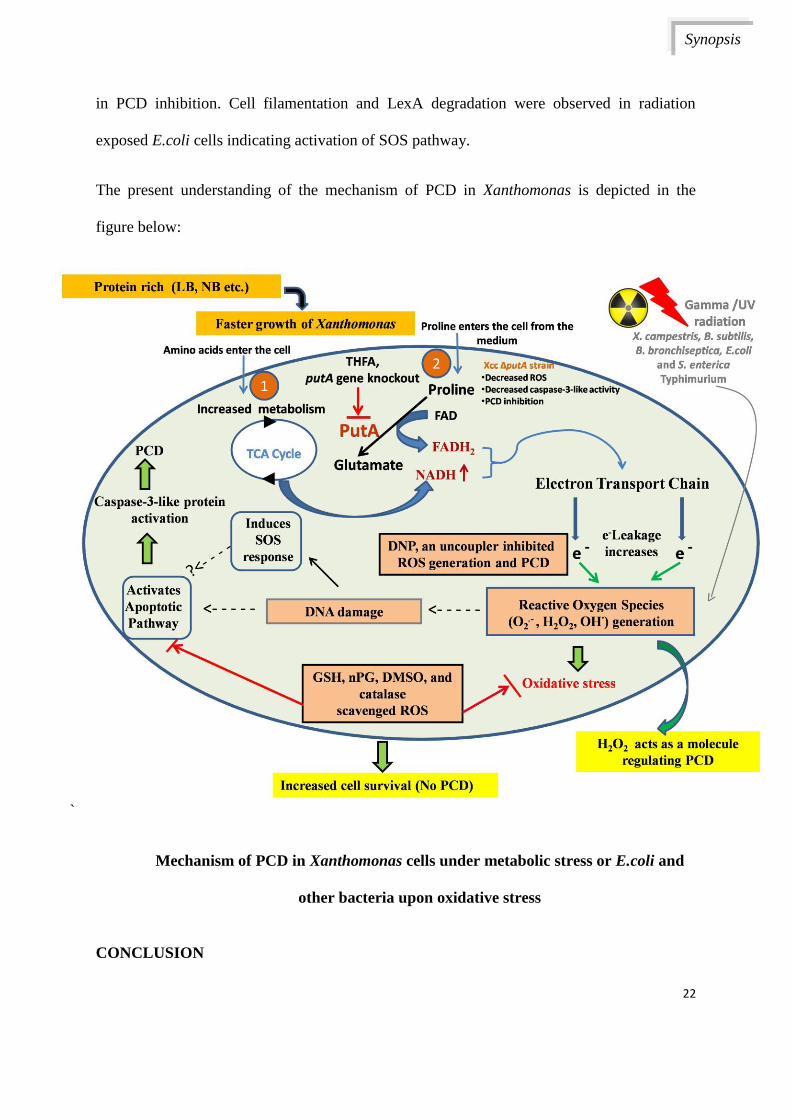
Enhanced level of NADH in Xanthomonas cells undergoing PCD exhibited metabolic
stress
Xanthomonas campestris pv. glycines (Xcg) grown in LB medium, hence after referred to as
PCD inducing medium or PIM, accumulated intracellular NADH and ATP. This was
revealed by comparative HPLC analysis of intracellular NADH and ATP levels in PIM and
PNIM grown cells. High intracellular NADH resulted in enhanced reactive oxygen species
(ROS) generation in cells grown in PIM as confirmed by 2’, 7’- dichlorohydrofluorescein
diacetate (H2DCFDA) labeling and ESR spectroscopy. This eventually resulted in the
activation of caspase-3-like protein in Xcg leading to PCD. ROS scavengers like
dimethylsulfoxide, glutathione, n-propyl gallate and catalase significantly inhibited PCD,
caspase biosynthesis as well as caspase-like enzyme activity in this organism. Enhanced ROS
level was conferred as one of the possible reasons contributing to caspase activation. This
was confirmed by the addition of an electron transport chain (ETC) uncoupler, 2, 4-
dinitrophenol, that reduced ROS generation and increased the cell survival. Thus, these
results indicated that Xcg cells grown in PIM experience metabolic stress leading to electron
leakage during electron transfer in ETC which leads to generation of ROS and subsequent
activation of caspase-3-like protein, resulting in PCD.
Role of proline oxidase (PutA) in regulating PCD in Xanthomonas
LB medium has glutamate (15%) and proline (6%) in abundance (BD Bionutrients technical
manual, 2006; Sezonov et al., 2007). PCD was induced in Xanthomonas when it was grown
Synopsis
20
in PNIM supplemented with proline. However, this was not the case when Xanthomonas was
grown in PNIM in the presence of glutamate. Proline being a secondary amino acid is not
metabolized by transaminases and carboxylases but is oxidized by PutA {also called as
proline oxidase (POX) or proline dehydrogenase (PRODH)} (Liu and Phang, 2012). PutA
converts proline to glutamate through an intermediate P5C. Notably, Xanthomonas cells
grown in PIM were found to accumulate proline and have higher proline oxidase activity.
Moreover, cells grown in PNIM in the presence of higher levels of proline were also found to
undergo PCD. Tetrahydro-2-furoic acid (5mM), an inhibitor of PutA, was found to prevent
PCD in PIM growing Xcc cells. ETC inhibitors rotenone and antimycin were also found to
inhibit PutA activity in Xcc. To further confirm the role of PutA in PCD, a putA knockout
was constructed. Interestingly, PCD was abolished in Xcc ΔputA cells, and the phenotype
could be further restored upon complementation with a plasmid vector carrying wild type
PutA (pPutA). Contrary to Xcc wt cells, Xcc ΔputA cells showed diminished ROS generation
and reduced caspase-3-like activity as well as PCD inhibition. Xcc wt cells also displayed
cell filamentation and in situ caspase-3-like activity when treated with a fluorophore tagged
caspase-3 inhibitor (FITC-DEVD-FMK). PCD markers like DNA damage (determined by
TUNEL assay), phosphatidylserine (PS) externalization and membrane depolarization were
significantly less in Xcc ΔputA cells with respect to wt cells. The findings indicate that the
oxidation of proline by PutA is one of the contributing factors leading to an increase in ROS
levels and PCD of stressed Xanthomonas cells.
PCD in other bacteria and involvement of ROS
To evaluate the conserved existence of PCD-like process in other bacteria besides
Xanthomonas, the study was performed in Bacillus subtilis, Bordetella bronchiseptica,
Escherichia coli and Salmonella enterica sv. Typhimurium. Radiation was used as a means to
Synopsis
21
generate ROS. Irradiating these bacteria at their respective D10 in the presence of Ac-DEVD-
CMK and 3-aminobenzamide, the cell permeable inhibitors of caspase-3 and poly (ADP
ribose) polymerase (PARP) respectively, increased the cell survival significantly. FACS
analysis indicated an increase in phosphatidylserine (PS) externalization in the radiation
exposed bacteria and was found to be reduced in the cells pre-incubated with the cell
permeable caspase-3 inhibitor. Radiation-induced SOS response in E.coli was also alleviated
in the presence of caspase-3 inhibitor as indicated by decrease in LexA degradation and
reduced cell filamentation frequency. This might indicate a probable linkage between SOS
response and PCD in radiation exposed E. coli cells and needs to be confirmed by additional
evidences.
SUMMARY
Xanthomonas campestris was observed to experience metabolic stress when grown in a
medium (like Luria Bertani broth) where amino acids are the predominant source of carbon
and nitrogen. Reactive oxygen species (ROS) were found to be produced in Xanthomonas
cells undergoing PCD in PIM (PCD inducing medium) and leakage of electrons from
electron transport chain (ETC) was found to be a possible source of ROS generation. PutA
was also found to be involved in PCD of Xanthomonas growing in PIM. PutA enzyme
activity was found to be linked to ETC resulting in ROS generation during oxidation of
proline and induction of PCD in Xanthomonas. Besides Xanthomonas, PCD was also
observed in other bacteria like, B.subtilis, B. bronchiseptica, E.coli and S. Typhimurium
when they were exposed to oxidative stress caused by γ-radiation. Bacteria undergoing
radiation induced cell death displayed PS externalization and activation of caspase-3-like
protein. The presence of cell permeable caspase-3 inhibitor inhibited these processes resulting
Synopsis
22
in PCD inhibition. Cell filamentation and LexA degradation were observed in radiation
exposed E.coli cells indicating activation of SOS pathway.
The present understanding of the mechanism of PCD in Xanthomonas is depicted in the
figure below:
`
Mechanism of PCD in Xanthomonas cells under metabolic stress or E.coli and
other bacteria upon oxidative stress
CONCLUSION
Synopsis

23
Metabolism associated stress in Xanthomonas cells was found to lead to ROS generation and
caspase dependent PCD during which involvement of PutA activity was observed as well.
Moreover, other triggers of oxidative stress like radiation treatment also elicited a similar
response in different bacteria.
REFERENCES
1. Alexeyev MF (1999) Biotechniques 26: 824-826
2. Bates LS, et al. (1973) Plant soil 39: 205-207
3. Bayles KW (2014) Nat Rev Microbiol 12:63-69
4. BD Bionutrients technical manual (2006) Becton, Dickinson and Company, USA. 53
p
5. Caruso R, et al. (2004) Anal Biochem 330: 43-51
6. Dendinger S, Brill WJ (1970) J Bacteriol 103: 144-152
7. Garcı́a-Ochoa et al. (2000) Biotechnol Adv 18: 549-579
8. Gautam S, Sharma A (2002a) J Gen Appl Microbiol 48: 67-76
9. Gautam S, Sharma A (2002b) Mol Microbiol 44: 393-401
10. Gautam S, Sharma A (2005) Daya Publishing House, India, pp 122-157
11. Gautam S, et al. (2005) Research Sign Post, India, pp 1-39
12. Giannattasio S, et al. (2003) Brain Res Protoc 10: 168-174
13. Kovach ME, et al. (1995) Gene 166: 175-176
14. Liu W, Phang JM (2012) Biofactors 38: 398-406
15. Raju KK, et al. (2006) J Bacteriol 188: 5408-5416
16. Sezonov G, et al. (2007) J Bacteriol 189: 8746-8749
Publications in Refereed Journal:
Synopsis

24
a. Published
(i) Journals
Wadhawan S, Gautam S, Sharma A. Involvement of Proline Oxidase (PutA) in
Programmed Cell Death of Xanthomonas. PLoS ONE, 2014, 9(5):e96423.
Wadhawan S, Gautam S, Sharma A. Bacteria undergo programmed cell death upon
low dose gamma radiation exposure. 2014, Int.J.Curr.Microbiol.App.Sci. 3(12):276-
283.
Wadhawan S, Gautam S, and Sharma A. A component of gamma radiation induced
cell death in E. coli is programmed and interlinked with activation of caspase-3 and
SOS response. Archives of Microbiology, 2013, 195(8):545-57.
Wadhawan S, Gautam S, and Sharma A. Metabolic stress induced programmed cell
death in Xanthomonas. FEMS Microbiol. Letters., 2010, 312 (2):176-183.
(ii) BARC Newsletter
Wadhawan S, Gautam S., and Sharma, A. Radiation induced cell death in
bacteria is partially programmed. BARC Newsletter. Oct. 2013, 348-354.
b. Accepted: Four (published in journals)
c. Communicated: Nil
d. Other Publications: Four (Symposia and Newsletter)
Symposium/ Conference
Wadhawan S, Gautam S., and Sharma, A. Radiation induced cell death in bacteria is
partially programmed. XXXVI All India Cell Biology Conference and International
Symposium on “Stress Adaptive Response and Genome Integrity (SARGI)”,
Mumbai, Oct., 2012, 130 (Best poster award).
Synopsis

25
Sharma A, Gautam S, and Wadhawan S. Reactive oxygen species: Role in bacterial
Programmed Cell Death. The International conference SFRR 2011, Recent Trends in
Therapeutic Advancement of Free Radical Science, Chennai, Jan. 2011, 44.
Wadhawan S, Gautam S, and Sharma A. Involvement of oxidative stress during
programmed cell death in Xanthomonas. Proceedings of “50th Annual Conference of
Association of Microbiologists of India (Third Golden Era of Microbiology)” Pune,
Dec. 2009, 175.
Signature of Student:
Date:
Doctoral committee
S. No. Name Designation Signature Date
1. Dr. S. K. Apte Chairman
2. Dr. A. K. Sharma Guide & Convener
3. Dr. S Gautam Co-Guide
4. Dr. Vinay Kumar Member
5. Dr. H. S. Misra Member
6. Dr. Santosh Kumar Member
Synopsis