synergistic enhancement of nk cell-mediated cytotoxicity by combination of histone deacetylase...
TRANSCRIPT

RESEARCH Open Access
Synergistic enhancement of NK cell-mediatedcytotoxicity by combination of histonedeacetylase inhibitor and ionizing radiationCheol-Hun Son6*, Jin-Hee Keum1*, Kwangmo Yang6, Jiho Nam3, Mi-Ju Kim1,2, Sun-Hee Kim1,2, Chi-Dug Kang1,Sae-Ock Oh2,4, Chi-Dae Kim2,5, You-Soo Park6* and Jaeho Bae1,2*
Abstract
Background: The overexpression of histone deacetylase (HDAC) and a subsequent decrease in the acetylationlevels of nuclear histones are frequently observed in cancer cells. Generally it was accepted that the deacetylationof histones suppressed expression of the attached genes. Therefore, it has been suggested that HDAC mightcontribute to the survival of cancer cells by altering the NKG2D ligands transcripts. By the way, the translationalregulation of NKG2D ligands remaines unclear in cancer cells. It appears the modulation of this unclear mechanismcould enhance NKG2D ligand expressions and the susceptibility of cancer cells to NK cells. Previously, it wasreported that irradiation can increase the surface expressions of NKG2D ligands on several cancer cell types withoutincreasing the levels of NKG2D ligand transcripts via ataxia telangiectasia mutated and ataxia telangiectasia andRad3 related (ATM-ATR) pathway, and suggested that radiation therapy might be used to increase the translation ofNKG2D ligands.
Methods: Two NSCLC cell lines, that is, A549 and NCI-H23 cells, were used to investigate the combined effects ofionizing radiation and HDAC inhibitors on the expressions of five NKG2D ligands. The mRNA expressions of theNKG2D ligands were quantitated by multiplex reverse transcription-PCR. Surface protein expressions were measuredby flow cytometry, and the susceptibilities of cancer cells to NK cells were assayed by time-resolved fluorometryusing the DELFIA® EuTDA cytotoxicity kit and by flow cytometry.
Results: The expressions of NKG2D ligands were found to be regulated at the transcription and translation levels.Ionizing radiation and HDAC inhibitors in combination synergistically increased the expressions of NKG2D ligands.Furthermore, treatment with ATM-ATR inhibitors efficiently blocked the increased translations of NKG2D ligandsinduced by ionizing radiation but did not block the increased ligand translations induced by HDAC inhibitors. Thestudy confirms that increased NKG2D ligand levels by ionizing radiation and HDAC inhibitors could synergisticallyenhance the susceptibilities of cancer cells to NK-92 cells.
Conclusions: This study suggests that the expressions of NKG2D ligands are regulated in a complex manner at themultilevel of gene expression, and that their expressions can be induced by combinatorial treatments in lungcancer cells.
Keywords: NKG2D ligands, HDAC inhibitors, Ionizing radiation, Radioresistance
* Correspondence: [email protected]; [email protected]; [email protected]; [email protected] Research Center, Dongnam Institute of Radiological and MedicalSciences, Busan 619-953, South Korea1Department of Biochemistry, Pusan National University School of Medicine,Yangsan 626-870, South Korea2Medical Research Center for Ischemic Tissue regeneration, Pusan NationalUniversity, Busan 609-735, South KoreaFull list of author information is available at the end of the article
© 2014 Son et al.; licensee BioMed Central Ltd. This is an open access article distributed under the terms of the CreativeCommons Attribution License (http://creativecommons.org/licenses/by/2.0), which permits unrestricted use, distribution, andreproduction in any medium, provided the original work is properly cited.
Son et al. Radiation Oncology 2014, 9:49http://www.ro-journal.com/content/9/1/49
BackgroundIt is well known NK cells play a role in immune surveil-lance for cancer [1] and that their anticancer immunityis controlled by a balance of activating and inhibitorysignals [2]. NKG2D is a well characterized immunore-ceptor which mediates activating signals on NK cells andT cell subsets, such as, CD8+ and γδT lymphocytes [3].In humans, eight distinct NKG2D ligands, includingMHC class I chain-related gene A/B(MICA/B) andUL16-binding protein 1–6 (ULBP1-6 or RAET1I,H,N,E,G and L), have been described [4]. Furthermore, theinduction of NKG2D ligands by several methods, in-cluding treatment with anti-cancer drugs, ionizing ra-diation, heat shock, or proteasomal inhibition, has beenproposed as a strategy for eliciting anti-cancer immunity[5-8]. Radiotherapy is a widely used modality to treatcancer; it causes double-strand DNA breaks, and thus,induces cancer cell death. Although it has been reportedthat ionizing radiation can induce NKG2D ligands oncancer cells by activating the ATM-ATR pathway [9],the precise regulatory mechanism involved is unclear. Ofthe recently developed anti-cancer agents, HDAC inhibi-tors have been investigated in treatment of cancers, andit has been reported that several HDAC inhibitors, in-cluding suberoylanilide hydroxamic acid (SAHA), tricos-tatin A (TSA), valproic acid, and PCI-24781, enhancethe radiosensitivities of cancer cells [10-13]. BecauseHDAC inhibitors are known potent inducers of NKG2Dligands on many cancer cells [14,15], it is possible thatthe induced NKG2D ligands could overcome immunetolerance and make cancer cells sensitive to NK-cell me-diated cytotoxicity. Accordingly, we investigated whetherionizing radiation in combination with HDAC inhibitortreatment increases the expressions of NKG2D ligands,and ATM-ATR signaling is involved in this process, andthis expressional increases enhances the susceptibility ofcancer cell to NK cells.
Materials and methodsCell lines and reagentsTwo human non-small cell lung cancer cell lines, A549 andNCI-H23, were used in this study, and were obtained fromthe Korean Cell Line Bank (Seoul, Korea). These cell lineswere maintained in RPMI media supplemented with 10%fetal bovine serum (FBS) (Gibco, Grand Island, NY),2 mM L-glutamine, 100 μg/ml streptomycin, and 100 U/mlpenicillin. The NK-92 cell line was obtained from theAmerican Type Culture Collection (Rockville, MD, USA)and maintained in alpha-Minimum Essential Modifiedmedium supplemented with 12.5% (v/v) fetal bovine serum,12.5% (v/v) horse serum, 2 mM L-glutamine,0.1 mM2-mercaptoethanol, 200 U/mL of recombinant humaninterleukin-2, 100 μg/mL streptomycin, and 100U/mLpenicillin. All cells were cultured at 37°C in a humidified
atmosphere containing 5% CO2.Three HDAC inhibitors,apicidin, suberoylanilide hydroxamic acid (SAHA; vorino-stat) and tricostatin A (TSA), two ATM-ATR inhibitors,caffeine, and KU-55933, cycloheximide (CHX) were pur-chased from Sigma-Aldrich (St. Louis, MO, USA). To ir-radiate cancer cells, we used a ClinaciX Linear Accelerator(Varian Medical Systems, Inc. Palo Alto, CA, USA) withthe assistance of Dr. Jiho Nam (Pusan National UniversityYangsan Hospital).
Total RNA extraction and Multiplex Reverse Transcription(RT)-PCRTotal RNA extraction and RT-PCR were performed aspreviously described [16]. Briefly, total RNA was extractedfrom cells using the RNeasy® Mini Kit (Qiagen GmbH,Germany). One microgram of extracted total RNA wasused to synthesize cDNA using 100 pmol of randomprimers (Takara, Japan) and 100 U of M-MLV reversetranscriptase (Promega Co., Fitchburg, Wisconsin, USA).The resulting cDNA was used as template for PCR, whichwas conducted using the QIAGEN® Multiplex PCR Kit(Qiagen GmbH). Seven pairs of primer sets were used toinvestigate the expressions of the ribosomal protein L19(RPL19), MICA, MICB, ULBP1-3, and β-actin (ACTB)genes. ACTB and RPL19 were used as a loading controland a degradation marker, respectively. PCR productswere stained by ethidium bromide and separated by 2.0%agarose gel electrophoresis, and quantified using imageanalyzing software (Quantity One; Bio-Rad Laboratories,Inc., Hercules, CA, USA).
Flow cytometryTo determine the surface expressions of NKG2D ligandson cancer cells, the cells were incubated with mouseanti-MICA, anti-MICB, anti-ULBP1-3 (R&D systems,Minneapolis, MN, USA), anti-HLA-ABC (Clone W6/32,Serotec, Oxford, UK) or the corresponding isotype con-trols at 10 μg/ml and then incubated with goat anti-mouse-PE conjugated (BD Pharmingen Inc., San Diego,CA., USA). The analysis was performed on a FACS Sort®(Becton Dickinson, Mountain View, CA., USA) usingCell Quest software (Becton Dickinson), and cell surfaceexpressions were quantified using mean fluorescenceintensities (MFIs). Relative expression ratios were calcu-lated by dividing treated sample MFI by untreated sam-ple MFI without subtracting the MFI of the appropriateisotype control.
NK cell-mediated cytotoxicity assay using time-resolvedfluorometryNK cell-mediated cytotoxicity was determined using theDELFIA® EuTDA Cytotoxicity Reagents (PerkinElmer LifeSciences, Waltham, MA, USA), as described previously[17]. Briefly, target cells (1X10⁶ cells/ml) were incubated
Son et al. Radiation Oncology 2014, 9:49 Page 2 of 10http://www.ro-journal.com/content/9/1/49

with freshly prepared 10 μM BATDA (a fluorescenceenhancing ligand) in 2 ml of culture medium for 30 minat 37°C, and washed. Next, 100 μl of BATDA-labeledtarget cells (5,000 cells) were transferred into a round-bottom sterile plate and co-cultured with NK-92 cells for2 hours at effector/target ratios ranging from 2.5:1 to 10:1.After incubation, 20 μl of supernatant from each well wastransferred to the wells of flat-bottom 96 well plates.180 μl of europium (Eu) solution was then added to formhighly fluorescent and stable chelates (EuTDA), and thefluorescences of these chelates were measured by time-resolved fluorometry (Victor3, PerkinElmer). The percentof specific release was calculated using (experimentalrelease – spontaneous release)/(maximum release –spontaneous release) X 100(%). In blocking experiments,blocking anti-NKG2D mAb (R&D Systems) was pre-added to suspensions of NK-92 cells and incubated for30 min prior to co-cultured with target cells. All experi-ments were performed in triplicate.
NK cell-mediated cytotoxicity assay by flow cytometryFresh NK cells were obtained from normal healthy donorswith informed consent in accordance with the Declarationof Helsinki. Target cells (1X10⁵ cells/ml) were stainedusing the Vybrant® carboxyfluorescein diacetate, succini-midyl ester (CFSE) Cell Tracer Kit (Invitrogen, Eugene,OR, USA) and incubated with NK-92 cells or freshly iso-lated NK cells at selected effector/target ratios for 2 hoursin 5 ml round-bottomed tubes. These co-cultured targetcells and NK cells were then stained with 1 μg/ml propi-dium iodide (Sigma-Aldrich). The assay was performed onFACS Sort® (Becton Dickinson) by acquiring 3,000 targetcells. The percent if specific release was calculated by thenumber of PI+&CFSE+ cells/3,000 X 100 (%). All experi-ments were performed in triplicate.
Statistical analysisTo evaluate alterations in gene expression, the gene ex-pressions in treated cells were divided by those in un-treated controls (mean fold) and standard errors (SE)were calculated. To compare groups, we used the pairedStudent’s t-test. Statistical significance was accepted forp values < 0.05.
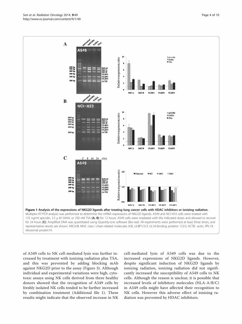
ResultsHDAC inhibitors increase the mRNA expressions ofNKG2D ligands but ionizing radiation minimally altersthese expressions in A549 cellsThe mRNA expressions of NKG2D ligands, MICA/Band ULBP1-3, were analyzed after treating A549 cellswith three HDAC inhibitors, that is, 125 ng/ml apicidin,2.5 μM SAHA, or 250 nM TSA, for 12 hours. Levels ofNKG2D ligands were significantly increased after treat-ment with two HDAC inhibitors, apicidin and TSA, in
A549 cells (Figure 1A). SAHA increased the mRNAlevels of only MICA and ULBP3. Although ULBP1 tendto increase, alterations of ULBP1 transcript levels werestatistically insignificant in apicidin or SAHA treatedA549 cells. The mRNA levels of the five NKG2D ligandswere also significantly increased after treatment with thethree HDAC inhibitors in NCI-H23 cells (Figure 1B).However, ionizing radiation did not significantly changethe mRNA levels of the five NKG2D ligands in A549cells (Figure 1C). Although previous reports have shownthat ionizing radiation significantly increases the mRNAand protein expressions of NKG2D ligands in severalcancer cells, including NCI-H23 cells [6], it was thoughtthat the induction of NKG2D ligands were differentfrom individual cancer cells. The reason for this lack ofresponsive in A549 cells to ionizing radiation is notclear.
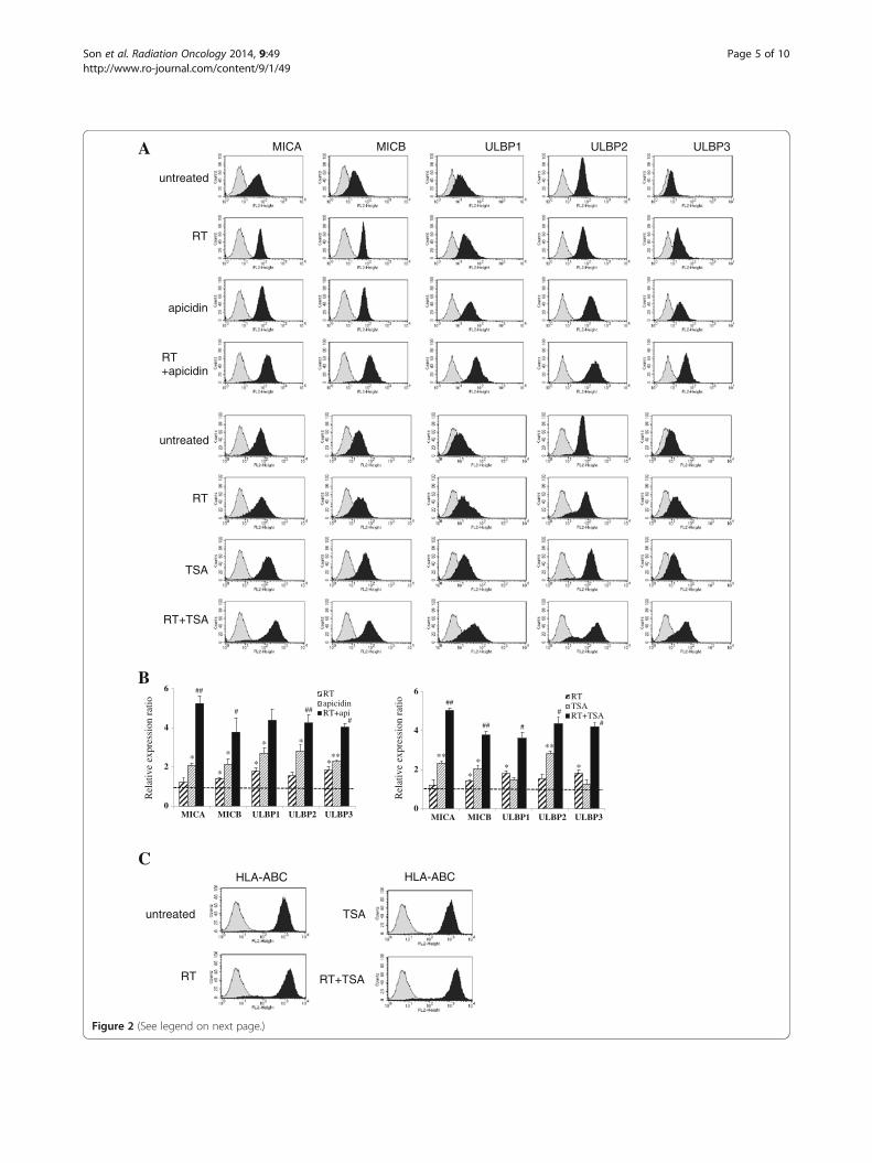
Combination of HDAC inhibitors and ionizing radiationprominently increases the surface expressions of NKG2DligandsFlow cytometric analysis showed that the cell surfaceprotein levels of the five NKG2D ligands were signifi-cantly increased after treatment with 125 ng/ml apicidinand 250 nM TSA for 18 hours following increased tran-scripts in A549 cells. Although alterations in the proteinlevels of MICA and ULBP2 were not significant, 8Gy ofirradiation and 24 hour-recovery incubation increasedthe surface protein expressions of NKG2D ligands des-pite little change in their mRNA levels. Relative ex-pression ratios were calculated by dividing the MFIratios of treated samples by those of untreated samples.These results suggested that the induction of NKG2Dligands was achieved at different levels of gene expres-sion by HDAC inhibitors and ionizing radiation differen-tially. When HDAC inhibitors plus ionizing radiationwere administered to A549 cells, surface NKG2D ligandprotein levels were further increased as compared withcells treated with HADC inhibitors or ionizing radiationonly (Figure 2A-B). We considered that this further in-duction of NKG2D ligands might be due to the promo-tion of transcription and post-transcription processes.On the other hand, the surface expression of HLA-ABC,which inhibits NK cells, was slightly increased after ion-izing radiation but was blocked by HDAC inhibitor(Figure 2C).
The susceptibility of A549 cells to NK cells issynergistically increased by HDAC inhibitors treatmentand ionizing radiation in combinationTo investigate whether treatment with HDAC inhibitorand irradiation increase the NK cell-mediated lysis ofcancer cells, cytotoxicity assays were performed usingDELFIA® EuTDA Cytotoxicity Reagents. The susceptibility
Son et al. Radiation Oncology 2014, 9:49 Page 3 of 10http://www.ro-journal.com/content/9/1/49
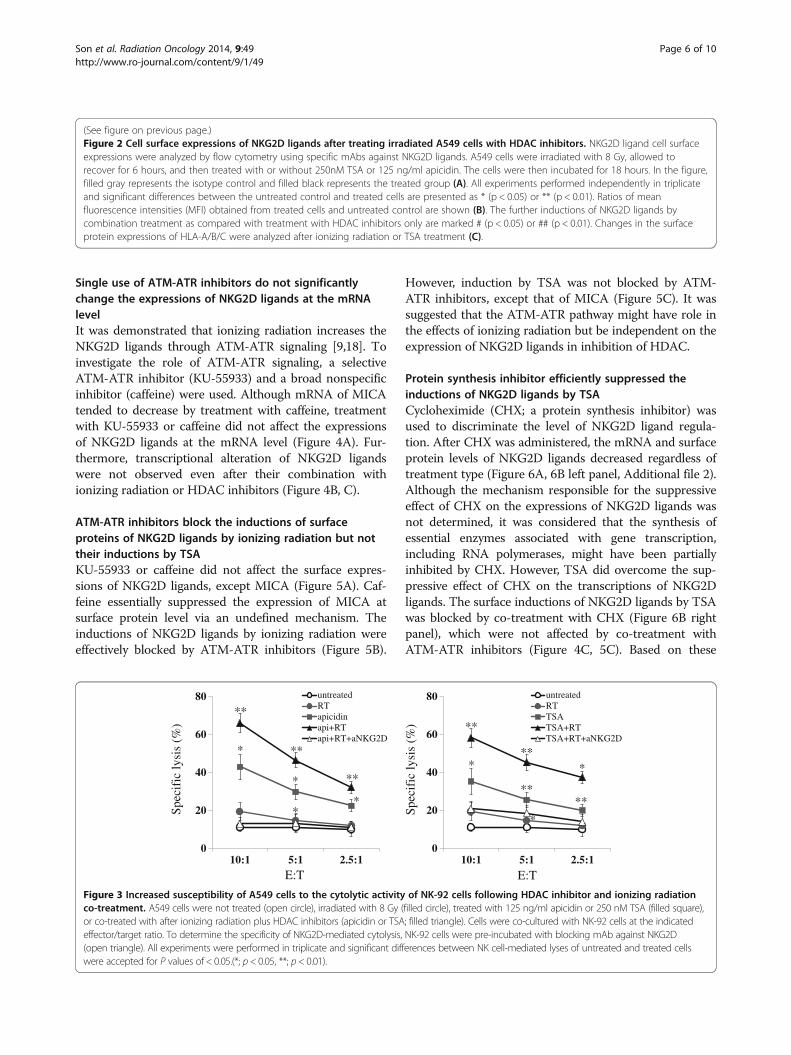
of A549 cells to NK cell-mediated lysis was further in-creased by treatment with ionizing radiation plus TSA,and this was prevented by adding blocking mAbagainst NKG2D prior to the assay (Figure 3). Althoughindividual and experimental variations were high, cyto-toxic assays using NK cells derived from three healthydonors showed that the recognition of A549 cells byfreshly isolated NK cells tended to be further increasedby combination treatment (Additional file 1). Theseresults might indicate that the observed increase in NK
cell-mediated lysis of A549 cells was due to theincreased expressions of NKG2D ligands. However,despite significant induction of NKG2D ligands byionizing radiation, ionizing radiation did not signifi-cantly increased the susceptibility of A549 cells to NKcells. Although the reason is unclear, it is possible thatincreased levels of inhibitory molecules (HLA-A/B/C)in A549 cells might have affected their recognition toNK cells. However this adverse effect of ionizing ra-diation was prevented by HDAC inhibitors.
A
B
C
Figure 1 Analysis of the expressions of NKG2D ligands after treating lung cancer cells with HDAC inhibitors or ionizing radiation.Multiplex RT-PCR analysis was performed to determine the mRNA expressions of NKG2D ligands. A549 and NCI-H23 cells were treated with125 ng/ml apicidin, 2.5 μ M SAHA, or 250 nM TSA (A, B) for 12 hours. A549 cells were irradiated with the indicated doses and allowed to recoverfor 24 hours (C). Amplified DNA was quantitated using Quantity-one software (Bio-rad). All experiments were performed at least three times, andrepresentative results are shown. MICA/B: MHC class I chain-related molecules A/B, ULBP1/2/3: UL16-binding proteins 1/2/3, ACTB: -actin, RPL19:ribosomal protein19.
Son et al. Radiation Oncology 2014, 9:49 Page 4 of 10http://www.ro-journal.com/content/9/1/49
A MICA MICB ULBP1 ULBP2 ULBP3
untreated
RT
apicidin
RT+apicidin
untreated
RT
TSA
RT+TSA
0
2
4
6
MICA MICB ULBP1 ULBP2 ULBP3
RTapicidinRT+api
0
2
4
6
MICA MICB ULBP1 ULBP2 ULBP3
RTTSART+TSA
****
** *
*
#
#
###
##
**
*
###
##
** *
***
#
Rel
ativ
e ex
pres
sion
rat
io
Rel
ativ
e ex
pres
sion
rat
io
B
CHLA-ABC
untreated
RT
TSA
RT+TSA
HLA-ABC
Figure 2 (See legend on next page.)
Son et al. Radiation Oncology 2014, 9:49 Page 5 of 10http://www.ro-journal.com/content/9/1/49
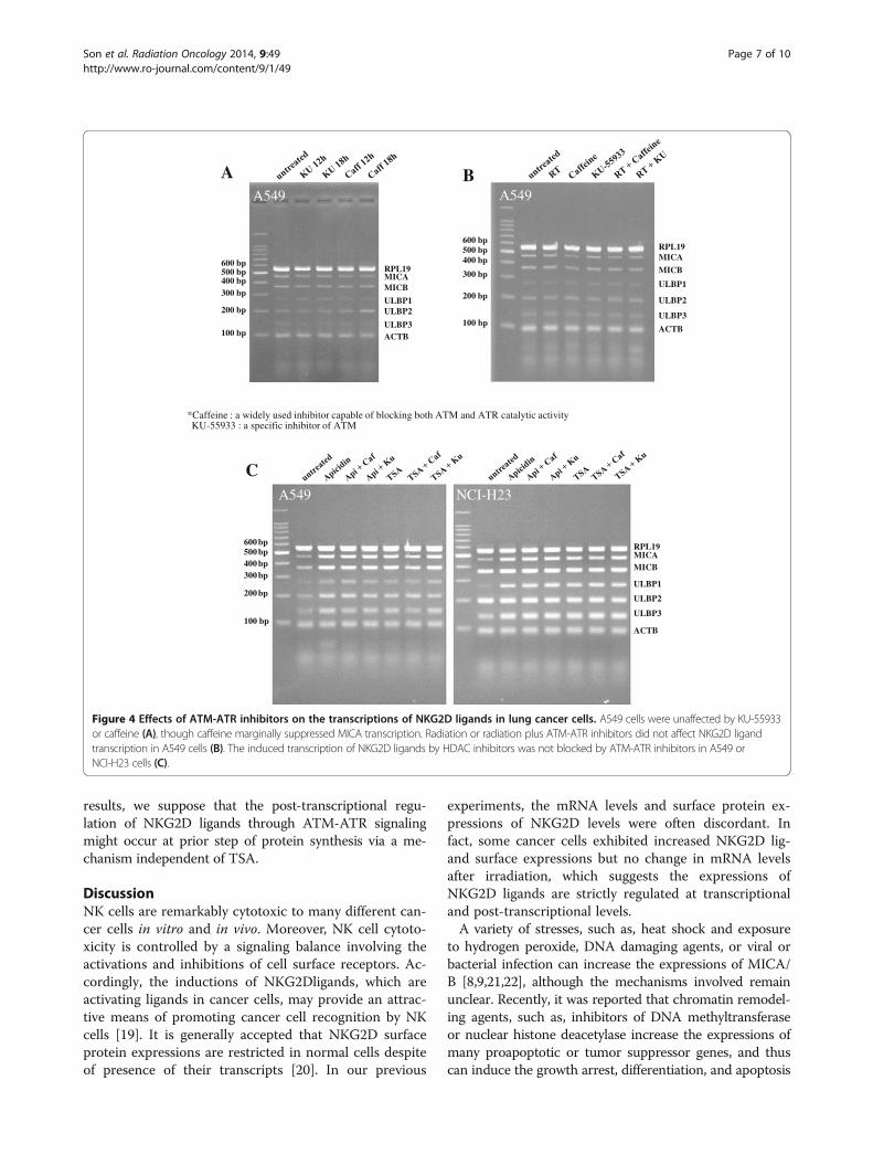
Single use of ATM-ATR inhibitors do not significantlychange the expressions of NKG2D ligands at the mRNAlevelIt was demonstrated that ionizing radiation increases theNKG2D ligands through ATM-ATR signaling [9,18]. Toinvestigate the role of ATM-ATR signaling, a selectiveATM-ATR inhibitor (KU-55933) and a broad nonspecificinhibitor (caffeine) were used. Although mRNA of MICAtended to decrease by treatment with caffeine, treatmentwith KU-55933 or caffeine did not affect the expressionsof NKG2D ligands at the mRNA level (Figure 4A). Fur-thermore, transcriptional alteration of NKG2D ligandswere not observed even after their combination withionizing radiation or HDAC inhibitors (Figure 4B, C).
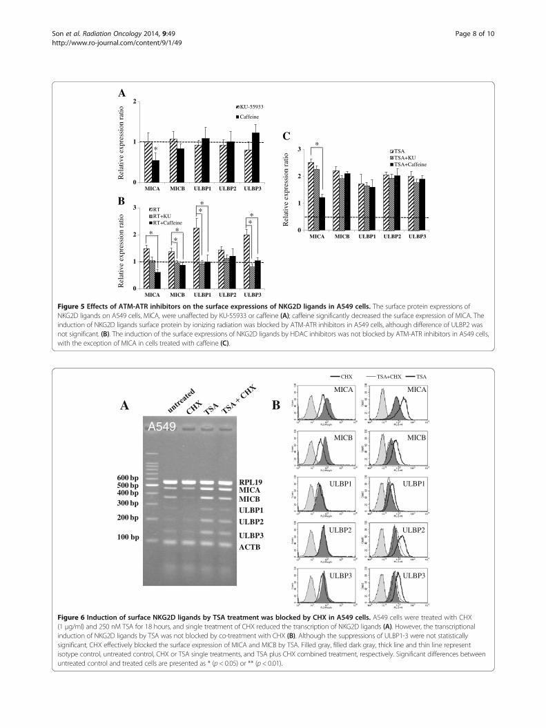
ATM-ATR inhibitors block the inductions of surfaceproteins of NKG2D ligands by ionizing radiation but nottheir inductions by TSAKU-55933 or caffeine did not affect the surface expres-sions of NKG2D ligands, except MICA (Figure 5A). Caf-feine essentially suppressed the expression of MICA atsurface protein level via an undefined mechanism. Theinductions of NKG2D ligands by ionizing radiation wereeffectively blocked by ATM-ATR inhibitors (Figure 5B).
However, induction by TSA was not blocked by ATM-ATR inhibitors, except that of MICA (Figure 5C). It wassuggested that the ATM-ATR pathway might have role inthe effects of ionizing radiation but be independent on theexpression of NKG2D ligands in inhibition of HDAC.
Protein synthesis inhibitor efficiently suppressed theinductions of NKG2D ligands by TSACycloheximide (CHX; a protein synthesis inhibitor) wasused to discriminate the level of NKG2D ligand regula-tion. After CHX was administered, the mRNA and surfaceprotein levels of NKG2D ligands decreased regardless oftreatment type (Figure 6A, 6B left panel, Additional file 2).Although the mechanism responsible for the suppressiveeffect of CHX on the expressions of NKG2D ligands wasnot determined, it was considered that the synthesis ofessential enzymes associated with gene transcription,including RNA polymerases, might have been partiallyinhibited by CHX. However, TSA did overcome the sup-pressive effect of CHX on the transcriptions of NKG2Dligands. The surface inductions of NKG2D ligands by TSAwas blocked by co-treatment with CHX (Figure 6B rightpanel), which were not affected by co-treatment withATM-ATR inhibitors (Figure 4C, 5C). Based on these
(See figure on previous page.)Figure 2 Cell surface expressions of NKG2D ligands after treating irradiated A549 cells with HDAC inhibitors. NKG2D ligand cell surfaceexpressions were analyzed by flow cytometry using specific mAbs against NKG2D ligands. A549 cells were irradiated with 8 Gy, allowed torecover for 6 hours, and then treated with or without 250nM TSA or 125 ng/ml apicidin. The cells were then incubated for 18 hours. In the figure,filled gray represents the isotype control and filled black represents the treated group (A). All experiments performed independently in triplicateand significant differences between the untreated control and treated cells are presented as * (p < 0.05) or ** (p < 0.01). Ratios of meanfluorescence intensities (MFI) obtained from treated cells and untreated control are shown (B). The further inductions of NKG2D ligands bycombination treatment as compared with treatment with HDAC inhibitors only are marked # (p < 0.05) or ## (p < 0.01). Changes in the surfaceprotein expressions of HLA-A/B/C were analyzed after ionizing radiation or TSA treatment (C).
0
20
40
60
80
10:1 5:1 2.5:1
untreatedRTTSATSA+RTTSA+RT+aNKG2D
0
20
40
60
80
10:1 5:1 2.5:1
untreatedRTapicidinapi+RTapi+RT+aNKG2D
*
*
*
****
*
**
**
**
**
**
***
Spec
ific
lysi
s (%
)
Spec
ific
lysi
s (%
)
E:T E:T
Figure 3 Increased susceptibility of A549 cells to the cytolytic activity of NK-92 cells following HDAC inhibitor and ionizing radiationco-treatment. A549 cells were not treated (open circle), irradiated with 8 Gy (filled circle), treated with 125 ng/ml apicidin or 250 nM TSA (filled square),or co-treated with after ionizing radiation plus HDAC inhibitors (apicidin or TSA; filled triangle). Cells were co-cultured with NK-92 cells at the indicatedeffector/target ratio. To determine the specificity of NKG2D-mediated cytolysis, NK-92 cells were pre-incubated with blocking mAb against NKG2D(open triangle). All experiments were performed in triplicate and significant differences between NK cell-mediated lyses of untreated and treated cellswere accepted for P values of < 0.05.(*; p < 0.05, **; p< 0.01).
Son et al. Radiation Oncology 2014, 9:49 Page 6 of 10http://www.ro-journal.com/content/9/1/49
results, we suppose that the post-transcriptional regu-lation of NKG2D ligands through ATM-ATR signalingmight occur at prior step of protein synthesis via a me-chanism independent of TSA.
DiscussionNK cells are remarkably cytotoxic to many different can-cer cells in vitro and in vivo. Moreover, NK cell cytoto-xicity is controlled by a signaling balance involving theactivations and inhibitions of cell surface receptors. Ac-cordingly, the inductions of NKG2Dligands, which areactivating ligands in cancer cells, may provide an attrac-tive means of promoting cancer cell recognition by NKcells [19]. It is generally accepted that NKG2D surfaceprotein expressions are restricted in normal cells despiteof presence of their transcripts [20]. In our previous
experiments, the mRNA levels and surface protein ex-pressions of NKG2D levels were often discordant. Infact, some cancer cells exhibited increased NKG2D lig-and surface expressions but no change in mRNA levelsafter irradiation, which suggests the expressions ofNKG2D ligands are strictly regulated at transcriptionaland post-transcriptional levels.
A variety of stresses, such as, heat shock and exposureto hydrogen peroxide, DNA damaging agents, or viral orbacterial infection can increase the expressions of MICA/B [8,9,21,22], although the mechanisms involved remainunclear. Recently, it was reported that chromatin remodel-ing agents, such as, inhibitors of DNA methyltransferaseor nuclear histone deacetylase increase the expressions ofmany proapoptotic or tumor suppressor genes, and thuscan induce the growth arrest, differentiation, and apoptosis
*Caffeine : a widely used inhibitor capable of blocking both ATM and ATR catalytic activityKU-55933 : a specific inhibitor of ATM
A B
ULBP2
RPL19
MICB
ULBP1
ULBP3
ACTB
MICA
600 bp500 bp400 bp
300 bp
200 bp
100 bp
A549
ULBP2
RPL19
MICB
ULBP1
ULBP3ACTB
MICA
600 bp500 bp400 bp300 bp
200 bp
100 bp
A549
A549 NCI-H23
ULBP2
RPL19
MICB
ULBP1
ULBP3
ACTB
MICA
600 bp500 bp400 bp300 bp
200 bp
100 bp
C
Figure 4 Effects of ATM-ATR inhibitors on the transcriptions of NKG2D ligands in lung cancer cells. A549 cells were unaffected by KU-55933or caffeine (A), though caffeine marginally suppressed MICA transcription. Radiation or radiation plus ATM-ATR inhibitors did not affect NKG2D ligandtranscription in A549 cells (B). The induced transcription of NKG2D ligands by HDAC inhibitors was not blocked by ATM-ATR inhibitors in A549 orNCI-H23 cells (C).
Son et al. Radiation Oncology 2014, 9:49 Page 7 of 10http://www.ro-journal.com/content/9/1/49
0
1
2
MICA MICB ULBP1 ULBP2 ULBP3
KU-55933
Caffeine
*
Rel
ativ
e ex
pres
sion
rat
io
*
0
1
2
3
MICA MICB ULBP1 ULBP2 ULBP3
RTRT+KURT+Caffeine
Rel
ativ
e ex
pres
sion
rat
io
* **
**
**
0
1
2
3
MICA MICB ULBP1 ULBP2 ULBP3
TSATSA+KUTSA+Caffeine
Rel
ativ
e ex
pres
sion
rat
io
*
B
A
C
Figure 5 Effects of ATM-ATR inhibitors on the surface expressions of NKG2D ligands in A549 cells. The surface protein expressions ofNKG2D ligands on A549 cells, MICA, were unaffected by KU-55933 or caffeine (A); caffeine significantly decreased the surface expression of MICA. Theinduction of NKG2D ligands surface protein by ionizing radiation was blocked by ATM-ATR inhibitors in A549 cells, although difference of ULBP2 wasnot significant. (B). The induction of the surface expressions of NKG2D ligands by HDAC inhibitors was not blocked by ATM-ATR inhibitors in A549 cells,with the exception of MICA in cells treated with caffeine (C).
ULBP2
RPL19
MICBULBP1
ULBP3
ACTB
MICA
600 bp500 bp400 bp300 bp
200 bp
100 bp
A549
A BMICA
MICB
ULBP1
ULBP2
ULBP3
MICA
MICB
ULBP1
ULBP2
ULBP3
CHX TSA+CHX TSA
Figure 6 Induction of surface NKG2D ligands by TSA treatment was blocked by CHX in A549 cells. A549 cells were treated with CHX(1 μg/ml) and 250 nM TSA for 18 hours, and single treatment of CHX reduced the transcription of NKG2D ligands (A). However, the transcriptionalinduction of NKG2D ligands by TSA was not blocked by co-treatment with CHX (B). Although the suppressions of ULBP1-3 were not statisticallysignificant, CHX effectively blocked the surface expression of MICA and MICB by TSA. Filled gray, filled dark gray, thick line and thin line representisotype control, untreated control, CHX or TSA single treatments, and TSA plus CHX combined treatment, respectively. Significant differences betweenuntreated control and treated cells are presented as * (p < 0.05) or ** (p < 0.01).
Son et al. Radiation Oncology 2014, 9:49 Page 8 of 10http://www.ro-journal.com/content/9/1/49

of cancer cells [23-25]. In addition, the expressions ofNKG2D ligands have been reported to be upregulated atthe transcription level in some cancer cells [14,15,26]. Onthe other hand, DNA damaging agents usually increasethe surface expressions of NKG2D ligands at post-tran-scriptional level in macrophage through ATM-ATR sig-naling [18]. Therefore, to further increase the expressionof NKG2D ligands in cancer cells, we co-treated cells withionizing radiation and HDAC inhibitors. We presumedthat HDAC inhibitors increase the transcription and ioni-zing radiation increases the translation of NKG2D ligandsvia different mechanisms.
The lung adenocarcinoma cell line A549 is resistant toionizing radiation and to cell-mediated killing [27,28]. Inthe present study, we found that ionizing radiation did notsignificantly increase NKG2D ligand transcript expressionin this cell line, but it did increase their protein levels. Onthe other hand, ionizing radiation significantly increasedthe expressions of NKG2D ligands at the mRNA and pro-tein levels in NCI-H23 cells (a radiosensitive lung adeno-carcinoma cell line). Although we did not investigate thereason for the different responses to ionizing radiation ofthese two lung cancer cells, it has been shown that theyexhibit different p53 activities [29]. Accordingly, our fin-dings suggest that ionizing radiation and HDAC inhibitorco-treatment increase NKG2D ligand expression and en-hance the susceptibility of cancer cells to NK-92 cells andfreshly isolated NK cells (Figure 3 and Additional file 1).To examine the effects of ionizing radiation and ofHDAC inhibitor treatment separately, we inhibitedATM-ATR signaling, which is activated by ionizing ra-diation and increased the NKG2D ligand expression [9].We choose two ATM-ATR inhibitors, that is, caffeineand KU-55933, and pretreated cancer cells with theseinhibitors prior to administering ionizing radiation orHDAC inhibitors. ATM-ATR inhibitors effectivelyblocked the induction of NKG2D ligands by ionizingradiation. However, not by HDAC inhibitors exceptMICA. These findings show that ionizing radiation andHDAC inhibitors differentially affect the ATM-ATRpathway and NKG2D ligand expression. More speci-fically, caffeine suppressed the expression of MICA atthe surface protein level (Figure 5), and although themechanism of MICA down-regulation by caffeine is notknown, it has been reported that MICA transcription isreduced via the inhibitions of PI3K and PKC, which re-gulators of MICA transcription [17,30] and caffeinemight affect the PI3K and PKC activities. CHX (aninhibitor of protein synthesis) treatment effectivelyblocked NKG2D ligand induction by HDAC inhibitors.We are of the opinion that ATM-ATR signaling pro-bably does not increase the protein synthesis of NKG2Dligands but rather promotes their translation at a priorstep of protein synthesis.
In previous studies, post-transcriptional and -translationalregulations were found to be involved in the control of thesurface protein levels of NKG2D ligands, and discre-pancies between the transcription and surface NKG2D ex-pressions of ligands have often been described [31-33].We found that co-treatment with ionizing radiation andHDAC inhibitors further increases NKG2D ligand expres-sions via independent mechanisms in lung cancer cells.BecauseA549 cells did not response to ionizing radiationwith respect to the transcriptions of NKG2D ligands andthese cells were essentially less susceptible to NK cells, itwould appear in this cell-line that by ionizing radiation inthe inductions of the surface protein expressions ofNKG2D ligands were limited. Although radioresistantlung cancer cells, such as, A549 cells, survive even high-doses irradiation, it appears that co-treatment with ioni-zing radiation and HDAC inhibitors might be helpful.
ConclusionsThis study suggests NKG2D ligands are regulated in acomplex, multi-level manner and that they can be inducedby ionizing radiation plus HDAC inhibitors in lung cancercells. We believe that such combination therapies offer anattractive means of improving the efficacy of NK cell-based cancer immunotherapy in patients with radioresis-tant cancer.
Additional files
Additional file 1: Increased susceptibility of A549 cells to thecytolytic activities of fresh isolated NK cells after treatment withionizing radiation plus TSA. A549 cells were co-cultured with NK-92cells or freshly isolated NK cells, the latter of which were obtained fromthree healthy donors after obtaining informed consent, at the indicatedeffector/target ratio. The cytotoxicity assay was performed by using flowcytometry and representative results were shown (A). Cytotoxicity assayresults were shown as marks (B). untreated (open circle), or irradiatedwith 8 Gy (filled circle), with 250 nM TSA (filled square), or with RT plusTSA (filled triangle). All experiments were performed in triplicate andsignificant differences between NK cell-mediated lyses of untreated andtreated cells were accepted for P values of <0.05.(* ;P < 0.05).
Additional file 2: Blockade of the radiation-induced surface expressionsof NKG2D ligands by CHX in A549 cells. A549 cells were irradiated with8 Gy, allowed to recover for 6 hours, and then treated with or without250nM TSA or 125 ng/ml apicidin. Cells were then incubated for18 hours. Filled gray represents the isotype control, filled dark gray theuntreated control, the thick line represents irradiated cells, and the thinline represents ionizing radiation plus CHX treated cells.
Competing interestsThe authors have no conflict of interest to declare.
Authors’ contributionsCHS and JHK carried out the studies and participated in experiments. SHK,CDK, SOO and CDK participated in the study design and helped to draft themanuscript. KY and JN conducted the irradiation experiments. MJKperformed the statistical analysis. All authors read and approved the finalmanuscript.
Authors’ informationCo-first authors: Cheol-Hun Son and Jin-Hee Keum.
Son et al. Radiation Oncology 2014, 9:49 Page 9 of 10http://www.ro-journal.com/content/9/1/49
AcknowledgementsThis research was supported by the National R&D Program through theDong-nam Institute of Radiological & Medical Sciences (DIRAMS) funded bythe Ministry of Education, Science and Technology (code: 50590-2013) andthe National Research Foundation of Korea (NRF) grant funded by the Koreagovernment(MSIP) (2005-0049416), and Pusan National University ResearchGrant, 2010, Korea.
Author details1Department of Biochemistry, Pusan National University School of Medicine,Yangsan 626-870, South Korea. 2Medical Research Center for Ischemic Tissueregeneration, Pusan National University, Busan 609-735, South Korea.3Department of Radiation Oncology, Pusan National University YangsanHospital, Yangsan 626-770, South Korea. 4Department of Anatomy, PusanNational University School of Medicine, Yangsan 626-870, South Korea.5Department of Pharmacology, Pusan National University School of Medicine,Yangsan 626-870, South Korea. 6Medical Research Center, Dongnam Instituteof Radiological and Medical Sciences, Busan 619-953, South Korea.
Received: 20 July 2013 Accepted: 6 December 2013Published: 10 February 2014
References1. Schmitt C, Ghazi B, Bensussan A: NK cells and surveillance in humans.
Reprod Biomed Online 2008, 16:192–201.2. Lanier LL: NK cell recognition. Annu Rev Immunol 2005, 23:225–274.3. Bauer S, Groh V, Wu J, Steinle A, Phillips JH, Lanier LL, Spies T: Activation of
NK cells and T cells by NKG2D, a receptor for stress-inducible MICA.Science 1999, 285:727–729.
4. Champsaur M, Lanier LL: Effect of NKG2D ligand expression on hostimmune responses. Immunol Rev 2010, 235:267–285.
5. Venkataraman GM, Suciu D, Groh V, Boss JM, Spies T: Promoter regionarchitecture and transcriptional regulation of the genes for the MHCclass I-related chain A and B ligands of NKG2D. J Immunol 2007,178:961–969.
6. Vales-Gomez M, Chisholm SE, Cassady-Cain RL, Roda-Navarro P, Reyburn HT:Selective induction of expression of a ligand for the NKG2D receptor byproteasome inhibitors. Cancer Res 2008, 68:1546–1554.
7. Butler JE, Moore MB, Presnell SR, Chan HW, Chalupny NJ, Lutz CT:Proteasome regulation of ULBP1 transcription. J Immunol 2009,182:6600–6609.
8. Kim JY, Son YO, Park SW, Bae JH, Chung JS, Kim HH, Chung BS, Kim SH,Kang CD: Increase of NKG2D ligands and sensitivity to NK cell-mediatedcytotoxicity of tumor cells by heat shock and ionizing radiation. Exp MolMed 2006, 38:474–484.
9. Gasser S, Orsulic S, Brown EJ, Raulet DH: The DNA damage pathwayregulates innate immune system ligands of the NKG2D receptor.Nature 2005, 436:1186–1190.
10. Chinnaiyan P, Vallabhaneni G, Armstrong E, Huang SM, Harari PM:Modulation of radiation response by histone deacetylase inhibition.Int J Radiat Oncol Biol Phys 2005, 62:223–229.
11. Yu J, Mi J, Wang Y, Wang A, Tian X: Regulation of radiosensitivity byHDAC inhibitor trichostatin A in the human cervical carcinoma cell lineHela. Eur J Gynaecol Oncol 2012, 33:285–290.
12. Shoji M, Ninomiya I, Makino I, Kinoshita J, Nakamura K, Oyama K,Nakagawara H, Fujita H, Tajima H, Takamura H, et al: Valproic acid, ahistone deacetylase inhibitor, enhances radiosensitivity in esophagealsquamous cell carcinoma. Int J Oncol 2012, 40:2140–2146.
13. Banuelos CA, Banath JP, MacPhail SH, Zhao J, Reitsema T, Olive PL:Radiosensitization by the histone deacetylase inhibitor PCI-24781.Clin Cancer Res 2007, 13:6816–6826.
14. Skov S, Pedersen MT, Andresen L, Straten PT, Woetmann A, Odum N:Cancer cells become susceptible to natural killer cell killing afterexposure to histone deacetylase inhibitors due to glycogen synthasekinase-3-dependent expression of MHC class I-related chain A and B.Cancer Res 2005, 65:11136–11145.
15. Armeanu S, Bitzer M, Lauer UM, Venturelli S, Pathil A, Krusch M, Kaiser S,Jobst J, Smirnow I, Wagner A, et al: Natural killer cell-mediated lysis ofhepatoma cells via specific induction of NKG2D ligands by the histonedeacetylase inhibitor sodium valproate. Cancer Res 2005, 65:6321–6329.
16. Park SW, Bae JH, Kim SD, Son YO, Kim JY, Park HJ, Lee CH, Park DY, Lee MK,Chung BS, et al: Comparison of level of NKG2D ligands between normaland tumor tissue using multiplex RT-PCR. Cancer Invest 2007, 25:299–307.
17. Bae JH, Kim JY, Kim MJ, Chang SH, Park YS, Son CH, Park SJ, Chung JS, Lee EY,Kim SH, et al: Quercetin enhances susceptibility to NK cell-mediated lysis oftumor cells through induction of NKG2D ligands and suppression ofHSP70. J Immunother 2010, 33:391–401.
18. Apetoh L, Ghiringhelli F, Tesniere A, Obeid M, Ortiz C, Criollo A, Mignot G,Maiuri MC, Ullrich E, Saulnier P, et al: Toll-like receptor 4-dependentcontribution of the immune system to anticancer chemotherapy andradiotherapy. Nat Med 2007, 13:1050–1059.
19. Ljunggren HG, Malmberg KJ: Prospects for the use of NK cells inimmunotherapy of human cancer. Nat Rev Immunol 2007, 7:329–339.
20. Eagle RA, Jafferji I, Barrow AD: Beyond Stressed Self: Evidence for NKG2DLigand Expression on Healthy Cells. Curr Immunol Rev 2009, 5:22–34.
21. Groh V, Rhinehart R, Randolph-Habecker J, Topp MS, Riddell SR, Spies T:Costimulation of CD8alphabeta T cells by NKG2D via engagement byMIC induced on virus-infected cells. Nat Immunol 2001, 2:255–260.
22. Borchers MT, Harris NL, Wesselkamper SC, Vitucci M, Cosman D: NKG2Dligands are expressed on stressed human airway epithelial cells. Am JPhysiol Lung Cell Mol Physiol 2006, 291:L222–L231.
23. Sandor V, Senderowicz A, Mertins S, Sackett D, Sausville E, Blagosklonny MV,Bates SE: P21-dependent g(1)arrest with downregulation of cyclin D1and upregulation of cyclin E by the histone deacetylase inhibitorFR901228. Br J Cancer 2000, 83:817–825.
24. Zhao Y, Tan J, Zhuang L, Jiang X, Liu ET, Yu Q: Inhibitors of histonedeacetylases target the Rb-E2F1 pathway for apoptosis inductionthrough activation of proapoptotic protein Bim. Proc Natl Acad Sci USA2005, 102:16090–16095.
25. Baylin SB, Esteller M, Rountree MR, Bachman KE, Schuebel K, Herman JG:Aberrant patterns of DNA methylation, chromatin formation and geneexpression in cancer. Hum Mol Genet 2001, 10:687–692.
26. Lopez-Soto A, Folgueras AR, Seto E, Gonzalez S: HDAC3 represses theexpression of NKG2D ligands ULBPs in epithelial tumour cells: potentialimplications for the immunosurveillance of cancer. Oncogene 2009,28:2370–2382.
27. Kim W, Youn H, Seong KM, Yang HJ, Yun YJ, Kwon T, Kim YH, Lee JY,Jin YW, Youn B: PIM1-activated PRAS40 regulates radioresistance innon-small cell lung cancer cells through interplay with FOXO3a, 14-3-3and protein phosphatases. Radiat Res 2011, 176:539–552.
28. Kim H, Kim SH, Kim MJ, Kim SJ, Park SJ, Chung JS, Bae JH, Kang CD: EGFRinhibitors enhanced the susceptibility to NK cell-mediated lysis of lungcancer cells. J Immunother 2011, 34:372–381.
29. Tetsuo M, Tomoko O, Shigeo S, Mikiko M, Yoshikazu S, Kanami Y, Jun-ichi H,Mitsuhiro T, Tetsuya M, Yuichi I, Yo K, Hiroshi T, Takao Y, Takashi T: p53-Defectivetumors with a functional apoptosome-mediated pathway: a new therapeutictarget. J Natl Cancer Inst 2005, 97:765–777.
30. Bae JH, Kim SJ, Kim MJ, Oh SO, Chung JS, Kim SH, Kang CD: Susceptibilityto natural killer cell-mediated lysis of colon cancer cells is enhanced bytreatment with epidermal growth factor receptor inhibitors throughUL16-binding protein-1 induction. Cancer Sci 2012, 103:7–16.
31. Groh V, Bahram S, Bauer S, Herman A, Beauchamp M, Spies T: Cell stress-regulated human major histocompatibility complex class I geneexpressed in gastrointestinal epithelium. Proc Natl Acad Sci USA 1996,93:12445–12450.
32. Molinero LL, Domaica CI, Fuertes MB, Girart MV, Rossi LE, Zwirner NW:Intracellular expression of MICA in activated CD4 T lymphocytes andprotection from NK cell-mediated MICA-dependent cytotoxicity.Hum Immunol 2006, 67:170–182.
33. Fuertes MB, Girart MV, Molinero LL, Domaica CI, Rossi LE, Barrio MM,Mordoh J, Rabinovich GA, Zwirner NW: Intracellular retention of theNKG2D ligand MHC class I chain-related gene A in human melanomasconfers immune privilege and prevents NK cell-mediated cytotoxicity.J Immunol 2008, 180:4606–4614.
doi:10.1186/1748-717X-9-49Cite this article as: Son et al.: Synergistic enhancement of NK cell-mediated cytotoxicity by combination of histone deacetylase inhibitorand ionizing radiation. Radiation Oncology 2014 9:49.
Son et al. Radiation Oncology 2014, 9:49 Page 10 of 10http://www.ro-journal.com/content/9/1/49