synchronous activation of gonadotropin-releasing … activation of gonadotropin-releasing hormone...
TRANSCRIPT

Synchronous activation of gonadotropin-releasinghormone gene transcription and secretionby pulsatile kisspeptin stimulationHan Kyoung Choea,b, Hee-Dae Kima,b, Sung Ho Parka,b, Han-Woong Leec, Jae-Yong Parkd, Jae Young Seonge,Stafford L. Lightmanf, Gi Hoon Sona,b,g, and Kyungjin Kima,b,1
aDepartment of Biological Sciences, Seoul National University, Seoul 151-742, Korea and bBrain Research Center for the 21st Frontier Program inNeuroscience, Seoul 151-742, Korea; cDepartment of Biochemistry, College of Life Science and Biotechnology, Yonsei University, Seoul 120-749, Korea;dDepartment of Physiology, Institute of Health Science and Medical Research Center for Neural Dysfunction, Biomedical Center (BK21), Gyeongsang NationalUniversity School of Medicine, Jinju 660-751, Korea; eGraduate School of Medicine and gDepartment of Legal Medicine, College of Medicine, KoreaUniversity, Seoul 136-705, Korea; and fHenry Wellcome Laboratories for Integrative Neuroscience and Endocrinology, University of Bristol, Bristol BS1 3NY,United Kingdom
Edited* by Joseph S. Takahashi, Howard Hughes Medical Institute, University of Texas Southwestern Medical Center, Dallas, TX, and approvedFebruary 28, 2013 (received for review August 6, 2012)
Pulsatile release of hypothalamic gonadotropin-releasing hormone(GnRH) is essential for pituitary gonadotrope function. Althoughthe importance of pulsatile GnRH secretion has been recognizedfor several decades, the mechanisms underlying GnRH pulse gener-ation in hypothalamic neural networks remain elusive. Here, wedemonstrate the ultradian rhythm of GnRH gene transcription insingle GnRH neurons using cultured hypothalamic slices preparedfrom transgenic mice expressing a GnRH promoter-driven desta-bilized luciferase reporter. Although GnRH promoter activity in eachGnRH neuron exhibited an ultradian pattern of oscillations with aperiod of ∼10 h, GnRH neuronal cultures exhibited partially syn-chronized bursts of GnRH transcriptional activity at ∼2-h intervals.Surprisingly, pulsatile administration of kisspeptin, a potent GnRHsecretagogue, evoked dramatic synchronous activation of GnRH genetranscription with robust stimulation of pulsatile GnRH secretion.We also addressed the issue of hierarchical interaction betweenthe circadian and ultradian rhythms by using Bmal1-deficient micewith defective circadian clocks. The circadian molecular oscillatorbarely affected basal ultradian oscillation of GnRH transcriptionbut was heavily involved in kisspeptin-evoked responses of GnRHneurons. In conclusion, we have clearly shown synchronous burstsof GnRH gene transcription in the hypothalamic GnRH neuronalpopulation in association with episodic neurohormone secretion,thereby providing insight into GnRH pulse generation.
biological rhythm | GnRH pulse generator | circadian rhythm |dynamic transcription
Diverse forms of biological oscillation are found in biochemicalreactions, cellular events, and physiological processes (1).
Intrinsic daily rhythms are generated by molecular circadianclockwork, which is based on transcription–translation feedbackloops composed of transcriptional activators such as the CLOCK:BMAL1 heterodimer and inhibitory factors including PERIODsand CRYPTOCHROMEs (2, 3). Dynamic regulation of the neu-roendocrine system occurs due to the integrative actions of multipletypes of biological oscillators. For example, corticosteroids in circu-lation exhibit robust circadian oscillation and hourly pulsatility (4).The pulsatile release of hypothalamic neurohormones into
the hypothalamic–pituitary portal vessels is a classic example ofultradian oscillation. Gonadotropin-releasing hormone (GnRH) isthe most extensively studied neurohormone. Knobil (5) elegantlydemonstrated that the pulsatile neurosecretion of GnRH is crucialfor normal reproductive function. Although the physiological im-portance of pulsatile GnRH secretion is recognized, the cellularmechanisms underlying GnRH pulse generation remain unclear.An intrinsic mechanism for generating pulsatile secretion withinindividual GnRH neurons may exist. Pulsatile GnRH release hasbeen demonstrated in immortalized GnRH-producing GT1 cells
and in embryonic GnRH neuronal cultures (6, 7). Intracellularcalcium oscillation (8), episodic gene expression coupled withexocytic activity (9, 10), voltage-dependent ion channel-mediatedsynchronization (11), and cellular circadian oscillators (12) havebeen implicated in pulsatile secretion of GnRH. Electrophysio-logical studies have suggested that a synchronization mechanismunderlies the autonomous pulse generation (13, 14). However, mostGnRH neuronal cell bodies are located in the preoptic area (POA)of the hypothalamus and direct contact between GnRH neuronalcell bodies is only occasionally found despite considerable dendro–dendritic interactions among them (14). Indeed, these anatomicalfeatures make it possible that scattered GnRH neurons formnetworks to coordinate their function along with neighboringnon-GnRH neurons.Kisspeptin, a neuropeptide encoded by the Kiss1 gene, is one
of the strongest secretagogues of GnRH and luteinizing hormone(LH) (15, 16). Mutations in the kisspeptin receptor (GPR54)gene are associated with hypogonadotropic hypogonadism (17).Kisspeptin regulates GnRH neurons during multiple processesincluding pubertal maturation and mammalian reproduction (15,17–19). Kisspeptin-producing cells reside in two distinct hypo-thalamic regions, the anteroventral periventricular nucleus and thearcuate nucleus, and extend their neurites adjacent to axon termi-nals and cell bodies of GnRH neurons (15, 18). Notably, kisspeptinrelease in the stalk-median eminence is pulsatile and exhibits astrong correlation with GnRH pulses (20). Furthermore, pulsatilekisspeptin administration drives gonadotropin secretion in juvenilemale monkeys primed with GnRH. However, continuous adminis-tration of kisspeptin abolishes gonadotropin secretion after an acutestimulatory effect, presumably owing to receptor desensitization(21–23). Thus, kisspeptin may participate in GnRH pulse genera-tion, although the underlying mechanisms remain elusive (24, 25).We used organotypic cultures of hypothalamic GnRH neurons
to elucidate the episodic pattern of GnRH gene expression andsecretion in response to kisspeptin stimulation. Further, we ex-amined the role of the molecular circadian clock in the ultradianrhythmicity of GnRH neurons.
Author contributions: G.H.S. and K.K. designed research; H.K.C., H.-D.K., S.H.P., and G.H.S.performed research; H.-W.L. contributed new reagents/analytic tools; H.K.C., J.-Y.P., J.Y.S.,and G.H.S. analyzed data; and H.K.C., S.L.L., G.H.S., and K.K. wrote the paper.
The authors declare no conflict of interest.
*This Direct Submission article had a prearranged editor.1To whom correspondence should be addressed. E-mail: [email protected].
This article contains supporting information online at www.pnas.org/lookup/suppl/doi:10.1073/pnas.1213594110/-/DCSupplemental.
www.pnas.org/cgi/doi/10.1073/pnas.1213594110 PNAS | April 2, 2013 | vol. 110 | no. 14 | 5677–5682
NEU
ROSC
IENCE

ResultsUltradian Rhythm of GnRH Promoter Activity in Single GnRH Neurons.We generated transgenic mice bearing a destabilized luciferasereporter under the control of the 3.0-kb rat GnRH promoter(GnRHp-dsLuc) (Fig. S1A). We compared expression of the lu-ciferase reporter with endogenous GnRH to validate the model.In the POA, most luciferase-immunoreactive neurons coexpressedthe GnRH decapeptide (Fig. S1B). The relative luciferase activitiescorrelated well with GnRH content. The strongest transgene ex-pression was found in the POA. Considerable expression was alsofound in the olfactory bulb and hippocampus (Fig. S1 C and D).Moreover, GnRH promoter-driven luciferase activity exhibited cy-clical changes similar to endogenous GnRH during the estrous cycle(Fig. S1 E and F). These observations clearly indicate that luciferaseexpression in GnRHp-dsLuc transgenic mice closely parallelsthe spatiotemporal regulation of endogenous GnRH biosynthesis.We prepared coronal slice cultures using the POA of transgenic
animals on postnatal days 5–7. These cultures were analyzed byreal-time bioluminescence imaging after 2–4 wk of cultivation(Fig. S1 G and H). The GnRH promoter activity in an individualneuron exhibited irregular but distinct ultradian oscillations (Fig.1 A and B and Fig. S2). The mean interpulse interval was ∼10 h(594.51 ± 13.49 min, n = 124 from five slices) and the amplitudewas 105.12 ± 4.66% of the average bioluminescence (n = 124from five slices). Similar ultradian profiles were observed in GnRHneurons in cultures of the diagonal band of Broca and sagittally cuthypothalamic slices (Fig. S3). The POA cultures, which wereprepared from adult transgenic mice, also exhibited an ultradianpattern of GnRH gene transcription. These data suggest that aneonatal culture model accompanied by maturation ex vivo maymimic the dynamic GnRH gene expression profiles in the adulttissues (Fig. S4). Furthermore, the episodic GnRH promoter ac-tivity was maintained in the presence of tetrodotoxin, a sodiumchannel blocker, and nimodipine, an L-type calcium channelblocker (Fig. S5). These results collectively indicate that the ultra-dian rhythm of spontaneous GnRH gene transcription with a periodof ∼10 h is an intrinsic and cell-autonomous feature of postmitoticGnRH neurons.The 10-h interpulse interval for the GnRH transcriptional ac-
tivity of a single cell is longer than endogenous episodic neuro-hormone secretion, which is known to be ∼30 min in rodents (13).
Because pulsatile GnRH release from the hypothalamus is a resultof coordinated discharge of the decapeptide from hypophysiotropicGnRH neurons, synchronization of individual rhythms in a givencultured brain slice (typically 20–40 luciferase-expressing cells) isworth examining. We found that a small but significant subset ofGnRH neurons (13.44 ± 0.80% of luciferase-positive cells fromsix independent experiments) formed synchronized peaks with ∼2-hintervals (118 ± 21 min) under basal conditions (Fig. 1C). Thisfinding suggests that in vivo hypothalamic GnRH gene expressionresults from coordinated activity of a subset of GnRH neurons andfollows an oscillation pattern with a shorter period.
Synchronous Activation of GnRH Transcription and Secretion byIntermittent Kisspeptin Administration. We analyzed the kineticsof GnRH gene transcription in association with secretion of theneurohormone in response to the secretagogue kisspeptin in hy-pothalamic GnRH neurons. Although inhibition of kisspeptinsignaling modulates pulsatile GnRH secretion in several species(24, 25), the role of kisspeptin in episodic GnRH gene expressionremains elusive. We examined the effects of kisspeptin pulses onultradian GnRH gene transcription in individual GnRH neuronsand examined neuronal synchronization and secretion of the de-capeptide. We treated POA cultures with kisspeptin-10, the physi-ologically active form (26). Cultures received either a single bolusof 10 nM kisspeptin for 15 min, six doses of 10 nM kisspeptingiven intermittently (15 min on, 45 min off), or chronic infusionof 2.5 nM kisspeptin for 6 h. GnRH promoter activity and se-cretion into the perifused media were simultaneously measuredfrom the same explants.More than 40% of luciferase-positive cells showed an immedi-
ate spike-like increase in GnRH promoter activity 10–15 min afterthe end of the initial kisspeptin pulse (Fig. 2 A–C and Fig. S6).Another 30–40% of cells exhibited an increment of luminescenceduring the first one or two boluses of episodic stimulation followedby pulsatile responses later. Thus, kisspeptin-evoked synchronousbursts of GnRH transcription in a slice culture were reinforcedin up to 80% of luciferase-positive cells (Fig. 2D). The remainingGnRH neurons were unresponsive even after six pulses ofkisspeptin. These results demonstrate the heterogeneous responseof GnRH neurons to kisspeptin. GnRH secretion was sharplyinduced to form distinct peaks, which seemed to coincide or pre-cede transcriptional activation of GnRH in single cells (Fig. 2E).Continuous infusion of kisspeptin was less effective at inducingepisodic bursts of GnRH secretion and synchronous transcriptionin comparison with a brief administration of kisspeptin owing tosustained and variable responses among GnRH neurons (Fig. 2B–E, last panels).Recently, a subpopulation of kisspeptin neurons in the arcuate
nucleus that coexpress neurokinin B and dynorphin A has beenfound to form an interconnective network with reciprocal co-operation between positive and negative regulators (27). Thisnetwork may be capable of producing episodic transmission ofkisspeptinergic signaling, which may modulate pulsatile GnRHsecretion (19, 27, 28). In contrast to kisspeptin, neither dynor-phin A nor senktide, a neurokinin B receptor agonist, had anyinfluence on GnRH transcription or secretion (Fig. S7), supportingthe idea that kisspeptin directly regulates triphenotypic neuronsto control GnRH neuronal pulsatility. The effect of kisspeptin wasdistinguished from effects induced by nonpeptidergic neuro-transmitters that influence GnRH secretion, including norepi-nephrine, glutamate, and γ-aminobutyric acid (Fig. S8). Ourfindings strongly suggest that kisspeptin contributes to GnRHpulse generation as a prominent and selective activator of GnRHtranscription and secretion.
G Protein-Coupled Receptor 54 (GPR54) and Protein Kinase C (PKC)Mediate the Responses to Kisspeptin Stimulation. Kisspeptin acti-vates G protein-coupled receptor 54, which is expressed in GnRH
Fig. 1. Ultradian rhythm of GnRHp-dsLuc under basal conditions. (A) Real-time bioluminescence in GnRHp-dsLuc mice. Representative time-lapse imagesof a GnRH neuron are shown on the right. (B) Quantitative luminescenceprofile of a GnRH neuron shown in A. (C) Spontaneous synchronization ofa GnRH neuronal population. Pulsatile peaks of 33 individual luciferase-positive cells in a POA culture are plotted. Each row represents an individualneuron (white dot, peak). Synchronization index (% Sync) is the percentageof luciferase-positive cells simultaneously reaching their peaks. Asterisksrepresent peaks identified by Cluster-8.
5678 | www.pnas.org/cgi/doi/10.1073/pnas.1213594110 Choe et al.

neurons (16) and works primarily through the Gq protein-initiatedpathway (26). PKC is a major mediator of Gq protein-initiatedsignaling. We pretreated GnRH neurons for 2 h with a recentlydeveloped GPR54 antagonist, 2-acylamino-4,6-diphenylpyr-idine derivative (15a) (29), or with a PKC inhibitor, Gö 6983,before stimulating with kisspeptin. Kisspeptin-induced luciferaseexpression was significantly attenuated in the presence of theseinhibitors, thereby suppressing the synchronous transcriptionalresponse of GnRH neurons (Fig. 3 A and B; Fig. S9 showseffects on unstimulated slices). Inhibition of GPR54 or PKCalso significantly impaired kisspeptin-evoked GnRH secretion(Fig. 3C). Transient transcriptional induction and secretion ofGnRH during consecutive kisspeptin applications were significantlyreduced to approximately half of the control level (Fig. 3 D–F).Although pharmacological manipulation did not completely abol-ish the effects of kisspeptin, these results suggest that activation ofkisspeptin–GPR54 may mediate episodic GnRH gene transcriptionand secretion in response to intermittent kisspeptin stimulation.
Involvement of de Novo Protein Synthesis and Secretory Pathway.Concomitant activation of episodic GnRH gene transcription andsecretion suggests that these processes may be directly related. Toaddress this issue, we inhibited de novo protein synthesis or secre-tory pathway during pulsatile applications of kisspeptin. Althoughprevious reports claimed that spontaneous episodic GnRH secre-tion or exocytic activity in GT1 cells require neither transcriptionnor translation (10, 30), pretreatment with cycloheximide for 4 h
completely abolished reporter expression (Fig. 4 A, B, and D;Fig. S9 shows effects on unstimulated slices). Episodic release ofneurohormones in response to kisspeptin was significantly impairedby cycloheximide (Fig. 4 C and E) in addition to a significant re-duction in cumulative GnRH secretion (Fig. 4F). Thus, GnRHbiosynthesis may be required to maintain kisspeptin-evoked pul-satile release, which may be related to a robust discharge effect onGnRH neurons (16).However, secretion may be important for maintaining synchro-
nized GnRH gene transcription after intermittent kisspeptin stim-ulation. Pretreatment of brefeldin A (BFA), an inhibitor of proteintrafficking and secretory pathway, led to a gradual attenuation inkisspeptin-induced episodic GnRH transcription in single GnRHneurons (Fig. 4A). As a result, synchronized bursts of GnRH genetranscription were reduced (Fig. 4 B and D). Neurohormone re-lease was significantly impaired in the presence of BFA (Fig. 4 C,E, and F). Consistent with a report that blockade of exocytosisimpairs spontaneous GnRH transcription pulses in GT1 cells (10),our results suggest that a secreted factor mediates the harmonizedtranscriptional response of GnRH neurons in response to kisspeptinpulses. Because autocrine feedback has been demonstrated forGnRH (31–33), we examined whether GnRH receptor inhibitionelicits effects similar to BFA. However, pretreatment with cetrorelix(CET), a GnRH antagonist, barely altered kisspeptin-mediatedeffects (Fig. 4). Therefore, mechanisms associated with secretoryFig. 2. Synchronous activation of GnRH promoter activity and secretion by
intermittent kisspeptin administration. (A) Time-lapse luminescence imagesof an individual GnRH neuron stimulated with intermittent kisspeptin pulses(10 nM, 15 min on, 45 min off for 6 h). Blue bar, kisspeptin administration. (Band C) Representative profiles of an individual GnRH neuron stimulatedwith vehicle (VEH, gray bar) or kisspeptin (KISS, blue bar) (continuous KISS,2.5 nM; single or intermittent KISS, 10 nM; VEH, 0.1% distilled water). Dataare shown as raw (B) or detrended profile (C). (D) Synchronization of GnRHneuronal population. Raster plot of normalized detrended luminescenceof a representative batch is color-coded according to scale shown at left(H, high; L, low). Each row represents an individual GnRH neuron. % Syncrepresents the percentage of luciferase-positive cells simultaneously reachingtheir peaks. Gray line, typical range of synchronization under basal conditions.(E) GnRH secretion in perifused media (gray bar, VEH; blue bar, kisspeptin). ForD–E, data are shown as the mean ± SEM (n = 61–92 cells from three to fourbatches per treatment). Asterisks represent peaks identified by Cluster-8.
Fig. 3. GPR54–PKC pathway in kisspeptin-induced synchronous activationof GnRH transcription and secretion. (A) Representative profiles of an in-dividual GnRH neuron stimulated with kisspeptin with vehicle or indicatedantagonist (15a, 30 μM GPR54 antagonist; Gö 6983, 10 μM PKC inhibitor;VEH, 0.1% dimethyl sulfoxide). Asterisks represent peaks identified byCluster-8. (B) Raster plot of GnRH promoter activity for a single POA culture.Each row represents an individual GnRH neuron. Normalized detrended valuesare color-coded according to the scale on left (H, high; L, low). % Sync is thepercentage of luciferase-positive cells simultaneously reaching their peaks.(C) GnRH secretion in perifused media (blue bar, kisspeptin pulse; line, vehicleor indicated drug). (D) Peak value of synchronization. Two-way repeatedmeasures analysis of variance (RM ANOVA), P < 0.01 (pharmacological agents),P < 0.01 (number of kisspeptin pulses), P < 0.01 (interaction). (E) Peak valueof GnRH secretion. Two-way RM ANOVA, P < 0.01 (pharmacological agents),P < 0.01 (number of kisspeptin pulses), P = 0.9710 (interaction). In D and E,*P < 0.05; **P < 0.01 vs. VEH, Bonferroni posttest. (F) Cumulative GnRH secretionduring 6 h after initiation of kisspeptin, **P < 0.01 vs. VEH, t test. In B–F,data are shown as mean ± SEM (n = 52–72 cells from three to four batchesper treatment).
Choe et al. PNAS | April 2, 2013 | vol. 110 | no. 14 | 5679
NEU
ROSC
IENCE

processes other than autocrine signaling may play an important rolein coupling GnRH transcription and secretion and in maintainingresponsiveness to kisspeptin.
Hierarchical Interaction Between Circadian and Ultradian Rhythms.Mutant mice bearing a defective allele of the Clock gene are sub-fertile and exhibit abnormal estrous cycles as a result of hypotha-lamic defects (12, 34). Cellular circadian clock machinery may berequired for spontaneous GnRH pulsatility in the GT1 cell line(12). The present study tried to elucidate the role of the molecularclockwork in spontaneous and kisspeptin-evoked GnRH transcrip-tion and secretion in cultured hypothalamic slices. We monitoredGnRH promoter activity and secretion in POA slice culturesobtained from GnRHp-dsLuc transgenic mice lacking functionalBMAL1 (GnRHp-dsLuc;Bmal1−/−), a key transcriptional regula-tor of the circadian molecular clock (35). Under basal conditions,ultradian oscillations of GnRH promoter activity in Bmal1−/− sliceswere similar to those in wild-type controls in terms of period(606.29 ± 15.31 min for Bmal1+/+ and 573.14 ± 18.16 min forBmal1−/−, P > 0.05; n = 77 and 58 cells with each genotype,respectively) and amplitude (100.57 ± 4.94% for wild-type and95.51 ± 5.75% for Bmal1−/−, P > 0.05; Fig. 5A). This result stronglysuggests that intrinsic ultradian GnRH gene expression is drivenby an unidentified oscillator distinct from the circadian clockwork.However, Bmal1-deficient cultures exhibited impaired responsesto pulsatile kisspeptin administration with slight but significantreductions in synchronous bursts of GnRH gene transcription (Fig.5 B and D). Episodic GnRH secretion in response to kisspeptinwas far more impaired in Bmal1−/− cultures (Fig. 5 C and E),
leading to a significant reduction in cumulative secretion duringkisspeptin administration (Fig. 5F).
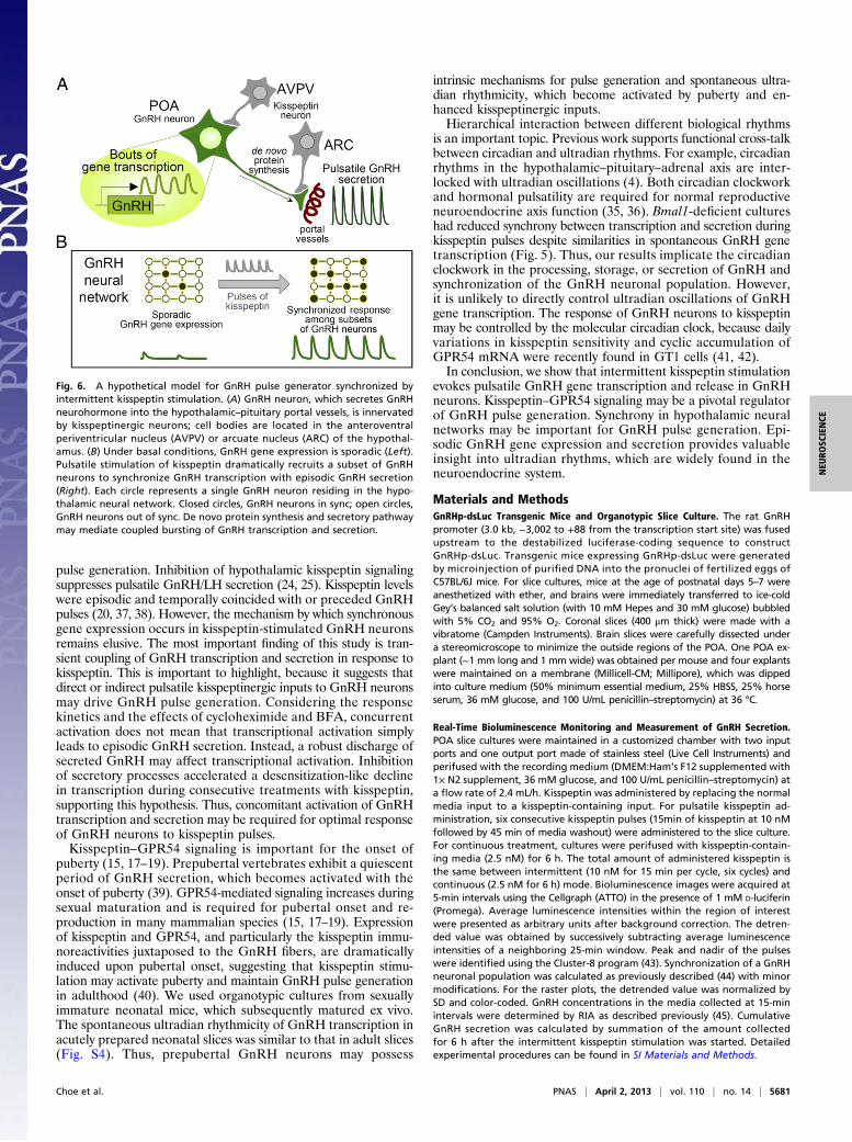
DiscussionThis study examined GnRH pulse generation, which is a classicexample of an ultradian biological rhythm. We characterizedspontaneous and kisspeptin-evoked ultradian GnRH gene tran-scription ex vivo in cultured hypothalamic slices derived fromGnRHp-dsLuc transgenic mice. We showed that pulsatile GnRHpromoter activity occurs in single GnRH neurons residing inhypothalamic neural networks, demonstrating their populationalproperties relative to GnRH secretion (Fig. 6).GnRH neurons may harbor intrinsic oscillatory mechanisms
that drive pulsatile GnRH secretion (13). We showed that GnRHpromoter activity exhibited spontaneous oscillations in single neu-rons. Despite stochastic synchronization at ∼2-h intervals in ∼10%of GnRH-expressing cells, the ultradian rhythm of GnRH genetranscription was not directly linked with episodic GnRH se-cretion because of differences in their periods. Moreover, thetranscriptional rhythm of single GnRH neurons was intrinsicand did not require voltage-gated ion channel activity, which isimportant for pulsatile GnRH release (32, 36). Thus, voltage-gated ion channels and intracellular calcium oscillation may reg-ulate neurohormone secretion by acting on axon terminals in themedian eminence. However, the intrinsic rhythmicity of GnRHneurons is still important, because ultradian gene transcription ineach cell and stochastic synchrony may contribute to GnRH pulsegeneration as demonstrated by repetitive kisspeptin stimulation.Hypothalamic GnRH neurons receive various inputs in vivo by
interacting with multiple cell types. Accumulating evidence sug-gests that kisspeptin–GPR54 signaling plays a key role in GnRH
Fig. 4. Effect of de novo protein synthesis and secretory pathway. (A)Representative profiles of an individual GnRH neuron with pulsatile kisspeptinstimulation with vehicle or indicated drug (BFA, brefeldin A, 10 μg/mL; CET,cetrorelix, 10 μM; CHX, cycloheximide, 100 μM; VEH, 0.1% dimethyl sulfoxide).Asterisks represent peaks identified by Cluster-8. (B) Synchronization of GnRHneuronal population. Raster plot of normalized detrended luminescence ofa single POA culture is shown as a pseudocolor scale (H, high; L, low). Each rowrepresents an individual GnRH neuron. % Sync is the percentage of luciferase-positive cells simultaneously reaching their peaks. (C) GnRH secretion in peri-fused media (blue bar, kisspeptin; line, vehicle or indicated drugs). (D) Peakvalue of synchronization. Two-way RM ANOVA, P < 0.01 (inhibitors), P < 0.01(number of kisspeptin pulses), P < 0.01 (interaction). (E) Peak value of GnRHsecretion. Two-way RM ANOVA, P < 0.05 (inhibitors), P < 0.01 (number ofkisspeptin pulses), P = 0.1977 (interaction). In D and E, *P < 0.05, **P < 0.01 vs.VEH, Bonferroni posttest. (F) Cumulative GnRH secretion during 6 h afterinitiation of kisspeptin. **P < 0.01 vs. VEH, t test. In B–F, data are shown asmean ± SEM (n = 65–89 cells from three to five batches per treatment).
Fig. 5. Ultradian rhythm of GnRHp-dsLuc and kisspeptin-induced synchro-nization in Bmal1 knock-out mice. (A) Representative luminescence profilesunder basal conditions in POA cultures obtained from GnRHp-dsLuc in WTor Bmal1−/− (Bmal1 KO) background. Asterisks represent peaks identifiedby Cluster-8. (B) Synchronization of GnRH neuronal population derived fromWT or Bmal1 KO mouse stimulated with kisspeptin. Raster plot of normal-ized detrended luminescence for a representative POA culture is shown as apseudocolor scale (H, high; L, low). Each row represents an individual GnRHneuron. % Sync is the percentage of luciferase-positive cells simultaneouslyreaching their peaks. (C) GnRH secretion in perifused media (blue bar, kisspeptinpulse). (D) Peak value of synchronization. Two-way RM ANOVA, P < 0.01(genotype), P < 0.01 (number of kisspeptin pulses), P = 0.7842 (interaction).(E) Peak value of GnRH secretion. Two-way RM ANOVA, P < 0.01 (genotype),P = 0.1245 (number of kisspeptin pulses), P = 0.6422 (interaction). In D and E,*P < 0.05 vs. VEH, Bonferroni posttest. (F) Cumulative GnRH secretion during6 h after initiation of kisspeptin. *P < 0.05 vs. VEH, t test. In B–F, data arepresented as mean ± SEM (n = 81–93 cells from four batches per genotype).
5680 | www.pnas.org/cgi/doi/10.1073/pnas.1213594110 Choe et al.
pulse generation. Inhibition of hypothalamic kisspeptin signalingsuppresses pulsatile GnRH/LH secretion (24, 25). Kisspeptin levelswere episodic and temporally coincided with or preceded GnRHpulses (20, 37, 38). However, the mechanism by which synchronousgene expression occurs in kisspeptin-stimulated GnRH neuronsremains elusive. The most important finding of this study is tran-sient coupling of GnRH transcription and secretion in response tokisspeptin. This is important to highlight, because it suggests thatdirect or indirect pulsatile kisspeptinergic inputs to GnRH neuronsmay drive GnRH pulse generation. Considering the responsekinetics and the effects of cycloheximide and BFA, concurrentactivation does not mean that transcriptional activation simplyleads to episodic GnRH secretion. Instead, a robust discharge ofsecreted GnRH may affect transcriptional activation. Inhibitionof secretory processes accelerated a desensitization-like declinein transcription during consecutive treatments with kisspeptin,supporting this hypothesis. Thus, concomitant activation of GnRHtranscription and secretion may be required for optimal responseof GnRH neurons to kisspeptin pulses.Kisspeptin–GPR54 signaling is important for the onset of
puberty (15, 17–19). Prepubertal vertebrates exhibit a quiescentperiod of GnRH secretion, which becomes activated with theonset of puberty (39). GPR54-mediated signaling increases duringsexual maturation and is required for pubertal onset and re-production in many mammalian species (15, 17–19). Expressionof kisspeptin and GPR54, and particularly the kisspeptin immu-noreactivities juxtaposed to the GnRH fibers, are dramaticallyinduced upon pubertal onset, suggesting that kisspeptin stimu-lation may activate puberty and maintain GnRH pulse generationin adulthood (40). We used organotypic cultures from sexuallyimmature neonatal mice, which subsequently matured ex vivo.The spontaneous ultradian rhythmicity of GnRH transcription inacutely prepared neonatal slices was similar to that in adult slices(Fig. S4). Thus, prepubertal GnRH neurons may possess
intrinsic mechanisms for pulse generation and spontaneous ultra-dian rhythmicity, which become activated by puberty and en-hanced kisspeptinergic inputs.Hierarchical interaction between different biological rhythms
is an important topic. Previous work supports functional cross-talkbetween circadian and ultradian rhythms. For example, circadianrhythms in the hypothalamic–pituitary–adrenal axis are inter-locked with ultradian oscillations (4). Both circadian clockworkand hormonal pulsatility are required for normal reproductiveneuroendocrine axis function (35, 36). Bmal1-deficient cultureshad reduced synchrony between transcription and secretion duringkisspeptin pulses despite similarities in spontaneous GnRH genetranscription (Fig. 5). Thus, our results implicate the circadianclockwork in the processing, storage, or secretion of GnRH andsynchronization of the GnRH neuronal population. However,it is unlikely to directly control ultradian oscillations of GnRHgene transcription. The response of GnRH neurons to kisspeptinmay be controlled by the molecular circadian clock, because dailyvariations in kisspeptin sensitivity and cyclic accumulation ofGPR54 mRNA were recently found in GT1 cells (41, 42).In conclusion, we show that intermittent kisspeptin stimulation
evokes pulsatile GnRH gene transcription and release in GnRHneurons. Kisspeptin–GPR54 signaling may be a pivotal regulatorof GnRH pulse generation. Synchrony in hypothalamic neuralnetworks may be important for GnRH pulse generation. Epi-sodic GnRH gene expression and secretion provides valuableinsight into ultradian rhythms, which are widely found in theneuroendocrine system.
Materials and MethodsGnRHp-dsLuc Transgenic Mice and Organotypic Slice Culture. The rat GnRHpromoter (3.0 kb, −3,002 to +88 from the transcription start site) was fusedupstream to the destabilized luciferase-coding sequence to constructGnRHp-dsLuc. Transgenic mice expressing GnRHp-dsLuc were generatedby microinjection of purified DNA into the pronuclei of fertilized eggs ofC57BL/6J mice. For slice cultures, mice at the age of postnatal days 5–7 wereanesthetized with ether, and brains were immediately transferred to ice-coldGey’s balanced salt solution (with 10 mM Hepes and 30 mM glucose) bubbledwith 5% CO2 and 95% O2. Coronal slices (400 μm thick) were made with avibratome (Campden Instruments). Brain slices were carefully dissected undera stereomicroscope to minimize the outside regions of the POA. One POA ex-plant (∼1 mm long and 1 mm wide) was obtained per mouse and four explantswere maintained on a membrane (Millicell-CM; Millipore), which was dippedinto culture medium (50% minimum essential medium, 25% HBSS, 25% horseserum, 36 mM glucose, and 100 U/mL penicillin–streptomycin) at 36 °C.
Real-Time Bioluminescence Monitoring and Measurement of GnRH Secretion.POA slice cultures were maintained in a customized chamber with two inputports and one output port made of stainless steel (Live Cell Instruments) andperifused with the recording medium (DMEM:Ham’s F12 supplemented with1× N2 supplement, 36 mM glucose, and 100 U/mL penicillin–streptomycin) ata flow rate of 2.4 mL/h. Kisspeptin was administered by replacing the normalmedia input to a kisspeptin-containing input. For pulsatile kisspeptin ad-ministration, six consecutive kisspeptin pulses (15min of kisspeptin at 10 nMfollowed by 45 min of media washout) were administered to the slice culture.For continuous treatment, cultures were perifused with kisspeptin-contain-ing media (2.5 nM) for 6 h. The total amount of administered kisspeptin isthe same between intermittent (10 nM for 15 min per cycle, six cycles) andcontinuous (2.5 nM for 6 h) mode. Bioluminescence images were acquired at5-min intervals using the Cellgraph (ATTO) in the presence of 1 mM D-luciferin(Promega). Average luminescence intensities within the region of interestwere presented as arbitrary units after background correction. The detren-ded value was obtained by successively subtracting average luminescenceintensities of a neighboring 25-min window. Peak and nadir of the pulseswere identified using the Cluster-8 program (43). Synchronization of a GnRHneuronal population was calculated as previously described (44) with minormodifications. For the raster plots, the detrended value was normalized bySD and color-coded. GnRH concentrations in the media collected at 15-minintervals were determined by RIA as described previously (45). CumulativeGnRH secretion was calculated by summation of the amount collectedfor 6 h after the intermittent kisspeptin stimulation was started. Detailedexperimental procedures can be found in SI Materials and Methods.
Fig. 6. A hypothetical model for GnRH pulse generator synchronized byintermittent kisspeptin stimulation. (A) GnRH neuron, which secretes GnRHneurohormone into the hypothalamic–pituitary portal vessels, is innervatedby kisspeptinergic neurons; cell bodies are located in the anteroventralperiventricular nucleus (AVPV) or arcuate nucleus (ARC) of the hypothal-amus. (B) Under basal conditions, GnRH gene expression is sporadic (Left).Pulsatile stimulation of kisspeptin dramatically recruits a subset of GnRHneurons to synchronize GnRH transcription with episodic GnRH secretion(Right). Each circle represents a single GnRH neuron residing in the hypo-thalamic neural network. Closed circles, GnRH neurons in sync; open circles,GnRH neurons out of sync. De novo protein synthesis and secretory pathwaymay mediate coupled bursting of GnRH transcription and secretion.
Choe et al. PNAS | April 2, 2013 | vol. 110 | no. 14 | 5681
NEU
ROSC
IENCE

ACKNOWLEDGMENTS. Bmal1-mutant mice were generously provided byDrs. Marina Antoch (Roswell Park Cancer Institute) and Karyn Esser (Universityof Kentucky). BioScience Writers provided English editing services for themanuscript. This work was supported by grants from the Korea Ministry
of Education, Science, and Technology (MEST) through the Brain ResearchCenter for the 21st Century Frontier Research and Development Program inNeuroscience. H.K.C. and H.K. were supported by Brain Korea 21 ResearchFellowships from MEST.
1. Goldbeter A (2008) Biological rhythms: Clocks for all times. Curr Biol 18(17):R751–R753.2. Takahashi JS, Hong HK, Ko CH, McDearmon EL (2008) The genetics of mammalian
circadian order and disorder: Implications for physiology and disease. Nat Rev Genet9(10):764–775.
3. Dibner C, Schibler U, Albrecht U (2010) The mammalian circadian timing system:Organization and coordination of central and peripheral clocks. Annu Rev Physiol 72:517–549.
4. Lightman SL, Conway-Campbell BL (2010) The crucial role of pulsatile activity of theHPA axis for continuous dynamic equilibration. Nat Rev Neurosci 11(10):710–718.
5. Knobil E (1980) The neuroendocrine control of the menstrual cycle. Recent Prog HormRes 36:53–88.
6. Martínez de la Escalera G, Choi AL, Weiner RI (1992) Generation and synchronizationof gonadotropin-releasing hormone (GnRH) pulses: Intrinsic properties of the GT1-1GnRH neuronal cell line. Proc Natl Acad Sci USA 89(5):1852–1855.
7. Terasawa E, Keen KL, Mogi K, Claude P (1999) Pulsatile release of luteinizing hormone-releasing hormone (LHRH) in cultured LHRH neurons derived from the embryonicolfactory placode of the rhesus monkey. Endocrinology 140(3):1432–1441.
8. Jasoni CL, Romanò N, Constantin S, Lee K, Herbison AE (2010) Calcium dynamics ingonadotropin-releasing hormone neurons. Front Neuroendocrinol 31(3):259–269.
9. Nuñez L, Faught WJ, Frawley LS (1998) Episodic gonadotropin-releasing hormonegene expression revealed by dynamic monitoring of luciferase reporter activity insingle, living neurons. Proc Natl Acad Sci USA 95(16):9648–9653.
10. Vazquez-Martinez R, et al. (2001) Pulsatile exocytosis is functionally associated withGnRH gene expression in immortalized GnRH-expressing cells. Endocrinology 142(12):5364–5370.
11. Vazquez-Martinez R, Shorte SL, Boockfor FR, Frawley LS (2001) Synchronized exo-cytotic bursts from gonadotropin-releasing hormone-expressing cells: Dual control byintrinsic cellular pulsatility and gap junctional communication. Endocrinology 142(5):2095–2101.
12. Chappell PE, White RS, Mellon PL (2003) Circadian gene expression regulates pulsatilegonadotropin-releasing hormone (GnRH) secretory patterns in the hypothalamicGnRH-secreting GT1-7 cell line. J Neurosci 23(35):11202–11213.
13. Moenter SM, DeFazio AR, Pitts GR, Nunemaker CS (2003) Mechanisms underlyingepisodic gonadotropin-releasing hormone secretion. Front Neuroendocrinol 24(2):79–93.
14. Campbell RE, Gaidamaka G, Han SK, Herbison AE (2009) Dendro-dendritic bundlingand shared synapses between gonadotropin-releasing hormone neurons. Proc NatlAcad Sci USA 106(26):10835–10840.
15. Oakley AE, Clifton DK, Steiner RA (2009) Kisspeptin signaling in the brain. Endocr Rev30(6):713–743.
16. Messager S, et al. (2005) Kisspeptin directly stimulates gonadotropin-releasinghormone release via G protein-coupled receptor 54. Proc Natl Acad Sci USA 102(5):1761–1766.
17. Seminara SB, et al. (2003) The GPR54 gene as a regulator of puberty. N Engl J Med349(17):1614–1627.
18. Roa J, Aguilar E, Dieguez C, Pinilla L, Tena-Sempere M (2008) New frontiers inkisspeptin/GPR54 physiology as fundamental gatekeepers of reproductive function.Front Neuroendocrinol 29(1):48–69.
19. Navarro VM, Tena-Sempere M (2012) Neuroendocrine control by kisspeptins: role inmetabolic regulation of fertility. Nat Rev Endocrinol 8(1):40–53.
20. Keen KL, Wegner FH, Bloom SR, Ghatei MA, Terasawa E (2008) An increase inkisspeptin-54 release occurs with the pubertal increase in luteinizing hormone-releasing hormone-1 release in the stalk-median eminence of female rhesus monkeysin vivo. Endocrinology 149(8):4151–4157.
21. Tovar S, et al. (2006) Effects of single or repeated intravenous administration ofkisspeptin upon dynamic LH secretion in conscious male rats. Endocrinology 147(6):2696–2704.
22. Plant TM, Ramaswamy S, Dipietro MJ (2006) Repetitive activation of hypothalamic Gprotein-coupled receptor 54 with intravenous pulses of kisspeptin in the juvenilemonkey (Macaca mulatta) elicits a sustained train of gonadotropin-releasing hormonedischarges. Endocrinology 147(2):1007–1013.
23. Seminara SB, Dipietro MJ, Ramaswamy S, Crowley WF, Jr., Plant TM (2006) Continuoushuman metastin 45-54 infusion desensitizes G protein-coupled receptor 54-inducedgonadotropin-releasing hormone release monitored indirectly in the juvenile maleRhesus monkey (Macaca mulatta): A finding with therapeutic implications. Endocri-nology 147(5):2122–2126.
24. Li XF, et al. (2009) Kisspeptin signalling in the hypothalamic arcuate nucleus regulatesGnRH pulse generator frequency in the rat. PLoS ONE 4(12):e8334.
25. Roseweir AK, et al. (2009) Discovery of potent kisspeptin antagonists delineatephysiological mechanisms of gonadotropin regulation. J Neurosci 29(12):3920–3929.
26. Ohtaki T, et al. (2001) Metastasis suppressor gene KiSS-1 encodes peptide ligand of aG-protein-coupled receptor. Nature 411(6837):613–617.
27. Lehman MN, Coolen LM, Goodman RL (2010) Minireview: kisspeptin/neurokininB/dynorphin (KNDy) cells of the arcuate nucleus: A central node in the control ofgonadotropin-releasing hormone secretion. Endocrinology 151(8):3479–3489.
28. Navarro VM, et al. (2009) Regulation of gonadotropin-releasing hormone secretionby kisspeptin/dynorphin/neurokinin B neurons in the arcuate nucleus of the mouse.J Neurosci 29(38):11859–11866.
29. Kobayashi T, et al. (2010) 2-acylamino-4,6-diphenylpyridine derivatives as novelGPR54 antagonists with good brain exposure and in vivo efficacy for plasma LH levelin male rats. Bioorg Med Chem 18(14):5157–5171.
30. Pitts GR, Nunemaker CS, Moenter SM (2001) Cycles of transcription and translationdo not comprise the gonadotropin-releasing hormone pulse generator in GT1 cells.Endocrinology 142(5):1858–1864.
31. Krsmanovi�c LZ, Stojilkovi�c SS, Mertz LM, Tomi�c M, Catt KJ (1993) Expression ofgonadotropin-releasing hormone receptors and autocrine regulation of neuropeptiderelease in immortalized hypothalamic neurons. Proc Natl Acad Sci USA 90(9):3908–3912.
32. Stojilkovic SS, Krsmanovic LZ, Spergel DJ, Catt KJ (1994) Gonadotropin-releasinghormone neurons: Intrinsic pulsatility and receptor-mediated regulation. TrendsEndocrinol Metab 5(5):201–209.
33. Xu C, Xu XZ, Nunemaker CS, Moenter SM (2004) Dose-dependent switch in responseof gonadotropin-releasing hormone (GnRH) neurons to GnRH mediated throughthe type I GnRH receptor. Endocrinology 145(2):728–735.
34. Miller BH, et al. (2004) Circadian clock mutation disrupts estrous cyclicity and main-tenance of pregnancy. Curr Biol 14(15):1367–1373.
35. Bunger MK, et al. (2000) Mop3 is an essential component of the master circadianpacemaker in mammals. Cell 103(7):1009–1017.
36. Krsmanovi�c LZ, et al. (1992) Calcium signaling and episodic secretion of gonadotropin-releasing hormone in hypothalamic neurons. Proc Natl Acad Sci USA 89(18):8462–8466.
37. Guerriero KA, Keen KL, Terasawa E (2012) Developmental increase in kisspeptin-54release in vivo is independent of the pubertal increase in estradiol in female rhesusmonkeys (Macaca mulatta). Endocrinology 153(4):1887–1897.
38. Kurian JR, Keen KL, Guerriero KA, Terasawa E (2012) Tonic control of kisspeptinrelease in prepubertal monkeys: implications to the mechanism of puberty onset.Endocrinology 153(7):3331–3336.
39. Sisk CL, Foster DL (2004) The neural basis of puberty and adolescence. Nat Neurosci7(10):1040–1047.
40. Clarkson J, Han SK, Liu X, Lee K, Herbison AE (2010) Neurobiological mechanismsunderlying kisspeptin activation of gonadotropin-releasing hormone (GnRH) neuronsat puberty. Mol Cell Endocrinol 324(1-2):45–50.
41. Zhao S, Kriegsfeld LJ (2009) Daily changes in GT1-7 cell sensitivity to GnRH secreta-gogues that trigger ovulation. Neuroendocrinology 89(4):448–457.
42. Tonsfeldt KJ, Goodall CP, Latham KL, Chappell PE (2011) Oestrogen induces rhythmicexpression of the Kisspeptin-1 receptor GPR54 in hypothalamic gonadotrophin-releasing hormone-secreting GT1-7 cells. J Neuroendocrinol 23(9):823–830.
43. Veldhuis JD, Johnson ML (1986) Cluster analysis: A simple, versatile, and robustalgorithm for endocrine pulse detection. Am J Physiol 250(4 Pt 1):E486–E493.
44. Moore JP, Jr., Shang E, Wray S (2002) In situ GABAergic modulation of synchronousgonadotropin releasing hormone-1 neuronal activity. J Neurosci 22(20):8932–8941.
45. Cho S, Cho H, Geum D, Kim K (1998) Retinoic acid regulates gonadotropin-releasinghormone (GnRH) release and gene expression in the rat hypothalamic fragments andGT1-1 neuronal cells in vitro. Brain Res Mol Brain Res 54(1):74–84.
5682 | www.pnas.org/cgi/doi/10.1073/pnas.1213594110 Choe et al.