synaptic connections, axonal and …mrcanu.pharm.ox.ac.uk/sites/default/files/pdfs/freund...gad,...
TRANSCRIPT
Neuroscience Vol. 19, No. 4, pp. 1133-1159, 1986 Printed in Great Britain
0306-4522/86 $3.00 + 0.00 Pergamon Journals Ltd
(t 1986lBRO
SYNAPTIC CONNECTIONS, AXONAL AND DENDRITIC PATTERNS OF NEURONS IMMlJNOREACTIVE FOR
CHOLECYSTOKININ IN THE VISUAL CORTEX OF THE CAT
T. F. FREUND,* Zs. MAGLOCZKY,* I. SOLTESZ* and P. SOMOGYI*t *lst Department of Anatomy, Semmelweis University Medical School, Budapest, Tiizolt6 u. 58,
H-1450 Hungary; and tMRC Anatomical Neuropharmacology Unit, Department of Pharmacology, Oxford University, South Parks Road, Oxford OXl 3QT, U.K.
Abstract-A subpopulation of y-aminobutyric acid (GABA) containing neurons was reported to contain cholecystokinin-immunoreactive material in the visual cortex of cat [Somogyi et al., J. Neurosci. (1984) 4, 2590-2603]. In the present study pre-embedding immunocytochemistry was used to identify which of the several types of presumed GABAergic nonpyramidal cells in areas 17 and 18 contain cholecystokinin immunoreactivity. Most of the cholecystokinin-immunoreactive somata were found in layers II-III, they were less frequent in layers I and VI, and relatively rare in layers IV and V. The distribution and density of the axon terminals resembled that of the cell bodies.
Two well defined types of cholecystokinin-immunoreactive neuron were distinguished: (1) double bouquet cells in layers II-III with vertically projecting axons, and (2) small basket cells with local axons either restricted to layers II-III, or descending to layer V. Additional cholecystokinin-positive cells showed features of bitufted or multipolar neurons in layers II-VI and horizontal cells in layer I, but these cells could be defined less well due to partial staining. Cholecystokinin-immunoreactive dendrites were found to run horizontally in layer I for several hundred micrometers. Some of the cholecystokininimmunoreactive cells in layer VI had very long dendrites ascending radially up to layer Ill, as did their axons. A few cholecystokinin-immunoreactive cells appeared to have two axons and this was confirmed by electron microscopy. All cholecystokinin-immunoreactive neurons and terminals were separated from the basal lamina of blood vessels by glial endfeet. Random samples of boutons from each layer as well as identified terminals traced to their origin from local neurons were examined in the electron microscope. All of the boutons established symmetrical (type Il) synaptic contacts with dendritic shafts, spines or somata. Quantitative electron microscopy of the postsynaptic targets of double bouquet cells and small basket cells demonstrated clear differences between these two types of neuron; basket cells having a higher proportion of their terminals in synaptic contact with somata.
The findings that several distinct types of cortical neurons, as defined by their synaptic connections, contain cholecystokinin-immunoreactive material and that identified axons of all examined neurons form type II synaptic contacts suggests that the majority, if not all cholecystokinin-positive boutons forming type II contacts originate from local cortical cells. The distribution of targets postsynaptic to cholecystokinin-positive neurons is compared to those of cells labelled by other methods.
In the central nervous system the highest concentration of cholecystokinin (CCK)-immunoreactive peptides is found in the cerebral cortex.1.2.10,23.35.55.78 The most abundant form is the carboxyterminal octapeptide, CCK_8 1O
•55 and there is a wealth of data
in favour of its involvement in interneuronal communication (for reviews see Refs 15 and 56). Thus, CCK-immunoreactive (CCK-I) peptides are found in cortical neurons and nerve terminals; 13.25.40,49.68 CCK-8-like material is present in cortical synaptosomes and synaptic vesicles; 14.53 CCK-8 is synthesized in vivo by cortical tissue,23 and it is released in vitro from
Abbreviations: CCK, cholecystokinin; CCK-I, cholecystokinin-immunoreactive; GABA, gamma-aminobutyrate; GAD, glutamate decarboxylase; GAD-I, glutamate decarboxylase-immunoreactive; HRP, horseradish peroxidase; IgG, immunoglobulin; NGS, normal goat serum; NPY, neuropeptide Y; P, pyramidal; PBS, phosphate-buffered saline; S, stellate.
cortical slices by depolarization. 14 Presumed CCK receptors have also been localized in cortex by biochemical and ligand-binding studies. 58.79.80 lontophoretically applied CCK -8 was shown to change, usually to depolarize, the membrane potential of corticaF8.52 and hippocampal neurons. 12 The above findings suggest a transmitter role for CCK, but at present nothing is known about its physiological role in the cortex. Nevertheless its localization is of interest because many CCK-I neurons also contain GABA68 or glutamate decarboxylase (GAD)?6 The inhibitory action of GABA33 and its role in shaping the receptive field properties of visual cortical neurons is well documented,6o.61 but its actions in different cortical operations have not been correlated with particular classes of GABAergic neurons. Since the peptides are localized to subpopulations of GABA cells, there is a possibility that through the identification of their input and output the role of the subpopulation can be delineated. It is possible that
1133

1134 T. F. FREUND et al.
each is present only in one class of cell as defined by their synaptic connections.
Immunocytochemical studies at the light and electron microscopic level provide clues for the position of CCK-I neurons in cortical networks. The structural features of the CCK-I boutons and synapses, and their distribution on the target neurons, e.g. cell body versus distal dendrites and spine heads, can be used in forming hypotheses for their possible function. 203 1.75.77 CCK-I neurons have not been studied from this point of view in the electrophysiologically most thoroughly explored cortical area, the visual cortex of the cat. In rat and monkey cortex,25,49 CCK-J boutons make symmetrical synapses with dendrites, spines and somata. However the origin of these CCK-I terminals from local neurons has not been demonstrated. Thus it is not known whether the CCK-I terminals reported earlier nated from local cortical neurons or from cortical and subcortical afferents that have been reported to contain CCK-I materiaL I6.42,59.72 Since the CCK-I terminals may from several sources laminar differences in their termination would also be expected.
Therefore, in the present experiments random samples of boutons were examined in each layer. In addition the fine structural features and postsynaptic targets of terminals, shown to originate from CCK-I local circuit neurons, were identified. We examined whether known types of neuron in the visual cortex of cat were immunoreactive for CCK. The light microscopic description of CCK-I neurons was combined with quantitative electron microscopic analysis of their synaptic boutons. The results are compared with similar data on cortical neurons identified by other marking methods.
EXPERIMENTAL PROCEDURES
Preparation of the animals: Five adult cats were used. They were anaesthetized with chloral hydrate (350 mg/kg Lp.) and per fused through the heart first with Tyrode's solution (gassed with a mixture of 95% O2 and 5% CO2)
and then with a fixative containing 4% paraformaldehyde (TAAB, Maidenhead, U.K.), 0.05% glutaraldehyde (TAAB) and approximately 0.2% picric acid dissolved in 0.1 M phosphate buffer (pH 7.4) as described previously.70 Two of the cats each received an injection of colchicine (BDH Chemicals, 6 flgifll dissolved in artificial cere-brospinal fluid) into the lateral 24 h before perfusion, under xylazine hydrochloride mg/kg; Rompun, Bayer) and ketamine hydrochloride mg/kg i.m.) anaesthesia. Colchicine was delivered through a micropipette with about 50 flm tip diameter. One penetration was made, and a total volume of 2 fll (12 flg) cholchicine was injected in 8-10 steps along a 10 mm long track. Following perfusion the brains were removed from the skull, dissected blocks of the lateral gyrus were placed into the fixative for 1-6 h, and then into 10 and 20% sucrose until they sank. The blocks were then frozen in liquid nitrogen, thawed in 0.1 M phosphate buffer at room temperature, and sectioned on a Vibratome (Oxford Instruments) at 80 tIm. The fixative was removed from the sections by washing them thoroughly in several changes of 0.1 M phosphate buffer (pH 7.4).
Immunocytochemical procedures Phosphate-buffered saline (PBS, ph 7.4) containing 1 %
normal goat serum (NGS) was used for all the washes and also for diluting the antisera, unless otherwise stated. The incubation procedure was carried out at room temperature in the following order: 1 h in 20% NGS (Miles); 2 x 30 min wash; 36 h or overnight at 4°C in the antiserum to CCK (diluted 1 :200); 3 x 30 min wash; 6 h in goat anti-rabbit (Miles) diluted I: 50; 3 x 30 min wash; overnight at rabbit peroxidase-antiperoxidase complex diluted 1: 100; 3 x 30 min wash in PBS only; 2 x 20 min wash in 0.05 M Tris-HCI buffer (pH 7.5). The sections were then preincubated for 30 min in a 0.05% solution of 3,3' -diaminobenzidine tetra HCI (Sigma) dissolved in the 0.05 M Tris-HCI buffer, and incubated further for 5-8 min after adding H20 2 to a final concentration of 0.01 %. During all the above steps the sections were continuously agitated on a shaker. After washing in Tris buffer and then in phosphate buffer the sections were postfixed for I h in 1 % OS04 dissolved in 0.1 M PBS.
Antiserum. Antiserum to CCK (Code No. Ll12) was kindly donated by Dr G. J. Dockray, and it was raised to the C-terminal tetrapeptide of CCK/gastrin coupled to bovine thyroglobulin. The immunological and immunocytochemical characteristics of this antiserum have been published earlier. 11
Controls. To test the specificity of the antiserum and the incubation procedure, some sections were incubated in normal rabbit serum (diluted to I: 200) instead of the CCK antiserum, or in the same diluted CCK antiserum, which was preincubated overnight with sulphated CCK-8 (Sigma, 10-5 M) prior to use. No specific staining was seen in these sections. Peroxidase reaction end product could be seen only in structures which are known to contain endogenous peroxidase activity. A homogenous faint reaction endproduct covered the surface of each section, which was due to nonspecific binding of the antibodies.
Correlated light and electron microscopy
Following osmication the sections were dehydrated in ethanol (1 % uranyl acetate was included in the 70% ethanol stage for 40 min), mounted on slides in resin (Durcupan ACM, Fluka) and cured for 2 days at 56°C. Selected neurons were drawn using a attachment, photo-graphed in the light microscope re-embedded into plastic capsules for further ultrathin sectioning as described earlier.70 The serial ultrathin sections were mounted on Formvar-coated single slot grids and stained with lead citrate. 57 Electron micrographs were taken at 80 k V using 20 or 30 flm objective apertures.
RESULTS
Light microscopy of cholecystokinin -immunoreactive structures in the lateral gyrus
CCK-I cells and axon terminals could be found in each layer of the lateral gyrus in the cat. The cellular pattern of CCK immunoreactivity was very similar in areas 17 and 18 and in the colchicine-treated and in the normal animals. Slight differences were found only in the subcellular distribution and intensity of the staining. In the colchicine-treated animals immunoreactive cell bodies were found with approximately the same frequency as in normal. Their staining was stronger but axons could not be followed from the perikarya beyond the axon initial segment, which in most cases appeared swollen. This contrasted with the CCK-J cells in normal animals,

Synaptic connections of CCK-J neurons in visual cortex 1135
where even the faintly staining cells had axons, which could be followed for variable distances, occasionally for as long as 2 mm. Apart from neuron NI of Fig. 1, which was found in colchicine-injected cortex, distant from the injection site, all cells shown in the drawings (Figs 1-5) derived from normal animals, as indicated by their extensive axon arborizations. The frequency and distribution of CCK-I varicosities in the neuropil was similar in the treated and untreated animals. The varicosities were usually isolated from each other with the exception of the main axons and their proximal collaterals, which in the normal animals had a continuous staining.
CCK-I varicosities were found scattered in each layer with the highest density in the supragranular layers, and the lowest in layer V. A modest number of boutons was seen in layers IV and VI. It was apparent at first sight that the density of CCK-I varicosities in any layer was much lower than that of the glutamate decarboxylase immunoreactive (GAD-I) varicosities reported previously.17 It is noteworthy that CCK-I varicosities, just as GAD-I terminals, were frequently seen in contact with cell bodies in all layers, but especially in layers I1, III and VI. In the neuropil of layer I, besides the axon terminals a large number of long horizontally running varicose dendrites were also stained for CCK.
The frequency of CCK-I somata closely resembled that of the CCK-I axon terminals, i.e. they were most numerous in layers II-I1I, and sparsest in layer V. Within a given layer they appeared to be scattered randomly, but occasionally small groups of 2-3 cells could also be detected. A selection of extensively immunostained cells is shown in Fig. 1-5. Although the dendritic and axonal arborizations of these neurons were only partially revealed, the details were sufficient to identify two major characteristic CCK-I cell types. These were the double bouquet cells, and the small basket cells of layers Il-III. The somata of these identified neurons were located invariably in layers II-III. The majority of the other CCK-I cells in layers II-III could not be put in any established category mainly because of their partial staining. Some of them showed the features of bitufted, or more rarely of multipolar cells, but these categories are not well defined. Many CCK-I cells were found in layer I, but they usually lacked stained axons. Their dendritic tree was generally elongated in the horizontal direction, or had a multipolar appearance. In contrast with similar studies in rats and monkeys25A9 in the cat neurons with typical features of bipolar cells have not been seen to be immunoreactive for CCK.
Double bouquet cells
These had a prominent upper and a usually smaller lower group of dendrites. The dendrites of the upper bunch often penetrated into layer I, where they ran horizontally sometimes as far as 300-400 flm. Their main axon and the majority of its collaterals were
radially oriented. Similar, but single, radially coursing varicose axon segments immunoreactive for CCK were frequently encountered in most of our sections. The axons when followed from the cells did not form narrow bundles (Fig. 1) like the horsetail-shaped axons of double bouquet cells in monkey.64 The perikarya of CCK-I double bouquet cells were elongated and had diameters of 8-15 fl m. They were invariably located in layers II-IIL
On the basis of their axon morphology double bouquet cells were divided into two subtypes: (1) "local circuit" type, with an axon consisting of 2-4 radially oriented collaterals, which were densely studded with boutons and could not be traced beyond layer V (Figs 1, NI-5; 6A,B). (2) "Projection" cells, with a main axon, usually free of varicosities, and running straight towards the white matter (Fig. 2). Of the latter type boutons appeared only on the recurrent axon collaterals, which emerged from the main axons in any of the layers, and passed obliquely towards the upper layers. The main axons could frequently be followed to the border of the white matter, where they were lost because of the dark background of the osmium-stained myelin or possibly because they became myelinated. This type of axon is typical of the supragranular pyramidal cells, some of which are the corti co-cortical projection neurons. 5
.22 In spite of the similarity in axon mor
phology, none of the CCK-I "projection" cells had dendritic trees similar to pyramidal cells, i.e. they had no prominent apical dendrites, and the dendritic shafts were free of spines.
Small basket cells in layers II-III
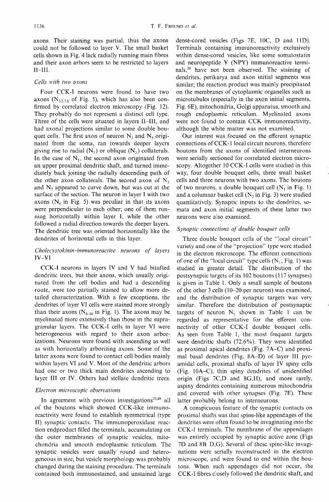
The somata of small basket cells were 8-12 flm in diameter and they were also situated in the supragranular layers. On the basis of dendritic morphology they could not be distinguished from the double bouquet cells (Figs 3 and 4). Most of them had an upper and lower tuft of dendrites, some of which reached layer I turning parallel to the pia. Their axon was characteristically formed of pericellular arrays of 3-6 boutons around somata in layers II--V (Figs 3, 4 and 11). Some of the cells had vertically oriented main axons, which could be followed as far as layer V, but their collaterals arborized most profusely in layers II-IlI (Figs 3 and 4). This was in contrast to the double bouquet cells, which only rarely made contacts onto perikarya (Figs 9 and 10) and never had extensive arbors in the tangential directions. The axon of one of the basket cells (Fig. 3, N2 ) arborized in layers I1-III then descended and contacted a few more somata in lower layer IV, but gave no collaterals or boutons in the upper half of layer IV. This axon arbor is similar to that of the columnar basket cells described by Szentagothai,75 although the true extent of the coIlaterals could not be established in immunostained material. Other small basket cells in
3 might also belong to the same type of CCK-I neuron since they also have vertically oriented main
1136 T. F. FREUND et al.
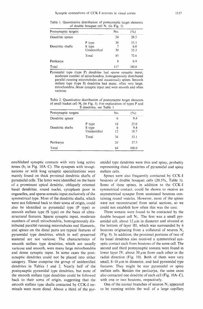
axons. Their stammg was partial, thus the axons could not be followed to layer V. The small basket cells shown in Fig. 4 lack radially running main fibres and their axon arbors seem to be restricted to layers II-III.
Cells with two axons
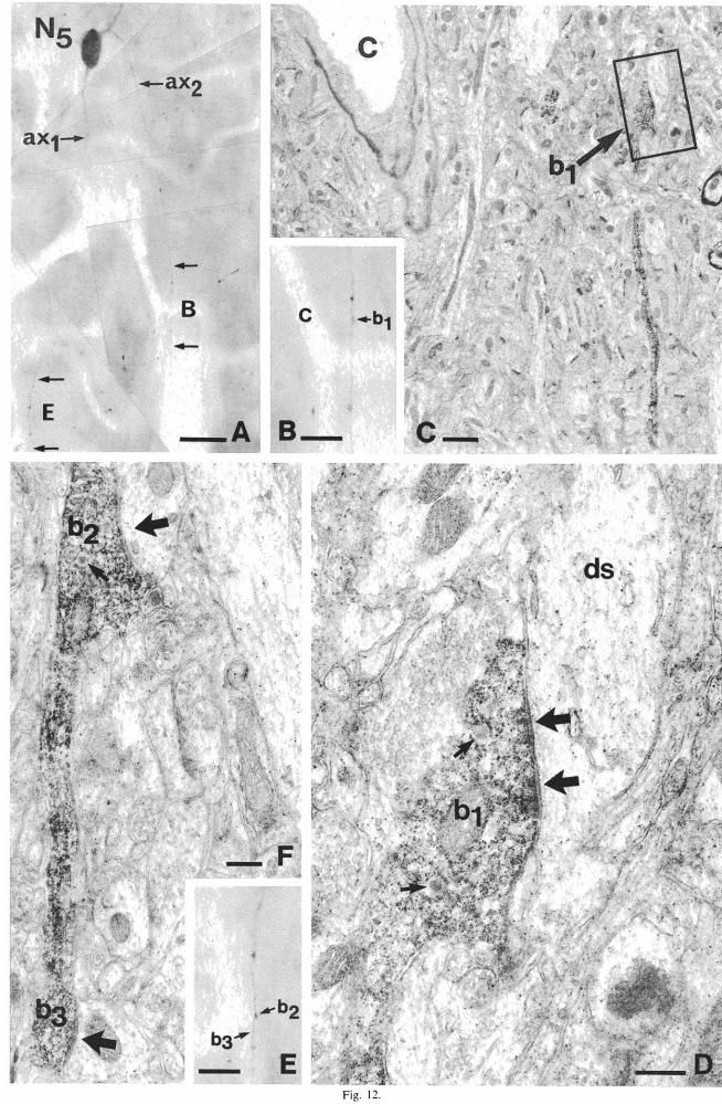
Four CCK-I neurons were found to have two axons (N3.5.7•8 of Fig. 5), which has also been confirmed by correlated electron microscopy (Fig. 12). They probably do not represent a distinct cell type. Three of the cells were situated in layers Il-IIl, and had axonal projections similar to some double bouquet cells. The first axon of neuron N3 and Ns originated from the soma, ran towards deeper layers giving rise to radial (Ns) or oblique (N3) collaterals. In the case of N 5, the second axon originated from an upper proximal dendritic shaft, and turned immediately back joining the radially descending path of the other axon collaterals. The second axon of N3 and appeared to curve down, but was cut at the surface of the section. The neuron in layer I with two axons (Ns in 5) was peculiar in that its axons were perpendicular to each other; one of them running horizontally within layer I, while the other followed a radial direction towards the deeper layers. The dendritic tree was oriented horizontally like the dendrites of horizontal cells in this layer.
Cholecystokinin -immunoreactive neurons of layers IV-VI
CCK-I neurons in layers IV and V had bitufted dendritic trees, but their axons, which usually originated from the cell bodies and had a descending route, were too partially stained to allow more detailed characterization. With a few exceptions, the dendrites of layer VI cells were stained more strongly than their axons (N6- IO in Fig. 1). The axons may be myelinated more extensively than those in the supragranular layers. The CCK-I cells in layer VI were heterogeneous with regard to their axon arborizations. Neurons were found with ascending as well as with horizontally arborizing axons. Some of the latter axons were found to contact cell bodies mainly within layers VI and V. Most of the dendritic arbors had one or two thick main dendrites ascending to layer III or IV. Others had stellate dendritic trees.
Electron microscopic observations
In agreement with previous investigations25.49 all
of the boutons which showed CCK-like immunoreactivity were found to establish symmetrical (type Il) synaptic contacts. The immunoperoxidase reaction endproduct filled the terminals, accumulating on the outer membranes of synaptic vesicles, mitochondria and smooth endoplasmic reticulum. The synaptic vesicles were usually round and heterogeneous in size, but vesicle morphology was probably changed during the staining procedure. The terminals contained both immunostained, and unstained large
dense-cored vesicles (Figs 7E, lOC, D and 11 D). Terminals containing immunoreactivity exclusively within dense-cored vesicles, like some somatostatin and neuropeptide Y (NPY) immunoreactive terminals,26 have not been observed. The staining of dendrites, perikarya and axon initial segments was similar; the reaction product was mainly precipitated on the membranes of cytoplasmic organelles such as microtubules (especially in the axon initial segments, Fig. 6E), mitochondria, Golgi apparatus, smooth and rough endoplasmic reticulum. Myelinated axons were not found to contain CCK immunoreactivity, although the white matter was not examined.
Our interest was focused on the efferent synaptic connections ofCCK-llocal circuit neurons, therefore boutons from the axons of identified interneurons were serially sectioned for correlated electron microscopy. Altogether 10 CCK -I cells were studied in this way, four double bouquet cells, three small basket cells and three neurons with two axons. The boutons of two neurons, a double bouquet cell (N 1 in Fig. 1) and a columnar basket cell (N2 in Fig. 3) were studied quantitatively. Synaptic inputs to the dendrites, somata and axon initial segments of these latter two neurons were also examined.
Synaptic connections of double bouquet cells
Three double bouquet cells of the "local circuit" variety and one of the "projection" type were studied in the electron microscope. The efferent connections of one of the "local circuit" type cells (N I, Fig. 1) was studied in greater detail. The distribution of the postsynaptic targets of its 102 boutons (117 synapses) is given in Table 1. Only a small sample of boutons of the other 3 cells (10-20 per neuron) was examined, and the distribution of synaptic targets was very similar. Therefore the distribution of postsynaptic targets of neuron N I shown in Table 1 can be regarded as representative for the efferent connectivity of other CCK-I double bouquet cells. As seen from Table 1, the most frequent targets were dendritic shafts (72.6%). They were identified as proximal apical dendrites (Fig. 7 A-C) and proximal basal dendrites (Fig. 8A-D) of layer III pyramidal cells, proximal shafts of layer IV spiny cells (Fig. lOA-C), thin spiny dendrites of unidentified origin (Figs 7C,D and SG,H), and more rarely, aspiny dendrites containing numerous mitochondria and covered with other synapses (Fig. 7E). These latter probably belong to interneurons.
A conspicuous feature of the synaptic contacts on proximal shafts was that spine-like appendages of the dendrites were often found to be invaginating into the CCK-I terminals. The membrane of the appendages was entirely occupied by synaptic active zone (Figs 7D and SB-D,G). Several of these spine-like invaginations were serially reconstructed in the electron microscope, and were found to end within the boutons. When such appendages did not occur, the CCK-I fibres closely followed the dendritic shaft, and
Synaptic connections of CCK-I neurons in visual cortex 1137
Table 1. Quantitative distribution of postsynaptic target elements of double bouquet cell N I (in Fig. I)
Postsynaptic targets No. (%)
Dendritic spines 24 20.5
P type 39 33.3 Dendritic shafts S type 7 6.0
Unidentified 39 33.3
Total 85 72.6
Perikarya 8 6.9
Total 117 100.0
Pyramidal type (type P) dendrites had sparse synaptlc mput, moderate number of mitochondria, homogeneously distributed parallel running microtubules and occasionally spines. Smooth stellate type (type S) dendrites had many, often very large, mitochondria, dense synaptic input and were smooth and often varicose.
Table 2. Quantitative distribution of postsynaptic of small basket cell N2 (in Fig. 3). For explanation
S dendrites, see Table 1
elements types P and
Postsynaptic targets No. (%)
Dendritic spines 6 9.4
P type 16 25.0 Dendritic shafts S type 6 9.4
Unidentified 12 18.7
Total
Perikarya
Total
established synaptic contacts with very long active zones (b2 in Fig. lOA-C). The synapses with invaginations or with long synaptic specializations were mainly found on thick proximal dendritic shafts of pyramidal cells. The latter were identified on the basis of a prominant apical dendrite, obliquely oriented basal dendrites, round nuclei, cytoplasm poor in organelIes, and sparse somatic input exclusively of the symmetrical type. Most of the dendritic shafts, which were not followed back to their soma of origin, could also be identified as pyramidal type (P type) or smooth stellate type (S type) on the basis of ultrastructural features. Sparse synaptic input, moderate numbers of small mitochondria, homogeneously distributed parallel running microtubules and filaments, and spines on the distal parts are typical features of pyramidal type dendrites, which in well preserved material are not varicose. The characteristics of smooth stellate type dendrites, which are usually varicose and smooth, were many large mitochondria and dense synaptic input. In some cases the postsynaptic dendrites could not be placed into either category. These comprise the group of unidentified dendrites in Tables 1 and 2. Nearly half of the postsynaptic pyramidal type dendrites, but none of the smooth stellate type dendrites could be followed back to their soma of origin, suggesting that the smooth stellate type shafts contacted by CCK-I terminals were more distal. About a third of the pyr-
34 53.1
24 37.5
64 100.0
amidal type dendrites were thin and spiny, probably representing distal dendrites of pyramidal and spiny stellate cells.
Spines were also frequently contacted by CCK-I boutons of double bouquet cells (20.5%, Table 1). Some of these spines, in addition to the CCK-I symmetrical contact, could be shown to receive an asymmetrical synapse from unstained boutons containing round vesicles. However, most of the spines were not reconstructed from serial sections, so we could not establish how often this was the case.
Three somata were found to be contacted by the double bouquet cell NI' The first was a small pyramidal cell, about 12 /lm in diameter and situated at the bottom of layer Ill, which was surrounded by 6 boutons originating from a collateral of neuron NI (Fig. 9). In addition, the proximal portions of two of its basal dendrites also received a symmetrical synaptic contact each from boutons of the same cell. The second and third postsynaptic somata were found in lower layer IV, about 50/lm from each other in the radial direction (Fig. 10). Both of them were very small, 8-10 /lm in diameter, and had pyramidal type features. They might be star pyramidal or spiny stellate cells. Besides the perikarya, the same axon also contacted one dendrite of each cell (Fig. lOA-C), with one or two boutons, respectively.
One of the axonal branches of neuron N I appeared to be running within the wall of a large capillary

1138 T. F. FREUND et al.
when viewed in the light microscope (Figs 6A,B and 8E-H). It was of special interest to examine whether the boutons of this collateral were in contact with the basal lamina of the blood vessel, as reported earlier25
or whether they made similar synaptic connections to the rest of the boutons of this cell. Electron microscopy showed that the boutons of this axon collateral were separated by at least glial endfeet from the basal lamina of the capillary, and that they made conventional synaptic contacts with dendrites and spines (Fig. 8E-H). We found no evidence for direct contact or special association between CCK-I profiles, e.g. cell bodies, dendritic or axonal processes, and blood vessels in this study.
Boutons located on the recurrent collaterals of a "projection" double bouquet cell have also been examined, and they were found to establish symmetrical synaptic contacts with dendritic shafts. Thus, in spite of the similarity in the axon morphology at the light microscopic level of these CCK-I cells (Fig. 2) and pyramidal cells in layer Ill, their synaptic specializations differ since pyramidal cells make asymmetrical synapses. 36
The synaptic input to neuron NI has also been examined. Its soma received only a very small number of synapses, some of which were asymmetrical (Fig. 6D). This is characteristic of non pyramidal cell bodies. Its proximal dendrites, which had a very irregularly shaped outline, were densely covered by synapses of both the symmetrical and asymmetrical types (Fig. 6C). The axon initial segment of neuron N I was reconstructed from its origin to the first efferent bouton, and no afferent synaptic contacts were found on it. A glial process was found to climb along it on one side of its entire length (Fig. 6E).
Synaptic connections of small basket cells
Three small basket cells were studied at the electron microscopic level. Two of them (NI and N 2 ,
Fig. 3) had descending branches while the third (N2' Fig. 4) had an axon restricted to layers Il-IIl. Sixtyfour of the boutons of neuron N2 (Fig. 3, Table 2) were studied while only about 10-15 boutons of each of the other two cells were examined. The latter had similar postsynaptic targets to N2 shown in Table 2.
As already described at the light microscopic level above, boutons of these CCK-I small basket cells frequently contacted somata mainly in layers Il-IlI. This was confirmed by electron microscopy; all the boutons were found to establish symmetrical synaptic contacts on the perikarya they surrounded. Quantitative analysis of the postsynaptic targets of neuron N2 shows that a high proportion (37.5%) of its boutons made synapses with cell bodies, which were mainly pyramidal cells (Fig. 1 lA-D). Occasionally, contacts on somata of non pyramidal cells were also observed. The soma of N2 also received input from a CCK-I basket-like axon (Fig. IIA,F), which, unfortunately could not be followed back to its parent cell body. Other CCK-I cell bodies mainly in layers
Il-IIl were also found to be surrounded by CCK positive boutons, but the axons of these postsynaptic CCK-I somata were not stained, therefore they could not be classified by their efferent connections. The number of boutons given by the CCK-I basket cell, N2, to single postsynaptic perikarya could not always be determined. The main reason was that in some cases, as the CCK-I collateral reached the cell body, staining became discontinuous and the boutons were not connected by preterminal axons. Nevertheless, from the examples with continuously stained collaterals, the number of boutons given to a single cell was between 3 and 6. Rarely the number was higher, thus three pyramidal neurons postsynaptic to basket cell NI (Fig. 3) were contacted by 12-16 boutons each on their somata and along the proximal apical dendrites from this single CCK-I neuron.
More than half of the boutons of N2 (Fig. 3) established synaptic contacts with dendritic shafts, which were mainly of the pyramidal type (25% of all targets) or could not be identified on the basis of our criteria (18.7% of all targets). Only 6 boutons (9.4% of all targets) contacted nonpyramidal (S type) dendritic shafts, and the percentage of contacts on dendritic spines (9.4% of all targets) was also small (Table 2).
The main difference between the efferent connections of double bouquet cells and small basket cells was that only a small number of somata were contacted by double bouquet cells, while they represented one of the major targets of the small basket cells. Dendrites were more frequent targets of the double bouquet cells, but they were also a significant target for CCK -I small basket cells.
Synapses formed by cholecystokinin -immunoreactive cells with two axons
Three such cells (N 5.7,8 of Fig. 5) were examined in the electron microscope. Only a small part of the axons of two of them were studied (N7 and Ns) to confirm that they were indeed giving synaptic contacts. The third cell (N 5) also had two axons that were presynaptic (Fig. 12) and this neuron was studied in more detail. The postsynaptic targets of 16 boutons of this cell were examined, and their distribution was found to be very similar to the most extensively studied double bouquet cell (N I, Table 1). Dendritic shafts, including some large proximal dendrites of the pyramidal type, were the targets of the majority (13) of the terminals. Two of the boutons were found to terminate on dendritic spines, and only one on a pyramidal cell soma. No major differences in postsynaptic elements could be revealed between the two axons of this CCK-I neuron.
The processes of this cell identified as dendrites resembled conventional dendrites, in that they were only postsynaptic, received synaptic input of a density similar to the dendrites of neuron NI (Fig. 6C) and contained no synaptic vesicles.

PIA
11.
Ill.
NA
NB
VA
VI
Wm
~\ I)
~~ I ~(
~
\ I
-Fig. l. CCK-I neurons in the cat lateral gyrus, drawn from 80 j.1m thick sections. The neurons (N I _5) in layers II-III have vertically projecting axon bundles and belong to the class of double bouquet cells. Most of the cells in layer VI (N(rIO) have ascending axons and/or dendrites, and only the axon of N6 appears to stay within layer VI. Neuron N I has been processed for correlated electron microscopy (see Figs 6-10).
Bar = 50 j.1m.
1139
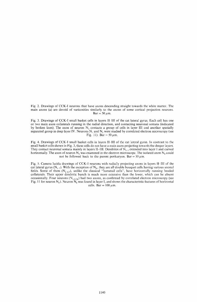
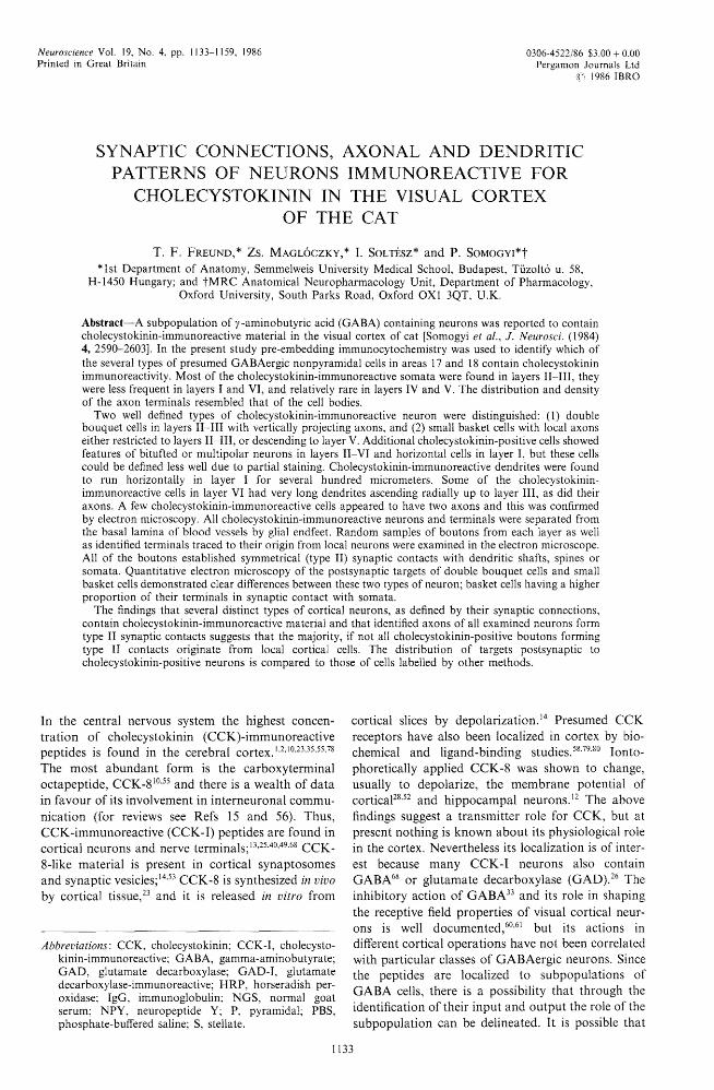
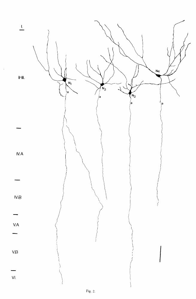
Fig. 2. Drawings of CCK-I neurons that have axons descending straight towards the white matter. The main axons (a) are devoid of varicosities to the axons of some cortical projection neurons.
flm.
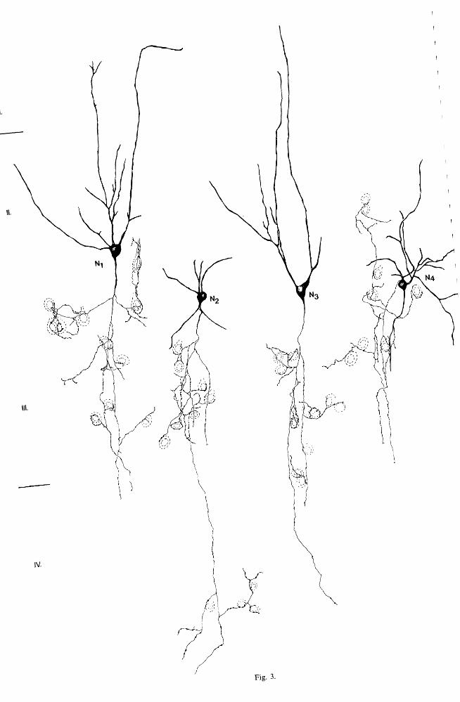
Fig. 3. Drawings of CCK-I small basket cells in Il-IIl of the cat lateral gyrus. Each cell has one or two main axon collaterals running in the radial and contacting neuronal somata (indicated by broken lines). The axon of neuron N2 contacts a group of cells in layer III and another spatially separated group in deep layer IV. Neurons NI and N2 were studied by correlated electron microscopy (see
Fig. 11). Bar 50 flm.
Fig. 4. Drawings of CCK-I small basket cells in II-III of the cat lateral gyrus. In contrast to the small basket cells shown in Fig. 3, these cells do not a main axon projecting towards the deeper layers. They contact neuronal somata mainly in layers II-III. Dendrites ofNl-J extended into layer I and curved horizontally_ The axon of neuron N2 was examined in the electron microscope. The isolated axon N4 could
not be followed back to the parent perikaryon. Bar 50 flm.
Fig. 5. Camera lucida drawings of CCK-I neurons with radially projecting axons in layers fI- III or the cat lateral gyrus (N 1-7)' With the exception of Ng , are all double bouquet cells having various axonal fields. Some of them (N2-4.7)' unlike the classical cells", have horizontally running beaded collaterals. Their upper dendritic bunch is much more extensive than the lower, which can be absent occasionally. Four neurons (N,s 7 8) had two axons, as confirmed by correlated electron microscopy (see Fig. It for neuron Ns). Neuron'N~ was found in layer I, and shows the characteristic features of horizontal
cells. Bar 100 flm.
1140
I.
11-111.
IV. A
IV.B
V.A
V.B
I
VI.
Fig. 2.
I.
1\.
IV.
Fig. 3.
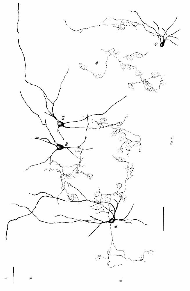
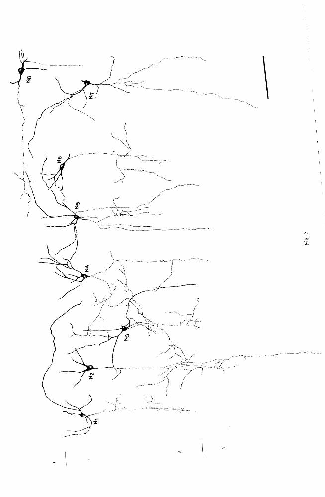

Fig. 6. (A,B) Light of a double bouquet cell (N!, also seen in Fig. I), as revealed by immunocytochemical CCK. Some of the dendrites penetrate into layer I, and the axon originates from a lower The axon branches (arrow) follow a path perpendicular to the pia. A large capillary (c), with two endogenous peroxidase reactive pericytes seen in both (A) and (B) indicates matching parts of the two photographs, which show different branches of the same axon at different focal depths. Portions of the axon (1,2,3) are shown at the electron microscopic level in Figs 7, 8 and 9, respectively. (C) Electron micrograph of a proximal dendritic shaft (ds) of the cell N!, seen in (A) and (B), with dense input from boutons (asterisks) establishing symmetrical or asymmetrical contacts. (D) An asymmetrical synaptic contact (arrow) on the cell body of neuron N!. (E) The axon initial segment (IS) of neuron N! covered by glial processes (arrows). No synaptic contacts were found on it. Bars: (A,B)
20 tLm; (C,E) 0.5 tLm; (D) 0.2 tLm.
Fig. 7. (A) Part of the axon of neuron N I (labelled I in Fig. 6A) is shown here enlarged. The low (B) and high power (C,D) correlated electron micrographs of the numbered boutons demonstrate that they establish symmetrical synaptic contacts (arrows in C and D) with dendritic shafts; b! on a thin shaft, b2
on a thick apical dendrite (ad), b3 on a spiny medium caliber dendrite (dp ' in D). The spine-like appendage (small arrows in D), with synaptic specialization was completely invaginated into the terminal. A basal dendritic shaft (bd) is contacted by b4 in B. (E) A bouton of the same axon is in synaptic contact (arrow) with a smooth varicose dendritic shaft (d,), also receiving asymmetrical synaptic contacts (asterisk). Bars:
(A) 10 tLm; (B) 2 tLm; CC-E) 0.2 tLm.
Fig. 8. (A-D) A bouton (b!) from the axon of neuron N! (portion 2 in Fig. 6A) is shown in light CA) and electron micrographs (B-D). Two and PI) and a capillary (c) serve for correlation. (B) The bouton b! is in contact with the (bd) of the pyramidal cell (PI) having a prominent apical dendrite (ad). (C,D) Serial sections of b! with spine-like of the basal dendritic shaft invaginating into it. (E) Light micrograph of a collateral of neuron. It to run within the wall of a blood vessel (c), (F-H) Electron of the same area the large vessel (c) and a pyramidal cell body (p). The CCK-I bouton by an arrow in CE) is framed in (F), and is shown in (G). (G) The immunoreactive bouton is in symmetrical contact (arrows) with a spine-like appendage of a pyramidal-type dendrite next to the wall (E). The CCK-I axon was always separated from the basal lamina by at end feet. (H) Another bouton of the same axon near the blood vessel (c) is in symmetrical synaptic contact (open with a small dendrite, also receiving an asymmetrical synaptic contact from an unstained bouton Bars: (A,E) 10 tLm; (B,F)
2 tLm; (C,D,G,H) 0.5 tLm.
Fig. 9. An axon segment (labelled 3 in Fig. 6A) of neuron NI is shown at both light (A and E) and electron microscopic level. (A and E) Boutons (b!, b2 in A and b3 in E) of a collateral are surrounding a pyramidal-shape perikaryon (PI)' (B) Low power electron micrograph of the axon collateral in contact with the pyramidal cell body (PI)' The large vessel (C) serves for correlation. The framed boutons are shown in (C) and (D), establishing symmetrical synaptic contacts (arrows) with the pyramidal soma (PI)' (F) The third bouton (b3 ) of the same collateral is also in symmetrical synaptic contact (arrow) with the
cell body. Bars: (A,E) 10 tLm; (B) 2 tLm; (C,D,F) 0.25 tLm.
1145


Fig. 10. (A) Light micrograph of the axon of neuron N! (Fig. I) in layer IV. Two branches running parallel give rise to varicosities (e.g. b l and b3), which are in contact with somata (SI and SJ. The capillary (c) in (A) and (B) help the correlation. (B) Electron micrograph of SI receiving synapses from on its soma as well as from another bouton of the same axon (b2 ) on its dendritic shaft. These boutons (b l
and are shown at higher magnification in (D) and (C). where the symmetrical synaptic contacts are clearly visible. (E) Symmetrical synaptic contact (arrow) between bouton b) and the other
cell body (S}) also shown in (A). Bars: (A) 10 /lm; (B) 2 J!m; (C-E) 0.2/lm.
Fig. 11. (A,B) and electron (C-F) microscopy of a CCK-I columnar basket cell (N}, Fig. 3) from layer III area 17. (A) Its perikaryon (N 2 ) is surrounded by CCK-I boutons (long arrow) not connected to its own axon. The boutons of N2 (small arrows) surrounding unstained cell bodies (asterisks) are clearly visible, while the preterminal axons are faintly stained. On the right of the figure is a capillary (Cl in B and C). (B) Axon collateral of N2 contacting two somata (PI and P) with some of its boutons (e.g. b l ).
Most of its terminals (e.g. b2, b3 and b4 ) do not seem to be associated with cell bodies. Electron micrograph of b l is shown in (C), capillaries (C! and C 2 ) help the correlation. (D) The framed bouton (b l ) in C is shown to form a symmetrical synaptic contact (arrow) with the perikaryon (PI). (E) Electron micrograph of b2 , b3 and also seen in (B), establishing synaptic contacts (arrows) with dendritic shafts (b3 and b4 )
and a spine (F) Symmetrical contact (arrows) on the cell body of N2 established by a bouton also immunoreactive for CCK. (A) 20/lm; (B) 10 pm, (C) 2/lm; (O,F) 0.25 /lm; (E) 0.5 /lm.
Fig. 12. (A) Light microscopic montage of a cell with two axons immunoreactive for CCK (N, of Fig. 5) in layer H. The first axon (ax l ) originates directly from the cell body, and after branching it had a straight course towards the deeper layers. The second axon (ax2) from a proximal dendrite, curves back immediately and has a path parallel to the collaterals of other axon. The portions ofax l
and aX2 between arrows (labelled E and B) are shown at higher magnification in (E) and (B), respectively, and also electron micrographs in (F), (C) and (D). and (C) The bouton b l can be identified in the correlated electron micrograph in (C) on the basis relative position to the capillary (c). The framed area is seen also in (D), showing the symmetrical synaptic contact (large arrows) between b l (belonging to ax2 ) and a pyramidal-type dendritic shaft (ds). The immunoreactive bouton contained a few unstained large dense core vesicles (small arrows). (F) Two boutons (b2 and b3 ) of E) are seen to establish symmetrical synaptic contacts (large arrows) with medium-calibre shafts. The boutons also contained unstained large dense core vesicles (small arrow). Bars: (A) 25 /lm; (B,E) 10 /lm; (C) 1 /lm; (O,F)
0.25 pm.
1150
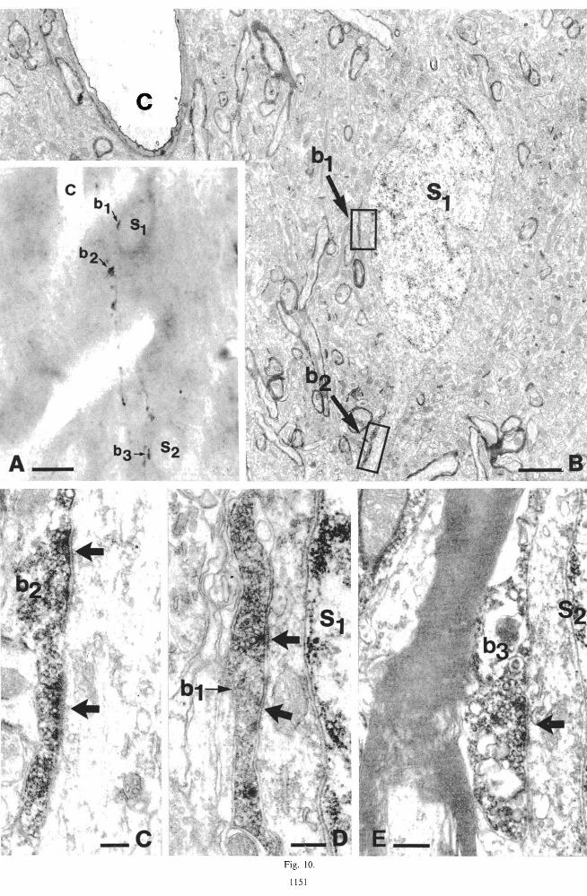
Fig. 10.
1151

* \.. .. 'f t t
A F ig. 11.
Synaptic connections of CCK-I neurons in visual cortex 1155
Synaptic connections of CCK-I neurons of other layers, like cells in layer VI (Fig. 1) with ascending projections and with pericellular terminal arrangements, were not examined in the electron microscope.
DISCUSSION
Four major findings emerge from this study: (1) Evidence is provided that CCK-I boutons, making synapses on somata, dendrites and spines, originate from local cortical neurons. (2) CCK-I boutons invariably form symmetrical synaptic contacts exclusively with neuronal elements. (3) Cortical CCK, as revealed by immunocytochemistry, is present in a heterogeneous group of neurons. (4) Some of the CCK-I cells were identified as double bouquet cells others as small basket cells in layers II-III, and these two types had distinct efferent synaptic connections.
Origin of the cholecystokinin -immunoreactive terminals in the visual cortex
CCK-I synaptic terminals could originate from three sources that have been reported to have CCK-I neuronal cell bodies. First, a considerable proportion of boutons could originate locally, since there are CCK-I somata in all cortical areasy,J4,27 The local origin is also supported by the ability of cortical tissue to synthesize CCK-8 in vivo.23 In this study we proved the local origin of some CCK-I boutons by tracing their origin from local somata. All the other CCK-I synaptic terminals may have the same origin since the terminals with identified origin and those found randomly in previous studies25.49 had the same fine structural characteristics and synaptic targets.
The second possibility is that some CCK-I terminals originate from cortico-cortical afferents. Several lines of circumstantial evidence suggest that CCK-I nonpyramidal neurons in layers JI-III may project to other cortical areas. Thus, Seroogy et al. 59 reported occasional CCK-I neurons that were thought to be non pyramidal cells which could be labelled retrogradely from the contralateral hemisphere. Some double bouquet cells in this study, on the basis of their smooth radial axons descending to the white matter, were called "projection" type neurons. Their main axons did not have a natural end at the border of the white matter, but could not be followed longer, probably because of the poor penetration of antibodies and the abundance of optically dense myelin. The course of these axons may explain the occurrence of CCK-I fibres in the white matter reported earlier.24.49.59
An alternative explanation, which represents the third possible origin of some CCK-I boutons in the visual cortex, is that some subcortical afferents contain immunoreactive CCK. Cortically projecting neurons in the ventral posterior medial nucleus of the thalamus have been shown to be immunoreactive for CCK42 and more recently the intralaminar nuclei of
NSC 194--G
the thalamus, that are known to project to the cortex,41 have also been reported to contain CCK-I neuronal cell bodies.72 The thalamic origin of some CCK-I material is also supported by a study of Fallon and Seroogy,16 who reported that nearly onefourth of the relay cells in the rat dorsal lateral geniculate nucleus (LGN) were immunoreactive for eCK. Furthermore, the density of CCK binding sites was found to be the highest in cortical layers III and IV,79 where a high proportion of the terminals are of thalamic origin.21 ,37 However it is difficult to reconcile these results with the fine structural features of thalamocortical afferents that, unlike CCK-I terminals~25.49 establish exclusively asymmetrical synapses in both rat and cat visual cortex.18.19.21.50 In addition, one would expect a much higher density of CCK-I terminals in layers IV and VI, if one-fourth of the geniculocortical afferents contained CCK (for density estimates in cat see Ref. 19).
At present several possible explanations of these controversial results may be given. Firstly, it could be argued that there are many thalamocortical afferents establishing symmetrical synapses in the cortex, but these have not been recognized so far. This is unlikely because even on a thalamocortical axon that made a few symmetrical synapses the overwhelming majority of boutons established asymmetrical contacts. 18,19
Another possibility is that CCK-l peptides are synthesized in relay cells of the LGN, but in the geniculocortical terminals they may be present in a molecular form not recognized by antibodies raised against the C-terminal tetrapeptide of CCK. The CCK immunoreactivity found in the relay cells in colchicine injected animals may also be a result of trauma caused by the injection, as also pointed out by Fallon and Seroogy.16 Clearly further work is necessary to establish the contribution of subcortical afferents to the CCK content of cortex.
Identity of the cholecystokinin -immunoreactive cell types
Double bouquet cells. The CCK-I neurons identified as double bouquet cells in this study might include neurons with a wide range of axonal morphology described in earlier studies. Thus, double bouquet cells with horsetail-shaped axons/4,64.65.73.74 bitufted neurons with horsetail or cone-shaped axons,45,46 or sparsely spinous bitufted neurons with vertical dendritic trees51 have been described in layers II-III. All of them have descending axon arbors of various shapes and diameters. From our CCK immunocytochemical material the true extent of the axonal arborizations of neurons could not be determined. Nevertheless, even this partial picture of CCK-I neurons allowed the identification of certain types of cell known from Golgi material.
Earlier electron microscopic studies on the termination of Golgi impregnated double bouquet cells emphasized the prevalence of nonpyramidal cells

1156 T. F. FREUND et al.
among the postsynaptic targets. 04•6
:' In the present study the cell bodies postsynaptic to the CCK-I double bouquet cell had features typical of pyramidal neurons. Only a proportion of the target dendrites CS type) could be identified as originating from nonpyramidal cells. This discrepancy might be due to several reasons. The criteria used for the identification of pyramidal versus nonpyramidal dendrites (for references see Refs 48 and 69) and somata6J6 may not be reliable in every case. It is also possible that the population of double bouquet cells with horsetailshaped axons is heterogeneous with regard to their efferent connectivity.
Small basket cells. Small basket cells localized mainly in layers II and upper III were first described by Ramon y Cajal,54 and further studied by Szentagothai. 73-
75 More recently several other authors8.45.51,76 demonstrated these cells in the cat. Small basket cells are characterized by a "short range" axon, which frequently contacts the somata of neurons in the same layer.45.51,74,75,76 Some small basket cells, also seen in our CCK material, have descending axons contacting cell bodies in a slab about 100 pm in diameter, and these cells may correspond to the columnar basket cells. 75 This type of neuron has also been recovered in experiments using intracellular recording and intracellular injection of horseradish peroxidase (HRP) (K. A. C. Martin and D. Whitteridge, personal communication). The lateral and radial dimensions of the axon will be revealed only by the intracellular marking techniques since neither in Golgi, nor in immunocytochemical material, was a complete reconstruction possible.
Golgi-electron microscopic analysis of superficial small basket cells,8 similar to our CCK -I cells, led to the conclusion that the main targets of these cells were neuronal somata. Our quantitative results demonstrated that dendrites were more frequent targets than somata. Nevertheless the proportion of somatic contacts is similar to that of large basket cells terminating also in layer III,69 and somewhat larger than the proportion of somatic efferent contacts of clutch cells terminating in layer IV.30 Among the target perikarya of CCK-I small basket cells, just as in the case of other basket cells,3o.69 we also found a few non pyramidal cells recognized on the basis of ultrastructural criteria.6.36 This could be expected because, just as in the hippocampus,47 many CCK-I cell bodies in the upper layers were surrounded by CCK-I varicosities, and received synapses from CCK-I boutons. Dendrites postsynaptic to CCK-I varicosities were mainly of the pyramidal type, but smooth stellate type shafts have also been found. The overall quantitative distribution of synaptic targets to the two previously analysed types of basket cells30,69
and the CCK-I small basket cells of layers II-III is very similar. However in spite of this similarity in efferent connections, it is unlikely that large basket cells and clutch cells were among the CCK-I neurons. Both of the latter neurons have somata of around
20-30 pm in diameter, which is larger than most CCK-I somata. Furthermore clutch cells and large basket cells in the cat, contrary to CCK-I neurons, have a very dense synaptic input on their somata. 30,69
Cells with two axons. Meyer44 described eight cells with two axon-like processes in a light microscopic Golgi study of the cat visual cortex. At least one of the cells illustrated in her study, the bitufted cell in layer Ill, is similar to one shown here in Fig. 11 to be immunoreactive for CCK. The group of cells with two axons is morphologically heterogenous, as also suggested by Meyer.44 However, our results indicate that they may be a homogeneous popUlation chemically, all being able to synthesize immunoreactive CCK. Cells with two axons in the cortex have not been reported in immunocytochemical studies for other peptides.7,9,13.l539 The presence of two axons may be a developmental curiosity which preferentially occurs amongst CCK-I neurons. The two axons are unlikely to have different functions because both in the Golgi44 and in the CCK-I material the territories occupied by the two axons were similar. The postsynaptic targets of the two axons of neuron Ns of this study (Fig. 11) were also qualitatively similar.
The rest of the CCK-I neurons had axons stained too incompletely to allow their proper classification. They correspond to bitufted or multipolar cells in layers II-VI, and horizontal cells in layer I, the latter also described by Demeulemeester et al. 9 Very little is known about the input-output characteristics of these heterogeneous cell classes, thus further work is necessary to establish whether they represent distinct cell populations.
Other cell types not immunoreactive for cholecystokinin. Basket and clutch cells have already been discussed above. Although, negative results in immunocytochemistry should be treated with caution, it is unlikely that the chandelier (or axo-axonic) cells contained immunoreactive CCK (see also Refs 25, 40 and 49). They are easy to exclude, because CCK-I was not found in terminals contacting axon initial segments, the only target of chandelier cells.63 It is noteworthy that the terminals of chandelier cells contain large dense-cored vesicles.67 In other terminals these are usually associated with peptide immunoreactivity, so chandelier cells may contain in addition to GABA 71 as yet unidentified peptides.
The peptide content of bipolar cells in the neocortex is not entirely clear. In the rat most immunocytochemical studies agree that vasoactive intestinal peptide (VIP) immunoreactivity is contained mainly in bipolar cells. 7,39 Bipolar cells were also reported to be among the much more heterogeneous group of neurons containing CCK immunoreactivity.13,}4,25,27.38.40,49 In the cat, CCK-I neurons
with the typical features of bipolar cells were not reported,9 and none of the cells in the present study were similar to those reported in the raL The difference is probably connected to differences in cortical organization between the two species.
Synaptic connections of CCK-I neurons in visual cortex 1157
Possible role of cholecystokinin as a neurotransmitter or modulator in the neocortex
CCK appears to fulfill many criteria for a role as neurotransmitter (see Introduction). However, it has been argued (for review see Ref. 34) that the synthesis of peptides such as CCK is energetically expensive. As the synthesis takes place in the cell body, its rate can only be regulated at the level of soma, and any such change is likely to be long term. These factors may limit the participation of peptides in the mediation of rapid inhibitory and excitatory s}gnals. 34
The coexistence of immunoreactive CCK with the inhibitory neurotransmitter GABA in neurons of the hippocampus32
.68 and cerebral cortex26
.68 is consis
tent with the morphological features of cortical (see above, and also Refs 25 and 49) and hippocampal47
CCK-I cells and terminals. It also suggests that the two neuroactive substances may interact if released together. Such an interaction is indicated by the results of Bradwejn and de Montigny3 who found that excitation by CCK was selectively antagonized by benzodiazepines in the hippocampus. Benzodiazepine receptors are assumed to be associated with GABA receptors, thus the CCK-antagonistic effect of benzodiazepines may have been mediated through the GABA receptor complex. This suggests that GABA and CCK, stored in the same interneurons, might interact on the postsynaptic cells.
It is difficult to reconcile the generally recorded excitatory effect of iontophoretically applied CCK-8
on cortical or hippocampal neurons4,12,28.52 with its localization in GABAergic cells. Only inhibitory effects of GABA have been reported in the visual cortex (for references see Ref. 62). Our present findings also suggest an inhibitory role for the CCK-I interneurons. Their boutons establish exclusively symmetrical synaptic contacts, which are found in locations thought to be ideal for inhibitory synapses, such as the somatic region and the proximal main dendrites.43 One possible explanation for the discrepancy between the anatomical data and the electrophysiological results is that the two techniques deal with different populations of synapses. It is possible that the immunocytochemical technique does not visualize the population of CCK neurons and terminals that are providing the input to receptors preferentially affected in iontophoretic experiments. The effect of CCK released from the terminals of GABAergic neurons, and preferentially revealed by current immunocytochemical methods, may not be detected without the simultaneous application of GABA.
Acknowledgements-We thank Mrs Klara Boczk6 for her excellent technical assistance, Edina Varga for her drawings and Dr J. Szentagothai for his helpful comments on the manuscript. We are grateful to Dr G. J. Dockray for the gift of an antiserum to CCK. The participation of Dr H. Takagi (Osaka, Japan) at the preliminary stages of these experiments is also acknowledged. This study was supported by the Hungarian Academy of Sciences.
REFERENCES
1. Beinfeld M. C. (1981) An HPLC and RIA analysis of the cholecystokinin peptides in rat brain. Neuropeptides 1, 203-207.
2. Beinfeld M. c., Mayer D. K., Eskay R. L., Jensen R. T. and Brownstein M. J. (1981) The distribution of cholecystokinin immunoreactivity in the central nervous system of the rat as determined by radioimmunoassay. Brain Res. 212,51-57.
3. Bradwejn J. and de Montigny C. (1984) Benzodiazepines antagonize cholecystokinin-induced activation of rat hippocampal neurones. Nature 312, 363-364.
4. Brooks P. A. and Kelly J. S. (1985) Cholecystokinin as a potent excitant of neurons of the dentate gyrus of rats. In Neuronal Cholecystokinin (eds Vanderhaeghen J.-J. and Crawley J. N.), Ann N. Y. A cad. Sci. 448, 361-374.
5. Bullier J., Kennedy H. and Salinger W. (1984) Branching and laminar origin of projections between visual cortical areas in the cat. J. comp. Neurol. 228, 329-341.
6. Colonnier M. (1968) Synaptic patterns on different cell types in the different laminae of the cat visual cortex. An electron-microscope study. Brain Res. 9, 268-287.
7. Connor J. R. and Peters A. (1984) Vasoactive intestinal polypeptide-immunoreactive neurons in rat visual cortex. Neuroscience 12, 1027-1044.
8. DeFelipe J. and Fairen A. (1982) A type of basket cell in superficial layers of the cat visual cortex. A Golgi--electron microscope study. Brain Res. 244, 9-16.
9. Demeulemeester H., Vandesande F. and Orban G. A. (1985) Immunocytochemical localization of somatostatin and cholecystokinin in the visual cortex. Brain Res. 332, 361-364.
10. Dockray G. J. (1980) Cholecystokinins in rat cerebral cortex: identification, purification and characterization by immunochemical methods. Brain Res. 188, 155-165.
11. Dockray G. J., Williams R. G. and Zhu W.-Y. (1981) Development of region-specific antisera for the C-terminal tetrapeptide of gastrin/cholecystokinin and their use in studies of immunoreactive forms of cholecystokinin in rat brain. Neurochem. Int. 3, 281-288.
12. Dodd J. and Kelly J. S. (1981) The actions of cholecystokinin and related peptides on pyramidal neurones of the mammalian hippocampus. Brain Res. 205, 337-350.
13. Emson P. C. and Hunt S. P. (1984) Peptide-containing neurons of the cerebral cortex. In Cerebral Cortex, Vol. 2. Functional Properties of Cortical Cells (eds Jones E. G. and Peters A.), pp. 145-169. Plenum Press, New York.
14. Emson P. c., Lee C. M. and Rehfeld C. F. (1980) Cholecystokinin octapeptide: vesicular localization and calcium dependent release from rat brain in vitro. L((e Sei. 26, 2157-2163.
15. Emson P. C. and Marley P. D. (1983) Cholecystokinin and vasoactive intestinal polypeptide. In Handbook of Psychopharmacology, Vol. 16 (eds Iversen L. L., Iversen S. D. and Snyder S. H.), pp. 255-306.
16. Fallon J. H. and Seroogy K. B. (IYM) VIsual and audItory pathways contain cholecystokinin: bVIdence from immunofluorescence and retrograde tracing. Neurosci. Let!. 45, 81-87.

1158 T. F. FREUND et al.
17. Freund T. F., Martin K. A. C, Smith A. D. and Somogyi P. (1983) Glutamate decarboxylase-immunoreactive terminals of Golgi-impregnated axoaxonic cells and of presumed basket cells in synaptic contact with pyramidal neurons of the cat's visual cortex. J. camp. Neural. 221, 263-278.
18. Freund T. F., Martin K. A. C and Whitteridge D. (1985) Innervation of cat visual areas 17 and 18 by physiologically identified X- and V-type thalamic afferents. I. Arborization patterns and quantitative distribution of postsynaptic elements. J. camp. Neural. 242, 263-274.
19. Freund T. F., Martin K. A. C, Somogyi P. and Whitteridge D. (1985) Innervation of cat visual areas 17 and 18 by physiologically identified X- and V-type thalamic afferents. n. Identification of postsynaptic targets by GABA immunocytochemistry and Golgi impregnation. J. camp. Neural. 242, 275-291.
20. Freund T. F., Powell J. F. and Smith A. D. (1984) Tyrosine hydroxylase-immunoreactive boutons in synaptic contact with identified striatonigral neurons, with particular reference to dendritic spines. Neurascience 13, 1189-1215.
21. Garey L. J. and Powell T. P. S. (1971) An experimental study of the termination of the lateral geniculo-cortical pathway in the cat and monkey. Proc. R. Soc. Land. B179, 41-63.
22. Gilbert C D. and Kelly J. P. (1975) The projections of cells in different layers of the cat's visual cortex. J. camp. Neural. 163, 81-106.
23. Golterman N. R., Rehfeld J. F. and Roigaard-Petersen H. (1980) In vivo biosynthesis of cholecystokinin in rat cerebral cortex. J. bioi. Chem. 255, 6181-6185.
24. Greenwood R. S., Godar S. E., Reaves T. A. and Hayward J. N. (1981) Cholecystokinin in hippocampal pathways. J. comp. Neurol. 203, 335-350.
25. Hendry S. H. C, Jones E. G. and Beinfeld M. C (1983) Cholecystokinin-immunoreactive neurons in rat and monkey cerebral cortex make symmetric synapses and have intimate associations with blood vessels. Proc. natn. A cad. Sci. U.s.A. 80, 2400-2404.
26. Hendry S. H. C, Jones E. G., DeFelipe J., Schmechel D., Brandon C and Emson P. C (1984) Neuropeptide-containing neurones of the cerebral cortex are also GABAergic. Proc. natn. A cad. Sci. U.S.A. 81, 6562-6530.
27. Innis R. 8., Correa F. M. A., Uhl G. R., Schneider 8. and Snyder S. (1979) Cholecystokinin octapeptide-like immunoreactivity: histochemical localization in rat brain. Proc. natn. A cad. Sci. U.S.A. 76, 521-525.
28. Ishibashi S., Oomura Y., Okajima T. and Shibata S. (1979) Cholecystokinin, motilin, and secretin effects on the central nervous system. Physiol. Behav. 23, 401-403.
29. Jones E. G. (1975) Varieties and distribution of non-pyramidal cells in the somatic sensory cortex of the squirrel monkey. J. comp. Neural. 160, 205-268.
30. Kisvarday Z. F., Martin K. A. C., Whitteridge D. and Somogyi P. (1985) Synaptic connections of intracellularly filled clutch cells: a type of small basket cell in the visual cortex of the cat. J. comp. Neurol. 241, 111-137.
31. Koch C and Poggio T. (1983) A theoretical analysis of electrical properties of spines. Proc. R. Soc. B218, 455-477.
32. Kosaka T., Kosaka K., Tateishi K., Hamoaka Y., Yanaihara N., Wu J.-y' and Hama K. (1985) GABAergic neurons containing CCK-8-like and for VIP-like immunoreactivities in the rat hippocampus and dentate gyrus. J. comp. Neural. 239, 420-430.
33. Krnjevic K. and Schwartz S. (1967) The action of gamma-aminobutyric acid on cortical neurones. Expl Brain Res. 3, 320-336.
34. Krnjevic K. (1984) Neurotransmitters in cerebral cortex, a general account. In Cerebral Cortex, Vo!. 2. Functional Properties of Cortical Cells (eds Jones E. G. and Peters A.), pp. 39-61. Plenum Press, New York.
35. Larsson L. I. and Rehfeld J. F. (1979) Localization and molecular heterogeneity of cholecystokinin in the central and peripheral nervous system. Brain Res. 165, 201-218.
36. LeVay S. (1973) Synaptic patterns in the visual cortex of the cat and monkey. Electron microscopy of Golgi preparations. J. comp. Neurol. 150, 53-86.
37. LeVay S. and Gilbert C. D. (1976) Laminar patterns of geniculocortical projection in the cat. Brain Res. 113, \-19. 38. Loren I., Alumets J., Hakanson R. and Sundler F. (1979) Distribution of gastrin and CCK-like peptides in rat brain.
An immunocytochemical study. Histochemistry 59, 249-257. 39. McDonald J. K., Parnavelas J. G., Karamanlidis A. and Brecha N. (1982) The morphology and distribution of
peptide-containing neurons in the adult and developing visual cortex of the rat. n. Vasoactive intestinal polypeptide. J. Neurocytol. 11, 825-837.
40. McDonald 1. K., Parnavelas J. G., Karamanlidis A. N., Rosenquist G. and Brecha N. (1982) The morphology and . distribution of peptide-containing neurons in the adult and developing visual cortex of the rat. Ill. Cholecystokinin. J. Neurocytol. 11, 881-895.
41. Macchi G., Bentivoglio M., Molinai M. and Minciacchi D. (1984) The thalamo-caudate versus thalamo-cortical projections as studied in the cat with fluorescent retrograde double labelling. Expl Brain Res. 54, 225-239.
42. Mantyh P. W. and Hunt S. P. (1984) Neuropeptides are present in projection neurones at all levels in visceral and taste pathways: from periphery to sensory cortex. Brain Res. 299, 297-311.
43. Martin K. A. C (1984) Neuronal circuits in cat striate cortex. In Cerebral Cortex, Vo!. 2. Functional Properties of Cortical Cells (eds Jones E. G. and Peters A.), pp. 241-284. Plenum Press, New York.
44. Meyer G. (1982) Short-axon neurons with two axon-like processes in the visual cortex of the cat. A Golgi study. Brain Res. 232, 455-459.
45. Meyer G. (1983) Axonal patterns and topography of short-axon neurons in visual areas 17, 18 and 19 of the cat. J. camp. Neurol. 220,405-428.
46. Meyer G. and Ferres-Torres R. (1984) Postnatal maturation of nonpyramidal neurons in the visual cortex of the cat. J. comp. Neural. 228, 226-244.
47. Nunzi M. G., Gorio A., Milan F., Freund T. F., Somogyi P. and Smith A. D. (1985) Cholecystokinin-immunoreactive cells form symmetrical synaptic contacts with pyramidal and non-pyramidal neurons in the hippocampus. J. camp. Neurol. 237, 485-505.
48. Peters A. and Jones E. G. (1984) Classification of cortical neurons. In Cellular Components of the Cerebral Cortex. Cerebral Cortex, Vo!. 1 (eds Peters A. and Jones E. G.), pp. 107--121. Plenum Press, New York.
49. Perers A., Miller M. and Kimerer L. M. (1983) Cholecystokinin-like immunoreactive neurons in rat cerebral cortex. Neuroscience 8, 431-448.
Synaptic connections of CCK-I neurons in visual cortex 1159
50, Peters A" Proskauer C. c., Feldman M, Land Kimerer L (1979) The projection of the lateral geniculate nucleus to area 17 of the rat cerebral cortex, V, Degenerating axon terminals synapsing with Golgi impregnated neurons, J, Neurocytol, 8, 331-357,
51. Peters A. and Regidor J, (1981) A reassessment of the forms of nonpyramidal neurons in area 17 of cat visual cortex, J, camp, Neurol. 203, 685-716,
52. Phillis J. N. and Kirkpatrick J, R, (1980) The actions of motilin, luteinizing-hormone-releasing hormone, cholecystokinin, somatostatin, vasoactive intestinal peptide and other pep tides on rat cerebral cortical neurons. Can. J, Physiol. Pharmac. 58, 612-623.
53, Pinget M" Straus E. and Yalow R. S. (1978) LocalizatiQn of cholecystokinin-like immunoreactivity in isolated nerve terminals. Proc. natn. A cad. Sci, USA. 75, 6324--6326.
54, Ramon y Cajal S, (1911) His/%gie du Systerne Nerveux de I'Homme et des Vertebres, Vol. 2. Maloine, Paris, 55, Rehfeld J. F. (1978) Immunochemical studies on cholecystokinin n. Distribution and molecular heterogeneity in the
central nervous system and small intestine of man and hog, J. bioI. Chem, 253, 4022-4030, 56, Rehfeld J, F. (1985) Neuronal cholecystokinin: One or multiple transmitters? J. Neurochem, 44, 1-10. 57, Reynolds E. S, (1963) The use of lead citrate at high pH as an electron opaque stain in electron microscopy, J, Cell
Bioi, 17, 208-212. 58, Saito A., Sankaran H" Goldfine L D. and Williams J, A. (1980) Cholecystokinin receptors in the brain: Characterization
and distribution. Science 208, 1155-1156, 59, Seroogy K. B" Fallon J. H., Loughlin S. E. and Leslie F, M, (1985) Few cortical cholecystokinin immunoreactive
neurones have long projections, Exp/ Brain Res, 59, 533-542, 60, Sillito A, M, (1975) The contribution of inhibitory mechanisms to the receptive field properties of neurones in the striate
cortex of the cat. J. Physio/" Land. 250, 305-329. 61. Sillito A. M. (1977) Inhibitory processes underlying the directional specificity of simple, complex and hypercomplex
cells in the cat's visual cortex. J, Physioi., Land. 271, 699-720. 62. Sillito A. M. (1984) Functional considerations of the operation of GABAergic inhibitory processes in the visual cortex.
In Cerebral Cortex, Vol. 2. Functional Properties of Cortical Cells (eds Jones E. G. and Peters A.), pp. 91-117. Plenum Press, New York.
63. Somogyi P. (1977) A specific "axo-axonal" interneuron in the visual cortex of the rat. Brain Res. 136, 345-350. 64. Somogyi P. and Cowey A. (1981) Combined Golgi and electron microscopic study on the synapses formed by double
bouquet cells in the visual cortex of the cat and monkey. J. comp. Neural. 195, 547--566. 65. Somogyi P. and Cowey A. (1984) Double bouquet cells. In Cerebral Cortex, Vol. 1. Cellular Components of the Cerebral
Cortex (eds Peters A. and Jones E. G.), pp. 337-360. Plenum Press, New York. 66. Somogyi P., Cowey A., Halasz N. and Freund T. F. (1981) Vertical organization of neurones accumulating [3H]GABA
in visual cortex of rhesus monkey. Nature 294, 761-763. 67. Somogyi P., Freund T. F. and Cowey A. (1982) The axo-axonic interneuron in the cerebral cortex of the rat, cat and
monkey. Neuroscience 7, 2577-2607. 68. Somogyi P., Hodgson A. J., Smith A. D., Nunzi M. G., Gracia M., Gorio A. and Wu J.-Y. (1984) Different populations
of GABAergic neurons in the visual cortex and hippocampus of the cat contain somatostatin- or cholecystokininimmunoreactive material. J, Neurosci. 4, 2590--2603.
69. Somogyi P., Kisvarday Z. F., Martin K. A. C. and Whitteridge D. (1983) Synaptic connections of morphologically identified and physiologically characterized large basket cells in the striate cortex of cat. Neuroscience 10, 261-294.
70. Somogyi P. and Takagi H. (1982) A note on the use of picric acid-paraformaldehyde-glutaraldehyde fixative for correlated light and electron microscopic immunocytochemistry. Neuroscience 7, 1779-1783.
71. Somogyi P., Freund T. F., Hodgson A. J., Somogyi J., Beroukas D. and Chubb I. W. (1985) Identified axo-axonic cells are immunoreactive for GABA in the hippocampus and visual cortex of the cat. Brain Res. 332, 143-149.
72. Sugimoto T., Itoh K., Yasui Y., Kaneko T. and Mizuno N. (1985) Coexistence of neuropeptides in projection neurons of the thalamus in the cat. Brain. Res. 347, 381-384.
73. Szentagothai J. (1969) Architecture of the cerebral cortex. In Basic Mechanisms of the Epi/epsies (eds Jasper H. H., Ward A. A. and Pope A.), pp. 13-28. Little, Brown and Co, Boston.
74. Szentagothai J. (1973) Synaptology of the visual cortex. In Handbook of Sensory Physiology, VIIj3B. (ed. Jung R.), pp. 269-324. Springer-Verlag, Berlin.
75. Szentagothai J. (1975) The "module-concept" in cerebral cortex architecture. Brain Res. 95, 475-496. 76. Tombol T. (1978) Comparative data on the Golgi architecture of interneurons of different cortical areas in cat and
rabbit. In Architectonics of the Cerebral Cortex (eds Brazier M. A. B. and Petsche H.), pp. 59-76. Raven Press, New York.
77. Uchizono K. (1965) Characteristics of excitatory and inhibitory synapses in the central nervous system of the cat. Nature 207, 642-643.
78. Vanderhaeghen J. J., Signeau J. C. and Gepts W. (1975) New peptide in the vertebrate CNS reacting with antigastrin antibodies. Nature 257, 604--605.
79. Van Oijk A., Richards J. G., Trzeciak A., Gillessen D. and Mohler H. (1984) Cholecystokinin receptors: Biochemical demonstration and autoradiographicallocalization in rat brain and pancreas using [3H]cholecystokining as radioligand. J. New'osci. 4, 1021-1033.
80. Zarbin M. A., Innis R. B., Wamsley J. K., Snyder S. H. and Kuhar M. J. (1983) Autoradiographic localization of cholecystokinin receptors in rodent brain. J. Neurosci. 3, 877-906.
(Accepted 15 May 1986)