symposium papers - respiratory · pdf filesymposium papers oxygen therapy in the neonatal care...
TRANSCRIPT
Symposium Papers
Oxygen Therapy in the Neonatal Care Environment
Brian K Walsh RRT-NPS, Toni M Brooks RRT, and Barry M Grenier RRT-NPS
IntroductionPhysiologic Effects of Oxygen Therapy: Benefits and Adverse Effects
Treatment of HypoxiaOxidative StressRetinopathy of PrematurityChronic Lung DiseaseLong-Term OutcomesOxygen During Resuscitation
Oxygen Delivery DevicesBlow-By OxygenOxygen HoodLow-Flow Nasal CannulaHigh-Flow Nasal Cannula
Device-Related ComplicationsAdvances in Oxygen Therapy
Closed-Loop FIO2Regulation
New-Generation Pulse OximetryDiscussion
Unresolved QuestionsFuture of Neonatal Oxygen Therapy
The use of oxygen in the treatment of neonates with respiratory distress has been reported for morethan a century. Oxygen therapy is generally titrated to one or more measures of blood oxygenationand administered to reverse or prevent hypoxia. Individual responses to oxygen therapy varygreatly, depending on the particular cause of hypoxia and the degree of impairment. Despite thisfocused purpose, oxygen administration in this patient population has become complex. The longerwe deliver this drug, the more we discover its beneficial and detrimental effects. New and innovativeways to deliver and monitor this therapy have improved outcomes. Despite this vast experiencethere still remain some unanswered questions regarding the use of oxygen in the neonatal envi-ronment. Nonetheless, oxygen is a major staple in our treatment arsenal for neonates. Key words:oxygen; neonatal; infant, newborn; retinopathy of prematurity; oxygen inhalation therapy. [Respir Care2009;54(9):1193–1202. © 2009 Daedalus Enterprises]
Introduction
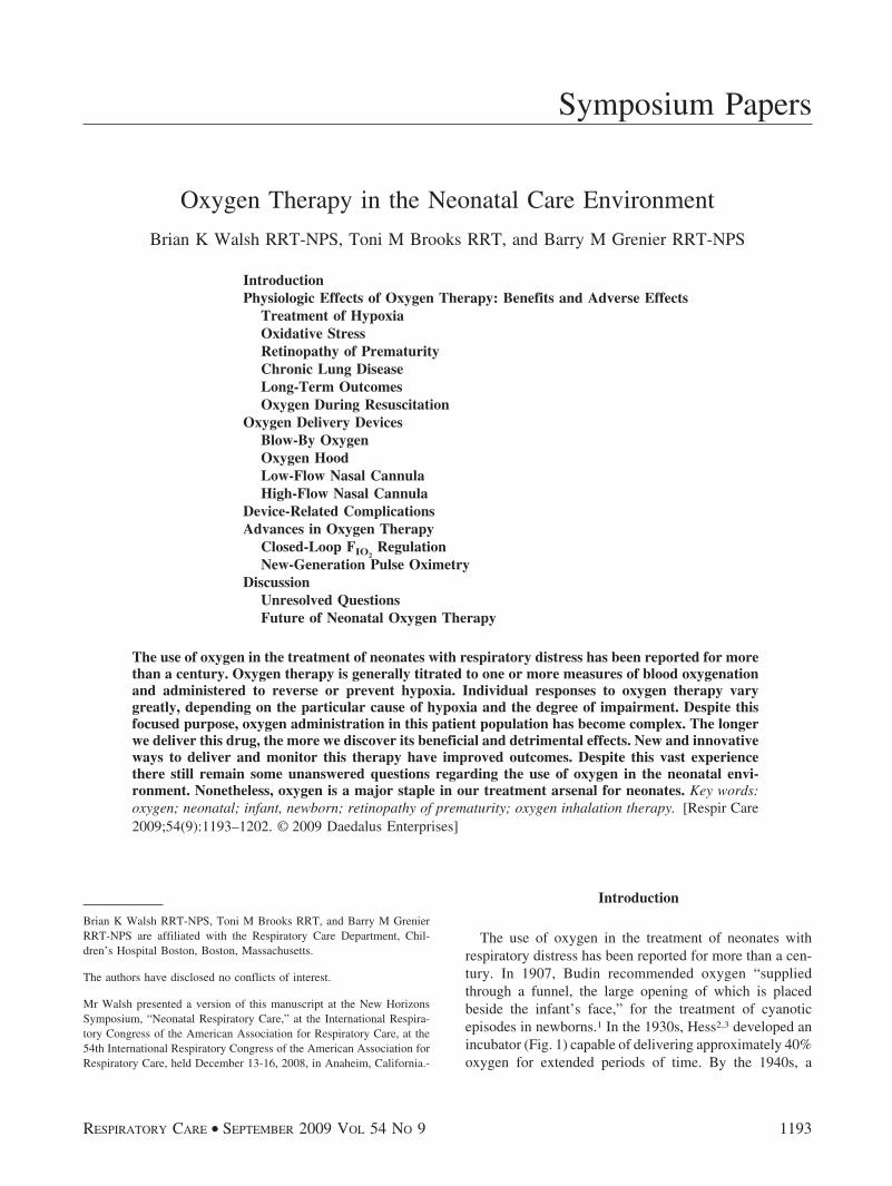
The use of oxygen in the treatment of neonates withrespiratory distress has been reported for more than a cen-tury. In 1907, Budin recommended oxygen “suppliedthrough a funnel, the large opening of which is placedbeside the infant’s face,” for the treatment of cyanoticepisodes in newborns.1 In the 1930s, Hess2,3 developed anincubator (Fig. 1) capable of delivering approximately 40%oxygen for extended periods of time. By the 1940s, a
Brian K Walsh RRT-NPS, Toni M Brooks RRT, and Barry M GrenierRRT-NPS are affiliated with the Respiratory Care Department, Chil-dren’s Hospital Boston, Boston, Massachusetts.
The authors have disclosed no conflicts of interest.
Mr Walsh presented a version of this manuscript at the New HorizonsSymposium, “Neonatal Respiratory Care,” at the International Respira-tory Congress of the American Association for Respiratory Care, at the54th International Respiratory Congress of the American Association forRespiratory Care, held December 13-16, 2008, in Anaheim, California.-
RESPIRATORY CARE • SEPTEMBER 2009 VOL 54 NO 9 1193

commercially available incubator capable of providing ahigh concentration of oxygen facilitated the liberal use ofoxygen for the treatment of cyanosis, apnea, and periodicbreathing in newborns.1,4 Throughout this time, oxygenadministration was guided by the clinical observations ofskin color, as well as the rate, regularity, and work ofbreathing. It wasn’t until the 1960s and 1970s that tech-nology—micro-sampling of blood gases, transcutaneousoxygen monitoring, and, later, pulse oximetry—becameavailable for more precise monitoring of physiologic ef-fect.
The overall goal of oxygen therapy is to achieve ade-quate oxygenation using the lowest concentration of in-spired oxygen. However, achieving this goal is compli-cated by a number of factors. Despite over 75 years ofroutine oxygen administration to newborn infants, the op-timal level of oxygenation—one that avoids the detrimen-tal effects of hypoxia on the one hand, and those caused by
hyperoxia on the other—has not yet been clearly defined,5-7
leading to wide variations in practice.8 Even the term “ad-equate oxygenation” is not clear.9 Other complicating fac-tors in achieving the goals of neonatal oxygen therapyinclude patient size, tolerance of delivery devices, andvariability in the use of delivery devices, which suggestthat clinicians often lack adequate knowledge in the use ofoxygen delivery equipment,10 and the lack of training inthe concepts of neonatal oxygenation and equipment usedto monitor the effects of oxygen therapy.11
Physiologic Effects of Oxygen Therapy:Benefits and Adverse Effects
Despite its universal acceptance as a life-saving therapyfor newborns, oxygen administration is associated withnumerous physiologic effects, particularly when used totreat premature infants.
Treatment of Hypoxia
While oxygen therapy is generally titrated to some mea-sure of arterial oxygenation in response to an abnormally
Correspondence: Brian K Walsh RRT-NPS, Respiratory Care Depart-ment, Children’s Hospital Boston, 300 Longwood Avenue, MA-861,Boston MA 02115. E-mail: [email protected].
Fig. 1. Hess bed equipped with an oxygen therapy unit (A-side view). 1: Pressure gauge. 2: Oxygen flow regulator. 3: Flow meter. 4: Glassand metal hinged door for feeding purposes. 5: Thermometer window. 6: Metal hinged door for purposes of body care of the infant.7: Ventilator with small and large exit openings. 8–12:Controls for maintaining temperature in water-jacket of the incubator. (From Refer-ence 3, with permission.)
OXYGEN THERAPY IN THE NEONATAL CARE ENVIRONMENT
1194 RESPIRATORY CARE • SEPTEMBER 2009 VOL 54 NO 9

low level of blood oxygen, or hypoxemia, oxygen is ad-ministered to the neonate to reverse or prevent hypoxia.Hypoxia is defined as a deficit of oxygen at the cellularlevel, and is commonly caused by one or more of thefollowing: the reduced availability of oxygen at the alve-olar level, due to pulmonary disease (hypoventilation, un-even matching of ventilation to perfusion, diffusion de-fects); intrapulmonary shunts or “right to left” cardiacshunts; reduced oxygen carrying capacity due to anemia orabnormal blood hemoglobin; or impaired oxygen deliverydue to shock, heart failure, or localized decreases in per-fusion.12,13 Left untreated, hypoxia can lead to serious andpermanent brain injury and death.12
Individual responses to oxygen therapy vary greatly,depending on the particular cause of hypoxia and the de-gree of impairment. Hypoxia caused by hypoventilationand ventilation-perfusion anomalies associated with pul-monary disease will be most responsive to oxygen therapy.Even large increases in FIO2
will produce only small in-creases in available oxygen if hypoxia is caused by cardiacshunts, shock, and hemoglobin deficiency/dysfunction.12,13
It should be stressed, however, that even small increases inoxygen availability may prevent life-threatening decom-pensation in the hypoxic neonate.
Oxidative Stress
The role of oxygen and oxidative stress in the develop-ment of a number of neonatal diseases has generated muchinterest. Oxidative stress has been defined as an imbalancebetween pro-oxidant and anti-oxidant forces in the body.14
Pro-oxidants include oxygen radicals or reactive oxygenspecies, which can be cytotoxic because of their ability toalter cellular components and function. Reactive oxygenspecies are generated as a result of normal mitochondrialrespiration, but also during the reperfusion phase of hy-poxic tissue injury and in association with infection andinflammation.15,16 Oxygen is “toxic” because of the pro-duction of reactive oxygen species; thus oxygen adminis-tration increases oxidative stress.
Antioxidant defenses include the enzymes superoxidedismutase, catalase, and glutathione. Nonenzymatic anti-oxidants start to cross the placenta in late gestation, andinclude vitamins A, C, E, and ubiquinone. Premature in-fants are at particular risk from oxidative stress becauseboth endogenous and passively acquired exogenous anti-oxidant defense systems do not accelerate in maturationuntil late in the third trimester.15,17,18 Investigators haveattempted to reverse or prevent the damage associated withreactive oxygen species not only by appropriate oxygenadministration but also by administering antioxidants; how-ever, this therapy has not shown to be effective.19 Saugstadhas suggested the term oxygen radical disease of neona-tology to encompass a variety of newborn diseases whose
pathogenesis involves oxidative stress and injury, whichinclude retinopathy of prematurity, bronchopulmonary dys-plasia, necrotizing enterocholitis, and intraventricular hem-orrhage.20
Retinopathy of Prematurity
Though long recognized as a complication of oxygentherapy, retinopathy of prematurity remains a major causeof morbidity for premature infants.21 Retinopathy of pre-maturity is a disease limited almost exclusively to prema-ture infants and is characterized by abnormal vasculariza-tion of the retina, causing a range of vision impairment,including blindness. Much has been described in the lit-erature regarding the role of supplemental oxygen in thedevelopment of retinopathy of prematurity.22-24 The al-tered regulation of vascular endothelial growth factor hasbeen suggested25,26 as one of the factors in the pathogen-esis of retinopathy of prematurity (Fig. 2). In premature
Fig. 2. The proposed role of vascular endothelial growth factor(VEGF). A: It is hypothesized that normal retinal vessel develop-ment is stimulated by production of VEGF (red) anterior to thedeveloping vasculature. In addition, maintenance of some retinalvessels is dependent on VEGF. B: In the first phase of retinopathyof prematurity, exposure to relative hyperoxia after birth interruptsthe gradient of physiologic hypoxia in the immature retina, leadingto downregulation of VEGF production, with associated vaso-obliteration and cessation of vessel growth. C: As the metabolicdemand of the developing retina increases, the nonperfused por-tions of the retina become hypoxic and overproduce VEGF. D: Neo-vascularization occurs in response to overproduction of VEGF,producing retinopathy of prematurity. If VEGF production persists,then the retinopathy of prematurity will progress. (From Refer-ence 26, with permission.)
OXYGEN THERAPY IN THE NEONATAL CARE ENVIRONMENT
RESPIRATORY CARE • SEPTEMBER 2009 VOL 54 NO 9 1195
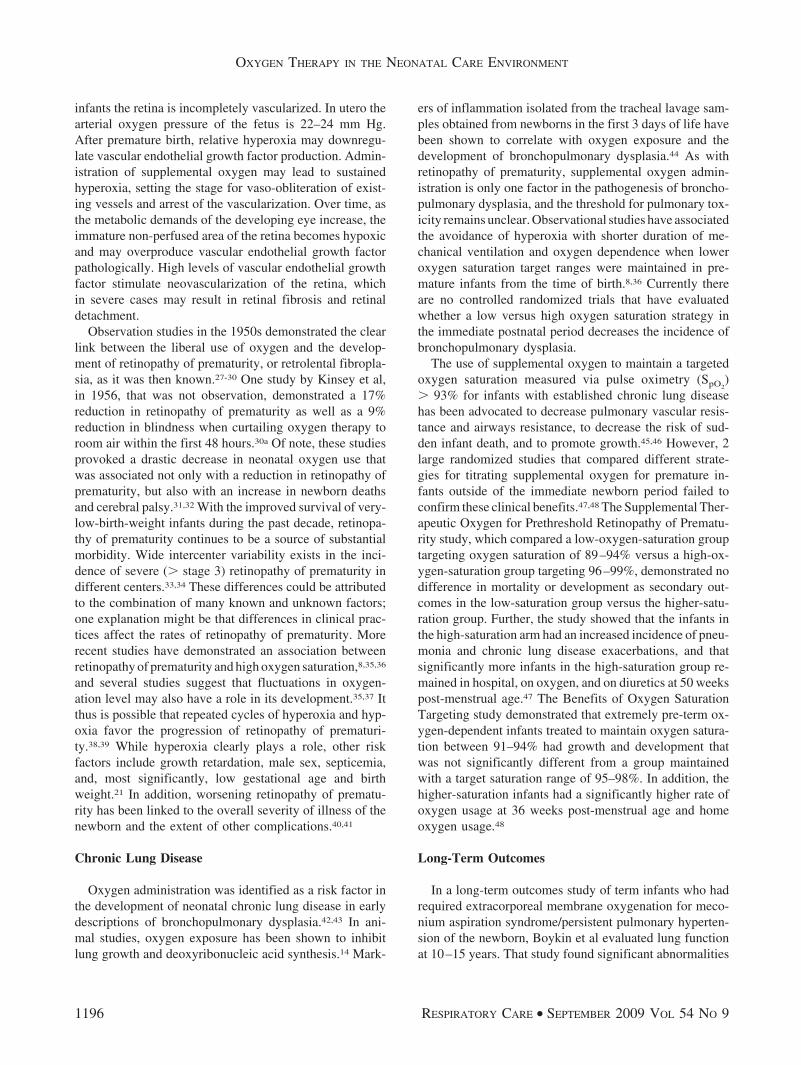
infants the retina is incompletely vascularized. In utero thearterial oxygen pressure of the fetus is 22–24 mm Hg.After premature birth, relative hyperoxia may downregu-late vascular endothelial growth factor production. Admin-istration of supplemental oxygen may lead to sustainedhyperoxia, setting the stage for vaso-obliteration of exist-ing vessels and arrest of the vascularization. Over time, asthe metabolic demands of the developing eye increase, theimmature non-perfused area of the retina becomes hypoxicand may overproduce vascular endothelial growth factorpathologically. High levels of vascular endothelial growthfactor stimulate neovascularization of the retina, whichin severe cases may result in retinal fibrosis and retinaldetachment.
Observation studies in the 1950s demonstrated the clearlink between the liberal use of oxygen and the develop-ment of retinopathy of prematurity, or retrolental fibropla-sia, as it was then known.27-30 One study by Kinsey et al,in 1956, that was not observation, demonstrated a 17%reduction in retinopathy of prematurity as well as a 9%reduction in blindness when curtailing oxygen therapy toroom air within the first 48 hours.30a Of note, these studiesprovoked a drastic decrease in neonatal oxygen use thatwas associated not only with a reduction in retinopathy ofprematurity, but also with an increase in newborn deathsand cerebral palsy.31,32 With the improved survival of very-low-birth-weight infants during the past decade, retinopa-thy of prematurity continues to be a source of substantialmorbidity. Wide intercenter variability exists in the inci-dence of severe (� stage 3) retinopathy of prematurity indifferent centers.33,34 These differences could be attributedto the combination of many known and unknown factors;one explanation might be that differences in clinical prac-tices affect the rates of retinopathy of prematurity. Morerecent studies have demonstrated an association betweenretinopathy of prematurity and high oxygen saturation,8,35,36
and several studies suggest that fluctuations in oxygen-ation level may also have a role in its development.35,37 Itthus is possible that repeated cycles of hyperoxia and hyp-oxia favor the progression of retinopathy of prematuri-ty.38,39 While hyperoxia clearly plays a role, other riskfactors include growth retardation, male sex, septicemia,and, most significantly, low gestational age and birthweight.21 In addition, worsening retinopathy of prematu-rity has been linked to the overall severity of illness of thenewborn and the extent of other complications.40,41
Chronic Lung Disease
Oxygen administration was identified as a risk factor inthe development of neonatal chronic lung disease in earlydescriptions of bronchopulmonary dysplasia.42,43 In ani-mal studies, oxygen exposure has been shown to inhibitlung growth and deoxyribonucleic acid synthesis.14 Mark-
ers of inflammation isolated from the tracheal lavage sam-ples obtained from newborns in the first 3 days of life havebeen shown to correlate with oxygen exposure and thedevelopment of bronchopulmonary dysplasia.44 As withretinopathy of prematurity, supplemental oxygen admin-istration is only one factor in the pathogenesis of broncho-pulmonary dysplasia, and the threshold for pulmonary tox-icity remains unclear. Observational studies have associatedthe avoidance of hyperoxia with shorter duration of me-chanical ventilation and oxygen dependence when loweroxygen saturation target ranges were maintained in pre-mature infants from the time of birth.8,36 Currently thereare no controlled randomized trials that have evaluatedwhether a low versus high oxygen saturation strategy inthe immediate postnatal period decreases the incidence ofbronchopulmonary dysplasia.
The use of supplemental oxygen to maintain a targetedoxygen saturation measured via pulse oximetry (SpO2
)� 93% for infants with established chronic lung diseasehas been advocated to decrease pulmonary vascular resis-tance and airways resistance, to decrease the risk of sud-den infant death, and to promote growth.45,46 However, 2large randomized studies that compared different strate-gies for titrating supplemental oxygen for premature in-fants outside of the immediate newborn period failed toconfirm these clinical benefits.47,48 The Supplemental Ther-apeutic Oxygen for Prethreshold Retinopathy of Prematu-rity study, which compared a low-oxygen-saturation grouptargeting oxygen saturation of 89–94% versus a high-ox-ygen-saturation group targeting 96–99%, demonstrated nodifference in mortality or development as secondary out-comes in the low-saturation group versus the higher-satu-ration group. Further, the study showed that the infants inthe high-saturation arm had an increased incidence of pneu-monia and chronic lung disease exacerbations, and thatsignificantly more infants in the high-saturation group re-mained in hospital, on oxygen, and on diuretics at 50 weekspost-menstrual age.47 The Benefits of Oxygen SaturationTargeting study demonstrated that extremely pre-term ox-ygen-dependent infants treated to maintain oxygen satura-tion between 91–94% had growth and development thatwas not significantly different from a group maintainedwith a target saturation range of 95–98%. In addition, thehigher-saturation infants had a significantly higher rate ofoxygen usage at 36 weeks post-menstrual age and homeoxygen usage.48
Long-Term Outcomes
In a long-term outcomes study of term infants who hadrequired extracorporeal membrane oxygenation for meco-nium aspiration syndrome/persistent pulmonary hyperten-sion of the newborn, Boykin et al evaluated lung functionat 10–15 years. That study found significant abnormalities
OXYGEN THERAPY IN THE NEONATAL CARE ENVIRONMENT
1196 RESPIRATORY CARE • SEPTEMBER 2009 VOL 54 NO 9
in lung function and that the most significant predictor oflong-term pulmonary outcomes was the duration of oxy-gen use post-extracorporeal-membrane-oxygenation de-cannulation.49
Oxygen During Resuscitation
The use of 100% oxygen during neonatal resuscitationhas also been challenged, on the premise that large andabrupt increases in blood oxygen level after birth can in-crease oxidative stress.20 Several studies have comparedthe use of 21% to 100% oxygen during resuscitation. Threerecent meta-analyses of these data concluded that the useof room air during the resuscitation of depressed newbornsresulted in a significantly reduced risk of neonatal mortal-ity.50-52 The studies found no significant difference in theincidence of severe hypoxic encephalopathy between the21% oxygen and 100% oxygen groups. Limitations to someof the studies in these analyses include a lack of blindingin some studies, and the exclusion of stillbirths.9 In onesmall recent study, the resuscitation of premature new-borns with 50% versus 100% oxygen did not reduce theincidence of bronchopulmonary dysplasia or improve othershort-term outcomes.53 The results of a recent study byEscrig et al54 indicate that extremely premature newbornscan be safely resuscitated with a low initial oxygen con-centration. Related to the use of oxygen in the deliveryroom for resuscitation, limited evidence suggests that theexposure of newborns to oxygen for 3 min or longer im-mediately after birth increases the risk of childhood can-cer.55,56
Oxygen Delivery Devices
Blow-By Oxygen
Blow-by oxygen delivery is the simplest and least cum-bersome form of available devices to provide oxygen ther-apy to the neonate, but it is also the least reliable in de-livering a specific FIO2
. Blow-by oxygen can be achievednumerous ways, but is most commonly done by means oflarge-bore or oxygen tubing connected to a face tent orsimple mask that is placed a relatively short distance from,and directed toward, the patient’s face. This type of oxy-gen delivery is ideal for patients who cannot tolerate morecumbersome oxygen delivery devices and/or require a lesseramount of oxygen. There is limited evidence that suggeststhat blow-by therapy can deliver low concentrations ofoxygen (0.3–0.4 at 10 L/min of flow) to an area largeenough to provide some level of oxygen therapy to theneonate, assuming adequate positioning of the device.57
Therefore, this type of therapy should be reserved for in-fants who do not a require high inspired oxygen concen-tration but may require short-term or intermittent oxygentherapy.
Oxygen Hood
An oxygen hood (cube) is a plastic enclosure that sur-rounds the head of the neonate, to which a continuous flowof humidified oxygen is supplied by means of an air-entrainment device or an air-oxygen blender. Fixed oxy-gen concentrations from 0.21 to 1.0 can be maintainedwith a minimum of 7 L/min oxygen flow into the hood.This minimum gas flow also ensures that exhaled carbondioxide is flushed out and not rebreathed. Although anoxygen hood can theoretically deliver 1.0 FIO2
, this deviceis best suited for patients who require less than 0.5 FIO2
.Patients requiring higher FIO2
can be managed in a hood,but it becomes increasingly difficult to maintain higheroxygen concentrations with the large neck opening and aless than optimal seal around the edges.58-60 An oxygenhood is an ideal method of oxygen delivery for neonateswho require higher fractional inspired oxygen concentra-tions but cannot tolerate more cumbersome oxygen deliv-ery devices.
Low-Flow Nasal Cannula
Low-flow nasal cannula remains one of the most com-mon and widely used neonatal oxygen delivery devices.This low-flow device delivers a fractional concentration ofoxygen to the patient through 2 soft prongs that rest in thepatient’s anterior nares. The distal end of the cannula tub-ing is then attached to either a 100% oxygen source flowmeter or to an air-oxygen blender. Finer et al found thatoxygen concentrations delivered to the neonate via nasalcannula varied from 22% to 95% with a maximum flowrate of 2 L/min.61 The precise FIO2
actually delivered to thepatient is contingent upon a number of factors, but mostspecifically on the set flow through the nasal cannula andits relation to the patient’s inspiratory flow demand. Aninspiratory flow demand greater than that supplied by thenasal cannula causes the exact FIO2
delivered to the patientto be a blend of the nasally inhaled oxygen with entrainedroom air through the nares and mouth.10,58,61 While actualoxygen concentrations delivered to the patient are vari-able, a nasal cannula remains a fairly trusted and effectivemethod of offering oxygen therapy to the neonate.
High-Flow Nasal Cannula
Nasal cannula oxygen therapy is a staple and continuesto be redefined to improve patient comfort, compliance,and outcomes. The concept of high flow and high humid-ity via a nasal cannula, however, is a fairly new conceptand was first introduced by Vapotherm to the respiratorycare community in the spring of 2002, after receiving Foodand Drug Administration 510K clearance in the fall of2001. Prior to high-flow nasal cannula, most clinicians
OXYGEN THERAPY IN THE NEONATAL CARE ENVIRONMENT
RESPIRATORY CARE • SEPTEMBER 2009 VOL 54 NO 9 1197
considered it uncomfortable to use a flow of greater than6 L/min via nasal cannula in adults; this was primarily dueto the lack of adequate humidification available via nasalcannula delivery. Little consensus existed in the neonatalpatient population on the parameters defining high-flownasal cannula, but for our discussion high-flow nasal can-nula is classified as a fixed-performance oxygen deliverysystem that is capable of delivering a specific oxygen con-centration at flows that meet or exceed the inspiratory flowdemand of the patient.58 This type of oxygen deliverydevice is composed of traditional nasal cannula style prongsthat rest in the patient’s anterior nares and allow heated,humidified oxygen to be delivered at flow rates of 1–8 L/min, while an air-oxygen blender allows FIO2
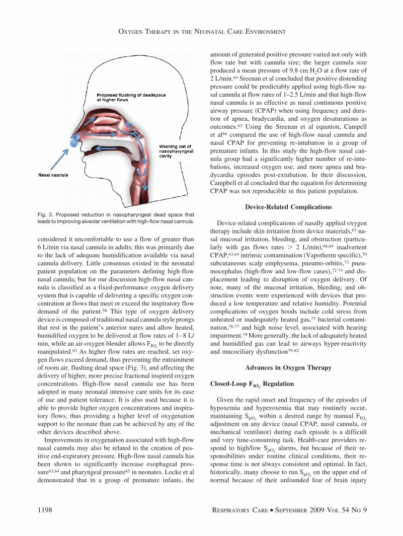
to be directlymanipulated.62 As higher flow rates are reached, set oxy-gen flows exceed demand, thus preventing the entrainmentof room air, flushing dead space (Fig. 3), and affecting thedelivery of higher, more precise fractional inspired oxygenconcentrations. High-flow nasal cannula use has beenadopted in many neonatal intensive care units for its easeof use and patient tolerance. It is also used because it isable to provide higher oxygen concentrations and inspira-tory flows, thus providing a higher level of oxygenationsupport to the neonate than can be achieved by any of theother devices described above.
Improvements in oxygenation associated with high-flownasal cannula may also be related to the creation of pos-itive end-expiratory pressure. High-flow nasal cannula hasbeen shown to significantly increase esophageal pres-sure63,64 and pharyngeal pressure65 in neonates. Locke et aldemonstrated that in a group of premature infants, the
amount of generated positive pressure varied not only withflow rate but with cannula size; the larger cannula sizeproduced a mean pressure of 9.8 cm H2O at a flow rate of2 L/min.64 Sreenan et al concluded that positive distendingpressure could be predictably applied using high-flow na-sal cannula at flow rates of 1–2.5 L/min and that high-flownasal cannula is as effective as nasal continuous positiveairway pressure (CPAP) when using frequency and dura-tion of apnea, bradycardia, and oxygen desaturations asoutcomes.63 Using the Sreenan et al equation, Campellet al66 compared the use of high-flow nasal cannula andnasal CPAP for preventing re-intubation in a group ofpremature infants. In this study the high-flow nasal can-nula group had a significantly higher number of re-intu-bations, increased oxygen use, and more apnea and bra-dycardia episodes post-extubation. In their discussion,Campbell et al concluded that the equation for determiningCPAP was not reproducible in this patient population.
Device-Related Complications
Device-related complications of nasally applied oxygentherapy include skin irritation from device materials,67 na-sal mucosal irritation, bleeding, and obstruction (particu-larly with gas flows rates � 2 L/min),68,69 inadvertentCPAP,63,64 intrinsic contamination (Vapotherm specific),70
subcutaneous scalp emphysema, pneumo-orbitis,71 pneu-mocephalus (high-flow and low-flow cases),72-74 and dis-placement leading to disruption of oxygen delivery. Ofnote, many of the mucosal irritation, bleeding, and ob-struction events were experienced with devices that pro-duced a low temperature and relative humidity. Potentialcomplications of oxygen hoods include cold stress fromunheated or inadequately heated gas,75 bacterial contami-nation,76,77 and high noise level, associated with hearingimpairment.78 More generally, the lack of adequately heatedand humidified gas can lead to airways hyper-reactivityand mucociliary dysfunction79-82
Advances in Oxygen Therapy
Closed-Loop FIO2Regulation
Given the rapid onset and frequency of the episodes ofhypoxemia and hyperoxemia that may routinely occur,maintaining SpO2
within a desired range by manual FIO2
adjustment on any device (nasal CPAP, nasal cannula, ormechanical ventilator) during each episode is a difficultand very time-consuming task. Health-care providers re-spond to high/low SpO2
alarms, but because of their re-sponsibilities under routine clinical conditions, their re-sponse time is not always consistent and optimal. In fact,historically, many choose to run SpO2
on the upper end ofnormal because of their unfounded fear of brain injury
Fig. 3. Proposed reduction in nasopharyngeal dead space thatleads to improving alveolar ventilation with high-flow nasal cannula.
OXYGEN THERAPY IN THE NEONATAL CARE ENVIRONMENT
1198 RESPIRATORY CARE • SEPTEMBER 2009 VOL 54 NO 9
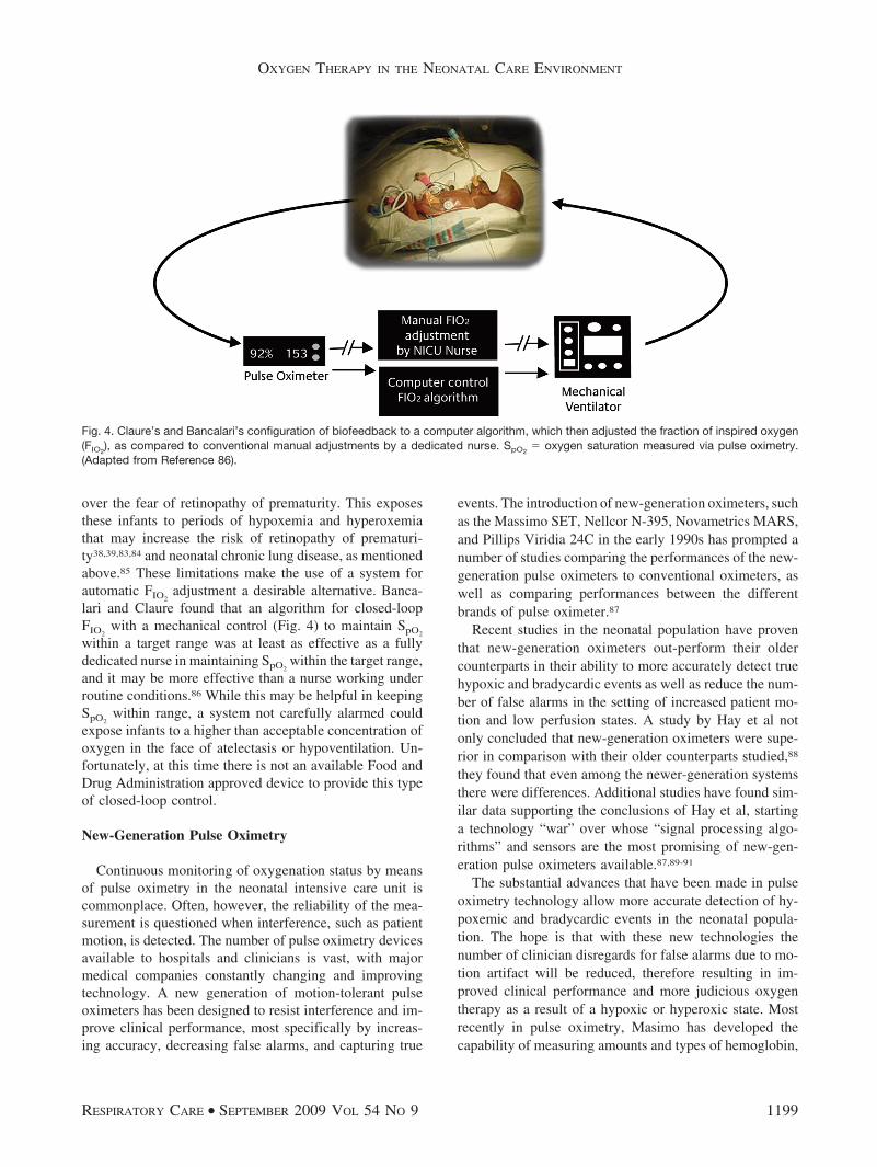
over the fear of retinopathy of prematurity. This exposesthese infants to periods of hypoxemia and hyperoxemiathat may increase the risk of retinopathy of prematuri-ty38,39,83,84 and neonatal chronic lung disease, as mentionedabove.85 These limitations make the use of a system forautomatic FIO2
adjustment a desirable alternative. Banca-lari and Claure found that an algorithm for closed-loopFIO2
with a mechanical control (Fig. 4) to maintain SpO2
within a target range was at least as effective as a fullydedicated nurse in maintaining SpO2
within the target range,and it may be more effective than a nurse working underroutine conditions.86 While this may be helpful in keepingSpO2
within range, a system not carefully alarmed couldexpose infants to a higher than acceptable concentration ofoxygen in the face of atelectasis or hypoventilation. Un-fortunately, at this time there is not an available Food andDrug Administration approved device to provide this typeof closed-loop control.
New-Generation Pulse Oximetry
Continuous monitoring of oxygenation status by meansof pulse oximetry in the neonatal intensive care unit iscommonplace. Often, however, the reliability of the mea-surement is questioned when interference, such as patientmotion, is detected. The number of pulse oximetry devicesavailable to hospitals and clinicians is vast, with majormedical companies constantly changing and improvingtechnology. A new generation of motion-tolerant pulseoximeters has been designed to resist interference and im-prove clinical performance, most specifically by increas-ing accuracy, decreasing false alarms, and capturing true
events. The introduction of new-generation oximeters, suchas the Massimo SET, Nellcor N-395, Novametrics MARS,and Pillips Viridia 24C in the early 1990s has prompted anumber of studies comparing the performances of the new-generation pulse oximeters to conventional oximeters, aswell as comparing performances between the differentbrands of pulse oximeter.87
Recent studies in the neonatal population have proventhat new-generation oximeters out-perform their oldercounterparts in their ability to more accurately detect truehypoxic and bradycardic events as well as reduce the num-ber of false alarms in the setting of increased patient mo-tion and low perfusion states. A study by Hay et al notonly concluded that new-generation oximeters were supe-rior in comparison with their older counterparts studied,88
they found that even among the newer-generation systemsthere were differences. Additional studies have found sim-ilar data supporting the conclusions of Hay et al, startinga technology “war” over whose “signal processing algo-rithms” and sensors are the most promising of new-gen-eration pulse oximeters available.87,89-91
The substantial advances that have been made in pulseoximetry technology allow more accurate detection of hy-poxemic and bradycardic events in the neonatal popula-tion. The hope is that with these new technologies thenumber of clinician disregards for false alarms due to mo-tion artifact will be reduced, therefore resulting in im-proved clinical performance and more judicious oxygentherapy as a result of a hypoxic or hyperoxic state. Mostrecently in pulse oximetry, Masimo has developed thecapability of measuring amounts and types of hemoglobin,
Fig. 4. Claure’s and Bancalari’s configuration of biofeedback to a computer algorithm, which then adjusted the fraction of inspired oxygen(FIO2
), as compared to conventional manual adjustments by a dedicated nurse. SpO2� oxygen saturation measured via pulse oximetry.
(Adapted from Reference 86).
OXYGEN THERAPY IN THE NEONATAL CARE ENVIRONMENT
RESPIRATORY CARE • SEPTEMBER 2009 VOL 54 NO 9 1199

which may lead to a more precise monitoring and controlof oxygen delivery noninvasively.92
Discussion
Unresolved Questions
Many unresolved questions remain when discussing neo-natal oxygen therapy, but one specific question that arisesis which SpO2
ranges are most appropriate for the newborn.This question is complex in that the most appropriate rangemay be different in different contexts. Many have con-ducted research with different SpO2
ranges, showing equiv-ocal if not better outcomes to higher SpO2
ranges; however,there have not been consistent ranges among the studies.In a recent review of resuscitation and ongoing manage-ment of pre-term infants, Finer discusses his recommen-dation that an SpO2
range of 85–93%, with alarms set �1–2%above and below that range, was most appropriate.17 How-ever, this does not answer the question for near-term, terminfants, or patients who have developed chronic lung dis-ease and are susceptible to pulmonary hypertension. Fur-ther studies are needed to fully answer this question.
Two other unresolved questions are whether or not ahigh-flow nasal cannula can substitute for nasal CPAP andwhether or not high-flow nasal cannula can replace thehigh FIO2
oxygen hood. It is fairly clear that high-flownasal cannula is a safe oxygen delivery device, as therehave been hundreds of infants studied, with few adverseevents.62,63,66,68,70,93-96 It appears to have the same compli-cations as traditional low-flow oxygen delivery devices,yet is able to provide a higher humidity content (mg/L),which is probably beneficial. It has been discovered thathigh-flow nasal cannula may provide positive pressure;however, it doesn’t appear to be well controlled or repli-cable.93 The real question lies in whether or not it needs tobe well controlled or replicable? If you are using it as aprimary CPAP device and attempting to develop treatmentprotocols for care, there needs to be additional randomizedmulticenter trials to better develop a flow algorithm forequivocal outcomes. That being said, if you decide to usehigh-flow nasal cannula as your primary CPAP deliverydevice, it must be monitored with alarms (disconnect, tubeocclusion, FIO2
). If you are using it as an alternative toCPAP due to skin breakdown, mother infant bonding, toimprove developmental care, or as a high FIO2
deliverydevice (for example oxygen hood), there appear to bemultiple levels of support for its use. However, adoptionhas been slow for multiple reasons. Initially, it was thelack of evidence to support its use over current therapies,but more recently it has probably been cost. Currently,high-flow nasal cannula is reimbursed at the same level asa traditional nasal cannula, but with a substantially highercost. Depending on which system you use, as well as on
manufacturer agreements, a high-flow nasal cannula sys-tem can cost a department approximately $18–80. In ad-dition, if a neonatal unit were to switch half of its CPAPpatients over to high-flow nasal cannula, it could loserelative-value-unit justification for its respiratory therapystaffing model when the infants are at the same if nothigher illness-severity level than some of their typical low-flow nasal cannula patients.
Future of Neonatal Oxygen Therapy
Oxygen is a drug that is essential in the treatment andprevention of neonatal hypoxia. However, the excessiveuse of oxygen can lead to serious and long-lasting adversesequelae. Appropriate administration of oxygen will de-pend on controlled trials defining optimal ranges of oxy-genation for the newborn targets that may change withdifferent pathologies and at different stages of develop-ment. The types of oxygen delivery devices seem not asimportant as monitoring the effects of this therapy. Im-provements in oxygen therapy monitoring technology helpto improve a clinician’s ability to most appropriately applyand deliver oxygen. If closed-loop FIO2
management be-comes available, it will be helpful; however, it needs tocome with carefully thought-out limits. Additional im-provements in humidification control and ease of use al-low us to recommend optimal humidification with all ox-ygen therapy devices. High-flow nasal cannula proves tobe an effective high-humidity, high-FIO2
delivery devicethat is able to improve comfort and therapeutically hydratethe airway. High-flow nasal cannula should be consideredas an alternative but not a primary replacement for CPAPuntil future studies can be conducted to prove otherwise.
REFERENCES
1. James S, Lanman JT. History of oxygen therapy and retrolentalfibroplasia. Prepared by the American Academy of Pediatrics, Com-mittee on Fetus and Newborn with the collaboration of specialconsultants. Pediatrics 1976;57(Suppl 2):591-642.
2. Silverman WA. Retrolental fibroplasia: a modern parable. NewYork: Grune & Stratton; 1980.
3. Hess J. Oxygen unit for premature and very young infants. Am JDis Child 1934;47:916-917.
4. Robertson AF. Reflections on errors in neonatology: I. The “hands-off” years, 1920 to 1950. J Perinatol 2003;23(1):48-55.
5. Cole CH, Wright KW, Tarnow-Mordi W, Phelps DL. Resolving ouruncertainty about oxygen therapy. Pediatrics 2003;112(6 Pt 1):1415-1419.
6. Tin W. Oxygen therapy: 50 years of uncertainty. Pediatrics 2002;110(3):615-616.
7. Finer NN, Rich WD. Neonatal resuscitation: raising the bar. CurrOpin Pediatr 2004;16(2):157-162.
8. Anderson CG, Benitz WE, Madan A. Retinopathy of prematurityand pulse oximetry: a national survey of recent practices. J Perinatol2004;24(3):164-168.
9. Higgins RD, Bancalari E, Willinger M, Raju TN. Executive sum-mary of the workshop on oxygen in neonatal therapies: controver-sies and opportunities for research. Pediatrics 2007;119(4):790-796.
OXYGEN THERAPY IN THE NEONATAL CARE ENVIRONMENT
1200 RESPIRATORY CARE • SEPTEMBER 2009 VOL 54 NO 9

10. Walsh M, Engle W, Laptook A, Kazzi SN, Buchter S, RasmussenM, et al. Oxygen delivery through nasal cannulae to preterm in-fants: can practice be improved? Pediatrics 2005;116(4):857-861.
11. Sola A, Saldeno YP, Favareto V. Clinical practices in neonataloxygenation: where have we failed? What can we do? J Perinatol2008;28(Suppl 1):S28-S34.
12. Guyton AC, Hall JE. Textbook of medical physiology, 10th edition.Philadelphia: WB Saunders; 2005.
13. West JB. Pulmonary pathophysiology: the essentials. Baltimore:Lippincott Williams & Wilkins; 2007.
14. Saugstad OD. Bronchopulmonary dysplasia-oxidative stress and an-tioxidants. Semin Neonatol 2003;8(1):39-49.
15. O’Donovan DJ, Fernandes CJ. Free radicals and diseases in pre-mature infants. Antioxidants and redox signaling 2004;6(1):169-176.
16. Saugstad OD. Oxygen for newborns: how much is too much? JPerinatol 2005;25(Suppl 2):S45-S50.
17. Finer N, Leone T. Oxygen saturation monitoring for the preterminfant: the evidence basis for current practice. Pediatr Res 2009;65(4):375-380.
18. Baba L, McGrath JM. Oxygen free radicals: effects in the newbornperiod. Adv Neonatal Care 2008;8(5):256-264.
19. Thomas W, Speer CP. Nonventilatory strategies for prevention andtreatment of bronchopulmonary dysplasia: what is the evidence?Neonatology 2008;94(3):150-159.
20. Saugstad OD. Oxidative stress in the newborn: a 30-year perspec-tive. Biol Neonate 2005;88(3):228-236.
21. Saugstad OD. Oxygen and retinopathy of prematurity. J Perinatol2006;26(Suppl 1):S46-S63.
22. Gaynon MW, Stevenson DK, Sunshine P, Fleisher BE, LandersMB. Supplemental oxygen may decrease progression of pre-thresh-old disease to threshold retinopathy of prematurity. J Perinatol 1997;17(6):434-438.
23. Phelps DL. Retinopathy of prematurity. Mead Johnson Symp Peri-nat Dev Med 1988(33):63-70.
24. Stuart MJ, Phelps DL, Setty BN. Changes in oxygen tension andeffects on cyclooxygenase metabolites: III. Decrease of retinal pros-tacyclin in kittens exposed to hyperoxia. Pediatrics 1988;82(3):367-372.
25. Pierce EA, Foley ED, Smith LE. Regulation of vascular endothelialgrowth factor by oxygen in a model of retinopathy of prematurity.Arch Ophthalmol 1996;114(10):1219-1228.
26. Robbins SG, Rajaratnam VS, Penn JS. Evidence for upregulationand redistribution of vascular endothelial growth factor (VEGF)receptors flt-1 and flk-1 in the oxygen-injured rat retina. GrowthFactors 1998;16(1):1-9.
27. Engle MA, Baker DH, Baras I, Freemond A, Laupus WE, NortonEW. Oxygen administration and retrolental fibroplasia. AMA Am JDis Child 1955;89(4):399-413.
28. Lanman JT, Guy LP, Dancis J. Retrolental fibroplasia and oxygentherapy. J Am Med Assoc 1954;155(3):223-226.
29. Patz A, Hoeck LE, De La Cruz E. Studies on the effect of highoxygen administration in retrolental fibroplasia. I. Nursery obser-vations. Am J Ophthalmol 1952;35(9):1248-1253.
30. Weintraub DH, Tabankin A. Relationship of retrolental fibroplasiato oxygen concentration. J Pediatr 1956;49(1):75-79.
30a. Kinsey VE. Retrolental fibroplasia: cooperative study of retrolentalfibroplasia and the use of oxygen. AMA Arch Ophthalmol 1956;56(4):481-543.
31. Avery ME. Recent increase in mortality from hyaline membranedisease. J Pediatr 1960;57:553-559.
32. McDonald AD. Cerebral palsy in children of very low birth weight.Arch Dis Child 1963;38:579-588.
33. Hussain N, Clive J, Bhandari V. Current incidence of retinopathy ofprematurity, 1989-1997. Pediatrics 1999;104(3):e26.
34. Gibson DL, Sheps SB, Uh SH, Schechter MT, McCormick AQ.Retinopathy of prematurity-induced blindness: birth weight-specificsurvival and the new epidemic. Pediatrics 1990;86(3):405-412.
35. Chow LC, Wright KW, Sola A. Can changes in clinical practicedecrease the incidence of severe retinopathy of prematurity in verylow birth weight infants? Pediatrics 2003;111(2):339-345.
36. Tin W, Milligan DW, Pennefather P, Hey E. Pulse oximetry, severeretinopathy, and outcome at one year in babies of less than 28weeks gestation. Arch Dis Child 2001;84(2):F106-F110.
37. Cunningham S, Fleck BW, Elton RA, McIntosh N. Transcutaneousoxygen levels in retinopathy of prematurity. Lancet 1995;346(8988):1464-1465.
38. Penn JS, Henry MM, Tolman BL. Exposure to alternating hypoxiaand hyperoxia causes severe proliferative retinopathy in the new-born rat. Pediatric Res 1994;36(6):724-731.
39. Saito Y, Omoto T, Cho Y, Hatsukawa Y, Fujimura M, Takeuchi T.The progression of retinopathy of prematurity and fluctuation inblood gas tension. Graefes Arch Clin Exp Ophthalmol 1993;231(3):151-156.
40. Gunn TR, Easdown J, Outerbridge EW, Aranda JV. Risk factors inretrolental fibroplasia. Pediatrics 1980;65(6):1096-1100.
41. Palmer EA, Hardy RJ, Davis BR, Stein JA, Mowery RL, Tung B,et al. Operational aspects of terminating randomization in the Mul-ticenter Trial of Cryotherapy for Retinopathy of Prematurity. Con-trol Clin Trials 1991;12(2):277-292.
42. Bancalari E, Gerhardt T. Bronchopulmonary dysplasia. Pediatr ClinNorth Am 1986;33(1):1-23.
43. Northway WH Jr, Rosan RC. Radiographic features of pulmonaryoxygen toxicity in the newborn: bronchopulmonary dysplasia. Ra-diology 1968;91(1):49-58.
44. Bourbia A, Cruz MA, Rozycki HJ. NF-kappaB in tracheal lavagefluid from intubated premature infants: association with inflamma-tion, oxygen, and outcome. Arch Dis Child 2006;91(1):F36-F39.
45. Poets CF. When do infants need additional inspired oxygen? Areview of the current literature. Pediatric Pulmonol 1998;26(6):424-428.
46. Kotecha S, Allen J. Oxygen therapy for infants with chronic lungdisease. Arch Dis Child 2002;87(1):F11-F14.
47. Supplemental therapeutic oxygen for prethreshold retinopathy ofprematurity (STOP-ROP), a randomized, controlled trial. I: primaryoutcomes. Pediatrics 2000;105(2):295-310.
48. Askie LM, Henderson-Smart DJ, Irwig L, Simpson JM. Oxygen-saturation targets and outcomes in extremely preterm infants. N EnglJ Med 2003;349(10):959-967.
49. Boykin AR, Quivers ES, Wagenhoffer KL, Sable CA, Chaney HR,Glass P, et al. Cardiopulmonary outcome of neonatal extracorporealmembrane oxygenation at ages 10-15 years. Crit Care Med 2003;31(9):2380-2384.
50. Rabi Y, Rabi D, Yee W. Room air resuscitation of the depressednewborn: a systematic review and meta-analysis. Resuscitation 2007;72(3):353-363.
51. Saugstad OD, Ramji S, Soll RF, Vento M. Resuscitation of new-born infants with 21% or 100% oxygen: an updated systematicreview and meta-analysis. Neonatology 2008;94(3):176-182.
52. Tan A, Schulze A, O’Donnell CP, Davis PG. Air versus oxygen forresuscitation of infants at birth. Cochrane Database Syst Rev 2005(2):CD002273.
53. Harling AE, Beresford MW, Vince GS, Bates M, Yoxall CW. Doesthe use of 50% oxygen at birth in preterm infants reduce lunginjury? Arch Dis Child 2005;90(5):F401-F405.
54. Escrig R, Arruza L, Izquierdo I, Villar G, Saenz P, Gimeno A, et al.Achievement of targeted saturation values in extremely low gestationalage neonates resuscitated with low or high oxygen concentrations: aprospective, randomized trial. Pediatrics 2008;121(5):875-881.
OXYGEN THERAPY IN THE NEONATAL CARE ENVIRONMENT
RESPIRATORY CARE • SEPTEMBER 2009 VOL 54 NO 9 1201
55. Naumburg E, Bellocco R, Cnattingius S, Jonzon A, Ekbom A.Supplementary oxygen and risk of childhood lymphatic leukaemia.Acta Paediatr 2002;91(12):1328-1333.
56. Spector LG, Klebanoff MA, Feusner JH, Georgieff MK, Ross JA.Childhood cancer following neonatal oxygen supplementation. J Pe-diatr 2005;147(1):27-31.
57. Davies P, Cheng D, Fox A, Lee L. The efficacy of noncontactoxygen delivery methods. Pediatrics 2002;110(5):964-967.
58. American Association for Respiratory Care. Clinical practice guide-line. Selection of an oxygen delivery device for neonatal and pe-diatric patients: 2002 revision and update. Respir Care 2002;47(6):707-716.
59. Cairo JM, Susan P. Mosby’s respiratory care equipment. St Louis:Mosby; 1999.
60. Whittaker K. Comprehensive perinatology and pediatric respiratorycare. Florence, KY: Delmar; 2001.
61. Finer NN, Bates R, Tomat P. Low flow oxygen delivery via nasalcannula to neonates. Pediatric Pulmonol 1996;21(1):48-51.
62. Holleman-Duray D, Kaupie D, Weiss MG. Heated humidified high-flow nasal cannula: use and a neonatal early extubation protocol. JPerinatol 2007;27(12):776-781.
63. Sreenan C, Lemke RP, Hudson-Mason A, Osiovich H. High-flownasal cannulae in the management of apnea of prematurity: a com-parison with conventional nasal continuous positive airway pres-sure. Pediatrics 2001;107(5):1081-1083.
64. Locke RG, Wolfson MR, Shaffer TH, Rubenstein SD, GreenspanJS. Inadvertent administration of positive end-distending pressureduring nasal cannula flow. Pediatrics 1993;91(1):135-138.
65. Spence KL, Murphy D, Kilian C, McGonigle R, Kilani RA. High-flow nasal cannula as a device to provide continuous positive air-way pressure in infants. J Perinatol 2007;27(12):772-775.
66. Campbell DM, Shah PS, Shah V, Kelly EN. Nasal continuous pos-itive airway pressure from high flow cannula versus Infant Flow forpreterm infants. J Perinatol 2006;26(9):546-549.
67. McLaughlin AJ Jr. Allergic contact dermatitis from oxygen cannu-las. Respir Care 1980;25(10):1024-1026.
68. Woodhead DD, Lambert DK, Clark JM, Christensen RD. Compar-ing two methods of delivering high-flow gas therapy by nasal can-nula following endotracheal extubation: a prospective, randomized,masked, crossover trial. J Perinatol 2006;26(8):481-485.
69. Kopelman AE. Airway obstruction in two extremely low birth-weight infants treated with oxygen cannulas. J Perinatol 2003;23(2):164-165.
70. Jhung MA, Sunenshine RH, Noble-Wang J, Coffin SE, St John K,Lewis FM, et al. A national outbreak of Ralstonia mannitolilyticaassociated with use of a contaminated oxygen-delivery device amongpediatric patients. Pediatrics 2007;119(6):1061-1068.
71. O’Brien BJ, Rosenfeld JV, Elder JE. Tension pneumo-orbitus andpneumocephalus induced by a nasal oxygen cannula: report on twopaediatric cases. J Paediatr Child Health 2000;36(5):511-514.
72. Frenckner B, Ehren H, Palmer K, Noren G. Pneumocephalus causedby a nasopharyngeal oxygen catheter. Criti Care Med 1990;18(11):1287-1288.
73. Campos JM, Boechat MC, Azevedo ZM, Garrido JR, RodriguesSL, Pone MV. Pneumocephalus and exophthalmos secondary toacute sinusitis and nasopharyngeal oxygen catheter. Clin Pediatr(Phila) 1994;33(2):127-128.
74. Jasin LR, Kern S, Thompson S, Walter C, Rone JM, YohannanMD. Subcutaneous scalp emphysema, pneumo-orbitis and pneumo-cephalus in a neonate on high humidity high flow nasal cannula. JPerinatol 2008;28(11):779-781.
75. Scopes JW, Ahmed I. Indirect assessment of oxygen requirementsin newborn babies by monitoring deep body temperature. Arch DisChild 1966;41(215):25-33.
76. Tablan O, Anderson LJ, Besser R, Bridges C, Hajjeh R. Guidelinesfor preventing health-care-associated pneumonia, 2003: recommen-dations of CDC and the Healthcare Infection Control Practices Ad-visory Committee. MMWR Recomm Rep 2004;53(RR-3):1-36.
77. Kopelman AE, Holbert D. Use of oxygen cannulas in extremelylow birthweight infants is associated with mucosal trauma and bleed-ing, and possibly with coagulase-negative staphylococcal sepsis. JPerinatol 2003;23(2):94-97.
78. Beckham RW, Mishoe SC. Sound levels inside incubators and ox-ygen hoods used with nebulizers and humidifiers. Respir Care 1982;27(1):33-40.
79. Tepper RS. Airway reactivity in infants: a positive response tomethacholine and metaproterenol. J Appl Physiol 1987;62(3):1155-1159.
80. Geller DE, Morgan WJ, Cota KA, Wright AL, Taussig LM. Airwayresponsiveness to cold, dry air in normal infants. Pediatric Pulmo-nol 1988;4(2):90-97.
81. Greenspan JS, DeGiulio PA, Bhutani VK. Airway reactivity asdetermined by a cold air challenge in infants with bronchopulmo-nary dysplasia. J Pediatr 1989;114(3):452-454.
82. Williams R, Rankin N, Smith T, Galler D, Seakins P. Relationshipbetween the humidity and temperature of inspired gas and the func-tion of the airway mucosa. Crit Care Med 1996;24(11):1920-1929.
83. Phelps DL, Rosenbaum AL. Effects of marginal hypoxemia onrecovery from oxygen-induced retinopathy in the kitten model. Pe-diatrics 1984;73(1):1-6.
84. Flynn JT, Bancalari E, Snyder ES, Goldberg RN, Feuer W, CassadyJ, et al. A cohort study of transcutaneous oxygen tension and theincidence and severity of retinopathy of prematurity. N Engl J Med1992;326(16):1050-1054.
85. O’Brodovich HM, Mellins RB. Bronchopulmonary dysplasia. Un-resolved neonatal acute lung injury. Am Rev Respir Dis 1985;132(3):694-709.
86. Claure N, Gerhardt T, Everett R, Musante G, Herrera C, BancalariE. Closed-loop controlled inspired oxygen concentration for me-chanically ventilated very low birth weight infants with frequentepisodes of hypoxemia. Pediatrics 2001;107(5):1120-1124.
87. Salyer JW. Neonatal and pediatric pulse oximetry. Respir Care2003;48(4):386-396.
88. Hay WW Jr, Rodden DJ, Collins SM, Melara DL, Hale KA, FashawLM. Reliability of conventional and new pulse oximetry in neonatalpatients. J Perinatol 2002;22(5):360-366.
89. Barker SJ. “Motion-resistant” pulse oximetry: a comparison of newand old models. Anesth Analg 2002;95(4):967-972.
90. Sahni R, Gupta A, Ohira-Kist K, Rosen TS. Motion resistant pulseoximetry in neonates. Arch Dis Child 2003;88(6):F505-F508.
91. Workie FA, Rais-Bahrami K, Short BL. Clinical use of new-gen-eration pulse oximeters in the neonatal intensive care unit. Am JPerinatol 2005;22(7):357-360.
92. Barker SJ, Badal JJ. The measurement of dyshemoglobins and totalhemoglobin by pulse oximetry. Curr Opin Anaesthesiol 2008;21(6):805-810.
93. Lampland AL, Plumm B, Meyers PA, Worwa CT, Mammel MC.Observational study of humidified high-flow nasal cannula com-pared with nasal continuous positive airway pressure. J Pediatr2009;154(2):177-182.
94. Finer NN, Mannino FL. High-flow nasal cannula: a kinder, gentlerCPAP? J Pediatr 2009;154(2):160-162.
95. Saslow JG, Aghai ZH, Nakhla TA, Hart JJ, Lawrysh R, Stahl GE,et al. Work of breathing using high-flow nasal cannula in preterminfants. J Perinatol 2006;26(8):476-480.
96. Shoemaker MT, Pierce MR, Yoder BA, DiGeronimo RJ. High flownasal cannula versus nasal CPAP for neonatal respiratory disease: aretrospective study. J Perinatol 2007;27(2):85-91.
OXYGEN THERAPY IN THE NEONATAL CARE ENVIRONMENT
1202 RESPIRATORY CARE • SEPTEMBER 2009 VOL 54 NO 9