syllabus(for(guts(lecture(on(amino(acids -...
TRANSCRIPT
Page | 1
Syllabus for GUTS lecture on Amino Acids I. Introduction.
Amino acids serve a variety of functions in cells. They are the building blocks for proteins composed of hundreds of amino acids. In addition amino acids can be metabolized to provide energy for cells, provide building blocks for other biologically important compounds such as glucose, fatty acids and neurotransmitters, and can function on their own as neurotransmitters. This GUTS lecture focuses on the structural and chemical features of amino acids that you need to know before learning in later lectures how amino acids serve these functions. The primary focus of this lecture is on the 20 amino acids commonly found in proteins.
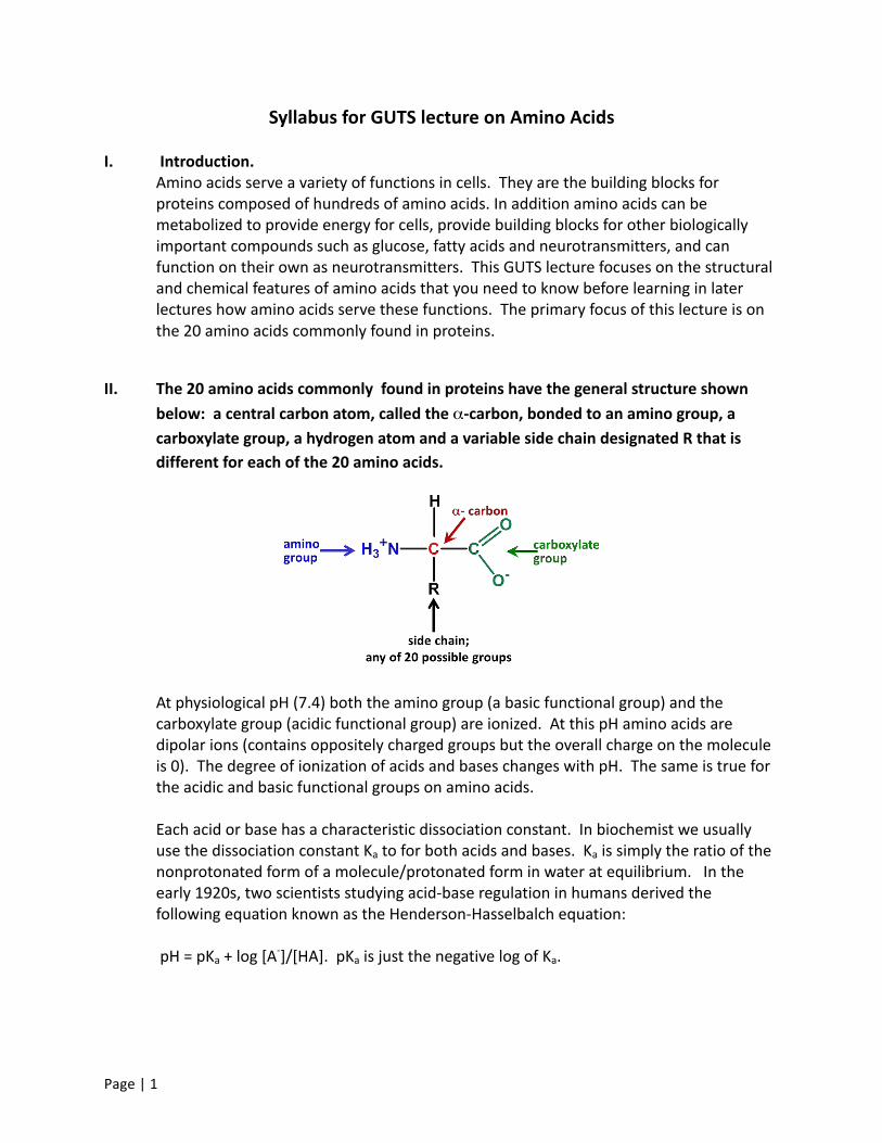
II. The 20 amino acids commonly found in proteins have the general structure shown below: a central carbon atom, called the α-‐carbon, bonded to an amino group, a carboxylate group, a hydrogen atom and a variable side chain designated R that is different for each of the 20 amino acids.
At physiological pH (7.4) both the amino group (a basic functional group) and the carboxylate group (acidic functional group) are ionized. At this pH amino acids are dipolar ions (contains oppositely charged groups but the overall charge on the molecule is 0). The degree of ionization of acids and bases changes with pH. The same is true for the acidic and basic functional groups on amino acids. Each acid or base has a characteristic dissociation constant. In biochemist we usually use the dissociation constant Ka to for both acids and bases. Ka is simply the ratio of the nonprotonated form of a molecule/protonated form in water at equilibrium. In the early 1920s, two scientists studying acid-‐base regulation in humans derived the following equation known as the Henderson-‐Hasselbalch equation: pH = pKa + log [A-‐]/[HA]. pKa is just the negative log of Ka.

Page | 2
Like Ka, pKa is a constant. Looking at this relationship you can see that when the ratio [A-‐
]/[HA] = 1.0, log of 1.0 = 0, and pH = pKa. Thus one way to think of pKa is that the value is equal to the pH at which 50% of an acid or base will be protonated and 50% will not.
A pH of approximately 2.2 is the average pKa for the α-‐carboxylic acid group on an amino acid. pH 7.4 is 5 orders of above the pKa for this group and so > 99.9999% of the COOH groups on the amino acid will have lost their protons at this pH and will be negatively charged. If acid is added, the carboxylate group will take up the protons and buffer the pH change. Now consider the α-‐amino group, for which the pKa (for NH3
+ losing a proton) is 9.4. At pH = 7.4, about 99.9% of α-‐amino groups will be in the NH3
+ form. At pH = 9.4, 50% of α-‐amino groups will have lost a proton to become NH2 and will no longer carry a charge. If base is added to the system, some of the NH3
+ will lose a proton and thus will buffer the pH change. From the above you should see that amino acids are effective buffers. They are also important buffers in cells because their concentration is very high there.
III. In proteins, amino acids are linked together by peptide bonds, formed in a
dehydration reaction (water is removed) between the α-‐carboxylate group on one amino acid and the α-‐amino group on another amino acid. In cells, the amino acid chain always grows left to right as drawn in the figure below. Since this always leaves a nonbonded amino group on the left end and a nonbonded carboxylate group on the right end, these ends are called the amino-‐terminal end and the carboxy-‐terminal end respectively. The direction of chain growth is from the amino-‐terminal end toward the carboxy-‐terminal end. In cells, the specific amino acid added during chain growth is dictated by the genetic code in DNA.
= peptide bond

Page | 3
Due to its proximity to a carbon with a double bond, the peptide bond has partial double bond character – specifically it is shorter than a single bond and is rigid and planar. This prevents rotation around the peptide bond. The rest of the bonds in the backbone of the chain, as well as the bond linking the R groups to the backbone, can rotate freely, allowing the chain to assume a variety of conformations (shapes) in space.
IV. Amino acids are generally classified on the basis of the properties of their side chains.
In biochemistry, the most useful classification relates to the presence or absence of charged or polar groups on the side chain. This distinction is important because polarity or its absence determines whether a side chain is hydrophilic or hydrophobic and whether it can form hydrogen bonds or ionic bonds with other side chains. The 3 categories of amino acids are amino acids with basic or acidic side chains, amino acids with uncharged polar side chains, and amino acids with nonpolar side chains. You are not expected be able to draw or recognize the individual amino acids described below, BUT YOU WILL NEED TO BE ABLE TO DIFFERENTIATE BETWEEN AN ACIDIC, BASIC, UNCHARGED POLAR AND HYDROPHOBIC AMINO ACID ON THE BASIS OF ITS STRUCTURE. YOU ARE ALSO EXPECTED TO KNOW WHICH AMINO ACID FALLS INTO EACH CATEGORY AND THE 3 LETTER ABBREVIATIONS FOR EACH AMINO ACID.
V. Amino acids with acidic or basic side chains – contain functional groups that are charged at or near physiological pH. These side chains are hydrophilic due to their charge. Histidine is the only one of the charged polar amino acids with a pKa somewhat near physiological range. This is because the imidazole side chain on histidine is weakly basic

Page | 4
and releases its proton at relatively low pH compared to the amine group found on other basic amino acid side chains. In solution at pH = 7.4, the side chain is almost 99% uncharged. If acid is added, histidine will take up some of the protons, thereby buffering the pH change in the cell or surrounding aqueous environment. This feature is very important in understanding how proteins in serum and especially in hemoglobin help to buffer the increased acidity found in metabolically active tissues and in some disease states.
VI. Amino acids with uncharged polar side chains – contain functional groups that have a net zero charge at physiological pH and that can form hydrogen bonds. These side chains are hydrophilic because their polar groups form hydrogen bonds with water. Most members of this group have a hydroxyl group (OH) or an amide group (H2N-‐C=O) on the side chain. With the exception of tyrosine, these groups do not gain or lose protons. Tyrosine is considered a group member because the pKa for loss of a proton from the hydroxyl group is far above physiological pH; the tyrosine side chain is uncharged in solution at physiological pH.
Cysteine is important in protein structure for a second reason. The thiol group on the cysteine side chain can form a covalent bond with other nearby cysteines; this type of bond is called a disulfide bond.
Page | 5
Disulfide bonds stabilize the three-‐dimensional structures of proteins, and in some cases the quaternary structures (see GUTS lecture on Protein Structure if you are not familiar with these terms). In eukaryotes, disulfide bonds are found only in secretory proteins, lysosomal proteins, and the exoplasmic domains of membrane-‐bound proteins. This is because the environment in the cytoplasm and nucleus favors reduction of the disulfide bond.
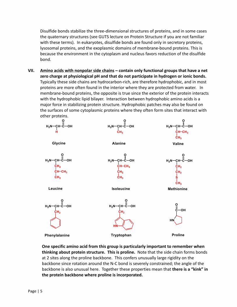
VII. Amino acids with nonpolar side chains – contain only functional groups that have a net zero charge at physiological pH and that do not participate in hydrogen or ionic bonds. Typically these side chains are hydrocarbon-‐rich, are therefore hydrophobic, and in most proteins are more often found in the interior where they are protected from water. In membrane-‐bound proteins, the opposite is true since the exterior of the protein interacts with the hydrophobic lipid bilayer. Interaction between hydrophobic amino acids is a major force in stabilizing protein structure. Hydrophobic patches may also be found on the surfaces of some cytoplasmic proteins where they often form sites that interact with other proteins.
One specific amino acid from this group is particularly important to remember when thinking about protein structure. This is proline. Note that the side chain forms bonds at 2 sites along the proline backbone. This confers unusually large rigidity on the backbone since rotation around the N-‐C bond is severely constrained; the angle of the backbone is also unusual here. Together these properties mean that there is a “kink” in the protein backbone where proline is incorporated.

Page | 6
VIII. The chirality of amino acids affects their functions. If you look back at the structures of the amino acids you will see that, with the exception of glycine, all amino acids have 4 different functional groups bound to the central α-‐carbon. The α-‐carbon is thus said to be chiral.
Amino acids that have a chiral α-‐carbon exist in two forms, called D and L, which are mirror images of each other. The D and L forms are stereoisomers, meaning that although they have the same functional groups attached to the central carbon they present their functional groups in different directions in space; in addition, the molecules cannot be superimposed in space simply by rotating the molecules. All amino acids found in human proteins have the L configuration. However both configurations occur in some plants and bacteria, so D-‐amino acids are part of our diets. Our bodies contain enzymes that can break down D-‐amino acids and then utilize the carbon skeletons for fuel. In addition, some D-‐amino acids are synthesized in neuronal cells and act as neurotransmitters.
To check your learning, go online and take the Post-‐Test for the GUTS lecture on Amino Acids. This is found at the end of the recorded lecture.