surgical management of complications following endoluminal grafting of abdominal aortic aneurysms
TRANSCRIPT

Eur J Vasc Endovasc Surg 10, 51-59 (1995)
Surgical Management of Complications Following Endoluminal Grafting of Abdominal Aortic Aneurysms
James May, Geoffrey H. White, Weiyun Yu, Richard C. Waugh, Michael S. Stephen, Timothy McGahan and John P. Harris
Department of Vascular Surgery, Royal Prince Alfred Hospital and Department of Surgery, University of Sydney, Australia.
Objective: The aim of this study was to report the outcome of endoluminal grafting of abdominal aortic aneurysms (AAA) with special reference to complications. Methods: Between May 1992 and August 1994 endoluminal repair of aneurysms was undertaken in 61 patients. In 53 the aneurysm was aortic and these are the basis of this report. In patients with AAA all procedures were elective and were performed in the operating room with the patient draped for an open repair in the event of failed endoluminal repair. The configuration of the endografts was tubular 36, tapered aortoiliac/aortofemoral 12 and bifurcated 5. Radiographic guidance was used to pass the endografts into the aorta via a delivery sheath introduced through the femoral or iliac arteries. Results: Successful endoluminaI repair of AAA was achieved in 43 of 53(81%) patients. In the remaining 10 patients, endoluminal repair was abandoned in favour of an open repair. There were 17(32%) local~vascular and 13(25%) systemic/ remote complications. The sum of these complications occurring in successful endoluminal repairs and those complications leading to failure of endoluminal repair was 40(75%). There were two cardiac deaths within 30 days in patients undergoing endoluminal repair (both procedure related) and four late deaths (unrelated to aneurysm repair). Three of the late deaths were in patients undergoing endoluminal repair and one endoluminal converted to open repair. Conclusion: Endoluminal repair of AAA in our experience has a low perioperative ( < 30 days) mortality rate (3.7%) but a high morbidity rate (75%). It is recommended that complications be classified into three groups: systemic~remote and local~vascular (following successful endoluminaI repair) plus those complications leading to failure of endoluminal repair. The first group is composed of medical complications while the latter two groups comprise those surgical complications directly related to the endoluminal technique.
Introduction that we have encountered and their surgical management.
There are now three groups with an experience of endoluminal AAA repair exceeding 50 patients each. These are Parodi and colleagues (Parodi, personal communication), ~'2 the authors of the present paper 3"4 and Endovascular Technologies (EVT), a California- based compan)~ who are conducting a multicentre, FDA-approved pilot study and randomised trial at 15 centres within the U.S.A. and five outside the U.S.A. (T. Koller, personal communication).
All three groups have clearly established the feasibility of endoluminal aneurysm repair. However, the risks and long-term outcome remain unclear. With the enthusiasm that accompanies any new technology there is a tendency for these to be overlooked. With this in mind we present our experience of endolumi- nal AAA with special emphasis on the complications
Please address all correspondence to: Professor James Ma~ Depart- ment of Surger~ University of Sydney, NSW 2006 Australia.
Material and Methods
Endovascular repair of aneurysms with materials currently used in vascular surgery is approved by the Institutional Review Board. Informed consent was obtained from each patient. Laboratory and clinical experience in the use of endoluminal grafts for aneurysms has previously been reported by the authors 3,4,5-17, 21
Since May 1992 endoluminal repair of aneurysms was undertaken in 61 patients. The aneurysm was aortic (n = 53), iliac (n -- 7) and subclavian (n = 1). In the aortic group there were 49 males and four females with a mean age of 70 years. In a previous publication 3 the authors have classified AAA into Type I (those with a proximal neck of length 2 cm or greater and
1078-5884/95/010051 + 09 $08"00/0 © 1995 W. B. Saunders Company Ltd.

52 J. May et aL
distal neck of length 1.5 cm or greater and iliac arteries of diameter 8 m m or greater) and Type II (any AAA not fitting the above criteria). In 29 patients the aneurysms were Type I and in 24 patients Type II. The mean diameter of the aneurysms was 5.2 cm (Type I) and 5.8 cm (Type II). Twenty-four patients had serious medical co-morbidities which led to them being rejected for open operation at other medical centres
Table 1. Medical co-morbidities leading to rejection for open surgical repair in 24 patients
Poor left ventricular function ( + renal impairment in 3) 16 Renal failure requiring dialysis 1 Bilateral thoracoplasties for tuberculosis I Chronic obstructive airways disease on oxygen 16 hours per day 1 Chronic liver disease (hepatitis C) with thrombocytopaenia 1 Hostile abdomen 4 Total 24
(Table 1). All procedures were elective and were performed
in the operating room with the patient draped for an open operation in the event of failed endoluminal repair. An on-table angiogram was used to identify the renal arteries and the aortic bifurcation. Radiographic guidance was used to pass the endografts into the aorta via a delivery sheath introduced through the femoral or iliac arteries. The configuration of the grafts was tubular (36), tapered aortoiliac/aortofemoral (12) and bifurcated (5). The grafts were anchored in the methods devised by Parodi 2 (10), White and Yu 14 (32), Chuter :s (1) and the Endovascular Technologies (EVT) grafting system :9 (10). The methods of endograft delivery and deployment have been described in detail in previous publications, s'14'18"~9
An on-table angiogram was performed at the completion of each procedure. All patients have been followed up by clinical examination, Duplex scan and computerised tomogram (CT) scan with contrast before discharge, at 6 months, 12 months and then annually. Complications have been recorded and classified in the manner recommended by the Ad Hoc Committee on Reporting Standards. 2°
Results
Endoluminal repair was achieved in 43 of 53(81%) patients with AAA and terminated in favour of an open repair in the remaining 10 patients. The causes of failed endoluminal repair are listed in Table 2 and have been analysed in detail together with sugges- tions for their avoidance in a previous publication. :6
Table 2. Causes of failed endoluminal repair of AAA
Problems with access 2 Balloon malfunction 1 Stent dislodgement 3 Graft thrombosis 1 Inability to deploy the contralateral limb of a bifurcated graft/twist in ipsilateral limb 3
There were two cardiac deaths within 30 days in patients undergoing endoluminal r epa i r - -bo th pro- cedure related. There have been four late deaths, one of which was cardiac, one due to liver failure, one due to hepatoma and one following spontaneous rupture of the oesophagus.
Complications
The 30 complications have been classified into 17 local/vascular and 13 systemic/remote and are sum-
Table 3. Complications following endoluminal AAA repair
Local~Vascular Femoral artery repair 3 Perforation of iliac artery 2 Graft stenosis 2 Common iliac artery occlusion 1 Proximal leak (corrected) 1 Distal leak (corrected) 2 Distal leak (persistent) 1 Bleeding (return to OR) 1 Bleeding (haematoma) 1 Lymph fistula 2 Wound infection 1 Total 17
Systemic~remote (Severity~outcome)* Renal insufficiency
obstructive (postoperative retention) (1) 1 contrast media induced (1,1,1,2,2) 5
Cardiac congestive failure (2,2) 2 myocardial infarction (2,2,3) 3 cardiac arrhythmia (1) 1
Stroke/TIA (1) 1 Total 13
marised in Table 3. These represent 32% and 25% respectively of total patients undergoing endoluminal repair.
Local~vascular complications
Femoral artery damage Femoral artery damage at the access site occurred in three patients. Two of these occurred early in our experience and resulted from
Eur J Vasc Endovasc Surg Vol 10, July 1995

Endovascular Grafting of Aneurysms 53
misguided attempts to pass a 24F delivery sheath through 6-7 mm diameter femoral and iliac arteries. Both injuries required vein patch repairs. Both patients have been followed-up regularly with clinical exam- ination and in the non-invasive vascular laboratory. Ankle/brachial pressure indices were unchanged from before operation. The third ~atient was noted to have an ischaemic leg (category 22°) at the completion of a successful endoluminal tube graft repair via a femoral arteriotomy. On reopening the arteriotom~ the artery was found to be thrombosed. A large calcified posterior plaque had become detached and moved in such a manner as to block the origins of both profunda and superficial femoral arteries. Thrombect- omy of the superficial femoral artery and endarter- ectomy of the common femoral artery restored the circulation of the leg to its preoperative state.
In a fourth patient a successful endoluminal tube graft repair of an AAA had been achieved. Access was via an arteriotomy in a small aneurysm in the left common femoral artery. On withdrawal of the deliv- ery sheath it was considered to be a safer option to replace the femoral aneurysm with a Dacron tube graft rather than close the arteriotomy.
Graft stenosis Graft stenoses occurred in two patients where endoluminal grafts of 10 mm diameter had been placed within iliac arteries of the same diameter. Post-procedure on-table aortograms showed normal flow through both grafts. A postoperative Duplex scan on the first patient demonstrated a small longitudinal
infolding of that portion of the graft within the common iliac artery (Fig. 1). This was not thought to be significant at the time, as the flow was normal on Duplex scan and clinically femoral pulses were palpa- ble. Twelve days after operation, over a period of 3 hours, however, the patient developed weakness of both lower limbs and signs of ischaemia (category 220).
An aortogram performed via the right brachial artery demonstrated a stenosis in the endoluminal aortofemoral graft corresponding to that section of the graft lying within the right common iliac artery (Fig. 2). The stenosis was corrected by percutaneous bal- loon dilatation using sequentially 6 mm diameter and 10 mm diameter balloons (Fig. 3a, b). Follow up with Duplex scan demonstrated normal flow in his endolu- minal and cross-over grafts.
The second patient had a similar problem follow- ing an endoluminal aortofemoral graft. In the first month after discharge he was readmitted with acute ischaemia of the right leg. In the absence of femoral pulses, urgent operation was undertaken. The endolu- minal aortofemoral graft was found to be thrombosed over that section lying within the common and external iliac arteries. It was not possible to re- establish flow with a balloon catheter. An extraper- itoneal approach to the right common iliac artery was made allowing this artery to be opened and flow re- established by means of an extraluminal graft running from the previously narrowed aortoiliac graft down to the common femoral artery. This procedure success- fully restored flow to the lower extremities. The
Fig. 1. Duplex scan of endoluminal aortofemoral graft showing infolding of the graft (arrow) just before entering the common iliac artery.
Eur I Vasc Endovasc Surg Vol 10, July 1995

54 J. May et aL
patient had a myocardial infarction three days later which resulted in his death.
Perforation of iliac artery Perforation of the iliac artery occurred in two patients. Both were caused by the delivery sheath. The first patient had previous irradia- tion for bladder cancer, and the field included the iliac arteries. The delivery sheath was introduced trans- femorally into the aorta with difficulty. Tachycardia and falling blood pressure indicated blood loss due to perforation. The blood loss was controlled via an iliac incision and the perforated external iliac artery liga- ted. A Dacron tube graft was placed end-to-side to the common iliac artery and brought down to be used as a conduit for the delivery sheath, which permitted successful placement of a tube endograft in the aorta. After withdrawal of the introducing sheath, the Dacron conduit was transected at the appropriate length and anastomosed end to side to the common femoral artery to revascularise the lower limb. This patient is also listed in the local/vascular complica- tions for return to the operating room for bleeding. No specific bleeding site was identified and the patient retained his endoluminal graft.
The second patient in which perforation of the iliac arteries occurred was one of the few women who fulfilled the criteria for endoluminal AAA repair. The introducing sheath was successfully placed in the aorta and a tube endograft was deployed immediately below the renal arteries. During withdrawal of the 24F delivery sheath the external iliac artery was avulsed from its origin. A similar procedure in the previous
case involving control of bleeding and placement of a Dacron graft from the common iliac to common femoral artery allowed successful revascularisation of the lower limb and retention of the aortic endograft. In addition to the two patients with perforation of the iliac artery there was a further patient who started to bleed heavily from the external iliac artery beneath the inguinal ligament following withdrawal through the femoral arteriotomy, of the introducing sheath. An extraperitoneal approach to the external iliac artery above the inguinal ligament was required to control
Fig. 2. Aortogram demonstrating stenosis in an endoluminal aortofemoral graft. Note revascularisation of left lower limb by femoro-femoral cross over graft.
Fig. 3. (a) Close up view of stenosis demonstrated in Fig. 2 before percutaneous dilatation. (b) Angiogram following 10 mm balloon dilatation.
Eur J Vasc Endovasc Surg Vol 10, July 1995

Endovascular Grafting of Aneurysms 55
the bleeding and allow suture repair of the bleeding point. No graft was required.
Iliac artery obstruction Obstruction of the common iliac artery was observed in one patient. A tube endograft was placed successfully in the aorta via a left-sided transfemoral approach. The post-procedure angio- gram demonstrated that the AAA had been excluded from the circulation but also demonstrated that the right common iliac artery was obstructed. The obstruction appeared to be due to compression of the contralateral atheromatous common iliac artery by the delivery sheath rather than an inappropriately placed inferior attachment device in the endograft. It was possible to pass a guidewire through the obstructed iliac artery into and beyond the endograft by the percutaneous route. Balloon dilatation followed by deployment of a Palmaz 8 mm stent (Johnson & Johnson Warren NJ) restored normal flow through the right iliac arterial system. Despite the presence of a right femoral pulse of normal amplitude, the patient developed paresis and ischaemia (category 22°) in the leg. Femoral artery bypass and fasciotomy were required in the first 48 hours post-aneurysm repair. Cardiac arrest, pulmonary oedema and renal failure led to death on the twelfth day postoperatively.
Incomplete seal between graft and aorta An incomplete seal between the graft and the aortic wall manifests itself in the on-table post-procedure aortogram as a leak of contrast from the lumen of the aorta into the aneurysmal sac. One such leak at the upper end of an endoluminal aortoiliac graft was successfully sealed by the introduction of a second stent which was completely covered bN and attached to, a Dacron graft (Fig. 4a-c).
Three further patients had a leak at the lower end of a tube endograft. Two of these were successfully sealed by a subsequent second endograft telescoped into the primary tube endograft (Fig. 5a, b). The third patient currently has a leak at the lower end of his endoluminal tube graft. The morphology of this aneurysm was well suited for transfemoral tube graft repair with proximal and distal necks of 2 cm in length. The leak is thought to be due to undersizing the diameter of the graft rather than deployment too far above the aortic bifurcation.
Wound complications A haematoma in the iliac fossa occurred in one patient following placement of an endoluminal aortoiliac graft via a conduit to the common iliac artery. This resolved spontaneously without operative intervention.
Two lymph fistulas have occurred following
Fig. 4. (a) On-table post-procedure angiogram demonstrating a leak of contrast at the upper end of the graft from the lumen into the aneurysmal sac. (b) Angiogram showing the insertion of a second covered stent mounted on a balloon catheter prior to deployment. (c) Angiogram demonstrating that the leak of contrast has been sealed by the second covered stent. Fig. 4. Reproduced with permission from the Journal of Endovascular Surgery.
Eur J Vasc Endovasc Surg Vol 10, July 1995

56 J. May et al.
transfemoral access. One patient was grossly obese and the other had a prolonged procedure due to heavy calcification in the iliac arteries.
A further patient had prolonged infection in both groin wounds following successful deployment of a bifurcated endoluminal graft. The organism cultured was Staphylococcus aureus and the length of hospital stay for the patient was 4 weeks.
Systemic~remote complications
Renal insufficiency was the commonest systemic complication observed. It occurred in five patients all of whom had renal impairment (mean serum crea- tinine 140 retools/l) before operation. In three of these the serum creatinine rose over 4 to 5 days to a mean of 290 mmols/1 before returning to the preoperative level over a further three to four days. In one of these patients the elevation in serum creatinine was accom- panied by an acute episode of pulmonary oedema on day four requiring intubation and ventilation for 24 hours followed by recovery associated with improved renal function. In the fourth patient the serum crea- tinine rose more rapidly in the first two postoperative days and was accompanied by myocardial infarction and congestion cardiac failure. Haemodialysis was required on four occasions before renal function improved to the preoperative level. In the fifth patient, the serum creatinine rose from 120 mmols/1 to 180 mmols/1 over the first 48 hours in the postoperative
period. At this point, the patient who had an ischae- mic limb (previously referred to under the heading of 'iliac artery obstruction') had a cardiac arrest. Renal function deteriorated further necessitating haemodial- ysis. He died 12 days after operation from a combina- tion of renal, cardiac and metabolic causes. The renal insufficiency in all five cases was considered to be contrast-medium induced. The mean dose of contrast medium was 285 ml. A further patient developed urinary retention due to prostatic hypertrophy. Con- servative management failed and he required transur- ethral resection of his prostate gland.
Congestive cardiac failure occurred in two patients. This was associated with renal insufficiency and was reversed with improvement in renal function. Three patients developed myocardial infarction in the immediate postoperative period. None required coro- nary angiography or surgical intervention. A further patient developed atrial fibrillation which responded to medication.
One patient developed a stroke due to infarction in the parietal area of the brain. Weakness in his left hand recovered rapidly but some residual personality change remains.
Discussion
Although endoluminal repair of AAA is less invasive than the standard transperitoneal operation, the mor- bidity associated with its performance must be taken
Fig. 5. (a) On-table pre-procedure angiogram confirming a known leak at the lower end of a tube endograft between the lumen and the aneurysmal sac. (b) On-table post-procedure angiogram demonstrating that the leak has been sealed by a second endograft telescoped into the primary tube endograft.
Eur J Vasc Endovasc Surg Vol 10, July 1995
Endovascular Grafting of Aneurysms 57
into account when assessing its merits. To obtain a clearer picture of the true morbidity those complica- tions which lead to failure of endoluminal repair and conversion to open operation must be added to the complications occurring in those with successful endoluminal repairs listed above. When this is done it can be seen that the combined complication rate is a substantial 75%. One-third of these complications were systemic/remote and can be attributed to the general medical condition of the patients. It is also important to note that centres known to undertake endoluminal AAA repair attract a high proportion of patients with medical co-morbidities. Not withstand- ing this, however, two-thirds of the complications are technical and are directly related to endoluminal method of AAA repair.
Those complications leading to failure of endolu- minal repair are of a higher order of importance since many patients undergoing endoluminal repair have been rejected as unfit for open surgical repair at other medical centres. These patients are exposed to the combined risk of a failed endoluminal procedure followed by a standard open operation. The fact that there have been no perioperative ( < 30 days) deaths in this group of at risk patients is a reflection of the quality of the anaesthetic and support services and an indication of how imprecise are our methods of risk assessment.
Some important lessons emerge from this experi- ence. The first is the recognition that atheromatous calcified iliac arteries of 6-7 mm diameter are unlikely to dilate sufficiently to accommodate a 24F internal diameter sheath which has an external diameter of almost 10 mm. This statement refers to uniformally small iliac arteries not those of 9-10 mm diameter with a focal stenosis which may be made accessible by balloon dilatation as reported by Parodi. 2 It is also important to note that in general females have smaller iliac arteries than males. We have recently developed a 20F delivery sheath in an attempt to extend the endoluminal technique to a larger number of females.
With regard to perforation of the iliac arteries it would be anticipated that previous irradiation would make them more susceptible to perforation. It is also generally appreciated that there is a risk of arterial perforation during the introduction of a large sheath. What is less obvious, however, is that the external iliac artery can be so tightly stretched over the sheath that the whole artery can be avulsed from its origin as the sheath is withdrawn. It is also important to recognise that damage to the iliac arteries may occur during introduction of the sheath but bleeding may not
become apparent until withdrawal of the sheath and removal of its tamponading effect.
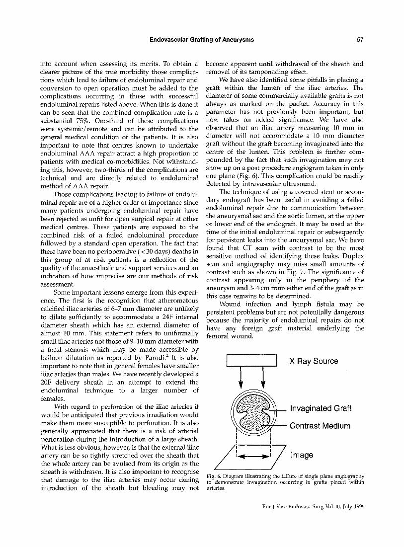
We have also identified some pitfalls in placing a graft within the lumen of the iliac arteries. The diameter of some commercially available grafts is not always as marked on the packet. Accuracy in this parameter has not previously been important, but now takes on added significance. We have also observed that an iliac artery measuring 10 mm in diameter will not accommodate a 10 mm diameter graft without the graft becoming invaginated into the centre of the lumen. This problem is further com- pounded by the fact that such invagination may not show up on a post procedure angiogram taken in only one plane (Fig. 6). This complication could be readily detected by intravascular ultrasound.
The technique of using a covered stent or secon- dary endograft has been useful in avoiding a failed endoluminal repair due to communication between the aneurysmal sac and the aortic lumen, at the upper or lower end of the endograft. It may be used at the time of the initial endoluminal repair or subsequently for persistent leaks into the aneurysmal sac. We have found that CT scan with contrast to be the most sensitive method of identifying these leaks. Duplex scan and angiography may miss small amounts of contrast such as shown in Fig. 7. The significance of contrast appearing only in the periphery of the aneurysm and 3-4 cm from either end of the graft as in this case remains to be determined.
Wound infection and lymph fistula may be persistent problems but are not potentially dangerous because the majority of endoluminal repairs do not have any foreign graft material underlying the femoral wound.
] X Ray Source
_ ~ Invaginated Graft
Contrast Medium i I i i
/'ql-----'~iF Image Fig. 6. Diagram illustrating the failure of single plane angiography to demonstrate invagination occurring in grafts placed within arteries.
Eur J Vasc Endovasc Surg Vol 10, July 1995

58 J. May et al.
It is n o t e w o r t h y tha t so far in o u r expe r i ence no p r o c e d u r e has b e e n c o m p l i c a t e d b y m i c r o e m b o l i . By con t r a s t P a r o d i ( p e r s o n a l c o m m u n i c a t i o n ) has exper i - e n c e d t w o in s t ances in 50 cases, one of w h i c h r e s u l t e d in the p a t i e n t ' s dea th . We a t t r i bu t e th is to t w o p o i n t s of t echn ique . F i r s t ly the i n t r o d u c t i o n of a r e g u l a r g u i d e w i r e in to the tho rac i c ao r t a w h i c h is t h e n e x c h a n g e d for an ex t ra stiff one. This m i n i m i s e s the r i sk of the ex t ra stiff g u i d e w i r e d i s t u r b i n g m u r a l t h r o m b u s . S e c o n d l y a p o l i c y of m a k i n g a s ingle p a s s of the i n t r o d u c i n g shea th w i t h i ts m a n d r i l to a p o s i t i o n in the ao r t a s u p e r i o r to the r ena l ar ter ies . F r o m this p o i n t on, the m a n d r i l a n d s h e a t h are r e m o v e d in sequence , l e a v i n g the e n d o g r a f t p o s i t i o n e d i m m e - d i a t e l y b e l o w the r ena l ar ter ies . At no p o i n t is the e n d o g r a f t a d v a n c e d w i t h a n y u n p r o t e c t e d p ro jec t ions w h i c h m i g h t d i s r u p t the m u r a l t h r o m b u s .
Rena l i m p a i r m e n t , the c o m m o n e s t s y s t e m i c / r e m o t e compl i ca t i on , has the p o t e n t i a l for r e d u c e d inc idence b y m o r e eff icient use of con t r a s t agent . This is p a r t i c u l a r l y i m p o r t a n t in those p a t i e n t s w i t h i m p a i r e d r ena l func t ion be fo re ope ra t i on . Ca rd i ac events , the s e c o n d c o m m o n e s t c o m p l i c a t i o n in th is g r o u p wi l l be diff icul t to r e d u c e b e c a u s e so m a n y p a t i e n t s w i t h A A A also h a v e a d v a n c e d ca rd i ac d i sease w h i c h is f r e q u e n t l y i n o p e r a b l e .
Fig. 7. CT scan with contrast seen within the endograft and in the periphery of the aneurysmal sac 3-4 cm from either end of the endograft.
References
1 PARODI JC, PALMAZ JC, BARONE HD. Transfemoral intraluminal graft implantation abdominal aortic aneurysms. Ann Vasc Surg 1991; 5: 491-499.
2 PARODI JC. Endovascular repair of abdominal aortic aneurysms In: WHITTEMORE A ed. Advances in Vascular Surgery Chicago: Mosby Year Book 1994.
3 MAY J, WHITE GH, Yu W, WAUGH RC, STEPHEN MS, HARRIS JP. Results of endoluminal grafting of abdominal aortic aneurysms are dependent on aneurysm morphology. J Vasc Surg; in press.
4 WHrrE GH, MAY J, Yu W. Development of a clinical program of endoluminal anenrysm grafting: experience in 60 patients. In: PARODI JC, VEITH F~ MARIN M eds. Endovascular Grafting Techniques
5 MAY J, WHITE G, YU W, WAUGH R, HARRIS JP. Treatment of complex abdominal aneurysms by a combination of endolumi- nal and extraluminal aorto-femoral grafts. J Vasc Surg 1994; 19: 924-933.
6 MAY J, WHITE G, Yu W, WAUGH R I STEPHEN M, HARRIS JP. Transluminal placement of aorto-iliac grafts for treatment of large abdominal aortic aneurysms. In: WEIMANN S z ed. Thoracic and Thoracoabdominal Aortic Aneurysm Bologna, Italy: Monduzzi Editore 1994; 249-255.
7 MAY J, WHITE G, Yu W, WAUGH R~ HARRIS JP. Endoluminal stent- grafts for intrathoracic and abdominal aortic aneurysm. In: LIERMANN DD ed. Proceedings of International Stent Symposium 3. Frankfurt, 1993.
8 MAY J, WHITE G. WAUGH R~ Yu Wz HARRIS JP. Advantages and limitations of intraluminal grafts for thoracic and abdominal aortic aneurysm. Angiology 1993; 44(Suppl 1): 21 (abstract).
9 MAY Jr WHITE G, WAUGH R, YU W, HARRIS JP. Endoluminal repair of large and small abdominal aortic aneurysm. J Interventional Cardiol 1994; 7(1): 109 (abstract).
10 MAY J, WHITE G, Yu W, WAUGH R, HARRIS JP. Intraluminal stents for thoracic and abdominal aortic aneurysm. J Cardiovasc and Interventional Radiol 1994; in press (abstract).
11 MAY J, WHITE G, WAUGH R~ Yu W, HARRIS JP. Transluminal placement of a prosthetic graft-stent device for treatment of subclavian aneurysm, f Vasc Surg 1993; 18: 1056-1059.
12 WHITE GH, Yu W, MAY J. Experimental endoluminal grafts and coated stents. Angiology 1993; 4(Suppl 1): 26 (abstract).
13 WHITE GH, MAY J, Yu W. Stented and non-stented grafts for aneurysmal disease: the Sydney experience. In: CHUTER % DONAYRE C, WHITE R I eds. Endoluminal Vascular Prostheses. Boston, Massachusetts: Little Brown & Compan3~ 199?.
14 WHITE GH, Yu W, MAY J, WAUGH RC, STEPHEN M. A new non stented endoluminal graft for straight or bifurcated repair of aneurysms, f Endovasc Surg; 1994; 1: 16-24.
15 MAY J, WHITE GH, WAUGH R, Yu W, STEPHEN MS, HARRIS JP. Endoluminal repair of abdominal aortic aneurysm. Med J Aust 1994; 161: 541-543.
16 MAY J, WHITE GH, Yu W, WAUGH RC, MCGAHAN T~ STEPHEN MS, HARRIS JE Endoluminal grafting of abdominal aortic aneurysms: causes of failure and their prevention. J Endovasc Surg 1994; 1" 44-52.
17 MAY J & WHITE GH. Specialised endovascular techniques: combined surgical and endovascular approaches. In: WHITE RA, FOGARTY TJ, Peripheral Endovascular Interventions Chicago: Mosby Year Book, 199.
18 CHUTER TAM, GREEN RM, OURIEL K, FIORE W, DE WEES~ JA. Transfemoral endovascular aortic graft replacement. J Vasc Surg 1993; 18: 185-197.
19 MOORE WS. Endovascular grafting technique: a feasibility study. In: YAO JST, PEARCE WH eds. Aneurysms, New Findings and Treatments. Norwalk, Connecticut: Appleton & Lange; 1944: 333-340.
Eur J Vasc Endovasc Surg Vol 10, July 1995

Endovascular Grafting of Aneurysms 59
20 Ad Hoc Committee on Reporting Standards RUTHERFORD RB, FLANIGAN DP, GUPTA SK et al. Suggested standards for reports dealing with lower extremity ischaemia. J Vasc Surg 1986; 4: 80-94.
Accepted 7 September 1994
Eur J Vasc Endovasc Surg Vol 10, July 1995