surgical correction of a class ii skeletal malocclusion ... · case report surgical correction of a...
TRANSCRIPT

CASE REPORT
Surgical correction of a Class II skeletalmalocclusion associated with anterior open biteand temporomandibular joint painJosé Augusto Mendes Miguel,a Julio Pedra e Cal-Neto,b and Henrique Martins da Silveirac
Rio de Janeiro, Brazil
This case report describes the treatment of a 25-year-old woman with anterior open bite, Class II skeletalmalocclusion, and a history of temporomandibular joint pain and sounds. She also had significantanteroposterior and vertical discrepancies and a convex profile with protrusive lips. Intraorally, she had ananterior open bite of 3 mm and an overjet of 5 mm. Mandibular surgical rotation, associated with mandibularincisor extraction, was performed to reduce the protrusion, close the open bite, and minimize the
temporomandibular joint disorder. (Am J Orthod Dentofacial Orthop 2007;132:400-7)Anterior open bite associated with a Class IIskeletal pattern in adults can be a challengingorthodontic problem. Basically, 3 treatment al-
ternatives are available: tooth extraction, molar intrusion,and distalization using a skeletal anchorage system ororthognathic surgical correction.1-4 Unfortunately, theoutcome obtained with nonsurgical extraction therapyor orthodontics using absolute anchorage has limitedimpact on the facial profile and in many cases poorstability. Correction with maxillary surgery has becomethe standard of care, although technical advances nowallow clinicians to close open-bite discrepancies usingbilateral split osteotomies to rotate the mandibulardistal segment counterclockwise.5
Patients with Class II malocclusion or mandibularretrognathia and an increased occlusal plane anglehave the highest incidence of temporomandibular joint(TMJ) problems.6 Currently, there is controversy re-garding the appropriate management of patients withpreexisting TMJ disorder (TMD) symptoms who re-quire orthognathic surgery. Some investigators that or-thognathic surgery helps reduce TMD symptoms,7-9
whereas others contend that orthognathic surgery in suchpatients causes further deleterious effects to the TMJ and
aProfessor, Department of Orthodontics, State University of Rio de Janeiro, Riode Janeiro, Brazil.bGraduate student, Department of Orthodontics, State University of Rio deJaneiro, Rio de Janeiro, Brazil.cProfessor, Department of Oral and Maxillofacial Surgery, State University ofRio de Janeiro, Rio de Janeiro, Brazil.Reprint requests to: Julio Pedra e Cal-Neto, Rua Carlos Góis 375 gr 511Leblon, 22440-040, Rio de Janeiro, Brazil 22440-033; e-mail,[email protected], July 2005; revised and accepted, January 2006.0889-5406/$32.00Copyright © 2007 by the American Association of Orthodontists.
doi:10.1016/j.ajodo.2006.01.033400
thus worsens the symptoms and dysfunction postsurgery,so this philosophy proposes surgical management of theTMJ pathology at an initial separate procedure or con-comitantly with the orthognathic surgery.10,11
The following case report illustrates the treatmentof a patient with Class II malocclusion complicated bydentoalveolar protrusion, anterior open bite of 3 mm,TMD symptoms, and a Class II skeletal pattern.
DIAGNOSIS AND ETIOLOGY
The patient was a 25-year-old white woman whosemain complaints were an anterior open bite, TMJ pain,and TMJ sounds (Figs 1-4). A facial evaluation showeda symmetric, elongated face; a retrognathic mandible; aprominent nose; and strain of the mentalis muscle(Fig 1). She had a Class II Division 1 malocclusionwith a 30-mm anterior open bite, a 5-mm overjet, andreduced gingival attachment on the mandibular rightcentral incisor. Space analysis showed approximately 2mm of crowding in the mandibular arch, with a mandib-ular midline deviation of 1 mm to the right. Cephalometricanalysis showed a skeletal Class II relationship (ANBangle, 6°) with mandibular retrusion (SNB angle, 76°), asteep mandibular plane (FMA, 36°; SN-GoGn, 39°),and protrusive incisors (interincisal angle, 104°; 31°from maxillary incisor to NA angle; 8 mm frommaxillary incisor to NA; 39° from mandibular incisorto NB angle; 10 mm from mandibular incisor to NB;IMPA, 102°).
TREATMENT OBJECTIVES
The primary objectives of treatment were to closethe anterior open bite, obtain Class I canine and molarrelationships with ideal overjet and overbite, and im-
prove facial esthetics. The complementary treatment
nt fac
graph
American Journal of Orthodontics and Dentofacial OrthopedicsVolume 132, Number 3
Miguel, Cal-Neto, and Silveira 401
objectives were to establish good functional and stableocclusion, avoid extrusion of the molars and clockwiserotation of the mandible during presurgical treatment,correct the axial inclinations of the maxillary andmandibular anterior teeth, enhance the facial profile andlip closure, improve smile characteristics and dentalesthetics, and improve the shape of both arches.
TREATMENT ALTERNATIVES
Three approaches were considered. Extracting only2 maxillary first premolars would reduce the overjet
Fig 1. Pretreatme
Fig 2. Pretreatment intraoral photo
and close the anterior open bite, resulting in a Class I
canine relationship; mandibular crowding would berelieved by interproximal reduction. This approach,however, would flatten the upper lip, jeopardizing theprofile, and a Class I molar relationship could not beachieved.
The third approach included surgically advancingand rotating the distal segment of the mandible coun-terclockwise to close the open bite. The diagnostic waxsetup showed that, after the mandibular advance, aClass I molar relationship could not be achieved be-cause of protrusion of the mandibular incisors. There-
ial photographs.
s show negative overbite of 3 mm.
fore, mandibular incisor extraction would also be

ment
American Journal of Orthodontics and Dentofacial OrthopedicsSeptember 2007
402 Miguel, Cal-Neto, and Silveira
needed to allow the large mandibular advancement andimprove the facial profile. Due to the TMD symptoms,surgical stabilization would be accomplished with non-
Fig 3. Pretreat
Fig 4. Pretreatment cephalometric tracing.
rigid fixation to decrease the load or stress on the TMJ.6
The second treatment possibility would be the use ofminiplates or microscrews for intrusion and distaliza-tion of upper and posterior teeth. The retraction ofupper incisors, whether caused by premolar extractionor distalization of posterior teeth would have the sameflattening effect on the upper lip.
TREATMENT PROGRESS
In order to obtain a balanced profile, the surgical-orthodontic alternative was chosen. The maxillary andmandibular third molars were extracted. The molarswere banded and the remaining teeth bonded withpreadjusted .022-in straight-wire fixed appliances. Themandibular right central incisor was chosen for extractionbecause of the loss of gingival attachment on the facialsurface. The root was removed from the crown with ahigh speed bur, and the crown was used as a prosthetic.Periodical interproximal reduction was performed on thecrown to allow closure of the extraction space. Initialleveling was accomplished with a .016-in nickel-titaniumarchwire in the maxillary arch and a .020 � .020-inBioForce NiTi (GAC, Bohemia, NY) archwire in themandibular arch. The remaining space in the mandibulararch was closed, and the incisors retracted with a .017 �.022-in stainless steel (SS) archwire by using slidingmechanics with a power chain. The maxillary arch wasaligned with a .016-in progressing to a .020-in SS arch-wire. The mandibular second molars were banded and
study models.
leveled with an .018-in SS boot loop superimposed

l intrao
ent fa
American Journal of Orthodontics and Dentofacial OrthopedicsVolume 132, Number 3
Miguel, Cal-Neto, and Silveira 403
archwire. Presurgical rectangular archwires were placed,the maxillary arch received a .019 � .025-in progressingto a .021 � .025-in SS archwire, and the mandibular archreceived a .019 � .025-in SS archwire. The presurgicalorthodontic phase lasted approximately 17 months. Sur-gical hooks were then soldered to the SS archwires andplaced in both arches (Fig 5).
The surgery involved sagittal split osteotomies withapproximately 5 mm advancement of the mandible andcounterclockwise rotation to allow overjet reductionand closure of the open bite. Nonrigid fixation wasselected to avoid more TMJ complications. Threemonths after surgery, a mandibular .018 � .025-inbeta-titanium (TMA, Ormco, Orange, Calif) archwireand a maxillary .018-in SS archwire were placed, andbilateral Class II elastic mechanics were initiated. Amaxillary continuous .018 � .025-in beta-titanium wirewas used during finishing, along with the elastics.Twenty-four months after initial bracket placement, theteeth were in acceptable positions, and the appliances
Fig 5. Presurgica
Fig 6. Posttreatm
were removed.
For retention, the patient was instructed to wear amaxillary circumferential Hawley retainer 24 hours a dayfor 2 years and at night for another 6 months. In addition,a fixed lingual mandibular retainer was bonded fromcanine to canine. Because of the potential for bite opening,the patient was highly motivated to comply with dailytongue exercises and received a modified Hawley retainerwith a palatal crib to be used at night.
TREATMENT RESULTS
Because of the skeletal pattern and the surgicalapproach that was chosen, excellent facial and occlusalresults were achieved. The most significant changeswere a dramatic decrease in TMD symptoms aftersurgery and an improvement in occlusal function.Esthetically, facial convexity decreased, the face be-came less retrognathic, and lower face height de-creased. Lip competency was improved significantly,and the patient was satisfied with the results of treat-ment. Well-established Class I canine and molar rela-
ral photographs.
cial photographs.
tionships were obtained, rotations were corrected, and
nt intr
tment
American Journal of Orthodontics and Dentofacial OrthopedicsSeptember 2007
404 Miguel, Cal-Neto, and Silveira
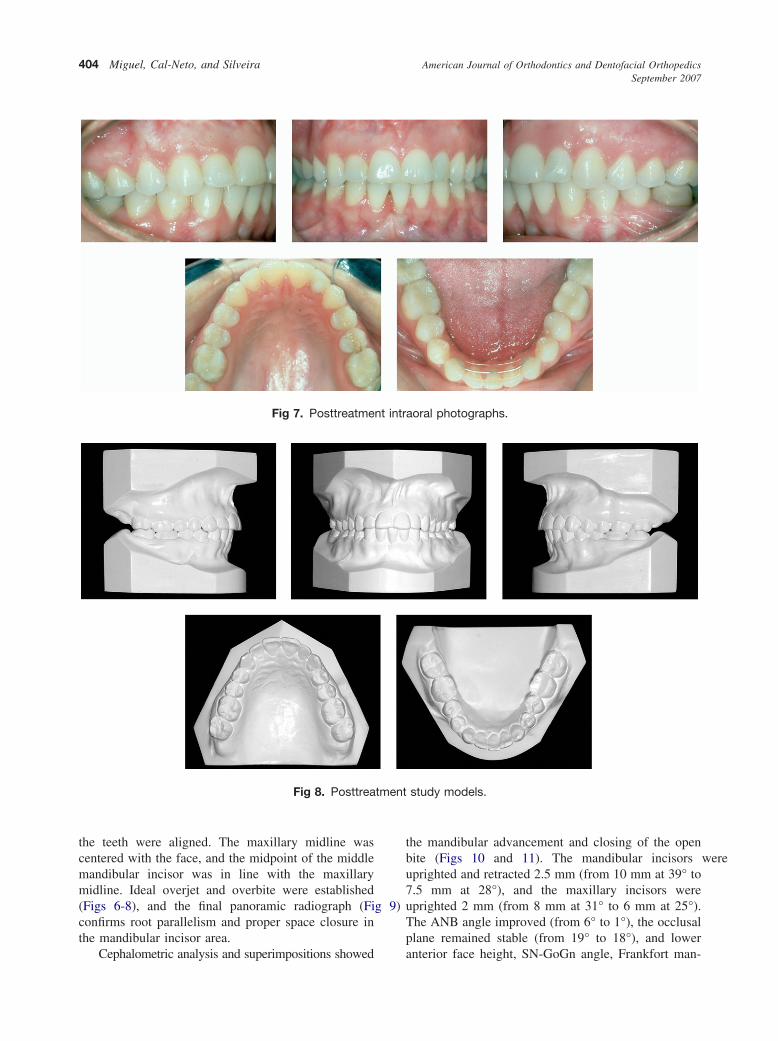
the teeth were aligned. The maxillary midline wascentered with the face, and the midpoint of the middlemandibular incisor was in line with the maxillarymidline. Ideal overjet and overbite were established(Figs 6-8), and the final panoramic radiograph (Fig 9)confirms root parallelism and proper space closure inthe mandibular incisor area.
Fig 7. Posttreatme
Fig 8. Posttrea
Cephalometric analysis and superimpositions showed
the mandibular advancement and closing of the openbite (Figs 10 and 11). The mandibular incisors wereuprighted and retracted 2.5 mm (from 10 mm at 39° to7.5 mm at 28°), and the maxillary incisors wereuprighted 2 mm (from 8 mm at 31° to 6 mm at 25°).The ANB angle improved (from 6° to 1°), the occlusalplane remained stable (from 19° to 18°), and lower
aoral photographs.
study models.
anterior face height, SN-GoGn angle, Frankfort man-

American Journal of Orthodontics and Dentofacial OrthopedicsVolume 132, Number 3
Miguel, Cal-Neto, and Silveira 405
dibular plane angle, and Y-axis to SN angle were allreduced (Table). The corrected occlusion at 5 yearsposttreatment shows excellent stability, esthetics, andperiodontal health (Figs 12 and 13), without signs orsymptoms of TMD.
DISCUSSION
Orthodontists are aware of the difficulty of manag-ing a Class II malocclusion with an open bite and askeletal Class II pattern. Faced with the limitations oforthodontic treatment, most orthodontists would agreethat this situation is best treated with a combination of
Fig 9. Posttreatment panoramic radiograph.
Fig 10. Posttreatment cephalometric tracing.
orthodontics and orthognathic surgery.12 The advan-
tages of orthognathic surgical treatment are that theprofile can be improved and relapse is less likely thanwith a nonsurgical option.12,13 Although maxillaryimpaction surgery has been considered the most stableorthognathic procedure, in this case, mandibular ad-vancement was chosen because of the maxilla’s goodposition and the reported stability of mandibular ad-vancement surgery.14,15
The TMJs are the foundation for stable results withorthognathic surgery. Although counterclockwise ad-vancement of the maxillomandibular complex might
Fig 11. Superimposed cephalometric tracings.
Table. Summary of cephalometric analysis
Standard Pretreatment Posttreatment
SNA angle (°) 82 82 82SNB angle (°) 80 76 80ANB angle (°) 2 6 2FMA (°) 25 36 30SNGoGn (°) 32 39 34.51/NA (°) 22 31 251-NA (mm) 4 8 5.61/NB (°) 25 39 281-NB (mm) 4 10 7.51/1 (°) 131 104.5 123IMPA (°) 93 102 92
further increase the loading of the TMJ by stretching

tion fa
n intr
American Journal of Orthodontics and Dentofacial OrthopedicsSeptember 2007
406 Miguel, Cal-Neto, and Silveira
the associated soft tissues, it is a stable procedure whenthe joints are healthy.16,17
Common symptoms of TMJ dysfunction includesounds, pain, headaches, limited movement, change inocclusion, masticatory difficulty, earaches, tinnitus, ver-tigo, and others.11 There is controversy concerning theadequate management of patients with preexisting TMDwho require orthognathic surgery to correct jaw deformi-ties and malocclusion. In a retrospective study, Wolfordet al11 concluded that orthognathic surgery in patientswith TMD can lead to the initiation or exacerbation of
Fig 12. Postreten
Fig 13. Postretentio
problems. However, several studies comparing TMD
symptoms before and after bilateral sagittal splitramus osteotomy suggest that in patients with severemaxillomandibular discrepancy, surgical-orthodontictherapy is a good choice of treatment for reducingmyogenous TMJ pain and discomfort.18-22
An alternative approach advocated by many prac-titioners is not to decrease the occlusal plane angle inpatients with Class II relationships but, rather, tomaintain or increase it. By minimizing load or stress onthe TMJ, the likelihood of problems and relapse isdecreased, and TMJ surgery is avoided. This concept can
cial photographs.
aoral photographs.
be supported by the success of techniques such as condy-

American Journal of Orthodontics and Dentofacial OrthopedicsVolume 132, Number 3
Miguel, Cal-Neto, and Silveira 407
lar repositioning via bilateral sagittal split ramus osteot-omy with nonrigid fixation to increase joint space.6
In our patient, orthognathic surgical correction ofthe Class II malocclusion with anterior open bite andTMJ pain was selected to improve the functional statusof the TMJ, reduce pain levels, and avoid posteriorsurgery.18,19,22 Despite concerns about stability, coun-terclockwise surgical advancement of the mandiblewith nonrigid fixation was performed to reduce stresson the TMJ, allowing the open bite to close and theoverjet to be reduced. In other attempts to avoid TMJsurgery, orthognathic surgery was conducted to mini-mize occlusal plane changes, and the occlusal planeangle decreased by only 1° (Table).
A mandibular incisor was extracted to maximize themandibular advancement and the open-bite closure. Adiagnostic wax setup of the arches was performed withextreme accuracy, indicating no need for interproximalreduction of the maxillary anterior teeth to allowmaxillary and mandibular arch coordination.23-26 Long-term stability can be considered an advantage of thisapproach. Riedel suggested that incisor extraction maygive greater stability in this area in the absence ofpermanent retention.27,28
The final outcome of treatment was a substantialimprovement in function and esthetics. Through thisapproach, the patient had excellent skeletal, dental, andocclusal improvements, with a significant decrease inTMJ pain. As an additional benefit, she has reportedimproved self-esteem and greater satisfaction with herappearance. At the retention check 60 months afterremoving the appliances, the occlusion was stable,without TMD symptoms (Fig 13). Long-term follow-upwill need to continue because open bites tend to relapsemore than most other types of malocclusion.
REFERENCES
1. Sugawara J, Kanzaki R, Takahashi I, Nagasaka H, Nanda R.Distal movement of maxillary molars in nongrowing patientswith skeletal anchorage system. Am J Othod Dentofacial Orhop2006;129:723-33.
2. Yamaguchi K, Nanda RS. The effects of extraction and nonex-traction treatment on the mandibular position. Am J OrthodDentofacial Orthop 1991;100:443-52.
3. Kim YH. Anterior open bite and its treatment with multiloopedgewise archwire. Angle Orthod 1987;57:290-321.
4. Epker BN, Fish LC. Surgical-orthodontic correction of open-bitedeformity. Am J Orthod 1977;71:278-99.
5. Joondeph DR, Bloomquist D. Open-bite closure with mandibularosteotomy. Am J Orthod Dentofacial Orthop 2004;126:296-8.
6. Cottrell DA. In discussion of: Wolford LM, Reiche-Fischel O,Mehra P. Changes in temporomandibular joint dysfunction afterorthognathic surgery. J Oral Maxillofac Surg 2003;61:655-60.
7. Karabouta I, Martis C. The TMJ dysfunction syndrome beforeand after sagittal split osteotomy of the rami. J Maxillofac Surg
1985;13:185-8.8. Magnusson T, Ahlborg G, Finne K, Nethander G, Svartz K.Changes in temporomandibular joint pain-dysfunction after sur-gical correction of dentofacial anomalies. Int J Oral MaxillofacSurg 1986;15:707-14.
9. Upton G, Scott R, Hayward J. Major maxillomandibular malre-lations and temporomandibular joint pain-dysfunction. J ProsthetDent 1984;51:586-9.
10. Onizawa K, Schmelzeisen R, Vogt S. Alteration of temporoman-dibular joint symptoms after orthognathic surgery: comparisonwith healthy volunteers. J Oral Maxillofac Surg 1995;53:117-21.
11. Wolford LM, Reiche-Fischel O, Mehra P. Changes in temporo-mandibular joint dysfunction after orthognathic surgery. J OralMaxillofac Surg 2003;61:655-60.
12. Hiller ME. Nonsurgical correction of Class II open bite malocclusonin an adult patient. Am J Orthod Dentofacial Orthop 2002;122:210-6.
13. Denison TF, Kokich VG, Shapiro PA. Stability of maxillarysurgery in open bite versus non-open bite malocclusions. AngleOrthod 1989;59:5-10.
14. Proffit WR, Turvey TA, Phillips C. Orthognathic surgery: a hierar-chy of stability. Int J Adult Orthod Orthognath Surg 1996;11:191-204.
15. Bailey LJ, Cevidanes LH, Proffit WR. Stability and predictabilityof orthognathic surgery. Am J Orthod Dentofacial Orthop 2004;126:273-7.
16. Satrom KD, Sinclair PM, Wolford LM. The stability of doublejaw surgery: a comparison of rigid versus wire fixation. Am JOrthod Dentofacial Orthop 1991;99:550-63.
17. Chemello PD, Wolford LM, Buschang PH. Occlusal planealteration in orthognathic surgery—part II: long-term stability ofresults. Am J Orthod Dentofacial Orthop 1994;106:434-40.
18. Panula K, Somppi M, Finne K, Oikarinen K. Effects of orthog-nathic surgery on temporomandibular joint dysfunction. A con-trolled prospective 4-year follow-up study. Int J Oral MaxillofacSurg 2000;29:183-7.
19. Egermark I, Blomqvist JE, Cromvik U, Isaksson S. Temporo-mandibular dysfunction in patients treated with orthodontics incombination with orthognathic surgery. Eur J Orthod 2000;22:537-44.
20. Westermark A, Shayeghi F, Thor A. Temporomandibular dys-function in 1,516 patients before and after orthognathic surgery.Int J Adult Orthod Orthognath Surg 2001;16:145-51.
21. Dervis E, Tuncer E. Long-term evaluations of temporomandib-ular disorders in patients undergoing orthognathic surgery com-pared with a control group. Oral Surg Oral Med Oral Pathol OralRadiol Endod 2002;94:554-60.
22. Pahkala R, Heino J. Effects of sagittal split ramus osteotomy ontemporomandibular disorders in seventy-two patients. Acta Od-ontol Scand 2004;62:238-44.
23. Bolton WA. The clinical application of a tooth size analysis.Am J Orthod 1962;48:504-29.
24. Tuverson DL. Anterior interocclusal relations. Part II. Am JOrthod 1980;78:371-93.
25. Kokich VG, Shapiro PA. Lower incisor extraction in orthodontictreatment. Angle Orthod 1984;54:139-53.
26. Valinoti JR. Mandibular incisor extraction therapy. Am J OrthodDentofacial Orthop 1994;105:107-16.
27. Riedel RA. Retention. In: Graber TM, editor. Current orthodon-tic concepts and techniques. Philadelphia: W. B. Saunders; 1969.
28. Riedel RA, Little RM, Bui TD. Mandibular incisor extraction—postretention evaluation of stability and relapse. Angle Orthod
1992;62:103-16.