surgery for colorectal liver metastases technical … · chapter 8 - long-term results of the...
TRANSCRIPT
SURGERY FOR COLORECTAL LIVER METASTASES
Technical aspects, prognostic factors & timing
Ninos Ayez

The studies in this thesis were performed at the Department of Surgical Oncology, Erasmus MC, University Medical Center, Rotterdam, The Netherlands.
ISBN 978-94-6299-305-1
Cover design: Ninos Ayez
© Copyright of the published articles is with corresponding journal or otherwise the author. No part of this book may be reproduced, stored, or transmitted in any form or by any means without prior permission from the author or corresponding journal.
SURGERY FOR COLORECTAL LIVER METASTASESTechnical aspects, prognostic factors & timing
Chirurgie van colorectale levermetastasenTechnische aspecten, prognostische factoren & timing
Proefschrift
Ter verkrijging van graad van doctor aan deErasmus Universiteit Rotterdam
op gezag van derector magnificus
Prof.dr. H.A.P. Polsen volgens besluit van het College voor promoties.
De openbare verdediging zal plaatsvinden op
Woensdag 16 Maart 2016 om 13:30 uur
door
Ninos Ayezgeboren te Malkieh, Syrië

PROMOTIECOMMISSIE
Promotoren: Prof.dr. C. Verhoef Prof.dr. A.M.M. Eggermont
Overige leden: Prof.dr. T.M. van Gulik Prof.dr. H.W. Tilanus Prof.dr. J.H.W. de Wilt
Copromotor: Dr. D.J. Grünhagen
Contents
Chapter 1 - Introduction 7
PART 1 - Technical aspects
Chapter 2 - Anatomical versus non-anatomical resection of colorectal liver metastases: is there a difference? 17
Chapter 3 - Outcome of microscopic incomplete resection (R1) of colorectal liver metastases in the era of neoadjuvant chemotherapy. 29
PART 2 - Prognostic factors
Chapter 4 - Is the clinical risk score for patients with colorectal liver metastases still useable in the era of effective neoadjuvant chemotherapy? 49
Chapter 5 - Preoperative FDG-PET-scan in patients with resectable colorectal liver metastases does not improve overall survival: a retrospective analyses stratified by clinical risk score. 65
Chapter 6 - The use of neoadjuvant chemotherapy in patients with resectable colorectal liver metastases: clinical risk score as possible discriminator. 79
Chapter 7 - Neo-adjuvant chemotherapy followed by surgery versus surgery alone in high-risk patients with resectable colorectal liver metastases The CHARISMA randomized multicenter clinical trial. 93
PART 3 - Timing
Chapter 8 - Long-term results of the ‘liver first’ approach in patients with locally advanced rectal cancer and synchronous liver metastases 109
Chapter 9 - Is restaging with chest and abdominal CT scan after neoadjuvant chemoradiotherapy for locally advanced rectal cancer necessary? 123
Chapter 10 - Surgery of the Primary Tumor in Stage IV Colorectal Cancer with Unresectable Metastases 135
PART 4 - Summery, general discussion and appendices
Chapter 11 - Summery 151
Chapter 12 - Nederlandse samenvatting 161
Dankwoord 171PhD Portfolio 173Curriculum Vitae 175

Chapter 1
Introduction
Chapter 18
Introduction
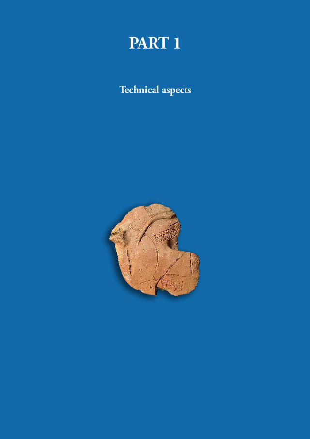
In the early beginnings of civilization mankind tried to predict the future. They tried to achieve this by divination. Divination is a technique that seeks to discover information about current and future conditions. In ancient Mesopotamia (now located in modern Iraq/Syria) diviners interpreted the shape and appearance of the liver of a sacrificed animal to predict outcome in ill patients.1 (Front cover; Clay model of a sheep’s liver, probably used for teaching divination, from Babylon c. 2000 BCE.) It seems far-fetched, however there is a similarity with research nowadays where we try to predict patient outcome. Of course this is now based on scientific evidence.
Colorectal cancer (CRC) is one of the two most commonly diagnosed cancers, with approximately 1.2 million new cases each year and more than 600,000 annual deaths estimated to occur worldwide.2 At diagnosis of CRC, approximately 20% of the patients present with synchronous metastatic CRC, and the liver is the predilection site in half these patients.3, 4 Liver resection is considered to be the best optimal treatment for colorectal liver metastases (CRLM) with 5-year survival rates up to 60% in highly selected patients. Until recently, only 10-20% of patients were considered suitable for attempted curative resection.5, 6 Due to improvements in surgical technique, the acceptance of smaller resection margins7, 8, the introduction of more effective systemic chemotherapy9, 10, the use of portal vein embolization (VPE)11, 12, radio frequency ablation (RFA)13, 14 and stereotactic body radiation (STBR)15 more patients are eligible for liver surgery. Many factors contribute to a better outcome, however surgery is fundamental in achieving long term survival. This thesis describes some factors that contribute to a better outcome in patient with colorectal metastatic disease. In three parts it will cover technical surgical aspects, prognostic factors and timing of therapy.
Part 1Due to the increased use of neoadjuvant chemotherapy and repeated liver resections for local recurrence, sufficient liver remnant is becoming more important. Therefore, strategies in liver resection that aim to preserve as much healthy liver tissue as possible are needed. Chapter 2 determines whether an anatomical or a non-anatomical approach affects morbidity, mortality, margin positivity, recurrence and survival in a single-institution series. If a non-anatomical approach is equally effective as the anatomical approach, this may lead to organ preserving surgery and expands the possibilities for further liver resections in case of local recurrence.
Introduction 9
Surgical margin status has been described as the major determinant of survival after resection, with R1 resections (microscopically incomplete) doing worse compared to R0 (microscopically complete) resections. However, the impact of the several proposed cutoff points regarding R0 resections remains controversial. Several studies demonstrated that a small resection margin width is not a contraindication for resection, providing a radical resection is performed. However, these studies did not evaluate the specific group of patients who received neoadjuvant chemotherapy. It might be possible that in some patients there is no prospect for an acceptable resection margin width. If these patients are treated with neoadjuvant chemotherapy a marginal resection margin width might be sufficient. In Chapter 3 we analyzed whether a resection margin of 0 mm is sufficient in patients that are treated with effective neoadjuvant chemotherapy.
Part 2Since we practice evidence based medicine, instead of looking at livers of sacrificed animals, nowadays we use clinical parameters of patients to predict outcome. Several clinical risk scores (CRSs) for the outcome of patients with CRLM have been published.16-25 A CRS is a predictive tool for patients with CRLM who undergo resection.17-21, 26-32 In addition, CRSs are used to stratify patients into risk categories, to compare patient cohorts from different studies and institutions, and to select patients for different treatment protocols. Acknowledging the fact that CRSs are far from perfect in predicting patient outcome, they provide highly valuable information and their predictive value has been validated. As most CRSs were developed prior to the introduction of effective chemotherapy, their predictive value in the specific group of patients receiving neoadjuvant chemotherapy before resection of CRLM is unknown. It is possible that the traditional CRSs, applied before administration of neoadjuvant chemotherapy, may no longer be capable of correctly predicting the outcome in patients receiving neoadjuvant chemotherapy. In Chapter 4, four widely used CRSs are applied in a cohort of patients with CRLM who received neoadjuvant chemotherapy before resection, to evaluate whether neoadjuvant chemotherapy influences the predictive value of CRSs.
Preoperative staging is important for the selection of patients who can potentially undergo resection of CRLM. To identify the number and location of colorectal metastases, contrast-enhanced CT or MRI of the liver is generally used. In addition, an abdominal and chest CT is usually performed to exclude extrahepatic disease. To further improve the selection of patients for surgery, fluorine-18-deoxyglucose positron emission tomography (FDG-PET) has been assessed in patients with CRLM.33

Chapter 110
Some studies suggest that a change in clinical management could be expected after FDG-PET33,
34, whereas other authors claim that the addition of staging with a FDG-PET/CT prior to planned liver resection has substantially less impact on surgical management.35 Chapter 5 analyzes whether this selection with FDG-PET would result in an improved outcome in surgically treated patients with CRLM, stratified by the CRS of Fong.
The concept of stratification by CRS has been proposed by several authors. Some authors demonstrated on actual 10-year survivors of liver surgery for CRLM that patients with a low CRS had a substantially higher cure rate compared to patients with a high CRS. 36 They suggest that this finding may be used to identify patients who might benefit from neo-adjuvant chemotherapy. It is remarkable that at present, no survival benefit has been demonstrated in patients with CRLM when chemotherapy was added to surgery for this disease. Although CRLM are generally regarded as having a high probability of recurrence, CRSs can help to identify those patients within this group with the highest risk. In Chapter 6 we hypothesize that patients with the highest risk of recurrence are most likely to benefit from chemotherapy prior to liver resection based on a retrospective analysis of patients treated for CRLM in our institute.
Based on our results in chapter 6, and supported by further data in literature37 we hypothesize that chemotherapy combined with surgery may actually lead to improved overall survival for these patients, provided that patients are selected based on the probability of recurrence. We therefore designed a protocol for a randomized controlled trial that is described in Chapter 7. This trial is currently accruing patients in the Netherlands
Part 3This part of the thesis describes the challenging decision making process of finding the appropriate treatment strategy in the synchronously metastasized colorectal cancer patient. In this process, questions about the curative or palliative intent of the treatment should be addressed and where appropriate be re-evaluated. Furthermore, the order of treatment (whether to treat the primary tumor or the metastases first) is subject of debate. Especially in the case of locally advanced rectal cancer with synchronous metastases, these issues become even more important. The treatment of these patients differs from patients with colon cancer and synchronous liver metastases because rectal cancer often requires long-course neoadjuvant radiotherapy to reduce local recurrence rates.38, 39 If no complications occur, synchronous liver metastases will traditionally be treated as early as three months after rectal surgery. However, complications following rectal surgery are common and often delay adequate therapy.
Introduction 11
The first report on the ‘liver first’ approach was described by Mentha et al. demonstrating the safety of this procedure.40 Other authors proved the feasibility of this approach. 41, 42 All of these studies have described the ‘liver first’ approach in patients with colon and rectal cancer who have advanced synchronous liver metastases. In Chapter 8 the ‘liver first’ approach is described in patients with locally advanced rectal cancer and synchronous liver metastases. This ‘liver first’ approach facilitates optimal treatment of the liver metastases and adequate neoadjuvant treatment for the primary tumor. This study reports on a large group of patients with a long-term follow-up.
During the long period of neoadjuvant treatment for locally advanced rectal carcinoma, metastases can develop that previously were too small to be detected, or were not present at all. Therefore it seems prudent to restage for distant metastases after radiotherapy and before commencing surgery, since new findings in this relatively long period might alter the treatment options. Chapter 9 evaluates the value of restaging patients with locally advanced rectal cancer with a CT-scan.
Since patients with incurable metastatic CRC only have a relatively limited life expectancy, and resection of the primary tumor is accompanied by both morbidity and mortality, it is under debate whether resection of the primary tumor has an effect on survival or quality of life. The rationale behind the resection strategy is that prophylactic surgery prevents future complications. With current new chemotherapy regimens, a relatively low number of patients with metastatic CRC require surgery for their primary tumor. Many studies concerning the management of incurable stage IV CRC have been performed and most studies suggest a survival benefit for patients undergoing surgical resection of the primary tumor compared with those who received palliative treatment. However, in stage IV CRC with unresectable metastases, the role of a palliative resection of the primary tumor has never been assessed properly. Chapter 10 describes the advantages and rational for resection of the primary tumor in unresectable synchronous metastatic CRC or treatment with chemotherapy first.
Chapter 112
References
1. Annus A. Divination and interpretation of signs in the ancient world. Vol. Number 6. Chicago: The university of Chicago, 2010.
2. Jemal A, Bray F, Center MM, et al. Global cancer statistics. CA Cancer J Clin 2011; 61(2):69-90.
3. Bengmark S, Hafstrom L. The natural history of primary and secondary malignant tumors of the liver. I. The prognosis for patients with hepatic metastases from colonic and rectal carcinoma by laparotomy. Cancer 1969; 23(1):198-202.
4. van der Pool AE, Lalmahomed ZS, Ozbay Y, et al. “Staged” liver resection in synchronous and metachronous colorectal hepatic metastases; differences in clinicopathological features and outcome. Colorectal Dis 2009; 12(10):e229–e235.
5. Simmonds PC, Primrose JN, Colquitt JL, et al. Surgical resection of hepatic metastases from colorectal cancer: a systematic review of published studies. Br J Cancer 2006; 94(7):982-99.
6. Geoghegan JG, Scheele J. Treatment of colorectal liver metastases. Br J Surg 1999; 86(2):158-69.
7. Muratore A, Ribero D, Zimmitti G, et al. Resection Margin and Recurrence-Free Survival After Liver Resection of Colorectal Metastases. Ann Surg Oncol 2009.
8. de Haas RJ, Wicherts DA, Flores E, et al. R1 resection by necessity for colorectal liver metastases: is it still a contraindication to surgery? Ann Surg 2008; 248(4):626-37.
9. Saltz LB, Cox JV, Blanke C, et al. Irinotecan plus fluorouracil and leucovorin for metastatic colorectal cancer. Irinotecan Study Group. N Engl J Med 2000; 343(13):905-14.
10. Adam R, Delvart V, Pascal G, et al. Rescue surgery for unresectable colorectal liver metastases downstaged by chemotherapy: a model to predict long-term survival. Ann Surg 2004; 240(4):644-57; discussion 657-8.
11. Azoulay D, Castaing D, Smail A, et al. Resection of nonresectable liver metastases from colorectal cancer after percutaneous portal vein embolization. Ann Surg 2000; 231(4):480-6.
12. Hemming AW, Reed AI, Howard RJ, et al. Preoperative portal vein embolization for extended hepatectomy. Ann Surg 2003; 237(5):686-91; discussion 691-3.
13. de Meijer VE, Verhoef C, Kuiper JW, et al. Radiofrequency ablation in patients with primary and secondary hepatic malignancies. J Gastrointest Surg 2006; 10(7):960-73.
14. Wong SL, Mangu PB, Choti MA, et al. American Society of Clinical Oncology 2009 clinical evidence review on radiofrequency ablation of hepatic metastases from colorectal cancer. J Clin Oncol 2010; 28(3):493-508.
15. Mendez Romero A, Wunderink W, Hussain SM, et al. Stereotactic body radiation therapy for primary and metastatic liver tumors: A single institution phase i-ii study. Acta Oncol 2006; 45(7):831-7.
16. Minagawa M, Yamamoto J, Kosuge T, et al. Simplified staging system for predicting the prognosis of patients with resectable liver metastasis: development and validation. Arch Surg 2007; 142(3):269-76; discussion 277.
Introduction 13
17. Nordlinger B, Guiguet M, Vaillant JC, et al. Surgical resection of colorectal carcinoma metastases to the liver. A prognostic scoring system to improve case selection, based on 1568 patients. Association Francaise de Chirurgie. Cancer 1996; 77(7):1254-62.
18. Fong Y, Fortner J, Sun RL, et al. Clinical score for predicting recurrence after hepatic resection for metastatic colorectal cancer: analysis of 1001 consecutive cases. Ann Surg 1999; 230(3):309-18; discussion 318-21.
19. Iwatsuki S, Dvorchik I, Madariaga JR, et al. Hepatic resection for metastatic colorectal adenocarcinoma: a proposal of a prognostic scoring system. J Am Coll Surg 1999; 189(3):291-9.
20. Nagashima I, Takada T, Matsuda K, et al. A new scoring system to classify patients with colorectal liver metastases: proposal of criteria to select candidates for hepatic resection. J Hepatobiliary Pancreat Surg 2004; 11(2):79-83.
21. Konopke R, Kersting S, Distler M, et al. Prognostic factors and evaluation of a clinical score for predicting survival after resection of colorectal liver metastases. Liver Int 2009; 29(1):89-102.
22. Schindl M, Wigmore SJ, Currie EJ, et al. Prognostic scoring in colorectal cancer liver metastases: development and validation. Arch Surg 2005; 140(2):183-9.
23. Rees M, Tekkis PP, Welsh FK, et al. Evaluation of long-term survival after hepatic resection for metastatic colorectal cancer: a multifactorial model of 929 patients. Ann Surg 2008; 247(1):125-35.
24. Lise M, Bacchetti S, Da Pian P, et al. Patterns of recurrence after resection of colorectal liver metastases: prediction by models of outcome analysis. World J Surg 2001; 25(5):638-44.
25. Ueno H, Mochizuki H, Hatsuse K, et al. Indicators for treatment strategies of colorectal liver metastases. Ann Surg 2000; 231(1):59-66.
26. Zakaria S, Donohue JH, Que FG, et al. Hepatic resection for colorectal metastases: value for risk scoring systems? Ann Surg 2007; 246(2):183-91.
27. Merkel S, Bialecki D, Meyer T, et al. Comparison of clinical risk scores predicting prognosis after resection of colorectal liver metastases. J Surg Oncol 2009; 100(5):349-57.
28. Reissfelder C, Rahbari NN, Koch M, et al. Validation of prognostic scoring systems for patients undergoing resection of colorectal cancer liver metastases. Ann Surg Oncol 2009; 16(12):3279-88.
29. Small RM, Lubezky N, Shmueli E, et al. Response to chemotherapy predicts survival following resection of hepatic colo-rectal metastases in patients treated with neoadjuvant therapy. J Surg Oncol 2009; 99(2):93-8.
30. Nikfarjam M, Shereef S, Kimchi ET, et al. Survival outcomes of patients with colorectal liver metastases following hepatic resection or ablation in the era of effective chemotherapy. Ann Surg Oncol 2009; 16(7):1860-7.
31. Arru M, Aldrighetti L, Castoldi R, et al. Analysis of prognostic factors influencing long-term survival after hepatic resection for metastatic colorectal cancer. World J Surg 2008; 32(1):93-103.
32. Mann CD, Metcalfe MS, Leopardi LN, et al. The clinical risk score: emerging as a reliable preoperative prognostic index in hepatectomy for colorectal metastases. Arch Surg 2004; 139(11):1168-72.
Chapter 114
33. Ruers TJ, Wiering B, van der Sijp JR, et al. Improved selection of patients for hepatic surgery of colorectal liver metastases with (18)F-FDG PET: a randomized study. J Nucl Med 2009; 50(7):1036-41.
34. Wiering B, Krabbe PF, Jager GJ, et al. The impact of fluor-18-deoxyglucose-positron emission tomography in the management of colorectal liver metastases. Cancer 2005; 104(12):2658-70.
35. Moulton CA, Gu CS, Law CH, et al. Effect of PET before liver resection on surgical management for colorectal adenocarcinoma metastases: a randomized clinical trial. JAMA 2014; 311(18):1863-9.
36. Tomlinson JS, Jarnagin WR, DeMatteo RP, et al. Actual 10-year survival after resection of colorectal liver metastases defines cure. J Clin Oncol 2007; 25(29):4575-80.
37. Rahbari NN, Reissfelder C, Schulze-Bergkamen H, et al. Adjuvant therapy after resection of colorectal liver metastases: the predictive value of the MSKCC clinical risk score in the era of modern chemotherapy. BMC Cancer 2014; 14:174.
38. Gerard JP, Conroy T, Bonnetain F, et al. Preoperative radiotherapy with or without concurrent fluorouracil and leucovorin in T3-4 rectal cancers: results of FFCD 9203. J Clin Oncol 2006; 24(28):4620-5.
39. Bosset JF, Collette L, Calais G, et al. Chemotherapy with preoperative radiotherapy in rectal cancer. N Engl J Med 2006; 355(11):1114-23.
40. Mentha G, Majno PE, Andres A, et al. Neoadjuvant chemotherapy and resection of advanced synchronous liver metastases before treatment of the colorectal primary. Br J Surg 2006; 93(7):872-8.
41. Brouquet A, Mortenson MM, Vauthey JN, et al. Surgical strategies for synchronous colorectal liver metastases in 156 consecutive patients: classic, combined or reverse strategy? J Am Coll Surg 2010; 210(6):934-41.
42. de Jong MC, van Dam RM, Maas M, et al. The liver-first approach for synchronous colorectal liver metastasis: a 5-year single-centre experience. HPB (Oxford) 2011; 13(10):745-52.
PART 1
Technical aspects

Chapter 2
Anatomical versus non-anatomical resection of colorectal liver metastases: is there a difference?
Zarina S. LalmahomedNinos Ayez
Anne E.M. van der PoolJoanne Verheij
Jan N.M. IJzermansCornelis Verhoef
World journal of surgery, 2011
Chapter 218
Abstract
BackgroundThe increased use of neoadjuvant chemotherapy and minimally invasive therapies for recurrence in patients with colorectal liver metastases (CRLM) makes a surgical strategy to save as much liver volume as possible pivotal. In this study, we determined the difference in morbidity and mortality and the patterns of recurrence and survival in patients with CRLM treated with anatomical (AR) and nonanatomical liver resection (NAR). MethodsFrom January 2000 to June 2008, patients with CRLM who underwent a resection were included and divided into two groups: patients who underwent AR, and patients who underwent NAR. Patients who underwent simultaneous radiofrequency ablation in addition to surgery and patients with extrahepatic metastasis were excluded. Patient, tumor, and treatment data, as well as disease-free and overall survival (OS) were compared. ResultsEighty-eight patients (44%) received AR and 113 patients (56%) underwent NAR. NAR were performed for significant smaller metastases (3 vs. 4 cm, P<0.001). The Clinical Risk Score did not differ between the groups. After NAR, patients received significantly less blood transfusions (20% vs. 36%, P=0.012), and the hospital stay was significantly shorter (7 vs. 8 days, P < 0.001). There were no significant differences in complications, positive resection margins, or recurrence. For the total study group, estimated 5-year disease-free and OS was 31 and 44%, respectively, with no difference between the groups. Conclusions Our study resulted in no significant difference in morbidity, mortality, recurrence rate, or survival according to resection type. NAR can be used as a save procedure to preserve liver parenchyma.

Anatomical versus nonantomical resection 19
Introduction
Colorectal cancer is the most common gastrointestinal malignancy worldwide, affecting nearly one million people each year.1 Half of these patients have or will develop hepatic metastases at some point during their life. Liver resection is considered to be the best treatment for colorectal liver metastases (CRLM) with 5-year survival rates up to 60% in highly selected patients.2 Until recently, only 10–20% of patients were considered suitable for attempted curative resection.3-4 Due to improvements in surgical techniques, the acceptance of resection margins <1 cm, the introduction of more effective systemic chemotherapeutics, the use of portal vein embolization (VPE), the addition of radiofrequency ablation (RFA), and stereotactic body radiation (STBR) to surgery, more patients are eligible for liver surgery.5-13 Moreover, the indications for liver resection have expanded during the past decade and there are only few limitations left, which include unresectable extrahepatic disease and insufficient future remnant liver. The question has shifted from “what can be resected” to “what will be left”.During this period, a change in surgical approach can be observed by an increase of nonanatomical resections.14 A nonanatomical resection maximizes the amount of residual liver parenchyma, which is important, in particular for patients who received neoadjuvant chemotherapy. Although chemotherapy increases resectability, it is associated with hepatic changes, which might increase the risk of progressive hepatic failure and death after resection.15-16 Moreover, in case of intrahepatic recurrences after partial liver resection in patients with CRLM, a sufficient liver residual can offer the opportunity for local treatment.17
Although anatomical hepatic resection has been reported to improve patient survival in hepatocellular carcinoma (HCC), the literature about CRLM is conflicting.18-20 The purpose of this study was to investigate the influence of a nonanatomical liver resection (NAR) compared with an anatomical resection (AR) on morbidity, mortality, margin positivity, disease-free, and OS.
Methods
All patients who underwent partial hepatic resection for CRLM at the Erasmus Medical Center from January 2000 to June 2008 were evaluated for inclusion in this study. Patients who underwent simultaneous AR and NAR or received additional RFA in addition to surgery as well as patients with extrahepatic metastasis were excluded.Patients were divided into two groups: patients who underwent an AR, and patients who underwent a NAR. An AR was defined as resection of two or more hepatic segments as described by Couinaud.21
Chapter 220
This includes bisegmentectomy, (extended) right hemihepatectomy, (extended) left hemihepatectomy, or a combination of these.22 NAR was defined as resection of the CRLM, including a rim of microscopically normal tissue. The choice of resection type was made in a multidisciplinary hepatobiliary working group, based on tumor number, location, and patient status.Information collected included demographic details, primary tumor stage (TNM-classification), maximum size, number and distribution of liver metastases on CT, plasma carcinoembryonic antigen (CEA) levels, neoadjuvant chemotherapy, Clinical Risk Score (CRS)23, type of liver surgery, transfusion data, overall duration of hospital stay, perioperative complications, radicality, site, and treatment of recurrence.Overall survival and disease-free survival (DFS) were calculated from the date of liver resection. Complications or death occurring within 30 days or before discharge were considered perioperative. We defined a positive surgical margin as the presence of vital tumor along the line of transection.After partial hepatic resection, patients routinely underwent a physical examination and determination of CEA level, abdominal/chest CT, or ultrasonography every 4 months for the first year, every 6 months the second year and once per year thereafter.Statistical analyses were conducted using SPSS (version 15, SPSS Inc., Chicago, IL). Categorical variables are presented as number (percentage). Continuous variables are presented as median (range). Categorical variables were compared with the chi-square test; continuous variables were compared with the Mann-Whitney U test. Actuarial survival was calculated using the Kaplan-Meier method from the date of resection of CRLM, and differences in survival were examined using the log-rank test. P < 0.05 (two-sided) was considered significant.
Results
Clinicopathological variables
Between January 2000 and June 2008, 308 patients underwent a partial hepatic resection for CRLM; 201 patients met the study inclusion criteria, including 126 men (63%) and 75 women (37%). The median age was 65 (range, 30–86) years. The primary tumor was located in the colon in 114 patients (57%) and rectum in 87 patients (43%). After resection of the initial tumor, positive lymph nodes were present in 114 patients (57%); synchronous liver metastases were identified in 78 patients (39%). The median disease-free interval for the remaining 123 patients was 20 (range, 4–193) months from the time of resection of the colorectal tumor. The median CEA level was 16 (range, 1–1,292) ng/ml at the time of liver resection. In 16 patients (8%), the CEA level exceeded 200 ng/ml.
Anatomical versus nonantomical resection 21
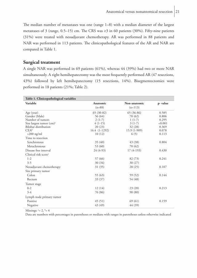
The median number of metastases was one (range 1–8) with a median diameter of the largest metastases of 3 (range, 0.5–15) cm. The CRS was ≥3 in 60 patients (30%). Fifty-nine patients (31%) were treated with neoadjuvant chemotherapy. AR was performed in 88 patients and NAR was performed in 113 patients. The clinicopathological features of the AR and NAR are compared in Table 1.
Surgical treatmentA single NAR was performed in 69 patients (61%), whereas 44 (39%) had two or more NAR simultaneously. A right hemihepatectomy was the most frequently performed AR (47 resections, 43%) followed by left hemihepatectomy (15 resections, 14%). Bisegmentectomies were performed in 18 patients (21%; Table 2).
Table 1. Clinicopathological variablesVariable Anatomic Non-anatomic p- value
(n=88) (n=113)
Age (year) 65 (30-82) 65 (36-86) 0.585Gender (Male) 56 (64) 70 (62) 0.806Number of tumors 2 (1-7) 1 (1-7) 0.295Size largest tumor (cm)a 4 (1-15) 3 (1-7) <0.001Bilobar distribution 20 (23) 32 (28) 0.369CEAb 16.4 (1-1292) 15.9 (1-909) 0.078 >200 ng/ml 10 (12) 6 (5) 0.113Time to resection Synchronous 35 (40) 43 (38) 0.804 Metachronous 53 (60) 70 (62)Disease free interval 24 (4-93) 17 (4-193) 0.430Clinical risk scorea
1-2 57 (66) 82 (73) 0.241 3-5 30 (34) 30 (27)Neoadjuvant chemotherapy 31 (35) 28 (25) 0.107Site primary tumor Colon 55 (63) 59 (52) 0.144 Rectum 33 (37) 54 (48)Tumor stage 0-2 12 (14) 23 (20) 0.213 3-4 76 (86) 90 (80)Lymph node primary tumor Positive 45 (51) 69 (61) 0.159 Negative 43 (49) 44 (39)
Missings: a= 2, b= 4Data are numbers with percentages in parentheses or medians with ranges in parentheses unless otherwise indicated
Chapter 222
Table 2. Type of resectionLiver resection Number of resections
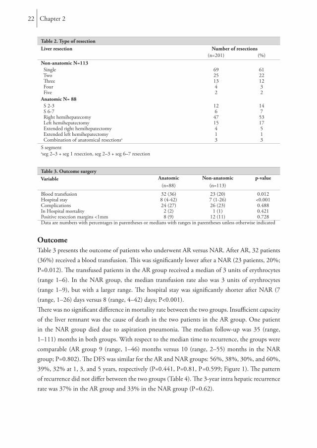
(n=201) (%)Non-anatomic N=113 Single 69 61 Two 25 22 Three 13 12 Four 4 3 Five 2 2Anatomic N= 88 S 2-3 12 14 S 6-7 6 7 Right hemihepatecomy 47 53 Left hemihepatectomy 15 17 Extended right hemihepatectomy 4 5 Extended left hemihepatectomy 1 1 Combination of anatomical resectionsa 3 3S segmentaseg 2–3 + seg 1 resection, seg 2–3 + seg 6–7 resection
Table 3. Outcome surgeryVariable Anatomic Non-anatomic p-value
(n=88) (n=113)
Blood transfusion 32 (36) 23 (20) 0.012Hospital stay 8 (4-42) 7 (1-26) <0.001Complications 24 (27) 26 (23) 0.488In Hospital mortality 2 (2) 1 (1) 0.421Positive resection margins <1mm 8 (9) 12 (11) 0.728Data are numbers with percentages in parentheses or medians with ranges in parentheses unless otherwise indicated
OutcomeTable 3 presents the outcome of patients who underwent AR versus NAR. After AR, 32 patients (36%) received a blood transfusion. This was significantly lower after a NAR (23 patients, 20%; P=0.012). The transfused patients in the AR group received a median of 3 units of erythrocytes (range 1–6). In the NAR group, the median transfusion rate also was 3 units of erythrocytes (range 1–9), but with a larger range. The hospital stay was significantly shorter after NAR (7 (range, 1–26) days versus 8 (range, 4–42) days; P<0.001). There was no significant difference in mortality rate between the two groups. Insufficient capacity of the liver remnant was the cause of death in the two patients in the AR group. One patient in the NAR group died due to aspiration pneumonia. The median follow-up was 35 (range, 1–111) months in both groups. With respect to the median time to recurrence, the groups were comparable (AR group 9 (range, 1–46) months versus 10 (range, 2–55) months in the NAR group; P=0.802). The DFS was similar for the AR and NAR groups: 56%, 38%, 30%, and 60%, 39%, 32% at 1, 3, and 5 years, respectively (P=0.441, P=0.81, P=0.599; Figure 1). The pattern of recurrence did not differ between the two groups (Table 4). The 3-year intra hepatic recurrence rate was 37% in the AR group and 33% in the NAR group (P=0.62).
Anatomical versus nonantomical resection 23
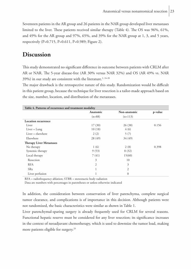
Seventeen patients in the AR group and 26 patients in the NAR group developed liver metastases limited to the liver. These patients received similar therapy (Table 4). The OS was 96%, 61%, and 49% for the AR group and 97%, 65%, and 39% for the NAR group at 1, 3, and 5 years, respectively (P=0.715, P=0.611, P=0.989; Figure 2).
Discussion
This study demonstrated no significant difference in outcome between patients with CRLM after AR or NAR. The 5-year disease-free (AR 30% versus NAR 32%) and OS (AR 49% vs. NAR 39%) in our study are consistent with the literature.2, 24-28
The major drawback is the retrospective nature of this study. Randomization would be difficult in this patient group, because the technique for liver resection is a tailor-made approach based on the size, number, location, and distribution of the metastases.
Table 4. Patterns of recurrence and treatment modalityAnatomic Non-anatomic p-value
(n=88) (n=113)Location recurrence Liver 17 (30) 26 (38) 0.156 Liver + Lung 10 (18) 4 (6) Liver + elsewhere 2 (2) 5 (7) Elsewhere 28 (49) 34 (49)Therapy Liver Metastases No therapy 1 (6) 2 (8) 0.398 Systemic therapy 9 (53) 8 (32) Local therapy 7 (41) 15(60) Resection 3 10 RFA 2 3 SRx 1 2 Liver perfusion 1 0
RFA = radiofrequency ablation; STBR = stereotactic body radiationData are numbers with percentages in parentheses or unless otherwise indicated
In addition, the consideration between conservation of liver parenchyma, complete surgical tumor clearance, and complications is of importance in this decision. Although patients were not randomized, the basic characteristics were similar as shown in Table 1.Liver parenchymal-sparing surgery is already frequently used for CRLM for several reasons. Functional hepatic reserve must be considered for any liver resection; its significance increases in the context of neoadjuvant chemotherapy, which is used to downsize the tumor load, making more patients eligible for surgery.29
Chapter 224
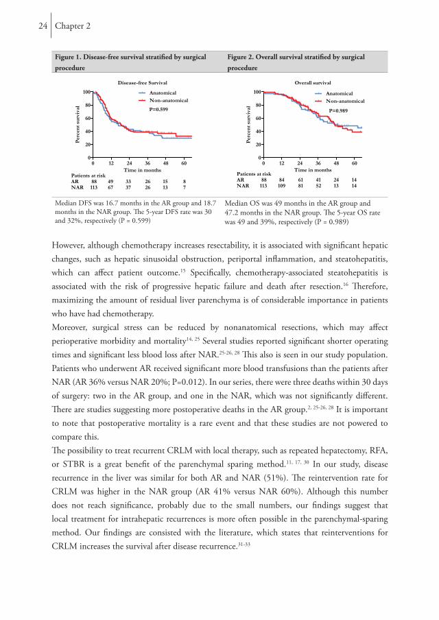
Figure 1. Disease-free survival stratified by surgical procedure
Figure 2. Overall survival stratified by surgical procedure
Median DFS was 16.7 months in the AR group and 18.7 months in the NAR group. The 5-year DFS rate was 30 and 32%, respectively (P = 0.599)
Median OS was 49 months in the AR group and 47.2 months in the NAR group. The 5-year OS rate was 49 and 39%, respectively (P = 0.989)
However, although chemotherapy increases resectability, it is associated with significant hepatic changes, such as hepatic sinusoidal obstruction, periportal inflammation, and steatohepatitis, which can affect patient outcome.15 Specifically, chemotherapy-associated steatohepatitis is associated with the risk of progressive hepatic failure and death after resection.16 Therefore, maximizing the amount of residual liver parenchyma is of considerable importance in patients who have had chemotherapy. Moreover, surgical stress can be reduced by nonanatomical resections, which may affect perioperative morbidity and mortality14, 25 Several studies reported significant shorter operating times and significant less blood loss after NAR.25-26, 28 This also is seen in our study population. Patients who underwent AR received significant more blood transfusions than the patients after NAR (AR 36% versus NAR 20%; P=0.012). In our series, there were three deaths within 30 days of surgery: two in the AR group, and one in the NAR, which was not significantly different. There are studies suggesting more postoperative deaths in the AR group.2, 25-26, 28 It is important to note that postoperative mortality is a rare event and that these studies are not powered to compare this.The possibility to treat recurrent CRLM with local therapy, such as repeated hepatectomy, RFA, or STBR is a great benefit of the parenchymal sparing method.11, 17, 30 In our study, disease recurrence in the liver was similar for both AR and NAR (51%). The reintervention rate for CRLM was higher in the NAR group (AR 41% versus NAR 60%). Although this number does not reach significance, probably due to the small numbers, our findings suggest that local treatment for intrahepatic recurrences is more often possible in the parenchymal-sparing method. Our findings are consisted with the literature, which states that reinterventions for CRLM increases the survival after disease recurrence.31-33
Anatomical versus nonantomical resection 25
For this reason, close surveillance of patients after NAR is essential. One of the possible disadvantages of NAR reported in the literature by DeMatteo et al. is the higher incidence of positive resection margins.24 In more recently published literature, it is advocated that a resection margin <1 cm is no longer a contraindication for curative resection. Moreover recent literature suggests that size of surgical margin does not correlate significantly with DFS or OS; even the need for R0 resections is being discussed.34-35 In a study by de Haas et al., the 5-year OS was similar for patients after a R0 or a R1 resection (61 versus 57%; P=0.27), although the recurrence was higher in the R1 group (28 versus 17%; P=0.004).6 In our study, the R1 resection rate was 9% in the AR group and 11% in the NAR group, which is comparable to the literature.6,
27 The concept of performing limited NAR with narrow margins is supported by the fact that micrometastases in the liver parenchyma surrounding CRLM are rare and are primarily confined to the immediate surrounding area of the tumor border.36-37
The second possible drawback of NAR, which is postulated in the literature, is the lack of vascular control.24 This is the opposite of what was published during the past years. Blood loss and blood transfusions are reported to be significantly less during and after NAR, which is confirmed by our results.25-26, 28 In contrast to CRLM, some studies report AR to be superior to NAR in HCCs.18-20 This difference may be explained by the variation in disease biology seen in primary versus metastatic liver tumors. Metastatic liver lesions develop from blood-borne tumor cells circulating throughout the body. AR may not offer the same advantage for these lesions as for HCC, which arise within a segment of the liver and might benefit from the removal of the complete functional liver unit.Multiple studies have been conducted to investigate which resection is favorable for patients with CRLM: anatomical or nonanatomical. Most authors similarly conclude that there is no significant difference between AR and NAR in disease-free and OSs. A disadvantage of the majority of studies is that the patient characteristics are not comparable between the two groups regarding tumor size and number, nodal status of the primary tumor, disease-free interval, and CEA blood levels.2, 14, 25-26 Our study contributes to this discussion due to the use of the CRS in which the previous described characteristics are incorporated. The CRS is the same for the AR and NAR, which indicates that the groups are comparable.Furthermore, the use of different neoadjuvant chemotherapy regimens during the years makes it difficult to compare the results of the studies.2, 14, 26-28 We started our patient selection after 2000, because Irinotecan and Oxaliplatin were added to the chemotherapeutic arsenal from this year forward, and all patients were treated with effective chemotherapeutics.We conclude that with a comparable complication rate, less blood transfusions, a significantly shorter hospital, and comparable disease-free and OS rates, a NAR is a safe technique for the resection of CRLM.
Chapter 226
References
1. Stangl R, Altendorf-Hofmann A, Charnley RM, et al. Factors influencing the natural history of colorectal liver metastases. Lancet 1994; 343(8910):1405-10.
2. Zorzi D, Mullen JT, Abdalla EK, et al. Comparison between hepatic wedge resection and anatomic resection for colorectal liver metastases. J Gastrointest Surg 2006; 10(1):86-94.
3. Simmonds PC, Primrose JN, Colquitt JL, et al. Surgical resection of hepatic metastases from colorectal cancer: a systematic review of published studies. Br J Cancer 2006; 94(7):982-99.
4. Geoghegan JG, Scheele J. Treatment of colorectal liver metastases. Br J Surg 1999; 86(2):158-69.
5. Muratore A, Ribero D, Zimmitti G, et al. Resection margin and recurrence-free survival after liver resection of colorectal metastases. Ann Surg Oncol 2010; 17(5):1324-9.
6. de Haas RJ, Wicherts DA, Flores E, et al. R1 resection by necessity for colorectal liver metastases: is it still a contraindication to surgery? Ann Surg 2008; 248(4):626-37.
7. Saltz LB, Cox JV, Blanke C, et al. Irinotecan plus fluorouracil and leucovorin for metastatic colorectal cancer. Irinotecan Study Group. N Engl J Med 2000; 343(13):905-14.
8. Adam R, Delvart V, Pascal G, et al. Rescue surgery for unresectable colorectal liver metastases downstaged by chemotherapy: a model to predict long-term survival. Ann Surg 2004; 240(4):644-57; discussion 657-8.
9. Azoulay D, Castaing D, Smail A, et al. Resection of nonresectable liver metastases from colorectal cancer after percutaneous portal vein embolization. Ann Surg 2000; 231(4):480-6.
10. Hemming AW, Reed AI, Howard RJ, et al. Preoperative portal vein embolization for extended hepatectomy. Ann Surg 2003; 237(5):686-91; discussion 691-3.
11. de Meijer VE, Verhoef C, Kuiper JW, et al. Radiofrequency ablation in patients with primary and secondary hepatic malignancies. J Gastrointest Surg 2006; 10(7):960-73.
12. Wong SL, Mangu PB, Choti MA, et al. American Society of Clinical Oncology 2009 clinical evidence review on radiofrequency ablation of hepatic metastases from colorectal cancer. J Clin Oncol 2010; 28(3):493-508.
13. Mendez Romero A, Wunderink W, Hussain SM, et al. Stereotactic body radiation therapy for primary and metastatic liver tumors: A single institution phase i-ii study. Acta Oncol 2006; 45(7):831-7.
14. Gold JS, Are C, Kornprat P, et al. Increased use of parenchymal-sparing surgery for bilateral liver metastases from colorectal cancer is associated with improved mortality without change in oncologic outcome: trends in treatment over time in 440 patients. Ann Surg 2008; 247(1):109-17.
15. Vauthey JN, Pawlik TM, Ribero D, et al. Chemotherapy regimen predicts steatohepatitis and an increase in 90-day mortality after surgery for hepatic colorectal metastases. J Clin Oncol 2006; 24(13):2065-72.
16. Zorzi D, Laurent A, Pawlik TM, et al. Chemotherapy-associated hepatotoxicity and surgery for colorectal liver metastases. Br J Surg 2007; 94(3):274-86.
Anatomical versus nonantomical resection 27
17. van der Pool AE, Lalmahomed ZS, de Wilt JH, et al. Local treatment for recurrent colorectal hepatic metastases after partial hepatectomy. J Gastrointest Surg 2009; 13(5):890-5.
18. Ueno S, Kubo F, Sakoda M, et al. Efficacy of anatomic resection vs nonanatomic resection for small nodular hepatocellular carcinoma based on gross classification. J Hepatobiliary Pancreat Surg 2008; 15(5):493-500.
19. Wakai T, Shirai Y, Sakata J, et al. Anatomic resection independently improves long-term survival in patients with T1-T2 hepatocellular carcinoma. Ann Surg Oncol 2007; 14(4):1356-65.
20. Hasegawa K, Kokudo N, Imamura H, et al. Prognostic impact of anatomic resection for hepatocellular carcinoma. Ann Surg 2005; 242(2):252-9.
21. Couinaud C. Liver anatomy: portal (and suprahepatic) or biliary segmentation. Dig Surg 1999; 16(6):459-67.
22. Strasberg SM. Nomenclature of hepatic anatomy and resections: a review of the Brisbane 2000 system. J Hepatobiliary Pancreat Surg 2005; 12(5):351-5.
23. Fong Y, Fortner J, Sun RL, et al. Clinical score for predicting recurrence after hepatic resection for metastatic colorectal cancer: analysis of 1001 consecutive cases. Ann Surg 1999; 230(3):309-18; discussion 318-21.
24. DeMatteo RP, Fong Y, Jarnagin WR, et al. Recent advances in hepatic resection. Semin Surg Oncol 2000; 19(2):200-7.
25. Kokudo N, Tada K, Seki M, et al. Anatomical major resection versus nonanatomical limited resection for liver metastases from colorectal carcinoma. Am J Surg 2001; 181(2):153-9.
26. Stewart GD, O’Suilleabhain CB, Madhavan KK, et al. The extent of resection influences outcome following hepatectomy for colorectal liver metastases. Eur J Surg Oncol 2004; 30(4):370-6.
27. Finch RJ, Malik HZ, Hamady ZZ, et al. Effect of type of resection on outcome of hepatic resection for colorectal metastases. Br J Surg 2007; 94(10):1242-8.
28. Sarpel U, Bonavia AS, Grucela A, et al. Does anatomic versus nonanatomic resection affect recurrence and survival in patients undergoing surgery for colorectal liver metastasis? Ann Surg Oncol 2009; 16(2):379-84.
29. Adam R, Wicherts DA, de Haas RJ, et al. Patients with initially unresectable colorectal liver metastases: is there a possibility of cure? J Clin Oncol 2009; 27(11):1829-35.
30. van der Pool AE, Mendez Romero A, Wunderink W, et al. Stereotactic body radiation therapy for colorectal liver metastases. Br J Surg 2010; 97(3):377-82.
31. Yamamoto J, Kosuge T, Shimada K, et al. Repeat liver resection for recurrent colorectal liver metastases. Am J Surg 1999; 178(4):275-81.
32. Shaw IM, Rees M, Welsh FK, et al. Repeat hepatic resection for recurrent colorectal liver metastases is associated with favourable long-term survival. Br J Surg 2006; 93(4):457-64.
33. Petrowsky H, Gonen M, Jarnagin W, et al. Second liver resections are safe and effective treatment for recurrent hepatic metastases from colorectal cancer: a bi-institutional analysis. Ann Surg 2002; 235(6):863-71.
34. Bodingbauer M, Tamandl D, Schmid K, et al. Size of surgical margin does not influence recurrence rates after curative liver resection for colorectal cancer liver metastases. Br J Surg 2007; 94(9):1133-8.
Chapter 228
35. Pawlik TM, Scoggins CR, Zorzi D, et al. Effect of surgical margin status on survival and site of recurrence after hepatic resection for colorectal metastases. Ann Surg 2005; 241(5):715-22, discussion 722-4.
36. Yamamoto J, Sugihara K, Kosuge T, et al. Pathologic support for limited hepatectomy in the treatment of liver metastases from colorectal cancer. Ann Surg 1995; 221(1):74-8.
37. Kokudo N, Miki Y, Sugai S, et al. Genetic and histological assessment of surgical margins in resected liver metastases from colorectal carcinoma: minimum surgical margins for successful resection. Arch Surg 2002; 137(7):833-40.
Chapter 3
Outcome of microscopic incomplete resection (R1) of colorectal liver metastases in the era of neoadjuvant
chemotherapy
Ninos Ayez
Zarina S. Lalmahomed
Alexander M.M. Eggermont
Jan N.M. Ijzermans
Jeroen de Jonge
Kees van Montfort
Cornelis Verhoef
Annals of surgical oncology, 2011
Chapter 330
Abstract
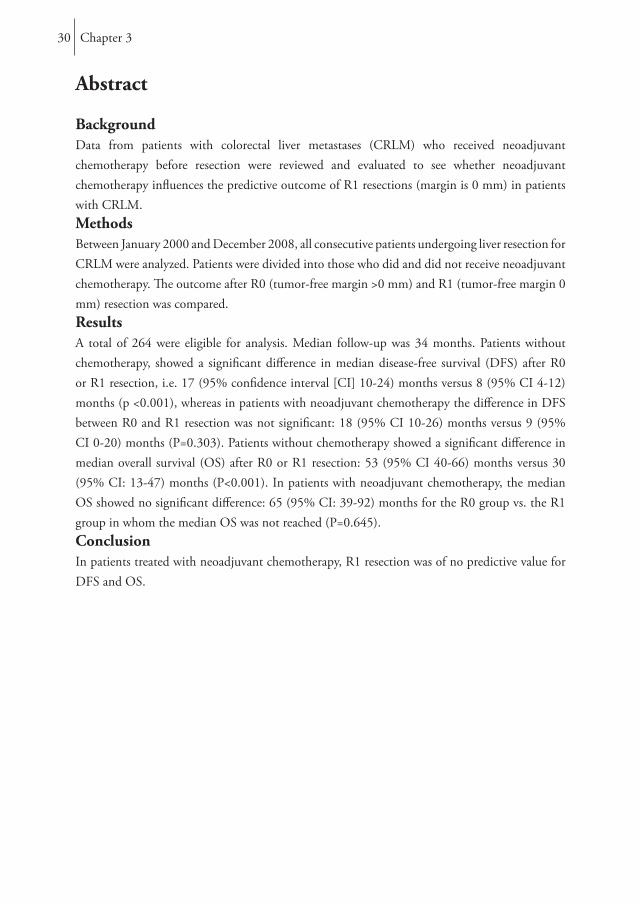
BackgroundData from patients with colorectal liver metastases (CRLM) who received neoadjuvant chemotherapy before resection were reviewed and evaluated to see whether neoadjuvant chemotherapy influences the predictive outcome of R1 resections (margin is 0 mm) in patients with CRLM.MethodsBetween January 2000 and December 2008, all consecutive patients undergoing liver resection for CRLM were analyzed. Patients were divided into those who did and did not receive neoadjuvant chemotherapy. The outcome after R0 (tumor-free margin >0 mm) and R1 (tumor-free margin 0 mm) resection was compared.ResultsA total of 264 were eligible for analysis. Median follow-up was 34 months. Patients without chemotherapy, showed a significant difference in median disease-free survival (DFS) after R0 or R1 resection, i.e. 17 (95% confidence interval [CI] 10-24) months versus 8 (95% CI 4-12) months (p <0.001), whereas in patients with neoadjuvant chemotherapy the difference in DFS between R0 and R1 resection was not significant: 18 (95% CI 10-26) months versus 9 (95% CI 0-20) months (P=0.303). Patients without chemotherapy showed a significant difference in median overall survival (OS) after R0 or R1 resection: 53 (95% CI 40-66) months versus 30 (95% CI: 13-47) months (P<0.001). In patients with neoadjuvant chemotherapy, the median OS showed no significant difference: 65 (95% CI: 39-92) months for the R0 group vs. the R1 group in whom the median OS was not reached (P=0.645).ConclusionIn patients treated with neoadjuvant chemotherapy, R1 resection was of no predictive value for DFS and OS.
Outcome of microscopic incomplete resection (R1) 31
Introduction
Without treatment, patients with colorectal liver metastases (CRLM) have a median survival of 5-8 months. 1-3 Unfortunately, only around 20-30% of patients with CRLM are resectable at the time of diagnosis.4-5 After resection 5-year survival rates of 21-48% have been reported.6-8 Nowadays, chemotherapy regimens are highly effective and can result in response rates of 50-80% and seem to convert 10-30% of the formerly irresectable CRLM to a resectable size or situation.6-7, 9-10
Several studies have described risk factors for the outcome of patients with CRLM.8, 11-17 Surgical margin status has been described as the major determinant of survival after resection, with R1 resections doing worse compared to R0 resections. However, the impact of the several proposed cut-off points regarding R0 resections remains controversial.Some older studies proposed a surgical margin of ≤ 1 cm as a contraindication for resection.16, 18-20 Others demonstrated that a resection margin ≤1 cm is not a contraindication, providing a radical resection (minimal margin of 1 mm) is performed.21-25
However, these studies did not evaluate the specific group of patients who received neoadjuvant chemotherapy before resection of CRLM. Therefore, the present study analyzes whether a resection margin of 0 mm is sufficient in the era of effective neoadjuvant chemotherapy.
Methods Between January 2000 and December 2008, all consecutive patients who underwent liver resection for CRLM were analyzed. Patients were eligible for this study if they fulfilled the following criteria: macroscopic complete resection; clear description of surgical margin status by the pathologist for each metastasis; no evidence of concomitant extrahepatic disease; no simultaneous use of local treatment modalities (radiofrequency ablation and/or cryotherapy).26 All patients underwent preoperative screening to assess the extent of the metastases by clinical examination and chest and abdominal imaging (ultrasonography, computed tomography [CT], magnetic resonance imaging). In our institute, positron emission tomography is not routinely used. Also, serum tumor marker levels (carcinoembryonic antigen [CEA]) and colonoscopy were performed preoperatively.
ChemotherapyWe are a tertiary referral hospital and we do not administer perioperative chemotherapy as a standard treatment protocol for patients with CRLM. Most patients had already received neoadjuvant chemotherapy in the referring hospital.
Chapter 332
In our clinic patients received neoadjuvant chemotherapy in case of initially difficult/unresectable liver metastases (poor location) or multiple synchronous metastases ≥4. Patients received a combination of 5-fluorouracil/capecitabine and oxaliplatin or irinotecan, with or without bevacizumab. The response to neoadjuvant chemotherapy was assessed after two or three cycles by CT scan and CEA levels. Further treatment was discussed according to the tumor response and extent of the disease. When the liver metastases were resectable, a laparotomy was planned more than three weeks after the last course of systemic neoadjuvant chemotherapy. Bevacizumab had to be excluded from the last course of chemotherapy to ensure an interval of at least 6 weeks. None of the patients received adjuvant chemotherapy as standard therapy after liver resection. The time period from 2000 was chosen because of the introduction of more effective chemotherapy and also because after 2000 the definition of resectability was not changed in our clinic.
Liver resectionHepatic parenchymal resection was performed using an ultrasonic surgical aspirator (Cavitron; Valleylab, Boulder, CO) and a monopolar coagulator. R0 was defined by the absence of microscopic tumor invasion of the resection margin (tumor-free margin >0 mm), and R1 was defined by the presence or microscopic tumor invasion of the resection margin (tumor-free margin 0 mm).
Follow upPostoperative follow-up consisted of clinical examination and measurement of CEA every 3 months. Abdominal imaging (ultrasound, CT of thorax and abdomen) was performed at 3, 6, 9 and 12 months in the first year; every 6 months the second year; and once per year thereafter. If recurrence disease recurred, a decision on whether to initiate chemotherapy treatment again was made by the multidisciplinary team.
OutcomeOverall survival (OS) was defined as the interval in months between resection of CRLM and death, or the date of last follow-up. Disease-free survival (DFS) was defined as the interval in months between resection of CRLM and recurrence, death without recurrence, or date of last follow-up without recurrence. Statistical AnalysisDescriptive values are expressed as median (range). Comparison between categorical variables was determined by the chi-square test or Fisher’s exact test as appropriate. Survival analysis was performed by the Kaplan-Meier method. Comparison between survival curves was made by log-rank tests. Univariate analysis was performed with Cox regression analysis.

Outcome of microscopic incomplete resection (R1) 33
For the multivariate analysis only parameters with a p value <0.25 in the univariate model were entered in the Cox regression model. Backward elimination was applied. Variables were included if p-values were ≤0.05 and were removed if p-values were >0.10. SPSS statistical software version 17.0 (SPSS, Chicago, IL), was used for statistical analysis; a p-value of ≤0.05 was considered statistically significant.
Results
Between January 2000 and December 2008, a total of 352 patients underwent liver resection for CRLM (Figure 1). Of these, 81 patients (23%) were excluded due to extrahepatic disease, concomitant local treatment and/or macroscopic incomplete liver resection. Seven patients
Figure 1. Flowchart of the study
Total nr.resected patients
352
Eligible patients264
Withoutchemotherapy
172
Extrahepaticdisease
23
RFA46
Extrahepatic +RFA
6
Withchemotherapy
92
R0150
R122
R081
R111
R26
Margin statusunknown
7
(2%) had unknown margin status. Finally, 264 (75%) patients were eligible for analysis. One patient was lost to follow up at 21 months. Neoadjuvant chemotherapy was given in 92 (35%) of 264 patients. Thirty eight patients (41%) received concomitant bevacizumab.Patient characteristics are listed in Tables 1 and 2. An R1 resection was found in 33 patients (13%). R1 resections in patients without chemotherapy and with chemotherapy were comparable: 13% versus 12% (P=0.845).The median follow-up was 34 (range 0-121) months. Five patients (1.9%) died postoperatively, 3 due to liver and kidney failure and 2 patients due to aspiration followed by sepsis. The median DFS was 14 (95% confidence interval [CI] 10-18) months for patients without chemotherapy and for patients with neoadjuvant chemotherapy it was 16 (95% CI 8-24) months (P=0.962).
Chapter 334
Table 1. Characteristics of patients by chemotherapy treatmentPatients without chemotherapy
Patients with chemotherapy
All patients
Value N=172
% or range
Value N=92
% or range
P-value Value N=264
% or range
Male 107 62 62 67 0.403 169 64Age 65 30-86 62 36-84 0.714 64 30-86Primary tumorRectal cancer 84 49 45 49 0.991 129 49T-stage 0.162 T3 T4 Missing data
12412
727
6682
7292
190202
7281
Positive lymph node Missing data
97 56 562
612
0.364 1532
581
Liver metastasesSynchronous 52 30 67 73 <0.001 119 45Diameter (cm) Missing data
3.32
0.9-151
3.52
0.5-182
0.068 3.44
0.5-182
Number of metastases Missing data
11
1-81
2.00
1-8 <0.001 21
1-80.5
Bilobar 43 25 36 39 0.017 79 30Resection type Anatomical Non anatomical Combined
786925
454015
413318
453620
0.54411910243
453916
R1 resection 22 13 11 12 0.845 33 13
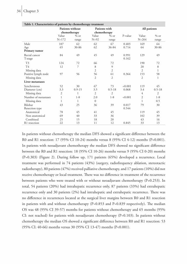
In patients without chemotherapy the median DFS showed a significant difference between the R0 and R1 resection: 17 (95% CI 10-24) months versus 8 (95% CI 4-12) months (P<0.001). In patients with neoadjuvant chemotherapy the median DFS showed no significant difference between the R0 and R1 resection: 18 (95% CI 10-26) months versus 9 (95% CI 0-20) months (P=0.303) (Figure 2). During follow up, 171 patients (65%) developed a recurrence. Local treatment was performed in 74 patients (43%) (surgery, radiofrequency ablation, stereotactic radiotherapy), 80 patients (47%) received palliative chemotherapy, and 17 patients (10%) did not receive chemotherapy or local treatment. There was no difference in treatment of the recurrence between patients who were treated with or without neoadjuvant chemotherapy (P=0.253). In total, 54 patients (20%) had intrahepatic recurrence only, 87 patients (33%) had extrahepatic recurrence only and 30 patients (2%) had intrahepatic and extrahepatic recurrence. There was no difference in recurrences located at the surgical liver margins between R0 and R1 resection in patients with and without chemotherapy (P=0.853 and P=0.839 respectively). The median OS was 48 (95% CI 39-57) months for patients without chemotherapy and 65 months (95% CI: not reached) for patients with neoadjuvant chemotherapy (P=0.103). In patients without chemotherapy the median OS showed a significant difference between R0 and R1 resection: 53 (95% CI: 40-66) months versus 30 (95% CI 13-47) months (P<0.001).

Outcome of microscopic incomplete resection (R1) 35
Table 2. Characteristics of patients by resection marginsR0 R1 All patients
Value N=231
% or range
Value N=33
% or range
P-value Value N=264
% or range
Male 152 66 17 52 0.123 169 64Age 64 31-86 62 30-77 0.499 64 30-86Primary tumorRectal cancer 117 51 12 36 0.139 129 49T-stage 0.682 T3 T4 Missing data
163182
7181
272
826
190202
7281
Positive lymph node Missing data
1272
551
26 79 0.011 1532
581
Liver metastasesSynchronous 106 46 13 39 0.483 119 45Diameter (cm) Missing data
3.24
0.5-182
3.9 1.6-7.0 0.048 3.44
0.5-182
Number of metastases Missing data
11
1-81
3.0 1-8 0.173 21
1-80.5
Bilobar 61 26 18 55 0.001 79 30Resection type Anatomical Non anatomical Combined
1078836
463816
12147
374221
0.51311910243
453916
ChemotherapyNeoadjuvant chemotherapy
81 35 11 33 P=0.845 92 35
In patients with neoadjuvant chemotherapy the median OS showed no significant difference between the R0 resection, 65 (95% CI 39-92) months, and the R1 resection, where the median OS was not reached (P=0.645) (Figure 2). A similar trend was found if a tumor-free margins of 0-2 mm versus >2mm and 0-5 mm versus >5 mm was chosen. The 5-year OS was 35% for patients without neoadjuvant chemotherapy who had R0 resection with ≤2 mm from the resection margin (n=42), whereas for patients who had a R0 resection with >2 mm from the resection margin (n=100) the 5-year OS was 51% (P=0.04). In patients with neoadjuvant chemotherapy this phenomenon could not be demonstrated: 65% (n=28) versus 45% (n=48) (P=0.564). When comparing 0-5 mm versus >5 mm, the 5 year OS was 55% versus 36% in patients without chemotherapy (P=0.062) and 44% vs. 63% in patients with chemotherapy (P=0.361). Predictive factors in univariate and multivariate analysis are depicted in Tables 3 and 4. In the total study population of 264 patients multivariate analysis showed 4 factors predictive for survival: T-stage primary tumor (hazard ratio [HR]=2.0 [1.1-3.5]; P=0.016), positive lymph nodes in primary tumor (HR=1.5 [1.0-2.2]; P=0.039), Number of metastases (≥4) (HR=1.8 [1.1-2.9]; P=0.028) and neoadjuvant chemotherapy (HR=0.62 [0.4-0.9]; p=0.027).
Chapter 336
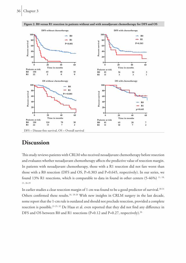
Figure 2. R0 versus R1 resection in patients without and with neoadjuvant chemotherapy for DFS and OS
DFS = Disease-free survival, OS = Overall survival
Discussion
This study reviews patients with CRLM who received neoadjuvant chemotherapy before resection and evaluates whether neoadjuvant chemotherapy affects the predictive value of resection margin. In patients with neoadjuvant chemotherapy, those with a R1 resection did not fare worst than those with a R0 resection (DFS and OS, P=0.303 and P=0.645, respectively). In our series, we found 13% R1 resections, which is comparable to data in found in other centers (5-46%) 11, 18,
21, 26-29
In earlier studies a clear resection margin of 1 cm was found to be a good predictor of survival.30-31 Others confirmed these results.16, 18-20 With new insights in CRLM surgery in the last decade, some report that the 1-cm rule is outdated and should not preclude resection, provided a complete resection is possible.21-25, 32 De Haas et al. even reported that they did not find any difference in DFS and OS between R0 and R1 resections (P=0.12 and P=0.27, respectively).26

Outcome of microscopic incomplete resection (R1) 37
Table 3. Data on univariate and multivariate analysis, Disease-free survivalWithout chemotherapy With chemotherapy
Variable MedianSurvival
(95% CI)
UnivariateHR (95% CI)
P-value
MultivariateHR (95%CI)
MedianSurvival
(95% CI)
UnivariateHR (95% CI)
MultivariateHR (95%CI)
Gender Male Female
14 (10-18)13 (7-19)
0.95 (0.65-1.39)P=0.789
NS 17 (6-28)14 (3-25)
1.03 (0.60-1.77)P=0.914
NS
Age < 60 years ≥ 60 years
14 (4-24)14 (10-18)
1.09 (0.72-1.64)P=0.692
NS 11 (6-16)21 (13-29)
0.87 (0.52-1.48)P=0.617
NS
Primary tumor
RectumColon
13 (10-16)15 (9-21)
0.86 (0.60-1.25)P=0.440
NS 17 (3-31)15 (4-26)
1.23 (0.73-2.08)P=0.440
NS
T stage primary tumor T1-3 T4
15 (11-19)6 (0-14)
2.00 (1.07-3.73)P=0.029
2.52 (1.33-4.76)P=0.004
18 (11-25)4 (1-7)
2.56 (1.16-5.67)P=0.021
2.4 (1.08-5.34)P=0.032
Lymph node Negative Positive
26 (0-57)10 (6-14)
1.70 (1.4-2.5)P=0.009
1.53 (1.02-2.29)P=0.042
16 (0-36)17 (8-26)
1.29 (0.74-2.24)P=0.371
2.35 (1.28-4.31)P=0.006
Hepatic metastases
Time diagnosis Metachronous Synchronous
15 (8-22)13 (8-18)
1.30 (0.9-1.9)P=0.174
NS 19 (8-30)14 (7-21)
1.10 (0.61-1.98)P=0.751
NS
Number of metastases ≤ 3 ≥ 4
26 (0-57)10 (6-14)
2.00 (1.1-3.5)P=0.024
NS21 (9-33)6 (5-7)
2.47 (1.37-4.44)P=0.003
NS
Largest metastasis size < 4 ≥ 4
16 (10-22)11 (8-140
1.10 (0.75-1.62)P=0.637
NS12 (6-18)23 (0-64)
0.74 (0.40-1.37)P=0.338
NS
Tumor distribution Unilobar Bilobar
16 (8-24)10 (8-12)
1.41 (0.94-2.10)P=0.093
NS 21 (0-44)11 (7-15)
1.80 (1.07-3.03)P=0.027
NS
CEA level < 50 ≥ 50
16 (11-21)8 (4-12)
1.39 (0.91-2.11)P=0.129
NS 15 (7-23)21 (0-50)
1.06 (0.48-2.35)P=0.880
NS
Resection margin R0 R1
17 (10-24)8 (4-12)
3.08 (1.90-5.01)P<0.001
2.86 (1.70-4.78)P<0.001
18 (10-26)9 (0-20)
1.44 (0.71-2.94)P=0.315
NS
NS = Not significant
In their study 74% of the patients received neoadjuvant chemotherapy and 26% did not receive neoadjuvant chemotherapy; however, they did not describe these two groups separately. In 83% of their patients, surgery was followed by adjuvant chemotherapy. The present study demonstrates comparable results when we divide the cohort into patients who did and did not receive neoadjuvant chemotherapy. However, one major difference is that none of our patients received adjuvant chemotherapy after CRLM surgery. As yet, to our knowledge, there are no randomized data to support adjuvant chemotherapy alone after liver resection. The liver represents the predominant site of cancer relapse after curative resection of CRLM, with up to 50% in the first 2 years.33-34 Thus there is a rational for perioperative chemotherapy, although controversy remains.
Chapter 338
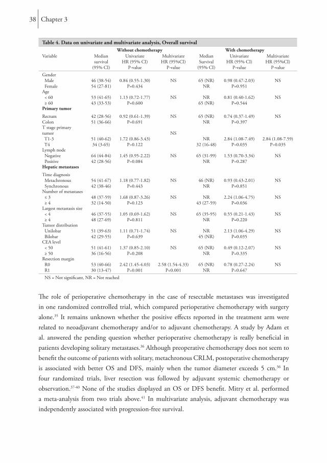
Table 4. Data on univariate and multivariate analysis, Overall survivalWithout chemotherapy With chemotherapy
Variable Mediansurvival
(95% CI)
UnivariateHR (95% CI)
P-value
MultivariateHR (95%CI)
P-value
MedianSurvival
(95% CI)
UnivariateHR (95% CI)
P-value
MultivariateHR (95%CI)
P-valueGender Male Female
46 (38-54)54 (27-81)
0.84 (0.55-1.30)P=0.434
NS 65 (NR)NR
0.98 (0.47-2.03)P=0.951
NS
Age < 60 ≥ 60
53 (41-65)43 (33-53)
1.13 (0.72-1.77)P=0.600
NS NR65 (NR)
0.81 (0.40-1.62)P=0.544
NS
Primary tumor
RectumColon
42 (28-56)51 (36-66)
0.92 (0.61-1.39)P=0.691
NS 65 (NR)NR
0.74 (0.37-1.49)P=0.397
NS
T stage primary tumor T1-3 T4
51 (40-62)34 (3-65)
1.72 (0.86-3.43)P=0.122
NSNR
32 (16-48)2.84 (1.08-7.49)
P=0.0352.84 (1.08-7.59)
P=0.035Lymph node Negative Positive
64 (44-84)42 (28-56)
1.45 (0.95-2.22)P=0.084
NS 65 (31-99)NR
1.53 (0.70-3.34)P=0.287
NS
Hepatic metastases
Time diagnosis Metachronous Synchronous
54 (41-67)42 (38-46)
1.18 (0.77-1.82)P=0.443
NS 46 (NR)NR
0.93 (0.43-2.01)P=0.851
NS
Number of metastases ≤ 3 ≥ 4
48 (37-59)32 (14-50)
1.68 (0.87-3.26)P=0.123
NS NR43 (27-59)
2.24 (1.06-4.75)P=0.036
NS
Largest metastasis size < 4 ≥ 4
46 (37-55)48 (27-69)
1.05 (0.69-1.62)P=0.811
NS 65 (35-95)NR
0.55 (0.21-1.43)P=0.220
NS
Tumor distribution Unilobar Bilobar
51 (39-63)42 (29-55)
1.11 (0.71-1.74)P=0.639
NS NR45 (NR)
2.13 (1.06-4.29)P=0.035
NS
CEA level < 50 ≥ 50
51 (41-61)36 (16-56)
1.37 (0.85-2.10)P=0.208
NS 65 (NR)NR
0.49 (0.12-2.07)P=0.335
NS
Resection margin R0 R1
53 (40-66)30 (13-47)
2.42 (1.45-4.03)P=0.001
2.58 (1.54-4.33)P<0.001
65 (NR)NR
0.78 (0.27-2.24)P=0.647
NS
NS = Not significant, NR = Not reached
The role of perioperative chemotherapy in the case of resectable metastases was investigated in one randomized controlled trial, which compared perioperative chemotherapy with surgery alone.35 It remains unknown whether the positive effects reported in the treatment arm were related to neoadjuvant chemotherapy and/or to adjuvant chemotherapy. A study by Adam et al. answered the pending question whether perioperative chemotherapy is really beneficial in patients developing solitary metastases.36 Although preoperative chemotherapy does not seem to benefit the outcome of patients with solitary, metachronous CRLM, postoperative chemotherapy is associated with better OS and DFS, mainly when the tumor diameter exceeds 5 cm.36 In four randomized trials, liver resection was followed by adjuvant systemic chemotherapy or observation.37-40 None of the studies displayed an OS or DFS benefit. Mitry et al. performed a meta-analysis from two trials above.41 In multivariate analysis, adjuvant chemotherapy was independently associated with progression-free survival.

Outcome of microscopic incomplete resection (R1) 39
Although there are strong theoretical arguments in favor of adjuvant systemic chemotherapy after resection of liver metastases, the evidence to suggest an improvement in survival after this regime is still lacking.42
Hou et al. found no difference in OS between R0 and R1 resections in patients with colorectal metastases (P=0.0776). In their study, patients did not receive neoadjuvant chemotherapy but were administered adjuvant hepatic arterial chemotherapy after resection. Furthermore, they used additional cryotherapy after resection of CRLM.43 Yan et al. also suggested that, after resection of CRLM, cryotherapy is useful; they found no difference between close or involved surgical margins (P=0.084).44 It is possible that in case of additional cryotherapy, the micrometastases that were not resected were eliminated by the edge cryotherapy, since edge cryotherapy can provide an area of tumor eradication of up to another 1 cm.To explain these results it is necessary to consider several characteristics of CRLM. One feature is micrometastases.45-47 Some found that approximately 95% of intrahepatic micrometastases are found in the close zone of < 1 cm from the gross hepatic tumor, and therefore suggested that a resection margin of 1 cm should remain the goal for resection of CRLM.47 Others harvested positive samples only within 4 mm of the tumor border.46, 48 Nanko et al. state that the bigger the macrometastases, the more micrometastases and the further away they are located.45 Yamamoto et al. could not demonstrate a relationship between distance and tumor size.49 Because there are several methods for detection of micrometastases, not all investigators report similar results on incidence. For example, when using genetic analysis, only 2% of micrometastases were detected, whereas with basic histopathology a detection rate of 56% was achieved, and with immunohistochemical staining, the detection rate is 58%.45-47 These differences in detection method might explain why micrometastases distribution is very different in these studies.It is widely accepted that neoadjuvant chemotherapy can decrease the size of metastases. But how neoadjuvant chemotherapy decreases the size of CRLM remains unclear. Ng et al. showed that cell death is randomly distributed, probably as a result of variations in chemosensitivity of tumor cells, and that tumor shrinkage occurs in a concentric manner.48 They also found that viable tumor cells were more frequent in the periphery of metastases, whether or not they were treated with chemotherapy. Mentha et al. reported similar results with the finding of a dangerous ‘halo’ (regrowth occurring at the periphery rather than in the center of the metastasis when chemotherapy is interrupted).50 They state that the surgeon should aim for a wider resection margin than 1 mm. Nevertheless, Klinger et al. rarely observed a dangerous halo.51 In their study the tumor glands were located mostly in the periphery of the metastases but were still covered by a layer of fibrotic tissue, and therefore parenchymal transection could be done within the new borders (after response to chemotherapy) without increasing the incidence of local recurrence.51 This phenomenon supports our results.
Chapter 340
Neoadjuvant chemotherapy is able to shrink the tumor, as described before. We believe that this is a concentric rather than a scattering response, as sometimes seen in primary tumors; perhaps micrometastases in the periphery of the tumor were destroyed.52 This could explain why recurrence was similar in R0 and R1 resection in patients with neoadjuvant chemotherapy. This also explains the successful downsizing of formerly unresectable metastases into resectable metastases with almost the same outcome in primary resectable cases. A concern of neoadjuvant chemotherapy might be the disappearance of smaller lesions after several lines of chemotherapy and the difficulty identifying these lesions during surgery. The need to resect all tumors seen on the prechemotherapy imaging was demonstrated previously.53-54 Another concern of chemotherapy is additional complications after liver resection. However it is known from the literature, that if limited cycles of chemotherapy are provided in the neoadjuvant setting, the morbidity or mortality of liver surgery is not increased.55-56 Therefore we administer our patients with <6 cycles of chemotherapy and evaluate after 2-3 cycles.Several authors state that the width of the negative margins (R0) does not show a significant correlation with survival.21-22, 25, 57 In all these studies a considerable proportion of the patients received neoadjuvant chemotherapy and/or adjuvant chemotherapy. In our study we demonstrated in both univariate and multivariate analysis, that the 5-year OS for patients without neoadjuvant chemotherapy showed a marked difference between R0 and R1 resection. Others proposed that margin widths of 2 mm and 5 mm, respectively, were acceptable and led to similar outcomes compared with 1-cm margin resection.28-29, 46 We also analyzed if margin width ≤ 2 mm or > 2 mm influenced survival. This proved to be the case (P=0.04) in patients who had a R0 resection without neoadjuvant chemotherapy. In patients with neoadjuvant chemotherapy this phenomenon could not be demonstrated (P=0.564). A similar trend was found if a tumor-free margin of 0-5 mm versus > 5 mm was chosen. It seems that the width of the resection margin still correlates with survival, but this only applies to patients who did not receive chemotherapy. This supports our hypothesis that neoadjuvant chemotherapy is able to destroy micrometastases in the periphery, and that in patients the same survival rate, regardless of the width of the negative margins, is achieved. Therefore, data should be revised and patients who did and did not receive chemotherapy should be investigated separately.Yamamoto et al. described another feature of some CRLM.49 They found that one third of their patients have a thick fibrous pseudocapsule and suggested that a generous surgical margin is not required for resection. Later, the same group reported on a larger cohort and demonstrated that a thicker pseudocapsule leads to fewer R1/R2 resections.58 Although not significant, there was some relation between the presence of a pseudocapsule and the ability to achieve complete resection. Pseudocapsule formation was not available in our dataset, so we could not compare this in the different chemotherapy groups.

Outcome of microscopic incomplete resection (R1) 41
Conclusion
In the present series, the DFS and OS in patients with neoadjuvant chemotherapy is similar for patients with either R0 or R1 resections.

Chapter 342
References
1. Bengmark S, Hafstrom L. The natural history of primary and secondary malignant tumors of the liver. I. The prognosis for patients with hepatic metastases from colonic and rectal carcinoma by laparotomy. Cancer 1969; 23(1):198-202.
2. Simmonds PC. Palliative chemotherapy for advanced colorectal cancer: systematic review and meta-analysis. Colorectal Cancer Collaborative Group. BMJ 2000; 321(7260):531-5.
3. Seymour MT, Stenning SP, Cassidy J. Attitudes and practice in the management of metastatic colorectal cancer in Britain. Colorectal Cancer Working Party of the UK Medical Research Council. Clin Oncol (R Coll Radiol) 1997; 9(4):248-51.
4. Adam R. Chemotherapy and surgery: new perspectives on the treatment of unresectable liver metastases. Ann Oncol 2003; 14 Suppl 2:ii13-6.
5. Scheele J, Stangl R, Altendorf-Hofmann A. Hepatic metastases from colorectal carcinoma: impact of surgical resection on the natural history. Br J Surg 1990; 77(11):1241-6.
6. Scheele J, Altendorf-Hofmann A. Resection of colorectal liver metastases. Langenbecks Arch Surg 1999; 384(4):313-27.
7. Adam R, Avisar E, Ariche A, et al. Five-year survival following hepatic resection after neoadjuvant therapy for nonresectable colorectal. Ann Surg Oncol 2001; 8(4):347-53.
8. Nordlinger B, Guiguet M, Vaillant JC, et al. Surgical resection of colorectal carcinoma metastases to the liver. A prognostic scoring system to improve case selection, based on 1568 patients. Association Francaise de Chirurgie. Cancer 1996; 77(7):1254-62.
9. Giacchetti S, Itzhaki M, Gruia G, et al. Long-term survival of patients with unresectable colorectal cancer liver metastases following infusional chemotherapy with 5-fluorouracil, leucovorin, oxaliplatin and surgery. Ann Oncol 1999; 10(6):663-9.
10. Nikfarjam M, Shereef S, Kimchi ET, et al. Survival outcomes of patients with colorectal liver metastases following hepatic resection or ablation in the era of effective chemotherapy. Ann Surg Oncol 2009; 16(7):1860-7.
11. Fong Y, Fortner J, Sun RL, et al. Clinical score for predicting recurrence after hepatic resection for metastatic colorectal cancer: analysis of 1001 consecutive cases. Ann Surg 1999; 230(3):309-18; discussion 318-21.
12. Iwatsuki S, Dvorchik I, Madariaga JR, et al. Hepatic resection for metastatic colorectal adenocarcinoma: a proposal of a prognostic scoring system. J Am Coll Surg 1999; 189(3):291-9.
13. Nagashima I, Takada T, Nagawa H, et al. Proposal of a new and simple staging system of colorectal liver metastasis. World J Gastroenterol 2006; 12(43):6961-5.
14. Konopke R, Kersting S, Distler M, et al. Prognostic factors and evaluation of a clinical score for predicting survival after resection of colorectal liver metastases. Liver Int 2009; 29(1):89-102.
15. Rees M, Tekkis PP, Welsh FK, et al. Evaluation of long-term survival after hepatic resection for metastatic colorectal cancer: a multifactorial model of 929 patients. Ann Surg 2008; 247(1):125-35.
Outcome of microscopic incomplete resection (R1) 43
16. Lise M, Bacchetti S, Da Pian P, et al. Patterns of recurrence after resection of colorectal liver metastases: prediction by models of outcome analysis. World J Surg 2001; 25(5):638-44.
17. Schindl M, Wigmore SJ, Currie EJ, et al. Prognostic scoring in colorectal cancer liver metastases: development and validation. Arch Surg 2005; 140(2):183-9.
18. Cady B, Jenkins RL, Steele GD, Jr., et al. Surgical margin in hepatic resection for colorectal metastasis: a critical and improvable determinant of outcome. Ann Surg 1998; 227(4):566-71.
19. Scheele J, Stang R, Altendorf-Hofmann A, et al. Resection of colorectal liver metastases. World J Surg 1995; 19(1):59-71.
20. Shirabe K, Takenaka K, Gion T, et al. Analysis of prognostic risk factors in hepatic resection for metastatic colorectal carcinoma with special reference to the surgical margin. Br J Surg 1997; 84(8):1077-80.
21. Pawlik TM, Scoggins CR, Zorzi D, et al. Effect of surgical margin status on survival and site of recurrence after hepatic resection for colorectal metastases. Ann Surg 2005; 241(5):715-22, discussion 722-4.
22. Figueras J, Burdio F, Ramos E, et al. Effect of subcentimeter nonpositive resection margin on hepatic recurrence in patients undergoing hepatectomy for colorectal liver metastases. Evidences from 663 liver resections. Ann Oncol 2007; 18(7):1190-5.
23. Elias D, Cavalcanti A, Sabourin JC, et al. Resection of liver metastases from colorectal cancer: the real impact of the surgical margin. Eur J Surg Oncol 1998; 24(3):174-9.
24. Elias D, Cavalcanti A, Sabourin JC, et al. Results of 136 curative hepatectomies with a safety margin of less than 10 mm for colorectal metastases. J Surg Oncol 1998; 69(2):88-93.
25. Bodingbauer M, Tamandl D, Schmid K, et al. Size of surgical margin does not influence recurrence rates after curative liver resection for colorectal cancer liver metastases. Br J Surg 2007; 94(9):1133-8.
26. de Haas RJ, Wicherts DA, Flores E, et al. R1 resection by necessity for colorectal liver metastases: is it still a contraindication to surgery? Ann Surg 2008; 248(4):626-37.
27. Choti MA, Sitzmann JV, Tiburi MF, et al. Trends in long-term survival following liver resection for hepatic colorectal metastases. Ann Surg 2002; 235(6):759-66.
28. Nuzzo G, Giuliante F, Ardito F, et al. Influence of surgical margin on type of recurrence after liver resection for colorectal metastases: a single-center experience. Surgery 2008; 143(3):384-93.
29. Poultsides GA, Schulick RD, Pawlik TM. Hepatic resection for colorectal metastases: the impact of surgical margin status on outcome. HPB (Oxford) 2010; 12(1):43-9.
30. Cady B, McDermott WV. Major hepatic resection for metachronous metastases from colon cancer. Ann Surg 1985; 201(2):204-9.
31. Ekberg H, Tranberg KG, Andersson R, et al. Determinants of survival in liver resection for colorectal secondaries. Br J Surg 1986; 73(9):727-31.
32. Pawlik TM, Vauthey JN. Surgical margins during hepatic surgery for colorectal liver metastases: complete resection not millimeters defines outcome. Ann Surg Oncol 2008; 15(3):677-9.
33. Fong Y, Cohen AM, Fortner JG, et al. Liver resection for colorectal metastases. J Clin Oncol 1997; 15(3):938-46.
Chapter 344
34. Petrelli NJ. Perioperative or adjuvant therapy for resectable colorectal hepatic metastases. J Clin Oncol 2008; 26(30):4862-3.
35. Nordlinger B, Sorbye H, Glimelius B, et al. Perioperative chemotherapy with FOLFOX4 and surgery versus surgery alone for resectable liver metastases from colorectal cancer (EORTC Intergroup trial 40983): a randomised controlled trial. Lancet 2008; 371(9617):1007-16.
36. Adam R, Bhangui P, Poston G, et al. Is perioperative chemotherapy useful for solitary, metachronous, colorectal liver metastases? Ann Surg 2010; 252(5):774-87.
37. Langer B BH, Labianca R, Shepherd L, Donato Nitti, Silvia Marsoni, Dongsheng Tu, Ann-Marie Sargeant, Anthony Fields. Fluorouracil (FU) plus l-leucovorin (l-LV) versus observation after potentially curative resection of liver or lung metastases from colorectal cancer (CRC): results of the ENG (EORTC/NCIC CTG/GIVIO) randomized trial. Proc Am Soc Clin Oncol 21: 2002 (abstr 592) 2002.
38. O’Connell MJ, Adson MA, Schutt AJ, et al. Clinical trial of adjuvant chemotherapy after surgical resection of colorectal cancer metastatic to the liver. Mayo Clin Proc 1985; 60(8):517-20.
39. Lopez-Ladron A. SJ, Bernabe R.,Bernardos A., Arriola E., Serrano J., Reina J.J.,Gomez M. A., Barneto I., Moreno-Nogueira J. A. Observation versus postoperative chemotherapy after resection of liver metastases in patients with advanced colorectal cancer. Proc Am Soc Clin Oncol 22: 2003 (abstr 1497) 2003.
40. Portier G, Elias D, Bouche O, et al. Multicenter randomized trial of adjuvant fluorouracil and folinic acid compared with surgery alone after resection of colorectal liver metastases: FFCD ACHBTH AURC 9002 trial. J Clin Oncol 2006; 24(31):4976-82.
41. Mitry E, Fields AL, Bleiberg H, et al. Adjuvant chemotherapy after potentially curative resection of metastases from colorectal cancer: a pooled analysis of two randomized trials. J Clin Oncol 2008; 26(30):4906-11.
42. Taylor I. Adjuvant chemotherapy after resection of liver metastases from colorectal cancer. Eur J Cancer 2008; 44(9):1198-201.
43. Hou RM, Chu F, Zhao J, et al. The effects of surgical margin and edge cryotherapy after liver resection for colorectal cancer metastases. HPB (Oxford) 2007; 9(3):201-7.
44. Yan TD, Padang R, Xia H, et al. Management of involved or close resection margins in 120 patients with colorectal liver metastases: edge cryotherapy can achieve long-term survival. Am J Surg 2006; 191(6):735-42.
45. Nanko M, Shimada H, Yamaoka H, et al. Micrometastatic colorectal cancer lesions in the liver. Surg Today 1998; 28(7):707-13.
46. Kokudo N, Miki Y, Sugai S, et al. Genetic and histological assessment of surgical margins in resected liver metastases from colorectal carcinoma: minimum surgical margins for successful resection. Arch Surg 2002; 137(7):833-40.
47. Wakai T, Shirai Y, Sakata J, et al. Appraisal of 1 cm hepatectomy margins for intrahepatic micrometastases in patients with colorectal carcinoma liver metastasis. Ann Surg Oncol 2008; 15(9):2472-81.
48. Ng JK, Urbanski SJ, Mangat N, et al. Colorectal liver metastases contract centripetally with a response to chemotherapy: a histomorphologic study. Cancer 2008; 112(2):362-71.

Outcome of microscopic incomplete resection (R1) 45
49. Yamamoto J, Sugihara K, Kosuge T, et al. Pathologic support for limited hepatectomy in the treatment of liver metastases from colorectal cancer. Ann Surg 1995; 221(1):74-8.
50. Mentha G, Majno PE, Andres A, et al. Neoadjuvant chemotherapy and resection of advanced synchronous liver metastases before treatment of the colorectal primary. Br J Surg 2006; 93(7):872-8.
51. Klinger M, Tamandl D, Eipeldauer S, et al. Bevacizumab improves pathological response of colorectal cancer liver metastases treated with XELOX/FOLFOX. Ann Surg Oncol 2010; 17(8):2059-65.
52. Burger JW, de Graaf EJ, Doornebosch PG, et al. Local excision of rectal cancer after chemoradiation: feasibility depends on the primary stage. Int J Colorectal Dis 2010; 25(9):1141-2.
53. Brouquet A, Abdalla EK, Kopetz S, et al. High survival rate after two-stage resection of advanced colorectal liver metastases: response-based selection and complete resection define outcome. J Clin Oncol 2011; 29(8):1083-90.
54. Benoist S, Brouquet A, Penna C, et al. Complete response of colorectal liver metastases after chemotherapy: does it mean cure? J Clin Oncol 2006; 24(24):3939-45.
55. Karoui M, Penna C, Amin-Hashem M, et al. Influence of preoperative chemotherapy on the risk of major hepatectomy for colorectal liver metastases. Ann Surg 2006; 243(1):1-7.
56. Allen PJ, Kemeny N, Jarnagin W, et al. Importance of response to neoadjuvant chemotherapy in patients undergoing resection of synchronous colorectal liver metastases. J Gastrointest Surg 2003; 7(1):109-15; discussion 116-7.
57. Hamady ZZ, Cameron IC, Wyatt J, et al. Resection margin in patients undergoing hepatectomy for colorectal liver metastasis: a critical appraisal of the 1cm rule. Eur J Surg Oncol 2006; 32(5):557-63.
58. Okano K, Yamamoto J, Kosuge T, et al. Fibrous pseudocapsule of metastatic liver tumors from colorectal carcinoma. Clinicopathologic study of 152 first resection cases. Cancer 2000; 89(2):267-75.

PART 2
Prognostic aspects
PART 2
Prognostic factors

Chapter 4
Is the clinical risk score for patients with colorectal liver metastases still useable in the era of effective
neoadjuvant chemotherapy?
Ninos AyezZarina S. Lalmahomed
Anne E.M. van der PoolYvonne Vergouwe
Kees van MontfortJeroen de Jonge
Alexander M.M. EggermontJan N.M. IJzermans
Cornelis Verhoef
Annals of surgical oncology 2011
Chapter 450
Abstract
BackgroundSeveral clinical risk scores (CRSs) for the outcome of patients with colorectal liver metastases have been validated, but not in patients undergoing neoadjuvant chemotherapy. Therefore, this study evaluates the predictive value of these CRSs in this specific group.MethodsBetween January 2000 and December 2008, all patients undergoing a metastasectomy were analyzed and divided into two groups: 193 patients did not receive neoadjuvant chemotherapy (group A) and 159 patients received neoadjuvant chemotherapy (group B). In Group B, the CRSs were calculated before and after administration of neoadjuvant chemotherapy. Results were evaluated using the CRSs proposed by Nordlinger et al., Fong et al., Nagashima et al., and Konopke et al.ResultsIn group A and B the overall median survival (OS) was 43 and 47 months, respectively (P=0.648). In group A, all CRSs used were of significant predictive value. Before administration of neoadjuvant chemotherapy, only the Nordlinger score was of predictive value. After administration of neoadjuvant chemotherapy all CRSs were of predictive value again, except for the Konopke score.ConclusionTraditional CRSs are not a reliable prognostic tool when used in patients before treatment with neoadjuvant chemotherapy. However, CRSs assessed after the administration of neoadjuvant chemotherapy are useful to predict prognosis.

Is the clinical risk score still useable? 51
Introduction
In patients with colorectal cancer, about 50-60% will develop metastatic disease. Synchronous metastases are present in 25% of CRC patients. 1-2 Nowadays, neoadjuvant chemotherapy is increasingly used for patients with colorectal liver metastases (CRLM). New systemic regimes are highly effective and response rates of 50-80% have been reported; they appear to convert 10-30% of the formerly irresectable CRLM to a resectable size.3-6 Several clinical risk scores (CRSs) for the outcome of patients with CRLM have been published.7-16 A CRS is a predictive tool for patients with CRLM who undergo resection.4, 8-12, 17-22 CRSs were initially used to predict the prognosis of patients with CRLM considered for surgery. In addition, CRSs are used to stratify patients into risk categories, to compare patient cohorts from different studies and institutions, and to select patients for different treatment protocols.However, the predictive value of these CRSs has not been assessed in the specific group of patients receiving neoadjuvant chemotherapy before resection of CRLM. It is possible that the traditional CRSs, applied before administration of neoadjuvant chemotherapy, may no longer be capable of correctly predicting the outcome in patients receiving neoadjuvant chemotherapy.4, 19-20 Therefore, in the present study, four widely used CRSs are applied in a cohort of patients with CRLM who received neoadjuvant chemotherapy before resection, to evaluate whether neoadjuvant chemotherapy influences the predictive value of CRSs.
Patients and Methods
Between January 2000 and December 2008, all consecutive patients who underwent liver resection for CRLM were analyzed. Patient characteristics were collected retrospectively from a prospectively recorded database. Two groups were created: group A (patients without neoadjuvant chemotherapy; n=193) and group B (patients with neoadjuvant chemotherapy; n=159). In Group B, the CRSs were calculated before (B1) and after (B2) administration of neoadjuvant chemotherapy.The prospective database comprises data on age, gender, primary tumor site, pathological primary tumor and lymph node stage, time between detection of primary tumor and liver metastases, type of surgery, location, maximum number and size of liver metastases on computed tomographic scan and pathology, carcinoembryonic antigen (CEA) levels, radicality of surgical margin and extrahepatic disease. Ours is a referral hospital, perioperative chemotherapy is not administered as a standard treatment protocol for patients with CRLM. Most of our patients have already received neoadjuvant chemotherapy in the referring hospital.
Chapter 452
In our center, the indication for neoadjuvant chemotherapy is twofold: in case of initially difficult/unresectable liver metastases, or in case of multiple synchronous metastases ≥4. It is our policy not to resect in case of tumor progression during chemotherapy. Neoadjuvant chemotherapy protocols comprise oxaliplatin-based combination therapies with or without bevacizumab. None of the patients in the present study received adjuvant chemotherapy. The duration of the chemotherapy was at minimum 3 cycles. If there were resectable metastases, chemotherapy was given to a maximum of 6 cycles or was stopped after 3 cycles in case of disappearing metastases. In case of unresectable disease, chemotherapy was given until the resectable status was achieved.
CRSsFour widely used CRSs were evaluated (Table 1).8-9, 11-12 The Nordlinger score includes seven risk factors and defines three risk groups but, as proposed by Nordlinger et al., we used only six risk factors. Fong’s score includes five risk factors and defines two risk groups. Nagashima’s score includes five risk factors and defines three risk groups. The Konopke score includes three risk factors and defines three risk groups. These four CRSs were applied on our data to evaluate each of the scores.
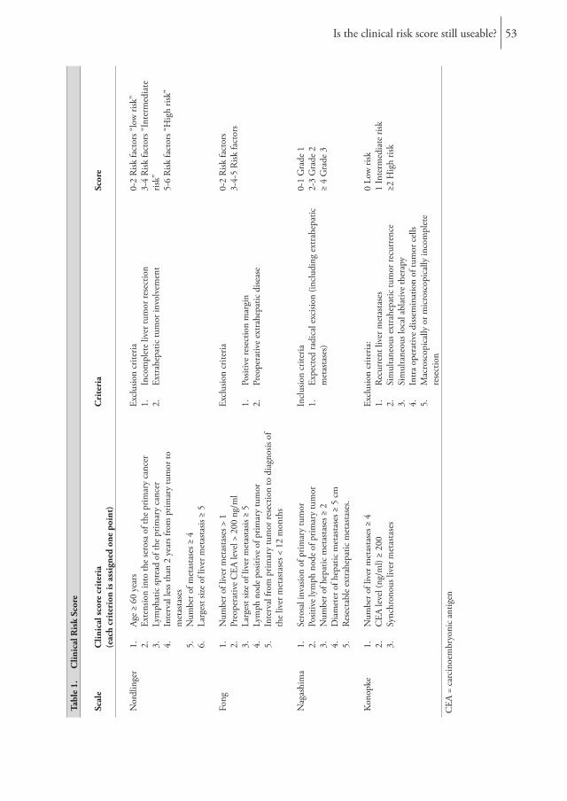
Is the clinical risk score still useable? 53
Tabl
e 1.
C
linic
al R
isk
Scor
e
Scal
eC
linic
al sc
ore
crit
eria
(e
ach
crit
erio
n is
ass
igne
d on
e po
int)
Cri
teri
aSc
ore
Nor
dlin
ger
1.
Age
≥ 60
yea
rs2.
Ex
tens
ion
into
the
sero
sa o
f the
prim
ary
canc
er3.
Ly
mph
atic
spre
ad o
f the
prim
ary
canc
er4.
In
terv
al le
ss th
an 2
yea
rs fr
om p
rimar
y tu
mor
to
met
asta
ses
5.
Num
ber o
f met
asta
ses ≥
46.
La
rges
t size
of l
iver
met
asta
sis ≥
5
Excl
usio
n cr
iteria
1.
Inco
mpl
ete
liver
tum
or re
sect
ion
2.
Extr
ahep
atic
tum
or in
volv
emen
t
0-2
Risk
fact
ors “
low
risk
”3-
4 R
isk fa
ctor
s “In
term
edia
te
risk”
5-6
Risk
fact
ors “
Hig
h ris
k”
Fong
1.
Num
ber o
f liv
er m
etas
tase
s > 1
2.
Preo
pera
tive
CEA
leve
l > 2
00 n
g/m
l3.
La
rges
t size
of l
iver
met
asta
sis ≥
54.
Ly
mph
nod
e po
sitiv
e of
prim
ary
tum
or5.
In
terv
al fr
om p
rimar
y tu
mor
rese
ctio
n to
dia
gnos
is of
th
e liv
er m
etas
tase
s < 1
2 m
onth
s
Excl
usio
n cr
iteria
1.
Posit
ive
rese
ctio
n m
argi
n2.
Pr
eope
rativ
e ex
trah
epat
ic d
iseas
e
0-2
Risk
fact
ors
3-4-
5 R
isk fa
ctor
s
Nag
ashi
ma
1.
Sero
sal i
nvas
ion
of p
rimar
y tu
mor
2.
Posit
ive
lym
ph n
ode
of p
rimar
y tu
mor
3.
Num
ber o
f hep
atic
met
asta
ses ≥
24.
D
iam
eter
of h
epat
ic m
etas
tase
s ≥ 5
cm
5.
Rese
ctab
le e
xtra
hepa
tic m
etas
tase
s.
Incl
usio
n cr
iteria
1.
Expe
cted
radi
cal e
xcisi
on (i
nclu
ding
ext
rahe
patic
m
etas
tase
s)
0-1
Gra
de 1
2-3
Gra
de 2
≥ 4
Gra
de 3
Kon
opke
1.
Num
ber o
f liv
er m
etas
tase
s ≥ 4
2.
CEA
leve
l (ng
/ml)
≥ 20
03.
Sy
nchr
onou
s liv
er m
etas
tase
s
Excl
usio
n cr
iteria
:1.
Re
curr
ent l
iver
met
asta
ses
2.
Sim
ulta
neou
s ext
rahe
patic
tum
or re
curr
ence
3.
Sim
ulta
neou
s loc
al a
blat
ive
ther
apy
4.
Intr
a op
erat
ive
diss
emin
atio
n of
tum
or c
ells
5.
Mac
rosc
opic
ally
or m
icro
scop
ical
ly in
com
plet
e re
sect
ion
0 Lo
w ri
sk1
Inte
rmed
iate
risk
≥2 H
igh
risk
CEA
= c
arci
noem
bryo
nic
antig
en
Chapter 454
OutcomeOverall survival (OS) was defined as the interval (in months) between resection of CRLM and death, or the date of last follow-up. Disease-free survival (DFS) was defined as the interval (in months) between resection of CRLM and intra and/or extrahepatic recurrence, death without recurrence, or date of last follow-up without recurrence.
Statistical analysisDescriptive statistics are expressed as median (range). Comparison between the categorical variables was made with the chi-square test. Pre- and post-chemotherapy variables are expressed as means (± SD) and compared with the paired t-test. Survival analysis was performed using the Kaplan-Meier method. Comparison between survival curves was made with log-rank tests. For missing values multiple imputation was used. The SPSS (version 17.0, Chicago, IL, USA) was used for statistical analysis, where a p-value ≤ 0.05 is considered statistically significant.
Results Between January 2000 and December 2008, 352 patients underwent liver resection for CRLM (Table 2). The median follow-up was 32 (range 0-121) months. Median age was 63 (range 30-86) years. The clinical characteristics are presented in Table 2. The median DFS was 11 (95% confidence [CI] 9-13) months, and the median OS was 46 (95% CI 39-53) months. Neoadjuvant chemotherapy was given to 159 patients (45.2%). Chemotherapy was given in a median of 6 (1-15) courses. In total, 43 patients received more than 6 courses with a median of 9 (7-15) courses.

Is the clinical risk score still useable? 55
Tabl
e 2.
Cha
ract
eris
tics
of t
he st
udy
pati
ents
Al
l pat
ient
sPa
tient
s with
out c
hem
othe
rapy
(Gro
up A
)Pa
tient
s with
che
mot
hera
py(G
roup
B)
Valu
e n=
352
% o
r ran
geVa
lue
n=19
3%
or r
ange
Valu
e n=
159
% o
r ran
geM
ale
218
6212
263
9660
Age
6330
-86
6430
-86
6236
-84
Prim
ary
tum
orRe
ctal
can
cer
167
4790
4777
48T
3T
4M
issin
g be
fore
impu
tatio
n
259
30 6
74 9 2
140
1573 8
119
1575 9
Posit
ive
lym
ph n
ode
Miss
ing
befo
re im
puta
tion
205 6
58 211
057
9258
Live
r m
etas
tase
sSy
nchr
onou
s17
249
5529
117
74D
iam
eter
(cm
)M
issin
g be
fore
impu
tatio
n3.
5 50.
5-18 1
3.5
0.9-
153.
40.
5-18
Num
ber o
f met
asta
ses
Miss
ing
befo
re im
puta
tion
2 21-
10 11
1-8
3.0
1-10
Bilo
bar
135
3852
2783
52Ex
tra
hepa
tic29
87
4422
14In
com
plet
e re
sect
ion
Miss
ing
befo
re im
puta
tion
72 721 2
4021
3220
Ove
rall
surv
ival
(in
mon
ths)
4695
% C
I 39-
5343
95%
CI 3
4-52
4795
% C
I 33-
61D
iseas
e-fre
e su
rviv
al (i
n m
onth
s)11
95%
CI 9
-13
1495
% C
I 11-
179
95%
CI 7
-11
CI =
con
fiden
ce in
terv
al
Chapter 456
Pre- and PostchemotherapyIn patients receiving neoadjuvant chemotherapy, the variables size of metastases, CEA level, number of metastases, bilobar disease and extrahepatic disease were analyzed before and after administration of neoadjuvant chemotherapy. Before neoadjuvant chemotherapy, the mean ± SD size, CEA level and number of metastases were 3.98 ± 2.62 cm, 171 ± 568 µg/l and 3.19 ± 1.95, respectively. Before neoadjuvant chemotherapy, 83 patients had bilobar disease and 22 had extrahepatic disease. After neoadjuvant chemotherapy, the mean ± SD size, CEA level, and number of metastases were 2.83 ± 2.45 cm, 21 ± 48 µg/l and 2.64 ± 1.96, respectively. After chemotherapy, 81 patients had bilobar disease and 22 patients still had extrahepatic disease. To determine the size of the metastases the postchemotherapy abdominal scans were assessed. To determine the number of metastases the postsurgery pathological report was examined. Only when complete response was reported did the number of metastases decrease. The difference between pre- and postneoadjuvant chemotherapy was significant for the size of metastases, CEA level, and the number of metastases (P<0.001, P=0.001 and P<0.001, respectively). No significant difference was found between bilobar disease and extrahepatic disease before and after neoadjuvant chemotherapy (P=0.832 and P=0.999, respectively).
NordlingerThe CRS of Nordlinger could be applied to 150 patients in group A and to 101 patients in group B (Table 1). In group A the median DFS was 16 (95% CI 12-20) months, and the median OS was 48 (95% CI 33-63) months (Tables 3, 4). In group A, the Nordlinger score was of statistically significant predictive value. There was a significant difference between the CRS subgroups for DFS and OS (P=0.028 and P=0.006, respectively). Because of the small numbers of patients, the CRS subgroup 3 (CRS 5-6) was pooled together with subgroup 2 (CRS 3-4). In group B the median DFS was 13 (95% CI 9-17) months and the median OS was 65 (95% CI 44-86) months. The Nordlinger score was of predictive value both before and after administration of neoadjuvant chemotherapy. In these CRS subgroups a significant difference was found in OS: P=0.007 before neoadjuvant chemotherapy and P=0.010 after neoadjuvant chemotherapy.
FongThe CRS of Fong could be applied to 150 patients in group A and to 101 patients in group B (Table 1). In group A the median DFS was 16.0 (95% CI 12-20) months, and the median OS was 48 (95% CI 33-63) months (Tables 3, 4). There was a significant difference between the CRS subgroups for DFS and OS (P<0.001 and P=0.001, respectively). In group B the median DFS was 13 (95% CI 9-17) months, and the median OS was 65 (95% CI 44-86) months. Fong’s score was not of significant predictive value when calculated before neoadjuvant chemotherapy.
Is the clinical risk score still useable? 57
In the CRS subgroups no significant difference was found for OS (P=0.592). After neoadjuvant chemotherapy, a significant difference (P=0.003) was found between the CRS subgroups for OS.
NagashimaThe CRS of Nagashima could be applied to 193 patients in group A and to 159 patients in group B (Table 1). In group A, the median DFS was 14 (95% CI 11-17) months and the median OS was 43 (95% CI 34-52) months (Tables 3, 4). In the CRS subgroups a significant difference was found for DFS and OS (P=0.001 and P=0.001, respectively). Due to the small numbers of patients, the CRS subgroup 3 (CRS≥4) was pooled together with subgroup 2 (CRS 2-3). In group B, the median DFS was 9 (95% CI 7-11) months and for OS the median was 47 (95% CI 33-61) months. When calculated before neoadjuvant chemotherapy Nagashima’s score was not of significant predictive value and no significant difference (P=0.122) was found between the CRS subgroups for OS. However, after neoadjuvant chemotherapy, a significant difference (P=0.001) was found between the CRS subgroups for OS.
KonopkeThe CRS of Konopke could be applied to 145 patients in group A and to 69 patients in group B (Table 1). In group A the median DFS was 16 (95% CI 11-21) months, and the median OS was 51 (95% CI 37-65) months (Tables 3, 4). Between the CRS subgroups a significant difference was found in DFS and OS (P=0.002 and P=0.024, respectively). In group B the median DFS was 21 (95% CI 3-39) months, and the median OS was 65 months (the 95% CI could not be calculated by the SPSS). There was no significant difference between the subgroups in OS, either before or after administration of neoadjuvant chemotherapy (P=0.092 and P=0.505, respectively).

Chapter 458
Tabl
e 3.
Kap
lan-
Mey
er a
naly
sis o
f dis
ease
-fre
e su
rviv
al in
pat
ient
s wit
h an
d w
itho
ut c
hem
othe
rapy
Wit
hout
Neo
adju
vant
che
mot
hera
py(G
roup
A)
Bef
ore
Neo
adju
vant
che
mot
hera
py(G
roup
B1)
Aft
er N
eoad
juva
nt c
hem
othe
rapy
(Gro
up B
2)Sc
orin
gSy
stem
nM
edia
n tim
e (m
onth
s)95
% CI
3-ye
ars
(%)
P-va
lue
nM
edia
n tim
e (m
onth
s)95
%
CI
3-ye
ars
(%)
P-va
lue
nM
edia
n tim
e (m
onth
s)95
%
CI
3-ye
ars
(%)
P-va
lue
Nor
dlin
ger
150
1612
-20
400.
028a
101
139-
1736
0.45
8a10
113
9-17
360.
173a
0-2
8718
0-43
4637
135-
2238
4714
7-23
403-
459
1511
-19
3460
136-
2036
5312
7-17
335-
64
40-
120
43
0-9
01
--
-Fo
ng15
016
12-2
040
<0.0
0110
113
9-17
360.
603
101
139-
1736
0.09
60-
212
321
3-39
4754
133-
2338
7014
6-22
393-
527
108-
1211
4712
7-17
3431
73-
1129
Nag
ashi
ma
193
1411
-17
350.
001b
159
97-
1126
0.03
0 b15
99
7-11
260.
001 b
0-1
112
1810
-26
4461
136-
2032
7214
8-20
342-
377
107-
1324
947
5-9
2284
65-
718
≥44
115-
170
46
-25
36
--
Kon
opke
145
1611
-21
410.
002
6921
3-39
450.
354
6921
3-39
450.
663
091
3712
-61
5113
41-
6115
410-
8359
147
1410
-18
2938
210-
4445
4620
6-34
41≥2
79
0-22
018
60-
1432
86
0-14
38
CI =
con
fiden
ce in
terv
al, C
RS
= cl
inic
al ri
sk sc
ore
a As a
resu
lt of
the
smal
l num
bers
of p
atie
nts,
CR
S su
bgro
up 3
(CR
S 5-
6) w
as p
oole
d to
geth
er w
ith su
bgro
up 2
(CR
S 3-
4)b As
a re
sult
of th
e sm
all n
umbe
rs o
f pat
ient
s, C
RS
subg
roup
3 (C
RS≥
4) w
as p
oole
d to
geth
er w
ith su
bgro
up 2
(CR
S 2-
3)

Is the clinical risk score still useable? 59
Tabl
e 4.
Kap
lan-
Mey
er a
naly
sis o
f ove
rall
surv
ival
in p
atie
nts w
ith
and
wit
hout
che
mot
hera
py
Wit
hout
Neo
adju
vant
che
mot
hera
py(G
roup
A)
Bef
ore
Neo
adju
vant
che
mot
hera
py(G
roup
B1)
Aft
er N
eoad
juva
nt c
hem
othe
rapy
(Gro
upB
2)Sc
orin
gSy
stem
nM
edia
n tim
e (m
onth
s)95
% CI
5-ye
ars
(%)
P-va
lue
nM
edia
n tim
e (m
onth
s)95
% CI
5-ye
ars
(%)
P-va
lue
nM
edia
n tim
e (m
onth
s)95
%
CI
5-ye
ars
(%)
P-va
lue
Nor
dlin
ger
150
4833
-63
450.
006a
101
6544
-86
530.
007a
101
6544
-86
530.
010a
0-2
8766
36-9
651
3765
NR
6647
6553
-77
653-
459
4223
-61
3960
4720
-74
4653
4629
-63
415-
64
180-
490
418
NR
01
--
-Fo
ng15
048
33-6
345
0.00
110
165
44-8
653
0.59
210
165
44-8
653
0.00
30-
212
364
40-8
852
5455
37-7
348
7065
NR
583-
527
3431
-37
2147
659-
121
6031
2916
-42
41N
agas
him
a19
343
34-5
238
0.00
1b15
947
33-6
147
0.12
2b15
947
33-6
147
0.00
1b
0-1
112
5438
-70
4761
5539
-71
4772
6543
-87
562-
377
3323
-43
2894
4318
-68
4784
335
26-4
438
≥44
3420
-48
04
238-
370
323
20-2
60
Kon
opke
145
5137
-65
450.
024
6965
NR
560.
092
6965
NR
560.
505
091
6639
-93
5213
NR
-60
15N
R-
631
4742
36-4
841
3865
44-8
660
4665
41-8
955
≥27
4118
-64
018
3227
-37
458
3212
-52
45
CI =
con
fiden
ce in
terv
al, N
R =
not
reac
hed,
CR
S =
clin
ical
risk
scor
eAs
a re
sult
of th
e sm
all n
umbe
rs o
f pat
ient
s, C
RS
subg
roup
3 (C
RS
5-6)
was
poo
led
toge
ther
with
subg
roup
2 (C
RS
3-4)
b As
a re
sult
of th
e sm
all n
umbe
rs o
f pat
ient
s, C
RS
subg
roup
3 (C
RS≥
4) w
as p
oole
d to
geth
er w
ith su
bgro
up 2
(CR
S 2-
3)
Chapter 460
Survival outcome after neoadjuvant chemotherapyPatients with a lower CRS after neoadjuvant chemotherapy, compared to before chemotherapy, had the same survival outcome as patients with the same score but who did not have chemotherapy. For Nagashima’s score, patients with a lower CRS after neoadjuvant chemotherapy had an even better survival outcome than patients with the same score who did not undergo chemotherapy (P=0.009).
Discussion
Until now, CRSs have not been evaluated for patients undergoing neoadjuvant chemotherapy before resection of CRLM. The present study evaluated CRSs in patients who received neoadjuvant chemotherapy prior to resection of liver metastases, and in patients who did not receive neoadjuvant chemotherapy.Our results confirm that the CRSs of Nordlinger, Fong, Nagashima and Konopke could be applied to patients without neoadjuvant C chemotherapy; however, when assessed before neoadjuvant chemotherapy, not all the CRSs are applicable.A recent study evaluated eight prognostic scoring systems whereas we examined only four CRSs.23 Our reason for not investigating more scoring systems was because we lacked data on some variables used for these scores. For example, the score of Ueno et al. uses a pathological factor ‘tumor budding’ which is not reported for all patients in our clinic.16 Rees et al. include the differentiation of the primary tumor in the score; however, because we are a referral center most patients had their primary tumor resected elsewhere and we were unable to obtain all information required for this score.14 Schindl et al. use specific laboratory findings in their score, but these variables were not available in our prospectively recorded database.13
Generally, CRSs are not used to determine the possibility of surgery in a patient with CRLM, but mainly to assess the prognosis of this group of patients after successful surgery. To compare results of different studies, it is helpful to assess outcome with knowledge of disease severity. The CRSs can be helpful in these cases and are often used.2, 24. However, use of effective neoadjuvant chemotherapy might influence the value of the widely used CRSs. Small et al. hypothesized that the power of prediction of Fong’s score is reduced due to the effects of chemotherapy.20 To our knowledge, the present study is the first to explore this hypothesis in a single-center database with four CRSs in patients treated with neoadjuvant chemotherapy. Our results support the finding that when the CRS is calculated before starting neoadjuvant chemotherapy it is of no predictive value; however, we demonstrate that the scores are applicable when the score is addressed after administration of neoadjuvant chemotherapy. Konopke et al. described 43 patients who received neoadjuvant chemotherapy.12
Is the clinical risk score still useable? 61
In their study, the factors concerning liver-related oncological status were determined intraoperatively. Konopke et al. also confirmed the prognostic value of their scoring system in the patients who received neoadjuvant chemotherapy. This means that the score was determined after receiving neoadjuvant chemotherapy so one would expect this score to be applicable. However, we could not demonstrate the same result in our group of patients.In the present study, chemotherapy downstaged the size and the CEA level. When the pathology report was consulted and complete response was reported, then the number of metastases also decreased significantly from a mean of 3.19 ± 1.95 to 2.64 ± 1.96 (p<0.001). This effect changes the CRS. Patients who had a higher risk score before chemotherapy became patients with a lower risk score after chemotherapy, with an associated improved survival. Bilobar disease showed no statistically significant change after administration of neoadjuvant chemotherapy and extrahepatic disease did not change at all.
ConclusionIn the era of effective neoadjuvant chemotherapy, the traditional CRSs may no longer be a reliable predictive tool. Based on our findings, if prediction of prognosis is required, all the traditional CRSs can be used if they are determined after treatment with neoadjuvant chemotherapy. If a prognosis is required before starting neoadjuvant therapy, only the Nordlinger CRS is of significant prognostic value.

Chapter 462
References
1. Bengmark S, Hafstrom L. The natural history of primary and secondary malignant tumors of the liver. I. The prognosis for patients with hepatic metastases from colonic and rectal carcinoma by laparotomy. Cancer 1969; 23(1):198-202.
2. van der Pool AE, Lalmahomed ZS, Ozbay Y, et al. “Staged” liver resection in synchronous and metachronous colorectal hepatic metastases; differences in clinicopathological features and outcome. Colorectal Dis 2009; 12(10):e229–e235.
3. Adam R, Avisar E, Ariche A, et al. Five-year survival following hepatic resection after neoadjuvant therapy for nonresectable colorectal. Ann Surg Oncol 2001; 8(4):347-53.
4. Nikfarjam M, Shereef S, Kimchi ET, et al. Survival outcomes of patients with colorectal liver metastases following hepatic resection or ablation in the era of effective chemotherapy. Ann Surg Oncol 2009; 16(7):1860-7.
5. Scheele J, Altendorf-Hofmann A. Resection of colorectal liver metastases. Langenbecks Arch Surg 1999; 384(4):313-27.
6. Giacchetti S, Itzhaki M, Gruia G, et al. Long-term survival of patients with unresectable colorectal cancer liver metastases following infusional chemotherapy with 5-fluorouracil, leucovorin, oxaliplatin and surgery. Ann Oncol 1999; 10(6):663-9.
7. Minagawa M, Yamamoto J, Kosuge T, et al. Simplified staging system for predicting the prognosis of patients with resectable liver metastasis: development and validation. Arch Surg 2007; 142(3):269-76; discussion 277.
8. Nordlinger B, Guiguet M, Vaillant JC, et al. Surgical resection of colorectal carcinoma metastases to the liver. A prognostic scoring system to improve case selection, based on 1568 patients. Association Francaise de Chirurgie. Cancer 1996; 77(7):1254-62.
9. Fong Y, Fortner J, Sun RL, et al. Clinical score for predicting recurrence after hepatic resection for metastatic colorectal cancer: analysis of 1001 consecutive cases. Ann Surg 1999; 230(3):309-18; discussion 318-21.
10. Iwatsuki S, Dvorchik I, Madariaga JR, et al. Hepatic resection for metastatic colorectal adenocarcinoma: a proposal of a prognostic scoring system. J Am Coll Surg 1999; 189(3):291-9.
11. Nagashima I, Takada T, Matsuda K, et al. A new scoring system to classify patients with colorectal liver metastases: proposal of criteria to select candidates for hepatic resection. J Hepatobiliary Pancreat Surg 2004; 11(2):79-83.
12. Konopke R, Kersting S, Distler M, et al. Prognostic factors and evaluation of a clinical score for predicting survival after resection of colorectal liver metastases. Liver Int 2009; 29(1):89-102.
13. Schindl M, Wigmore SJ, Currie EJ, et al. Prognostic scoring in colorectal cancer liver metastases: development and validation. Arch Surg 2005; 140(2):183-9.
14. Rees M, Tekkis PP, Welsh FK, et al. Evaluation of long-term survival after hepatic resection for metastatic colorectal cancer: a multifactorial model of 929 patients. Ann Surg 2008; 247(1):125-35.
15. Lise M, Bacchetti S, Da Pian P, et al. Patterns of recurrence after resection of colorectal liver metastases: prediction by models of outcome analysis. World J Surg 2001; 25(5):638-44.

Is the clinical risk score still useable? 63
16. Ueno H, Mochizuki H, Hatsuse K, et al. Indicators for treatment strategies of colorectal liver metastases. Ann Surg 2000; 231(1):59-66.
17. Zakaria S, Donohue JH, Que FG, et al. Hepatic resection for colorectal metastases: value for risk scoring systems? Ann Surg 2007; 246(2):183-91.
18. Merkel S, Bialecki D, Meyer T, et al. Comparison of clinical risk scores predicting prognosis after resection of colorectal liver metastases. J Surg Oncol 2009; 100(5):349-57.
19. Reissfelder C, Rahbari NN, Koch M, et al. Validation of prognostic scoring systems for patients undergoing resection of colorectal cancer liver metastases. Ann Surg Oncol 2009; 16(12):3279-88.
20. Small RM, Lubezky N, Shmueli E, et al. Response to chemotherapy predicts survival following resection of hepatic colo-rectal metastases in patients treated with neoadjuvant therapy. J Surg Oncol 2009; 99(2):93-8.
21. Arru M, Aldrighetti L, Castoldi R, et al. Analysis of prognostic factors influencing long-term survival after hepatic resection for metastatic colorectal cancer. World J Surg 2008; 32(1):93-103.
22. Mann CD, Metcalfe MS, Leopardi LN, et al. The clinical risk score: emerging as a reliable preoperative prognostic index in hepatectomy for colorectal metastases. Arch Surg 2004; 139(11):1168-72.
23. Sotiropoulos GC, Lang H. Clinical scoring systems for predicting outcome after surgery for colorectal liver metastases: towards a better multidisciplinary approach. Liver Int 2009; 29(1):6-9.
24. Mentha G, Terraz S, Morel P, et al. Dangerous halo after neoadjuvant chemotherapy and two-step hepatectomy for colorectal liver metastases. Br J Surg 2009; 96(1):95-103.

Chapter 5
Preoperative FDG-PET-scan in patients with resectable colorectal liver metastases does not improve overall
survival: a retrospective analyses stratified by clinical risk score
Ninos AyezJannemarie A. de Ridder
Bas WieringWim J. Oyen
Johannes H. de WiltCornelis Verhoef
Digestive surgery, 2013
Chapter 566
Abstract
BackgroundThe aim of this study was whether the selection with fluorine-18-deoxyglucose positron emission tomography (FDG-PET) imaging would result in an improved outcome in surgically treated patients with CRLM, stratified by the clinical risk score (CRS) of Fong. Patients and methodsBetween January 2000 and December 2009, all patients who underwent resection for CRLM from two different university teaching hospitals in the Netherlands were analyzed. Patients were stratified by the CRS.ResultsIn total 613 patients were eligible for analysis. There was no statistical difference in median DFS between patients with and without a FDG-PET-scan in both low CRS (17 months (95% CI: 12-22) versus 14 months (95% CI: 11-17), P=0.332) and high CRS 14 months (95% CI: 7-21) versus 9 months (95% CI: 8-10), P=0.073). There was no statistical difference in median OS between patients with and without a FDG-PET-scan in both low CRS (64 months (95% CI 54-74) versus 54 months (95% CI 42-66), P=0.663) and high CRS (39 months (95% CI 23-55) versus 41 months (95% CI 34-48), P=0.903).ConclusionsThe present study could not demonstrate that patients selected by a FDG-PET-scan before liver resection, and stratified by CRS, have an improvement in DFS or OS.
Preoperative FDG-PET-scan in patients with resectable colorectal liver metastases 67
Introduction
Colorectal cancer is one of the leading causes of cancer death world-wide.1 Approximately half of the patients with colorectal cancer will develop metastatic disease at some point during the course of the disease. If metastases are confined to the liver, resection of these metastases is at present the standard of care and it has a positive impact on survival.2, 3 After a curative resection of colorectal liver metastases (CRLM), cancer relapse is a common phenomenon, with approximately 50% of recurrences occurring in the first 2 years.4 In general, 5-year overall survival ranges between 20-60%, depending on tumor and patient characteristics. In an attempt to identify subgroups with a variable risk for relapse and survival, several clinical risk scores (CRSs) have been introduced. 5-9 The most widely used CRS was described by Fong et al. 10 and the prognostic value of this scoring system has been verified by independent investigators.11-13 Preoperative staging is important for the selection of patients who can potentially undergo resection of CRLM. To identify the number and location of colorectal metastases, contrast-enhanced CT or MRI of the liver is generally used. In addition, an abdominal and chest CT is usually performed to exclude extrahepatic disease. To further improve the selection of patients for surgery, fluorine-18-deoxyglucose positron emission tomography (FDG-PET) has been assessed in patients with CRLM.14 Some studies suggest that a change in clinical management could be expected after FDG-PET,14, 15 whereas other authors claim that the addition of staging with a FDG-PET/CT prior to planned liver resection has substantially less impact on surgical management than expected.16 If FDG-PET is able to identify the patients with extrahepatic disease who are unlikely to benefit from liver resection, the patients with a negative extrahepatic FDG-PET should represent a selected subgroup that is more likely to benefit from surgery. This might be reflected in an improved disease free and possibly overall survival compared to patients who have not undergone preoperative staging with FDG-PET.In the present study we analyzed whether this selection with FDG-PET would result in an improved outcome in surgically treated patients with CRLM, stratified by the CRS of Fong.
Patients and methods
Between January 2000 and December 2009, all patients who underwent liver resection for CRLM from two different university teaching hospitals in the Netherlands were analyzed retrospectively. In these two hospitals more than a quarter of all patients with colorectal liver metastases undergo surgery. Patients were assessed with the CRS according to Fong and excluded from the analysis if they had missing data to calculate the CRS.
Chapter 568
The criteria that are incorporated in this CRS system are: (1) nodal status of primary, (2) disease-free interval from the primary to discovery of the liver metastases <12 months, (3) number of tumors >1, (4) size of the largest tumor >5 cm, and (5) preoperative carcinoembryonic antigen (CEA) level >200 ng/ml.10 Each criterion is assigned one point in this CRS and we defined two risk groups: Low CRS (0-2 risk factors) and High CRS (3-5 risk factors).
Treatment protocolThe Erasmus MC University Medical Centre Rotterdam and Radboud University Nijmegen Medical Centre are tertiary referral hospitals for CRLM. All patients were discussed in multidisciplinary tumor boards. In their protocols, perioperative chemotherapy is not considered standard of care in all patients with primarily resectable CRLM (i.e. possibility of an R0 resection, the vascular inflow and outflow must be secured, as well as biliary drainage to the remaining segments, and a future liver remnant of at least 20-30%).Patients receive neoadjuvant chemotherapy in case liver metastases are initially unresectable or difficult to resect (due to ill location, close to vascular or biliary structures) or when multiple (≥4) synchronous metastases are present.A large proportion of patients in this study already received neoadjuvant chemotherapy in the referring hospitals, according to local treatment protocols. Patients treated with neoadjuvant chemotherapy received a combination of 5-fluorouracil (5-FU)/capecitabine and oxaliplatin or irinotecan, with or without bevacizumab. The response to neoadjuvant chemotherapy was assessed after 2 or 3 cycles by CT scan and CEA levels. Further treatment was considered depending on tumor response and extent of the disease. If liver metastases were considered resectable, a laparotomy was planned at least 3 weeks after the last course of systemic neoadjuvant chemotherapy. Bevacizumab had to be excluded from the last course of chemotherapy to ensure an interval of at least 6 weeks.FDG-PET was performed within 5 weeks before surgery in a selection of patients, based on a multicentre study or by physician’s choice. At laparotomy the abdomen was examined for extrahepatic disease. In case of extrahepatic disease (confirmed by frozen sections) any further surgical treatment was only carried out if all tumor deposits could be resected. A minority of patients received adjuvant chemotherapy as part of a trial in the Netherlands (HEPATICA) irrespective whether or not preoperative FDG-PET was performed.17
Postoperative follow-up consisted of clinical examination and measurement of CEA every 3 months. In the Erasmus MC University Medical Centre Rotterdam, abdominal imaging (ultrasound, CT of the chest and abdomen) was usually performed every 3 months in the first year and every 6 months the second year and once per year thereafter. In the Radboud University Nijmegen Medical Centre this was every 3 months in the first 3 years and every 6 months in the 4th and 5th year.
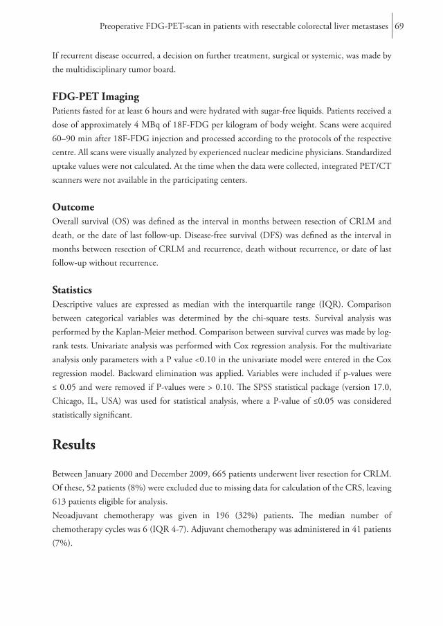
Preoperative FDG-PET-scan in patients with resectable colorectal liver metastases 69
If recurrent disease occurred, a decision on further treatment, surgical or systemic, was made by the multidisciplinary tumor board.
FDG-PET ImagingPatients fasted for at least 6 hours and were hydrated with sugar-free liquids. Patients received a dose of approximately 4 MBq of 18F-FDG per kilogram of body weight. Scans were acquired 60–90 min after 18F-FDG injection and processed according to the protocols of the respective centre. All scans were visually analyzed by experienced nuclear medicine physicians. Standardized uptake values were not calculated. At the time when the data were collected, integrated PET/CT scanners were not available in the participating centers.
OutcomeOverall survival (OS) was defined as the interval in months between resection of CRLM and death, or the date of last follow-up. Disease-free survival (DFS) was defined as the interval in months between resection of CRLM and recurrence, death without recurrence, or date of last follow-up without recurrence.
StatisticsDescriptive values are expressed as median with the interquartile range (IQR). Comparison between categorical variables was determined by the chi-square tests. Survival analysis was performed by the Kaplan-Meier method. Comparison between survival curves was made by log-rank tests. Univariate analysis was performed with Cox regression analysis. For the multivariate analysis only parameters with a P value <0.10 in the univariate model were entered in the Cox regression model. Backward elimination was applied. Variables were included if p-values were ≤ 0.05 and were removed if P-values were > 0.10. The SPSS statistical package (version 17.0, Chicago, IL, USA) was used for statistical analysis, where a P-value of ≤0.05 was considered statistically significant.
Results
Between January 2000 and December 2009, 665 patients underwent liver resection for CRLM. Of these, 52 patients (8%) were excluded due to missing data for calculation of the CRS, leaving 613 patients eligible for analysis. Neoadjuvant chemotherapy was given in 196 (32%) patients. The median number of chemotherapy cycles was 6 (IQR 4-7). Adjuvant chemotherapy was administered in 41 patients (7%).
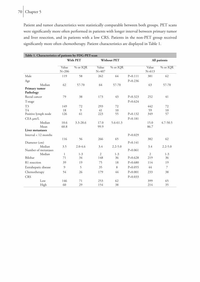
Chapter 570
Patient and tumor characteristics were statistically comparable between both groups. PET scans were significantly more often performed in patients with longer interval between primary tumor and liver resection, and in patients with a low CRS. Patients in the non-PET group received significantly more often chemotherapy. Patient characteristics are displayed in Table 1.
Table 1. Characteristics of patients by FDG-PET-scanWith PET Without PET All patients
ValueN=206
% or IQR ValueN=407
% or IQR ValueN=613
% or IQR
Male 119 58 262 64 P=0.111 381 62Age Median 62 57-70 64 57-70
P=0.23663 57-70
Primary tumorPathologyRectal cancer 79 38 173 43 P=0.323 252 41T-stage P=0.624T3T4
14918
729
29341
7210
44259
7210
Positive lymph node 126 61 223 55 P=0.132 349 57CEA µm/L Median Mean
10.660.8
3.3-28.6 17.099.9
5.6-61.3P=0.181
15.086.7
4.7-50.5
Liver metastasesInterval < 12 months
116 56 266 65P=0.029
382 62Diameter (cm) Median 3.5 2.0-4.6 3.4 2.2-5.0
P=0.1413.4 2.2-5.0
Number of metastases Median 1 1-3 2 1-3
P=0.0612 1-3
Bilobar 71 34 148 36 P=0.628 219 36R1 resection 39 19 75 18 P=0.680 114 19Extrahepatic disease 9 5 35 8 P=0.055 44 7Chemotherapy 54 26 179 44 P<0.001 233 38CRS Low High
14660
7129
253154
6238
P=0.033399214
6535
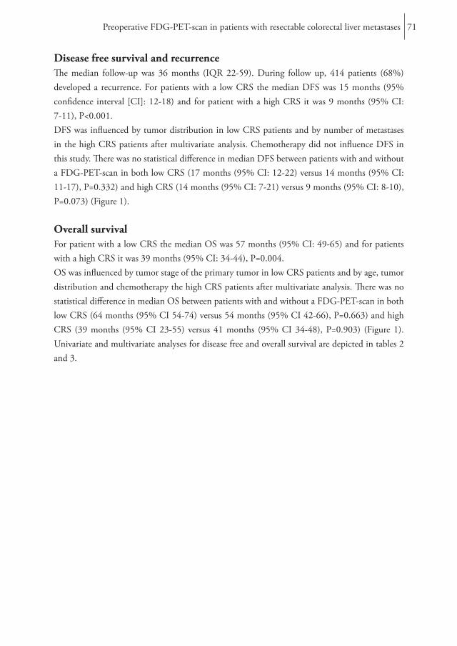
Preoperative FDG-PET-scan in patients with resectable colorectal liver metastases 71
Disease free survival and recurrenceThe median follow-up was 36 months (IQR 22-59). During follow up, 414 patients (68%) developed a recurrence. For patients with a low CRS the median DFS was 15 months (95% confidence interval [CI]: 12-18) and for patient with a high CRS it was 9 months (95% CI: 7-11), P<0.001. DFS was influenced by tumor distribution in low CRS patients and by number of metastases in the high CRS patients after multivariate analysis. Chemotherapy did not influence DFS in this study. There was no statistical difference in median DFS between patients with and without a FDG-PET-scan in both low CRS (17 months (95% CI: 12-22) versus 14 months (95% CI: 11-17), P=0.332) and high CRS (14 months (95% CI: 7-21) versus 9 months (95% CI: 8-10), P=0.073) (Figure 1).
Overall survivalFor patient with a low CRS the median OS was 57 months (95% CI: 49-65) and for patients with a high CRS it was 39 months (95% CI: 34-44), P=0.004.OS was influenced by tumor stage of the primary tumor in low CRS patients and by age, tumor distribution and chemotherapy the high CRS patients after multivariate analysis. There was no statistical difference in median OS between patients with and without a FDG-PET-scan in both low CRS (64 months (95% CI 54-74) versus 54 months (95% CI 42-66), P=0.663) and high CRS (39 months (95% CI 23-55) versus 41 months (95% CI 34-48), P=0.903) (Figure 1). Univariate and multivariate analyses for disease free and overall survival are depicted in tables 2 and 3.

Chapter 572
Figure 1. Low and high CRS in patients with and without neoadjuvant chemotherapy (CTx): disease-free survival (DFS) and overall survival (OS)
DFS = Disease free survival, OS = overall survival, CRS =Clinical risk score, PET = positron emission tomography

Preoperative FDG-PET-scan in patients with resectable colorectal liver metastases 73
Table 2. Univariate and multivariate analysis. Disease free survivalLow CRS High CRS
Variable Median survival
(95% CI)
UnivariateHR(95% CI)
P-value
MultivariateHR(95%CI)
P-value
Median survival
(95% CI)
UnivariateHR(95% CI)
P-value
MultivariateHR(95%CI)
P-value
GenderFemaleMale
14 (11-17)16 (12-20)
0.83 (0.64-1.07)P=0.146
NS 8 (6-10)9 (7-11)
1.01 (0.74-1.37)P=0.969
NS
Age< 60≥ 60
13 (10-16)17 (13-21)
0.85 (0.65-1.10)P=0.207
NS 9 (7-11)9 (7-11)
1.20 (0.88-1.64)P=0.248
NS
Primary tumorColonRectum
17 (12-22)13 (10-16)
0.86 (0.67-1.09)P=0.212
NS 9 (7-11)9 (7-11)
0.94 (0.69-1.28)P=0.707
NS
T stage primary tumorT1-3T4
16 (13-19)13 (8-18)
1.47 (0.99-2.17)P=0.057
NS 9 (7-11)9 (5-13)
1.44 (0.91-2.29)P=0.117
NS
Lymph nodeNegativePositive
16 (12-20)15 (11-19)
1.12 (0.87-1.43)P=0.377
NS 11 (6-16)9 (8-10)
1.10 (0.71-1.69)P=0.670
NS
Hepatic metastasesTime diagnosis> 12 months≤ 12 months
16 (11-21)14 (12-16)
1.12 (0.88-1.44)P=0.336
NS 8 (4-12)9 (7-11)
0.97 (0.57-1.65)P=0.907
NS
Number of metastases< 4≥ 4
17 (13-21)11 (8-14)
1.54 (1.11-2.13)P=0.010
NS 10 (7-13)8 (6-10)
1.51 (1.11-2.05)P=0.009
1.51 (1.07-2.05)P=0.009
Largest metastasis size< 5≥ 5
16 (13-19)14 (10-18)
1.32 (0.70-2.45)P=0.393
NS 9 (8-10)9 (5-13)
1.03 (0.81-1.29)P=0.835
NS
Tumor distributionUnilobarBilobar
19 (13-25)10 (7-13)
1.74 (1.33-2.27)P<0.001
1.71 (1.29-2.27)P<0.001
9 (6-12)9 (7-11)
1.24 (0.91-1.69)P=1.172
NS
CEA level< 200≥ 200
15 (12-18)12 (4-20)
1.62 (0.86-3.04)P=0.137
NS 9 (8-10)7 (4-10)
1.12 (0.77-1.64)P=0.551
NS
Resection marginR0R1
16 (12-20)11 (8-14)
1.35 (0.96-1.91)P=0.0.88
NS 10 (8-12)8 (6-10)
1.27 (0.90-1.80)P=0.178
NS
ChemotherapyNoYes
17 (13-21)12 (9-15)
1.08 (0.82-1.42)P=0.561
NS 9 (7-11)9 (7-11)
1.01 (0.75-1.37)P=0.936
NS
FDG-PET-scanNoYes
14 (11-7)17 (12-22)
0.88 (0.68-1.14)P=0.341
NS 9 (8-10)14 (7-21)
0.73 (0.51-1.04)P=0.084
NS
NS = Not significant, Univariate P value < 0.10 included in Multivariate analysis

Chapter 574
Table 3. Univariate and multivariate analysis, overall survivalLow CRS High CRS
Variable Median survival
(95% CI)
UnivariateHR(95% CI)
P-value
MultivariateHR(95%CI)
P-value
Median survival
(95% CI)
UnivariateHR(95% CI)
P-value
MultivariateHR(95%CI)
P-valueGenderFemaleMale
56 (45-67)58 (44-72)
0.99 (0.74-1.32)P=0.933
NS 35 (27-43)42 (33-51)
0.96 (0.68-1.37)P=0.832
NS
Age< 60≥ 60
64 (55-74)56 (41-71)
1.07 (0.79-1.43)P=0.679
NS 55 (32-78)35 (30-40)
1.52 (1.05-2.19)P=0.026
1.52 (1.05-2.20)P=0.027
Primary tumorColonRectum
58 (45-71)55 (44-66)
0.96 (0.73-1.28)P=0.787
NS 39 (28-50)39 (32-46)
1.02 (0.71-1.47)P=0.903
NS
T stage primary tumorT1-3T4
65 (55-75)33 (24-42)
1.96 (1.31-2.95)P=0.001
1.99 (1.32-2.99)P=0.001
42 (34-50)27 (19-35)
1.69 (1.01-2.83)P=0.044
NS
Lymph nodeNegativePositive
66 (53-79)49 (44-54)
1.28 (0.97-1.69)P=0.081
NS 46 (NR)39 (33-45)
1.38 (0.79-2.41)P=0.253
NS
Hepatic metastasesTime diagnosis > 12 months≤ 12 months 64 (50-78)
52 (38-66)1.06 (0.80-1.40)P=0.680
NS 34 (28-40)41 (34-48)
0.84 (0.46-1.52)P=0.562
NS
Number of metastases< 4≥ 4
58 (49-67)45 (28-62)
1.26 (0.87-1.85)P=0.226
NS 43 (31-55)34 (23-45)
1.26 (0.88-1.80)P=0.208
NS
Largest metastasis size< 5≥ 5
58 (50-66)49 (32-66)
1.08 (0.75-1.57)P=0.679
NS43 (32-54)32 (25-39)
1.39 (0.98-1.98)P=0.064
NS
Tumor distributionUnilobarBilobar 57 (46-68)
52 (35-69)1.16 (0.85-1.59)P=0.346
NS 46 (22-70)35 (29-41)
1.36 (0.95-1.95)P=0.097
1.47 (1.02-2.12)P=0.039
CEA level< 200≥ 200
57 (49-65)36 (23-49)
1.23 (0.58-2.63)P=0.588
NS 39 (33-45)43 (23-63)
0.97 (0.63-1.51)P=0.896
NS
Resection marginR0R1 65 (54-76)
53 (36-70)1.32 (0.89-1.95)P=0.165
NS 43 (31-55)35 (26-44)
2.82 (0.35-22.8)P=0.325
NS
ChemotherapyNoYes
61 (52-70)47 (65)
1.02 (0.74-1.40)P=0.903
NS 34 (28-40)65 (33-97)
0.65 (0.46-0.96)P=0.016
0.63 (0.44-0.89)P=0.10
FDG-PET-scanNoYes
54 (42-66)64 (54-74)
0.94 (0.71-1.25)P=0.665
NS 41 (34-48)39 (23-55)
1.02 (0.70-1.50)P=0.904
NS
NS = Not significant, NR = Not reached, Univariate P value < 0.10 included in Multivariate analysis
Discussion
FDG-PET is used for patients with colorectal cancer to demonstrate extrahepatic disease and as a consequence it may improve patient selection for surgical resection of the liver metastases.
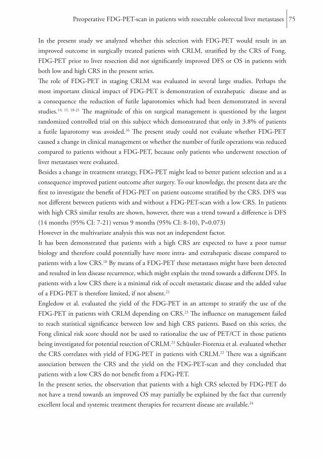
Preoperative FDG-PET-scan in patients with resectable colorectal liver metastases 75
In the present study we analyzed whether this selection with FDG-PET would result in an improved outcome in surgically treated patients with CRLM, stratified by the CRS of Fong. FDG-PET prior to liver resection did not significantly improved DFS or OS in patients with both low and high CRS in the present series.The role of FDG-PET in staging CRLM was evaluated in several large studies. Perhaps the most important clinical impact of FDG-PET is demonstration of extrahepatic disease and as a consequence the reduction of futile laparotomies which had been demonstrated in several studies.14, 15, 18-21 The magnitude of this on surgical management is questioned by the largest randomized controlled trial on this subject which demonstrated that only in 3.8% of patients a futile laparotomy was avoided.16 The present study could not evaluate whether FDG-PET caused a change in clinical management or whether the number of futile operations was reduced compared to patients without a FDG-PET, because only patients who underwent resection of liver metastases were evaluated.Besides a change in treatment strategy, FDG-PET might lead to better patient selection and as a consequence improved patient outcome after surgery. To our knowledge, the present data are the first to investigate the benefit of FDG-PET on patient outcome stratified by the CRS. DFS was not different between patients with and without a FDG-PET-scan with a low CRS. In patients with high CRS similar results are shown, however, there was a trend toward a difference is DFS (14 months (95% CI: 7-21) versus 9 months (95% CI: 8-10), P=0.073)However in the multivariate analysis this was not an independent factor. It has been demonstrated that patients with a high CRS are expected to have a poor tumur biology and therefore could potentially have more intra- and extrahepatic disease compared to patients with a low CRS.10 By means of a FDG-PET these metastases might have been detected and resulted in less disease recurrence, which might explain the trend towards a different DFS. In patients with a low CRS there is a minimal risk of occult metastatic disease and the added value of a FDG-PET is therefore limited, if not absent.22 Engledow et al. evaluated the yield of the FDG-PET in an attempt to stratify the use of the FDG-PET in patients with CRLM depending on CRS.23 The influence on management failed to reach statistical significance between low and high CRS patients. Based on this series, the Fong clinical risk score should not be used to rationalize the use of PET/CT in those patients being investigated for potential resection of CRLM.23 Schüssler-Fiorenza et al. evaluated whether the CRS correlates with yield of FDG-PET in patients with CRLM.22 There was a significant association between the CRS and the yield on the FDG-PET-scan and they concluded that patients with a low CRS do not benefit from a FDG-PET. In the present series, the observation that patients with a high CRS selected by FDG-PET do not have a trend towards an improved OS may partially be explained by the fact that currently excellent local and systemic treatment therapies for recurrent disease are available.24
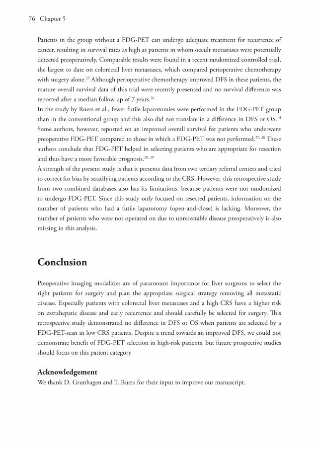
Chapter 576
Patients in the group without a FDG-PET can undergo adequate treatment for recurrence of cancer, resulting in survival rates as high as patients in whom occult metastases were potentially detected preoperatively. Comparable results were found in a recent randomized controlled trial, the largest to date on colorectal liver metastases, which compared perioperative chemotherapy with surgery alone.25 Although perioperative chemotherapy improved DFS in these patients, the mature overall survival data of this trial were recently presented and no survival difference was reported after a median follow up of 7 years.26 In the study by Ruers et al., fewer futile laparotomies were performed in the FDG-PET group than in the conventional group and this also did not translate in a difference in DFS or OS.14 Some authors, however, reported on an improved overall survival for patients who underwent preoperative FDG-PET compared to those in which a FDG-PET was not performed.27, 28 These authors conclude that FDG-PET helped in selecting patients who are appropriate for resection and thus have a more favorable prognosis.28, 29
A strength of the present study is that it presents data from two tertiary referral centers and tried to correct for bias by stratifying patients according to the CRS. However, this retrospective study from two combined databases also has its limitations, because patients were not randomized to undergo FDG-PET. Since this study only focused on resected patients, information on the number of patients who had a futile laparotomy (open-and-close) is lacking. Moreover, the number of patients who were not operated on due to unresectable disease preoperatively is also missing in this analysis.
Conclusion
Preoperative imaging modalities are of paramount importance for liver surgeons to select the right patients for surgery and plan the appropriate surgical strategy removing all metastatic disease. Especially patients with colorectal liver metastases and a high CRS have a higher risk on extrahepatic disease and early recurrence and should carefully be selected for surgery. This retrospective study demonstrated no difference in DFS or OS when patients are selected by a FDG-PET-scan in low CRS patients. Despite a trend towards an improved DFS, we could not demonstrate benefit of FDG-PET selection in high-risk patients, but future prospective studies should focus on this patient category
AcknowledgementWe thank D. Grunhagen and T. Ruers for their input to improve our manuscript.
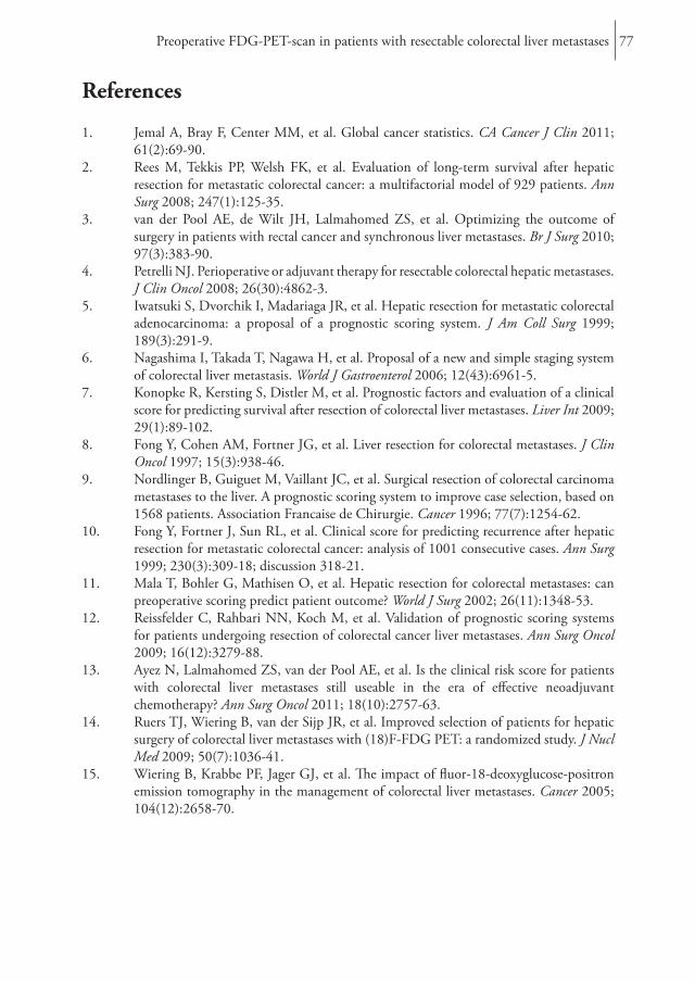
Preoperative FDG-PET-scan in patients with resectable colorectal liver metastases 77
References
1. Jemal A, Bray F, Center MM, et al. Global cancer statistics. CA Cancer J Clin 2011; 61(2):69-90.
2. Rees M, Tekkis PP, Welsh FK, et al. Evaluation of long-term survival after hepatic resection for metastatic colorectal cancer: a multifactorial model of 929 patients. Ann Surg 2008; 247(1):125-35.
3. van der Pool AE, de Wilt JH, Lalmahomed ZS, et al. Optimizing the outcome of surgery in patients with rectal cancer and synchronous liver metastases. Br J Surg 2010; 97(3):383-90.
4. Petrelli NJ. Perioperative or adjuvant therapy for resectable colorectal hepatic metastases. J Clin Oncol 2008; 26(30):4862-3.
5. Iwatsuki S, Dvorchik I, Madariaga JR, et al. Hepatic resection for metastatic colorectal adenocarcinoma: a proposal of a prognostic scoring system. J Am Coll Surg 1999; 189(3):291-9.
6. Nagashima I, Takada T, Nagawa H, et al. Proposal of a new and simple staging system of colorectal liver metastasis. World J Gastroenterol 2006; 12(43):6961-5.
7. Konopke R, Kersting S, Distler M, et al. Prognostic factors and evaluation of a clinical score for predicting survival after resection of colorectal liver metastases. Liver Int 2009; 29(1):89-102.
8. Fong Y, Cohen AM, Fortner JG, et al. Liver resection for colorectal metastases. J Clin Oncol 1997; 15(3):938-46.
9. Nordlinger B, Guiguet M, Vaillant JC, et al. Surgical resection of colorectal carcinoma metastases to the liver. A prognostic scoring system to improve case selection, based on 1568 patients. Association Francaise de Chirurgie. Cancer 1996; 77(7):1254-62.
10. Fong Y, Fortner J, Sun RL, et al. Clinical score for predicting recurrence after hepatic resection for metastatic colorectal cancer: analysis of 1001 consecutive cases. Ann Surg 1999; 230(3):309-18; discussion 318-21.
11. Mala T, Bohler G, Mathisen O, et al. Hepatic resection for colorectal metastases: can preoperative scoring predict patient outcome? World J Surg 2002; 26(11):1348-53.
12. Reissfelder C, Rahbari NN, Koch M, et al. Validation of prognostic scoring systems for patients undergoing resection of colorectal cancer liver metastases. Ann Surg Oncol 2009; 16(12):3279-88.
13. Ayez N, Lalmahomed ZS, van der Pool AE, et al. Is the clinical risk score for patients with colorectal liver metastases still useable in the era of effective neoadjuvant chemotherapy? Ann Surg Oncol 2011; 18(10):2757-63.
14. Ruers TJ, Wiering B, van der Sijp JR, et al. Improved selection of patients for hepatic surgery of colorectal liver metastases with (18)F-FDG PET: a randomized study. J Nucl Med 2009; 50(7):1036-41.
15. Wiering B, Krabbe PF, Jager GJ, et al. The impact of fluor-18-deoxyglucose-positron emission tomography in the management of colorectal liver metastases. Cancer 2005; 104(12):2658-70.
Chapter 578
16. Moulton C, Levine MN, Law C, et al. An Ontario Clinical Oncology Group (OCOG) randomized controlled trial (RCT) assessing FDG PET/CT in resectable liver colorectal adenocarcinoma metastases (CAM). J Clin Oncol 2011; 29: 2011 (suppl; abstr 3520) 2011.
17. Verhoef C, de Wilt JH, Burger JW, et al. Surgery of the primary in stage IV colorectal cancer with unresectable metastases. Eur J Cancer 2011; 47 Suppl 3:S61-6.
18. Briggs RH, Chowdhury FU, Lodge JP, et al. Clinical impact of FDG PET-CT in patients with potentially operable metastatic colorectal cancer. Clin Radiol 2011; 66(12):1167-74.
19. Ruers TJ, Langenhoff BS, Neeleman N, et al. Value of positron emission tomography with [F-18]fluorodeoxyglucose in patients with colorectal liver metastases: a prospective study. J Clin Oncol 2002; 20(2):388-95.
20. Desai DC, Zervos EE, Arnold MW, et al. Positron emission tomography affects surgical management in recurrent colorectal cancer patients. Ann Surg Oncol 2003; 10(1):59-64.
21. Imdahl A, Reinhardt MJ, Nitzsche EU, et al. Impact of 18F-FDG-positron emission tomography for decision making in colorectal cancer recurrences. Langenbecks Arch Surg 2000; 385(2):129-34.
22. Schussler-Fiorenza CM, Mahvi DM, Niederhuber J, et al. Clinical risk score correlates with yield of PET scan in patients with colorectal hepatic metastases. J Gastrointest Surg 2004; 8(2):150-7; discussion 157-8.
23. Engledow AH, Skipworth JR, Pakzad F, et al. The role of 18FDG PET/CT in the management of colorectal liver metastases. HPB (Oxford) 2012; 14(1):20-5.
24. van der Pool AE, Lalmahomed ZS, de Wilt JH, et al. Local treatment for recurrent colorectal hepatic metastases after partial hepatectomy. J Gastrointest Surg 2009; 13(5):890-5.
25. Nordlinger B, Sorbye H, Glimelius B, et al. Perioperative chemotherapy with FOLFOX4 and surgery versus surgery alone for resectable liver metastases from colorectal cancer (EORTC Intergroup trial 40983): a randomised controlled trial. Lancet 2008; 371(9617):1007-16.
26. Nordlinger B, Sorbye H, Glimelius B, et al. EORTC liver metastases intergroup randomized phase III study 40983: Long-term survival results. J Clin Oncol 2012; 30:(suppl; abstr 3508).
27. Fernandez FG, Drebin JA, Linehan DC, et al. Five-year survival after resection of hepatic metastases from colorectal cancer in patients screened by positron emission tomography with F-18 fluorodeoxyglucose (FDG-PET). Ann Surg 2004; 240(3):438-47; discussion 447-50.
28. Strasberg SM, Dehdashti F, Siegel BA, et al. Survival of patients evaluated by FDG-PET before hepatic resection for metastatic colorectal carcinoma: a prospective database study. Ann Surg 2001; 233(3):293-9.
29. Brennan MF. PET scanning in malignancy: infant, adolescent or mature citizen? Ann Surg 2001; 233(3):320-1.
Chapter 6
The use of neoadjuvant chemotherapy in patients with resectable colorectal liver metastases: clinical risk score
as possible discriminator
Ninos AyezErik van der StokDirk Grunhagen
J. RothbarthE. van Meerten
Alexander M.M. EggermontCornelis Verhoef
European journal of surgical oncology, 2015
Chapter 680
Abstract
BackgroundThe combination of chemotherapy (CTx) and hepatic resection is increasingly accepted as an effective treatment for patients with colorectal liver metastases (CRLM). However, controversy exists regarding the sequence of surgery and CTx and whether all patients with resectable CRLM benefit from perioperative CTx. We investigated whether overall survival was influenced by neo-adjuvant CTx in patients with resectable CRLM, stratified by the clinical risk score (CRS) described by Fong et al.. MethodsPatients who underwent liver resection for CRLM between January 2000 and December 2009 were analysed. We compared survival rates of patients with and without neo-adjuvant CTx stratified by the CRS. The CRS includes five risk factors and defines two risk groups: low CRS (0-2) and high CRS (3-5). ResultsIn total 363 patients (64% male) were included, median age 63 years (IQR 57-70). Prior to liver resection, 219 patients had a low CRS (65 received neo-adjuvant CTx) and 144 patients had a high CRS (88 received neo-adjuvant CTx). None of the patients received adjuvant CTx for CRLM. Median follow up was 47 months (IQR 25-82). In the low CRS group, there was no significant difference in median overall survival (OS) between patients with and without CTx (65 months (95% CI 39-91) vs. 54 months (95% CI 44-64), p=0.31). In the high CRS group, there was a significant difference in median OS between patients with and without CTx (46 months (95% CI 24-68) vs. 33 month (95% CI 29-37), p=0.004).ConclusionIn our series, patients with a high clinical risk profile benefit from neo-adjuvant CTx. In patients with a low risk profile, neo-adjuvant CTx might not be beneficial.
The use of neoadjuvant chemotherapie and clinical risk score 81
Introduction
Colorectal carcinoma is one of the leading causes of cancer death world-wide, mostly as a consequence of metastatic disease.1 Administration of combined chemotherapy regimens improves survival rates of patients with colorectal liver metastases (CRLM).2-4 If metastases are confined to the liver, surgical resection is the most effective therapy, providing the only potential for cure.5, 6 However, cancer relapse after curative resection of CRLM is a common phenomenon, with recurrence rates up to 50% in the first 2 years.7 In an attempt to reduce these recurrence rates, it has been proposed to combine liver resection with systemic chemotherapy, either pre-, peri- or postoperatively. Numerous studies have investigated the impact of adjuvant chemotherapy in addition to surgery for CRLM, but have failed to show survival benefit.8, 9 Recently, the mature results of the landmark EORTC 40983 trial, studying the impact of perioperative chemotherapy, were published showing no overall survival benefit for patients in the chemotherapy group.10,
11 Therefore, the exact role of systemic therapy in combination with liver resection for CRLM remains unclear. Nonetheless, a recent report has recommended to treat the majority of patients with CRLM with neo-adjuvant chemotherapy.12
In order to predict the likelihood of tumour recurrence and survival after resection for CRLM, several Clinical Risk Scores (CRS) have been developed.5, 13-18 The most widely used and validated CRS has been described by Fong et al. in 1999.15 In this publication, 5 independent clinical risk factors for survival after surgery for CRLM are described. Furthermore, 2 risk groups (high/low) are characterized. Although all CRLM may well be regarded as “high risk”, this CRS may play a role in explaining the relative lack of efficacy of systemic therapy when combined with surgery in the metastasized setting. It is not uncommon in other types of malignancies (e.g. breast, primary colon) to restrict the use of adjuvant chemotherapy to those patients with the most advanced disease (highest risk profile). The present study was conducted to retrospectively evaluate overall survival outcome in patients with and without neo-adjuvant chemotherapy, stratified by their clinical risk profile as described by Fong.
Patients and methods
Between January 2000 and December 2009, all consecutive patients who underwent liver resection for CRLM were analysed. Patients were assessed by Fong’s CRS and excluded from the analysis if they had missing data to calculate the CRS and/or extrahepatic disease. Calculation of the CRS was based on clinical data at presentation with CRLM.
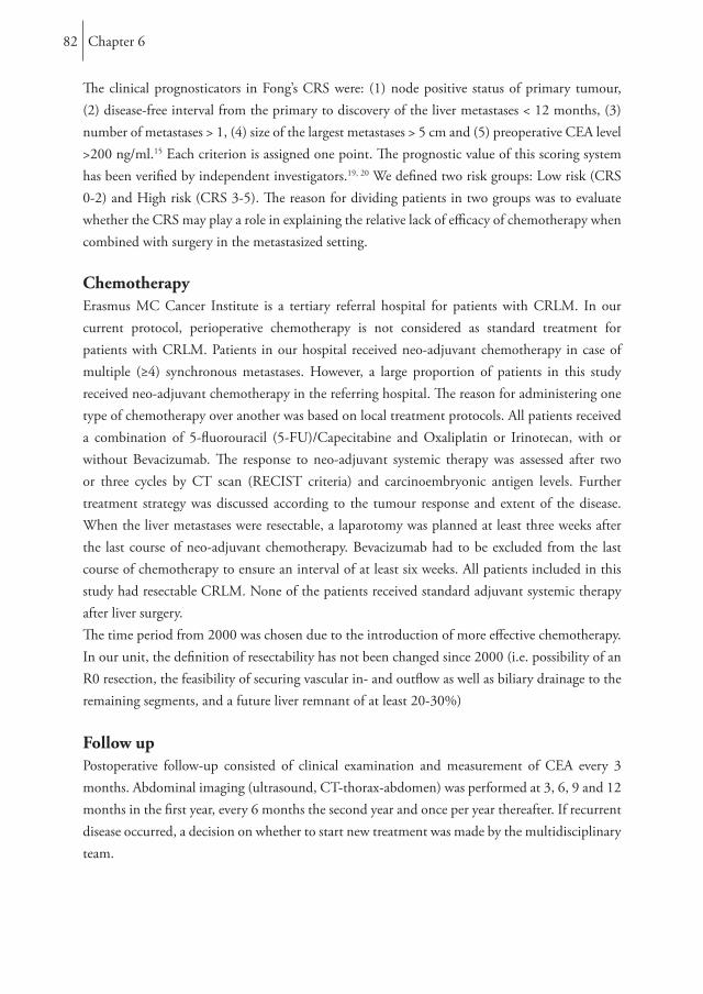
Chapter 682
The clinical prognosticators in Fong’s CRS were: (1) node positive status of primary tumour, (2) disease-free interval from the primary to discovery of the liver metastases < 12 months, (3) number of metastases > 1, (4) size of the largest metastases > 5 cm and (5) preoperative CEA level >200 ng/ml.15 Each criterion is assigned one point. The prognostic value of this scoring system has been verified by independent investigators.19, 20 We defined two risk groups: Low risk (CRS 0-2) and High risk (CRS 3-5). The reason for dividing patients in two groups was to evaluate whether the CRS may play a role in explaining the relative lack of efficacy of chemotherapy when combined with surgery in the metastasized setting.
ChemotherapyErasmus MC Cancer Institute is a tertiary referral hospital for patients with CRLM. In our current protocol, perioperative chemotherapy is not considered as standard treatment for patients with CRLM. Patients in our hospital received neo-adjuvant chemotherapy in case of multiple (≥4) synchronous metastases. However, a large proportion of patients in this study received neo-adjuvant chemotherapy in the referring hospital. The reason for administering one type of chemotherapy over another was based on local treatment protocols. All patients received a combination of 5-fluorouracil (5-FU)/Capecitabine and Oxaliplatin or Irinotecan, with or without Bevacizumab. The response to neo-adjuvant systemic therapy was assessed after two or three cycles by CT scan (RECIST criteria) and carcinoembryonic antigen levels. Further treatment strategy was discussed according to the tumour response and extent of the disease. When the liver metastases were resectable, a laparotomy was planned at least three weeks after the last course of neo-adjuvant chemotherapy. Bevacizumab had to be excluded from the last course of chemotherapy to ensure an interval of at least six weeks. All patients included in this study had resectable CRLM. None of the patients received standard adjuvant systemic therapy after liver surgery.The time period from 2000 was chosen due to the introduction of more effective chemotherapy. In our unit, the definition of resectability has not been changed since 2000 (i.e. possibility of an R0 resection, the feasibility of securing vascular in- and outflow as well as biliary drainage to the remaining segments, and a future liver remnant of at least 20-30%)
Follow up Postoperative follow-up consisted of clinical examination and measurement of CEA every 3 months. Abdominal imaging (ultrasound, CT-thorax-abdomen) was performed at 3, 6, 9 and 12 months in the first year, every 6 months the second year and once per year thereafter. If recurrent disease occurred, a decision on whether to start new treatment was made by the multidisciplinary team.

The use of neoadjuvant chemotherapie and clinical risk score 83
Outcome Overall survival (OS) was defined as the interval in months between resection of CRLM and death, or the date of last follow-up. Disease-free survival (DFS) was defined as the interval in months between resection of CRLM and recurrence, death without recurrence, or date of last follow-up without recurrence.
Statistics Descriptive values are expressed as median (interquartile range (IQR)). Variables were compared by means of chi-square analysis or Fischer’s exact test (depending on the sample size) or with the independent Student’s t test or Mann-Whitney U test when appropriate. Survival analysis was performed by the Kaplan-Meier method. Comparison between survival curves was made by log-rank tests. For the multivariate analysis only parameters with a p value < 0.10 in the univariate model were entered in the Cox regression model. The SPSS statistical package (version 21.0, Chicago, IL, USA) was used for statistical analysis; a p-value of ≤0.05 was considered statistically significant.
Results
Between January 2000 and December 2009, 442 patients underwent liver resection for CRLM. Of these, 77 patients (17%) were excluded due to extrahepatic disease and/or missing data for calculation of the CRS. 2 patients were lost to follow up, leaving 363 patients eligible for analysis (42 patients extrahepatic disease, 33 patients missing data and 2 patients with both, 2 lost to follow up). Neo-adjuvant chemotherapy was given in 153 (42%) patients. 51 patients received adjuvant chemotherapy for their primary colorectal cancer (30 patients in the low CRS group and 21 patients in the high CRS group, P=0.093). 15 of these patients received further neo-adjuvant chemotherapy for their liver metastases. Neo-adjuvant capecitabine monotherapy was administered in 5 patients and 5-FU/LV monotherapy was administered in 3 patients. The majority of patients receiving neo-adjuvant chemotherapy received either oxaliplatin-based chemotherapy (88%) or irinotecan-based chemotherapy (7%). Sixty-seven patients received concomitant bevacizumab (44%) mostly in combination with oxaliplatin (88%). The median number of chemotherapy cycles for all patients was 5 (IQR 4-6). The median number of chemotherapy cycles was also 5 (IQR 4-6) in patients with a low risk profile and 6 in patients with a high CRS (IQR 4-7).

Chapter 684
Table 1. Characteristics of patients by chemotherapy treatment. Low clinical risk score without CTx
(N=154)with CTx (N=65)
All patients (N=219)
value % or IQR value % or IQR P value value % or IQRMale 95 62% 47 72% 0,133 142 Age median 66 59-72 63 58-70 0,09 65 59-71Primary tumor Rectal cancer 70 46% 36 55% 0,179 106 48%T stage T3 107 70% 40 62% 147 67%T4 9 6% 7 11% 16 7%Positive lymph node 61 40% 46 71% 0,145 80 37%CEA median 15 6-49 16 6-47 0,726 16 6-48 mean 48 37 45 Liver metastases Synchronous 27 18% 40 62% < 0,001 67 31%Diameter (cm) median 3,2 2,4-4,4 2,6 2-3,8 0,016 3 2-4Number of mets median 1 1-2 2 1-4 < 0,001 1 1-2Bilobar 22 14% 27 42% < 0,001 49 22%R1 resection 21 14% 10 16% 0,743 31 14%
Patient characteristics are displayed in Tables 1, 2 and 3. Eighty-four patients (24%) had an R1 resection. The numbers of R1 resections in patients with and without neo-adjuvant chemotherapy were comparable: 28% vs. 20% (P=0.09), respectively. An R1 resection occurred more often in patients with a high CRS than in patients with a low CRS (38% versus 14%, P<0.001. The median follow-up was 47 months (IQR 25-82). Seven patients (1.9%) died postoperatively.
Disease Free Survival and recurrenceFor patients with neo-adjuvant chemotherapy the median DFS was 12 months (95% confidence interval (CI) 9-15) and for patients without chemotherapy it was 13 months ((95% CI 10-16), P=0.89). In patients with a low CRS there was no difference in the median DFS between patients with and without chemotherapy (13 months, (95% CI 9-17)) versus 16 months ((95% CI 10-22), P=0.86). The 5-year DFS was 33% versus 27% respectively. In patients with a high CRS there was a significant difference in median DFS between patients with and without chemotherapy (11 months, (95% CI 7-15)) versus 9 months ((95% CI 8-10), P=0.02). The 5-year DFS was 24% versus 9% respectively (Figure 1). During follow-up, 267 patients (74%) developed a recurrence. Local treatment was performed in 110 patients (41%) (surgery, radiofrequency ablation, stereotactic radiotherapy), 116 patients (44%) had palliative chemotherapy and 26 patients (10%) received neither chemotherapy nor local treatment.

The use of neoadjuvant chemotherapie and clinical risk score 85
Table 2. Characteristics of patients by chemotherapy treatment. High clinical risk score
without CTx
(N=56)with CTx (N=88)
all patients (N=144)
value % or IQR value % or IQR P value value % or IQRMale 36 64% 54 61% 0,724 90 63%Age median 61 57-69 61 56-68 0,397 61 56-68primary tumor Rectal cancer 26 46% 25 28% 0,028 51 35%T stage T3 43 77% 73 83% 116 81%T4 6 11% 7 8% 13 9%Positive lymph node 49 88% 68 77% 117 81%CEA median 26 8-70 55 8-229 0,246 34 8-202 mean 104 274 208 Liver metastases Synchronous 27 48% 72 82% < 0,001 99 69%Diameter (cm) median 3,5 2-5,5 4,5 3-6 0,033 4 2,5-5,8Number of mets median 3 2-3 4 2-5 0,004 3 2Bilobar 31 55% 55 63% 0,394 86 60%R1 resection 21 38% 32 38% 0,949 53 38%
Overall Survival For patients with neo-adjuvant chemotherapy the median OS was 57 months (95% CI 40-74) and for patients without chemotherapy it was 45 months (95% CI 38-52), P=0.08. In patients with a low CRS there was no difference in the median OS between patients with and without chemotherapy (65 months (95% CI 39-91) versus 54 months ((95% CI 44-64), HR 0.83, P=0.31). The 5-year OS was 52% versus 46% respectively. In patients with a high CRS the median OS was significantly higher in patients who received chemotherapy compared with those who did not (46 months (95% CI 24-68), versus 33 months (95% CI 29-37), HR 0.57, P=0.004). The 5-year OS was 46% versus 20% respectively (Figure 1).
Univariate and multivariate analyses In patients with a low CRS, the univariate analysis showed that 2 factors were prognostic for OS: T-stage primary tumour (T4) and positive resection margin (R1). In multivariate analysis these factors remained prognostic for OS (Table 4). In patients with a high CRS, 2 factors were prognostic for OS in univariate analysis: primary tumour location (colon vs. rectum) and administration of neo-adjuvant chemotherapy. In multivariate analysis, only administration of neo-adjuvant chemotherapy remained of significant influence on overall survival (Table 4).

Chapter 686
Table 3. Characteristics of patients by CRS
CRS low (N=219)
CRS high (N=144)
All patients (N=363)
value % or IQR value % or IQR P value value % or IQRMale 142 65% 90 63% 0,65 232 64%Age median 65 59-71 61 56-68 0,001 63 57-70Primary tumor rectal cancer 106 48% 51 35% 0,015 157 43%t stage t3 147 67% 116 81% 263 73%t4 16 7% 13 9% 29 8%positive lymph node 80 37% 117 81% < 0,001 197 54%CEA median 16 6-48 34 8-203 < 0,001 19 6-69 mean 45 208 110 Liver metastases synchronous 67 31% 99 69% < 0,001 166 46%Diameter (cm) median 3 2-4 4 3-6 < 0,001 3,4 2,2-5Number of mets median 1 1-2 3 2-4 < 0,001 2 1-3bilobar 49 22% 86 60% < 0,001 135 37%R1 resection 31 14% 53 38% < 0,001 84 24%Chemotherapy Yes 65 30% 88 61% < 0,001 153 42% Response
CR 5 8% 3 3% 8 5%PR 40 64% 61 69% 101 67%SD 18 29% 22 25% 40 27%PD 0 0 1 1% 1 1%
No 154 70% 56 39% 210 48%
Discussion
This is the first study to demonstrate that patients with primary resectable CRLM and a high clinical risk profile have significant overall survival benefit when adding neo-adjuvant chemotherapy to resection for metastases. All patients have a potential follow up of 4 years, as can be seen by the few censored cases in the Kaplan Meier curves. In the last decade, the development of modern chemotherapeutic agents and biologicals has significantly improved OS in patients with CRLM.2-4, 21-28 This success of systemic therapy in the palliative setting has prompted studies to evaluate the role of chemotherapy in combination with liver resection.9-11, 29 However, these studies often involve strict study protocol inclusion criteria. Consequently, patients with a high risk profile -who might benefit the most from chemotherapy- are often underrepresented in these studies. Since genuine survival benefit of multimodal therapies has not yet been demonstrated, could this low impact of chemotherapy on survival then be explained by the relatively low risk profile of the patients in these trials?The present role of perioperative chemotherapy for primary resectable metastases was established in a landmark randomized controlled trial, which compared perioperative chemotherapy with surgery alone 10.
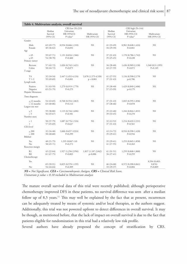
The use of neoadjuvant chemotherapie and clinical risk score 87
Table 4. Multivariate analysis, overall survival
CRS low (N=219) CRS high (N=144)MedianSurvival
(95% CI)
Univariate HR (95%CI)
P-value*Multivariate
HR (95% CI)
MedianSurvival
(95% CI)
UnivariateHR (95% CI)
P-value*Multivariate
HR (95% CI)
Gender
Male 63 (49-77) 0,934 (0,666-1,310) NS 41 (33-49) 0,961 (0,648-1,424) NS Female 49 (36-62) P=0,692 34 (29-39) P=0,841 Age
< 65 59 (47-71) 1,131 (0,816-1,566) NS 37 (31-43) 1,170 (0,786-1,742) NS > 65 54 (38-70) P=0,460 35 (25-45) P=0,438 Primary tumor
Rectum 57 (42-72) 1,026 (0,742-1,421) NS 34 (28-40) 1,436 (0,969-2,130) 1,340 (0,9-1,995) Colon 58 (44-72) P=0,875 43 (27-59) P=0,072 P=0,149T stage T4 32 (10-54) 2,447 (1,452-4,124) 2,670 (1,575-4,526) 41 (27-55) 1,134 (0,590-2,179) NS T 1-3 59 (49-69) P=0,001 p < 0,001 37 (31-43) p+0,706 Lymph node
Positive 51 (43-59) 1,279 (0,919-1,779) NS 35 (30-40) 1,429 (0,849-2,406) NS Negative 65 (51-79) P=0,279 57 (15-99) p=0,179 Hepatic Metastases
Time diagnosis
< 12 months 54 (43-65) 0,768 (0,554-1,063) NS 37 (31-43) 1,045 (0,595-1,836) NS > 12 months 63 (40-86) P=0,112 37 (28-46) P=0,878 Largest size met
> 5 59 (38-80) 1,123 (0,744-1,696) NS 35 (22-48) 1,244 (0,844-1,831) NS < 5 56 (45-67) P=0,581 39 (33-45) P=0,270 Number mets
> 1 58 (37-79) 1,087 (0,776-1,524) NS 32 (12-52) 1,214 (0,632-2,331) NS 1 57 (45-69) P=0,627 37 (31-43) P=0,561 CEA level
> 200 32 (16-48) 1,606 (0,657-3,924) NS 43 (14-72) 0,916 (0,590-1,420) NS < 200 58 (48-68) P=0,299 35 (29-41) P=0,916 Bilobair
Yes 48 (21-75) 1,235 (0,847-1,8) NS 35 (29-41) 1,253 (0,845-1,858) NS No 58 (45-71) P=0,272 41 (27-55) P=0,263 Resection margin
R1 43 (22-64) 1,927 (1,256-2,956) 1,837 (1,187-2,842) 41 (31-51) 1,259 (0,848-1,868) NS R0 61 (47-75) P=0,003 p=0,006 34 (27-41) P=0,259 Chemotherapy
Yes
65 (39-91) 0,825 (0,570-1,195) NS 46 (24-68) 0,572 (0,390-0,841)0,594 (0,403-
0,876) No 54 (44-64) P=0,309 33 (29-37) P=0,004 P=0,009
NS = Not Significant, CEA = Carcinoembryonic Antigen, CRS = Clinical Risk Score, Univariate p value < 0.10 included in Multivariate analysis
The mature overall survival data of this trial were recently published; although perioperative chemotherapy improved DFS in these patients, no survival difference was seen after a median follow up of 8,5 years.11 This may well be explained by the fact that at present, recurrences can be adequately treated by means of systemic and/or local therapies, as the authors suggest. Additionally, this trial was not powered upfront to detect differences in overall survival. It may be though, as mentioned before, that the lack of impact on overall survival is due to the fact that patients eligible for randomization in this trial had a relatively low risk profile. Several authors have already proposed the concept of stratification by CRS.
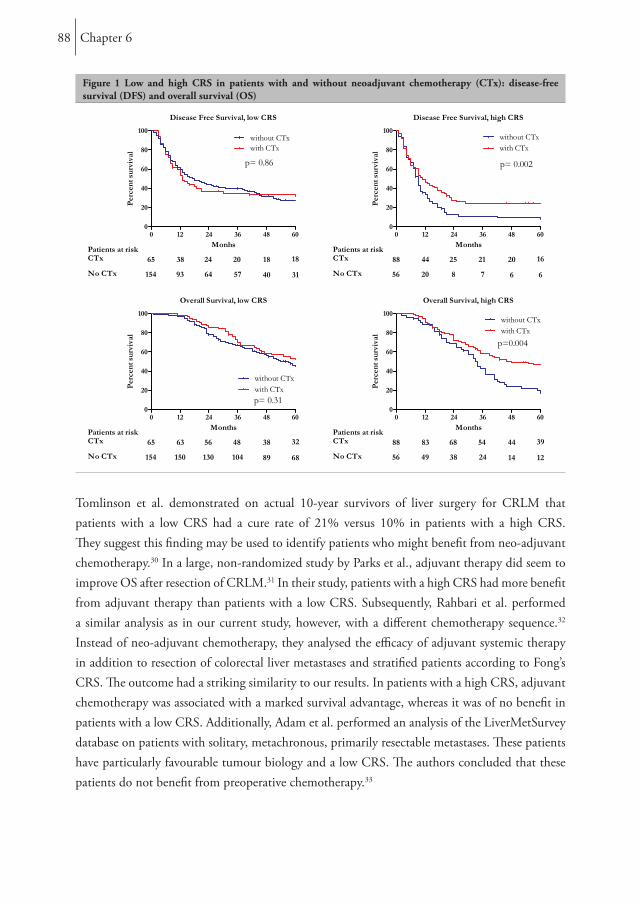
Chapter 688
Figure 1 Low and high CRS in patients with and without neoadjuvant chemotherapy (CTx): disease-free survival (DFS) and overall survival (OS)
Tomlinson et al. demonstrated on actual 10-year survivors of liver surgery for CRLM that patients with a low CRS had a cure rate of 21% versus 10% in patients with a high CRS. They suggest this finding may be used to identify patients who might benefit from neo-adjuvant chemotherapy.30 In a large, non-randomized study by Parks et al., adjuvant therapy did seem to improve OS after resection of CRLM.31 In their study, patients with a high CRS had more benefit from adjuvant therapy than patients with a low CRS. Subsequently, Rahbari et al. performed a similar analysis as in our current study, however, with a different chemotherapy sequence.32 Instead of neo-adjuvant chemotherapy, they analysed the efficacy of adjuvant systemic therapy in addition to resection of colorectal liver metastases and stratified patients according to Fong’s CRS. The outcome had a striking similarity to our results. In patients with a high CRS, adjuvant chemotherapy was associated with a marked survival advantage, whereas it was of no benefit in patients with a low CRS. Additionally, Adam et al. performed an analysis of the LiverMetSurvey database on patients with solitary, metachronous, primarily resectable metastases. These patients have particularly favourable tumour biology and a low CRS. The authors concluded that these patients do not benefit from preoperative chemotherapy.33
The use of neoadjuvant chemotherapie and clinical risk score 89
Finally, Sorby et al. demonstrated in an exploratory retrospective analysis of the EORTC Intergroup study 40983 that CEA was the strongest baseline predictive factor for the benefit of perioperative FOLFOX.34 They conclude that moderately and highly elevated CEA serum levels were both predictive for the benefit of perioperative chemotherapy; an obvious explanation would be that elevated CEA is a surrogate for more advanced disease.34 Again these results suggest a role for CRS’s when considering chemotherapy in addition to surgery for CRLM. This study might be biased due to its non-randomized, retrospective, single-centre design. However, patients were included from a prospective database and differences in terms of the main characteristics did not influence OS. Further, in our current protocol perioperative chemotherapy is not considered to be standard treatment for patients with CRLM. We consider patients for neo-adjuvant chemotherapy in case of initially difficult/irresectable liver metastases (ill location) or multiple (≥4) synchronous metastases. This implies that even within the high CRS group, patients are not comparable in terms of baseline characteristics. However, the “worst” patients within this group were treated with neo-adjuvant chemotherapy (see table 2), thus the significant overall survival benefit in this group is even more striking. On the other hand, patients within the low CRS had comparable survival rates. Patients in this group who received neo-adjuvant chemotherapy had significantly worse tumour characteristics (see table 1). Theoretically, these patients might have had poorer survival rates without neo-adjuvant chemotherapy. Therefore, this study at best suggests that neo-adjuvant chemotherapy will not improve survival in the low CRS group. Additional prospective studies are needed to explore the exact role of multimodal therapies in patients with a high risk profile. Since only patients coming to resection were included in this study, there is a potential bias of patients who have progressed on chemotherapy and were therefore not considered surgical candidates. This failure to comply with intention to treat principles is inherent to a retrospective study. However, progression on chemotherapy is rare - 7% in the EPOC trial 10, 11 - in the present era of modern chemotherapeutics and therefore we suggest that this phenomenon did not have a major impact on our conclusion. Finally, patients with a high risk profile might not have received chemotherapy for other reasons than pure oncologic risk, such as age and comorbidity. However, this is highly unlikely because all patients in this study were fit to undergo a partial liver resection.
Conclusion
In this study we demonstrate that stratifying patients with resectable CRLM according to their clinical risk profile, as described by Fong et al., could provide a useful tool for selecting patients who are most likely to obtain survival benefit from neo-adjuvant chemotherapy. Although the indication for neo-adjuvant chemotherapy may not solely be based on overall survival benefit, we believe it should be included in the decision making process.
Chapter 690
References
1. Jemal A, Bray F, Center MM, et al. Global cancer statistics. CA Cancer J Clin 2011; 61(2):69-90.
2. de Gramont A, Figer A, Seymour M, et al. Leucovorin and fluorouracil with or without oxaliplatin as first-line treatment in advanced colorectal cancer. J Clin Oncol 2000; 18(16):2938-47.
3. Douillard JY, Cunningham D, Roth AD, et al. Irinotecan combined with fluorouracil compared with fluorouracil alone as first-line treatment for metastatic colorectal cancer: a multicentre randomised trial. Lancet 2000; 355(9209):1041-7.
4. Saltz LB, Cox JV, Blanke C, et al. Irinotecan plus fluorouracil and leucovorin for metastatic colorectal cancer. Irinotecan Study Group. N Engl J Med 2000; 343(13):905-14.
5. Rees M, Tekkis PP, Welsh FK, et al. Evaluation of long-term survival after hepatic resection for metastatic colorectal cancer: a multifactorial model of 929 patients. Ann Surg 2008; 247(1):125-35.
6. van der Pool AE, de Wilt JH, Lalmahomed ZS, et al. Optimizing the outcome of surgery in patients with rectal cancer and synchronous liver metastases. Br J Surg 2010; 97(3):383-90.
7. Petrelli NJ. Perioperative or adjuvant therapy for resectable colorectal hepatic metastases. J Clin Oncol 2008; 26(30):4862-3.
8. Mitry E, Fields AL, Bleiberg H, et al. Adjuvant chemotherapy after potentially curative resection of metastases from colorectal cancer: a pooled analysis of two randomized trials. J Clin Oncol 2008; 26(30):4906-11.
9. Portier G, Elias D, Bouche O, et al. Multicenter randomized trial of adjuvant fluorouracil and folinic acid compared with surgery alone after resection of colorectal liver metastases: FFCD ACHBTH AURC 9002 trial. J Clin Oncol 2006; 24(31):4976-82.
10. Nordlinger B, Sorbye H, Glimelius B, et al. Perioperative chemotherapy with FOLFOX4 and surgery versus surgery alone for resectable liver metastases from colorectal cancer (EORTC Intergroup trial 40983): a randomised controlled trial. Lancet 2008; 371(9617):1007-16.
11. Nordlinger B, Sorbye H, Glimelius B, et al. Perioperative FOLFOX4 chemotherapy and surgery versus surgery alone for resectable liver metastases from colorectal cancer (EORTC 40983): long-term results of a randomised, controlled, phase 3 trial. Lancet Oncol 2013; 14(12):1208-15.
12. Benoist S, Nordlinger B. The role of preoperative chemotherapy in patients with resectable colorectal liver metastases. Ann Surg Oncol 2009; 16(9):2385-90.
13. Minagawa M, Yamamoto J, Kosuge T, et al. Simplified staging system for predicting the prognosis of patients with resectable liver metastasis: development and validation. Arch Surg 2007; 142(3):269-76; discussion 277.
14. Nordlinger B, Guiguet M, Vaillant JC, et al. Surgical resection of colorectal carcinoma metastases to the liver. A prognostic scoring system to improve case selection, based on 1568 patients. Association Francaise de Chirurgie. Cancer 1996; 77(7):1254-62.

The use of neoadjuvant chemotherapie and clinical risk score 91
15. Fong Y, Fortner J, Sun RL, et al. Clinical score for predicting recurrence after hepatic resection for metastatic colorectal cancer: analysis of 1001 consecutive cases. Ann Surg 1999; 230(3):309-18; discussion 318-21.
16. Iwatsuki S, Dvorchik I, Madariaga JR, et al. Hepatic resection for metastatic colorectal adenocarcinoma: a proposal of a prognostic scoring system. J Am Coll Surg 1999; 189(3):291-9.
17. Nagashima I, Takada T, Matsuda K, et al. A new scoring system to classify patients with colorectal liver metastases: proposal of criteria to select candidates for hepatic resection. J Hepatobiliary Pancreat Surg 2004; 11(2):79-83.
18. Konopke R, Kersting S, Distler M, et al. Prognostic factors and evaluation of a clinical score for predicting survival after resection of colorectal liver metastases. Liver Int 2009; 29(1):89-102.
19. Mala T, Bohler G, Mathisen O, et al. Hepatic resection for colorectal metastases: can preoperative scoring predict patient outcome? World J Surg 2002; 26(11):1348-53.
20. Reissfelder C, Rahbari NN, Koch M, et al. Validation of prognostic scoring systems for patients undergoing resection of colorectal cancer liver metastases. Ann Surg Oncol 2009; 16(12):3279-88.
21. Tournigand C, Andre T, Achille E, et al. FOLFIRI followed by FOLFOX6 or the reverse sequence in advanced colorectal cancer: a randomized GERCOR study. J Clin Oncol 2004; 22(2):229-37.
22. Seymour MT, Maughan TS, Ledermann JA, et al. Different strategies of sequential and combination chemotherapy for patients with poor prognosis advanced colorectal cancer (MRC FOCUS): a randomised controlled trial. Lancet 2007; 370(9582):143-52.
23. Koopman M, Antonini NF, Douma J, et al. Sequential versus combination chemotherapy with capecitabine, irinotecan, and oxaliplatin in advanced colorectal cancer (CAIRO): a phase III randomised controlled trial. Lancet 2007; 370(9582):135-42.
24. Hurwitz H, Fehrenbacher L, Novotny W, et al. Bevacizumab plus irinotecan, fluorouracil, and leucovorin for metastatic colorectal cancer. N Engl J Med 2004; 350(23):2335-42.
25. Saltz LB, Clarke S, Diaz-Rubio E, et al. Bevacizumab in combination with oxaliplatin-based chemotherapy as first-line therapy in metastatic colorectal cancer: a randomized phase III study. J Clin Oncol 2008; 26(12):2013-9.
26. Van Cutsem E, Kohne CH, Hitre E, et al. Cetuximab and chemotherapy as initial treatment for metastatic colorectal cancer. N Engl J Med 2009; 360(14):1408-17.
27. Douillard JY, Siena S, Cassidy J, et al. Randomized, phase III trial of panitumumab with infusional fluorouracil, leucovorin, and oxaliplatin (FOLFOX4) versus FOLFOX4 alone as first-line treatment in patients with previously untreated metastatic colorectal cancer: the PRIME study. J Clin Oncol 2010; 28(31):4697-705.
28. Douillard JY, Siena S, Cassidy J, et al. Final results from PRIME: randomized phase III study of panitumumab with FOLFOX4 for first-line treatment of metastatic colorectal cancer. Ann Oncol 2014; 25(7):1346-55.
29. O’Connell MJ, Adson MA, Schutt AJ, et al. Clinical trial of adjuvant chemotherapy after surgical resection of colorectal cancer metastatic to the liver. Mayo Clin Proc 1985; 60(8):517-20.
Chapter 692
30. Tomlinson JS, Jarnagin WR, DeMatteo RP, et al. Actual 10-year survival after resection of colorectal liver metastases defines cure. J Clin Oncol 2007; 25(29):4575-80.
31. Parks R, Gonen M, Kemeny N, et al. Adjuvant chemotherapy improves survival after resection of hepatic colorectal metastases: analysis of data from two continents. J Am Coll Surg 2007; 204(5):753-61; discussion 761-3.
32. Rahbari NN, Reissfelder C, Schulze-Bergkamen H, et al. Adjuvant therapy after resection of colorectal liver metastases: the predictive value of the MSKCC clinical risk score in the era of modern chemotherapy. BMC Cancer 2014; 14:174.
33. Adam R, Bhangui P, Poston G, et al. Is perioperative chemotherapy useful for solitary, metachronous, colorectal liver metastases? Ann Surg 2010; 252(5):774-87.
34. Sorbye H, Mauer M, Gruenberger T, et al. Predictive factors for the benefit of perioperative FOLFOX for resectable liver metastasis in colorectal cancer patients (EORTC Intergroup Trial 40983). Ann Surg 2012; 255(3):534-9.
Chapter 7
Neo-adjuvant chemotherapy followed by surgery versus surgery alone in high-risk patients with resectable
colorectal liver metastases
The CHARISMA randomized multicenter clinical trial
N. Ayez*, E.P. van der Stok*
J.H. de Wilt, S.A. Radema
R. van Hillegersberg, R.M. Roumen
G. Vreugdenhil, P.J. Tanis
C.J. Punt, C.H. Dejong
R.L. Jansen, H.M. Verheul
K.P. de Jong, G. Hospers
J.M. Klaase, M.C. Legdeur
E. van Meerten, F. Eskens
N. van der Meer, B. van der Holt
C. Verhoef, D.J. Grünhagen
* Both authors contributed equally.
BioMed Central, 2015
Chapter 794
Abstract
BackgroundEfforts to improve the outcome of liver surgery by combining curative resection with chemotherapy have failed to demonstrate definite overall survival benefit. This may partly be due to the fact that these studies often involve strict inclusion criteria. Consequently, patients with a high risk profile as characterized by Fong’s Clinical Risk Score (CRS) are often underrepresented in these studies. Conceptually, this group of patients might benefit the most from chemotherapy. The present study evaluates the impact of neo-adjuvant chemotherapy in high-risk patients with primary resectable colorectal liver metastases, without extrahepatic disease. Our hypothesis is that adding neo-adjuvant chemotherapy to surgery will provide an improvement in overall survival (OS) in patients with a high-risk profile.Methods/designCHARISMA is a multicenter, randomized, phase III clinical trial. Patients will be randomized to either surgery alone (standard treatment, arm A) or to 6 cycles of neo-adjuvant oxaliplatin-based chemotherapy, followed by surgery (arm B). Patients must be ≥ 18 years of age with liver metastases of histologically confirmed primary colorectal carcinoma. Patients with extrahepatic metastases are excluded. Liver metastases must be deemed primarily resectable. Only patients with a CRS of 3-5 are eligible. The primary study endpoint is OS. Secondary endpoints are progression free survival (PFS), quality of life, morbidity of resection, treatment response on neo-adjuvant chemotherapy, and whether CEA levels can predict treatment response.DiscussionCHARISMA is a multicenter, randomized, phase III clinical trial that will provide an answer to the question if adding neo-adjuvant chemotherapy to surgery will improve OS in a well-defined high-risk patient group with colorectal liver metastases.Trial registrationThe CHARISMA is registered at European Union Clinical Trials Register (EudraCT), number: 2013-004952-39.

CHARISMA 95
Background
Colorectal liver metastases: surgical treatmentColorectal cancer (CRC) is one of the leading causes of cancer death. It is in the top 3 most commonly diagnosed cancers, with over 1.2 million new cases and over 600,000 deaths estimated to have occurred in 2008 worldwide.1 In approximately 20% of patients distant metastases are present at time of diagnosis.2 The liver is the most common metastatic site. Approximately 50% of patients with early-stage disease will eventually develop colorectal liver metastases (CRLM).3, 4 When metastases of CRC patients are restricted to the liver, possible curative treatment can be obtained by surgical resection. Complete surgical resection of CRLM improves 5-year survival rates to around 35-60% in selected patients.5-8 However in only 10-20% of patients surgical resection of CRLM is feasible. Although surgery for CRLM provides the only potential for cure, cancer relapse is a common phenomenon, with a recurrence rate of up to 50% in the first 2 years after surgery.9
Chemotherapy for colorectal liver metastasesInitially, systemic treatment with 5-fluoruracil based regimens was standard of care in CRLM, improving OS from 6 to 10-12 months. The development of chemotherapeutic agents such as oxaliplatin and irinotecan has subsequently improved OS to a median of up to 24 months. Sequential treatment with all available cytotoxic agents, as well as the introduction of Epidermal Growth Factor receptor (EGFR) and Vascular Endothelial Growth Factor (VEGF) binding monoclonal antibodies have further increased overall survival.10-13 The high relapse rate after curative resection of CRLM, and the efficacy of modern systemic treatment in the metastatic setting, have prompted investigators to perform numerous studies to evaluate the potential role of systemic chemotherapy combined with liver resection. The purpose of both adjuvant and neo-adjuvant chemotherapy is to treat microscopic disease that is not addressed by surgery. This microscopic disease may be promoting the high relapse rate that is observed after liver surgery.9 Notably, current literature suggests that timing of additional chemotherapy (adjuvant vs. neo-adjuvant) seems to have no influence on outcome.14 The role of perioperative chemotherapy in case of resectable CRLM was established in a randomized controlled trial.15 In the mature OS analysis of this trial there was no significant effect on OS after a median follow up of 7 years.16
Stratification by clinical risk scoreIn the past, several clinical risk scores for the outcome of patients with CRLM have been published.7, 17-25 In 1999, Fong et al. described the most widely used CRS.19 This prognostic scoring system has been verified by independent investigators.26
Chapter 796
Several authors have proposed the concept of stratification by CRS in relation to the effects of a multimodal treatment strategy on OS. These authors suggest that patients with a high risk score have a worse prognosis and might therefore benefit more from chemotherapy compared to patients with a low risk score.27-29 These findings have prompted others and ourselves to retrospectively evaluate data on patients who have undergone liver resection for CRLM in the last decade with and without chemotherapy, stratified by CRS according to the Fong-criteria.30, 31
As described earlier, efforts to improve outcome of liver surgery by combining the resection with chemotherapy have failed to demonstrate definite OS benefit. This may partly be due to the fact that these studies often involve strict study protocol inclusion criteria. Consequently, patients with a high clinical risk score - which might benefit the most from chemotherapy - are often underrepresented in these studies. Since genuine survival benefit has not yet been demonstrated, could this low impact of chemotherapy on survival then be explained by the relatively low risk profile of the patients included in these trials?
Study aim and hypothesisThe CHARISMA randomized clinical trial will evaluate the effect on OS of neo-adjuvant chemotherapy in patients with primary resectable CRLM and a CRS (Fong) of 3-5, thereby bearing a poor prognosis. The primary aim of this study is to compare OS in patients with resectable liver metastases randomized for treatment with chemotherapy, consisting of capecitabine and oxaliplatin (XELOX), followed by surgery versus surgery alone. We hypothesize that neo-adjuvant chemotherapy will provide an improvement in OS in this high-risk patient group. Secondary endpoints in this study will be progression free survival (PFS), quality of life as assessed by QLQ-30 and MFI questionnaires, response to chemotherapy, morbidity of surgery and resection rate, and whether carcinoembryonic antigen (CEA) can predict for treatment response, PFS, and OS.
Study design
Patients with CRLM and a high CRS will be evaluated for inclusion by the local multidisciplinary team meeting. In this meeting, at least two surgeons with expertise in liver surgery should be present. In case of doubt, the imaging can be sent to a central expert panel. Patients are eligible for randomization if, in the opinion of a local expert panel, radical resection of the CRLM (R0-resection) is feasible.

CHARISMA 97
Patients will be randomized 1:1 to either (figure 1):Arm A: Surgery of the liver metastasesArm B: Neo-adjuvant oxaliplatin-based chemotherapy followed by surgery of the liver metastases
Figure 1. Study design
Inclusion
Randomisation
Baseline QoL (QIQ-C30, MFI) Baseline QoL (QIQ-C30, MFI)
Arm B1 Neoadjuvant chemotherapy (6x XELOX)· within 2 weeks after randomisation· within 4 weeks after last CT-scan· Assessment after 3 and 6 cycles: CT-scan· Assessment after 6 cycles: toxicity, ECOG performance
status, laboratory analysis incl CEA-level, QoL
2 Surgery
Arm ASurgery· within 4 weeks after randomisation· within 6 weeks after last CT-scan
· 1st visit after surgery (2-4 weeks after surgery) Surgical complications assessment· 1st and 2nd year after surgery Every 3 months; CEA, ECOG, CT-scan, QoL· 3rd and 5th year after surgery Every 6 months; CEA, ECOG, CT-scan
Follow up
Study population
Inclusion criteriaAge ≥ 18 years, ECOG performance status 0-1. Histologically confirmed primary colorectal carcinoma. Radiological confirmed and primary resectable CRLM. CRS of 3-5 (Fong). Adequate bone marrow, liver and renal functions.
Before any study related procedure will be pursued, written informed consent must be given according to ICH/GCP and national/local regulations.
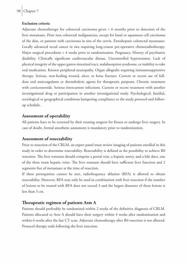
Chapter 798
Exclusion criteriaAdjuvant chemotherapy for colorectal carcinoma given < 6 months prior to detection of the liver metastases. Prior non colorectal malignancies, except for basal or squamous cell carcinoma of the skin, or patients with carcinoma in situ of the cervix. Extrahepatic colorectal metastases. Locally advanced rectal cancer in situ requiring long-course pre-operative chemoradiotherapy. Major surgical procedures < 4 weeks prior to randomization. Pregnancy. History of psychiatric disability. Clinically significant cardiovascular disease. Uncontrolled hypertension. Lack of physical integrity of the upper gastro-intestinal tract, malabsorption syndrome, or inability to take oral medication. Known peripheral neuropathy. Organ allografts requiring immunosuppressive therapy. Serious, non-healing wound, ulcer, or bone fracture. Current or recent use of full-dose oral anticoagulants or thrombolytic agents for therapeutic purposes. Chronic treatment with corticosteroids. Serious intercurrent infections. Current or recent treatment with another investigational drug or participation in another investigational study. Psychological, familial, sociological or geographical conditions hampering compliance to the study protocol and follow-up schedule.
Assessment of operabilityAll patients have to be screened by their treating surgeon for fitness to undergo liver surgery. In case of doubt, formal anesthetic assessment is mandatory prior to randomization.
Assessment of resectabilityPrior to resection of the CRLM, an expert panel must review imaging of patients enrolled in this study in order to determine resectability. Resectability is defined as the possibility to achieve R0 resection. The liver remnant should comprise a portal vein, a hepatic artery, and a bile duct, one of the three main hepatic veins. The liver remnant should have sufficient liver function and 2 segments free of metastases at the time of resection.If these prerequisites cannot be met, radiofrequency ablation (RFA) is allowed to obtain resectability. However, RFA may only be used in combination with liver resection if the number of lesions to be treated with RFA does not exceed 3 and the largest diameter of these lesions is less than 3 cm.
Therapeutic regimen of patients Arm APatients should preferably be randomized within 2 weeks of the definitive diagnosis of CRLM. Patients allocated to Arm A should have their surgery within 4 weeks after randomization and within 6 weeks after the last CT scan. Adjuvant chemotherapy after R0 resection is not allowed. Protocol therapy ends following the liver resection.
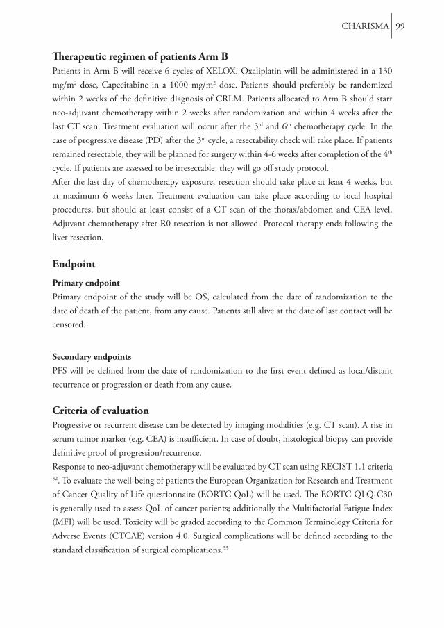
CHARISMA 99
Therapeutic regimen of patients Arm BPatients in Arm B will receive 6 cycles of XELOX. Oxaliplatin will be administered in a 130 mg/m2 dose, Capecitabine in a 1000 mg/m2 dose. Patients should preferably be randomized within 2 weeks of the definitive diagnosis of CRLM. Patients allocated to Arm B should start neo-adjuvant chemotherapy within 2 weeks after randomization and within 4 weeks after the last CT scan. Treatment evaluation will occur after the 3rd and 6th chemotherapy cycle. In the case of progressive disease (PD) after the 3rd cycle, a resectability check will take place. If patients remained resectable, they will be planned for surgery within 4-6 weeks after completion of the 4th cycle. If patients are assessed to be irresectable, they will go off study protocol. After the last day of chemotherapy exposure, resection should take place at least 4 weeks, but at maximum 6 weeks later. Treatment evaluation can take place according to local hospital procedures, but should at least consist of a CT scan of the thorax/abdomen and CEA level. Adjuvant chemotherapy after R0 resection is not allowed. Protocol therapy ends following the liver resection.
Endpoint
Primary endpointPrimary endpoint of the study will be OS, calculated from the date of randomization to the date of death of the patient, from any cause. Patients still alive at the date of last contact will be censored.
Secondary endpointsPFS will be defined from the date of randomization to the first event defined as local/distant recurrence or progression or death from any cause.
Criteria of evaluationProgressive or recurrent disease can be detected by imaging modalities (e.g. CT scan). A rise in serum tumor marker (e.g. CEA) is insufficient. In case of doubt, histological biopsy can provide definitive proof of progression/recurrence. Response to neo-adjuvant chemotherapy will be evaluated by CT scan using RECIST 1.1 criteria 32. To evaluate the well-being of patients the European Organization for Research and Treatment of Cancer Quality of Life questionnaire (EORTC QoL) will be used. The EORTC QLQ-C30 is generally used to assess QoL of cancer patients; additionally the Multifactorial Fatigue Index (MFI) will be used. Toxicity will be graded according to the Common Terminology Criteria for Adverse Events (CTCAE) version 4.0. Surgical complications will be defined according to the standard classification of surgical complications.33
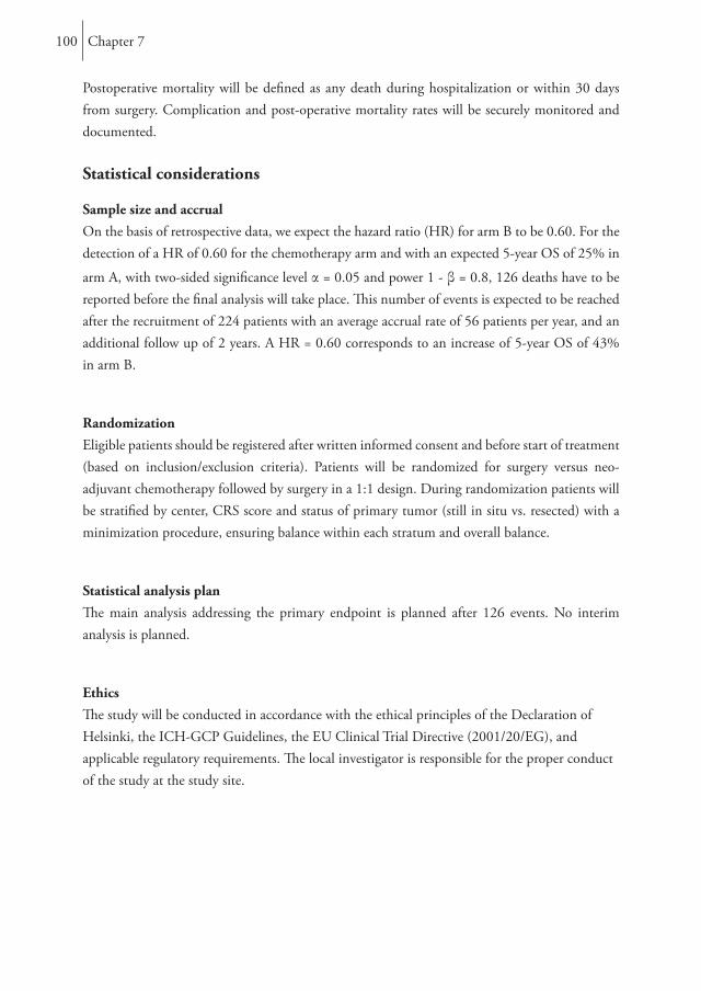
Chapter 7100
Postoperative mortality will be defined as any death during hospitalization or within 30 days from surgery. Complication and post-operative mortality rates will be securely monitored and documented.
Statistical considerations
Sample size and accrualOn the basis of retrospective data, we expect the hazard ratio (HR) for arm B to be 0.60. For the detection of a HR of 0.60 for the chemotherapy arm and with an expected 5-year OS of 25% in
arm A, with two-sided significance level α = 0.05 and power 1 - β = 0.8, 126 deaths have to be reported before the final analysis will take place. This number of events is expected to be reached after the recruitment of 224 patients with an average accrual rate of 56 patients per year, and an additional follow up of 2 years. A HR = 0.60 corresponds to an increase of 5-year OS of 43% in arm B.
RandomizationEligible patients should be registered after written informed consent and before start of treatment (based on inclusion/exclusion criteria). Patients will be randomized for surgery versus neo-adjuvant chemotherapy followed by surgery in a 1:1 design. During randomization patients will be stratified by center, CRS score and status of primary tumor (still in situ vs. resected) with a minimization procedure, ensuring balance within each stratum and overall balance.
Statistical analysis planThe main analysis addressing the primary endpoint is planned after 126 events. No interim analysis is planned.
EthicsThe study will be conducted in accordance with the ethical principles of the Declaration of Helsinki, the ICH-GCP Guidelines, the EU Clinical Trial Directive (2001/20/EG), and applicable regulatory requirements. The local investigator is responsible for the proper conduct of the study at the study site.
CHARISMA 101
Discussion
Currently, multimodal treatment is not incorporated in the standard of care for primary resectable colorectal liver metastases. To date, no definite evidence exists favoring administration of (neo) adjuvant chemotherapy in CRLM in addition to surgery. Considering the retrospective observations that pre-selection of patients by clinical prognostic characteristics may define a patient population expected to benefit from chemotherapy, CRS stratification provides the base for this randomized controlled trial. Preceding studies of peri-operative chemotherapy combined with liver surgery often engaged strict study protocol inclusion criteria. Consequently, patients with a high CRS - which might benefit the most from chemotherapy - are often underrepresented in these studies. Possibly, this low impact of chemotherapy on survival could be explained by the relatively low risk profile of the patients included in these trials. Recently, two reports on patients with relatively low risk for recurrence have been published. Adam et al. performed an analysis of the LiverMetSurvey database on patients with solitary, metachronous, primarily resectable metastases. These patients have particularly favorable tumor biology and a low CRS. The authors concluded that these patients do not benefit from preoperative chemotherapy.34 A recent systematic review of the literature by Lehmann et al. concludes that routine use of neo-adjuvant chemotherapy for patients with clearly resectable lesions limited to the liver is not recommended due to a lack of benefit on survival.35
As mentioned before, several authors have proposed the concept of stratification by CRS with regard to the effects of systemic therapy. Tomlinson et al. demonstrated on actual 10-year survivors of liver surgery for CRLM that patients with a low CRS had a cure rate of 21% and that patients with a high CRS had a cure rate of 10%.27 They suggest that this finding may be used to identify patients who might benefit from neo-adjuvant chemotherapy.27 In a large, non-randomized study by Parks et al., adjuvant therapy did seem to improve OS.28 In this study, patients with a high CRS had more benefit from adjuvant therapy than patients with a low CRS, again suggesting a role for CRS when considering chemotherapy.These reports have stimulated others and our own unit to retrospectively evaluate data on patients that underwent liver resection for CRLM in the last decade with and without chemotherapy, stratified by CRS according to the Fong-criteria.19 Rahbari et al. have evaluated the role of adjuvant chemotherapy in a cohort of 316 patients, of whom 43% were high-risk according to the “Memorial Sloan-Kettering Cancer Center CRS” (CRS>2). They found that adjuvant chemotherapy had a profound impact on OS in the high-risk population (HR=0.40), whereas in low-risk patients HR=0.90.31 In a recent manuscript by Hirokawa et al. similar results are described with de use of adjuvant chemotherapy.36 In our population of patients that underwent resection for CRLM in Rotterdam (N=365), we have focused on neo-adjuvant chemotherapy.

Chapter 7102
In this study, a pronounced improvement in OS was found in high-risk patients receiving neo-adjuvant chemotherapy versus no chemotherapy (median 67 months vs. 33 months, HR=0.55 [95% CI 0.35-0.84], p=0.006). This difference was absent in the low-risk group (median 65 months vs. 56 months, HR=0.89 [95% CI 0.57-1.40], p=0.62).30 Notably, these studies were retrospective and non-randomized. The sample size calculation of the present study is based on these retrospective data. In a recent editorial by Jarnagin et al. it is suggested that future trials should strongly consider stratification by some scoring system29, given the results of the retrospective studies as mentioned above. Our study will evaluate patients with resectable CRLM without extra hepatic disease and a CRS of 3-5 thereby bearing a poor prognosis. The primary aim of this study is to compare OS rates of patients with resectable liver metastases randomized for treatment with chemotherapy consisting of capecitabine and oxaliplatin (XELOX) followed by surgery, versus surgery alone. We hypothesize that adding neo-adjuvant chemotherapy to surgical resection of CRLM will provide an improvement in OS in patients with a high-risk profile. As secondary objectives we will study PFS, quality of life, treatment response on neoadjuvant chemotherapy, morbidity of surgery and resection rate, and whether CEA can predict for treatment response, PFS, and OS.
List of abbreviationsCEA = Carcinoembryonic antigenCRC = Colorectal cancerCRLM = Colorectal liver metastasesCRS = Clinical risk scoreECOG = Eastern cooperative oncology group OS = Overall survivalPFS = Progression free survivalRCT = Randomized controlled trial RFA = Radiofrequency ablationXELOX = chemotherapy consisting of capecitabine and oxaliplatin
Competing interestsThe authors declare that they have no competing interests.
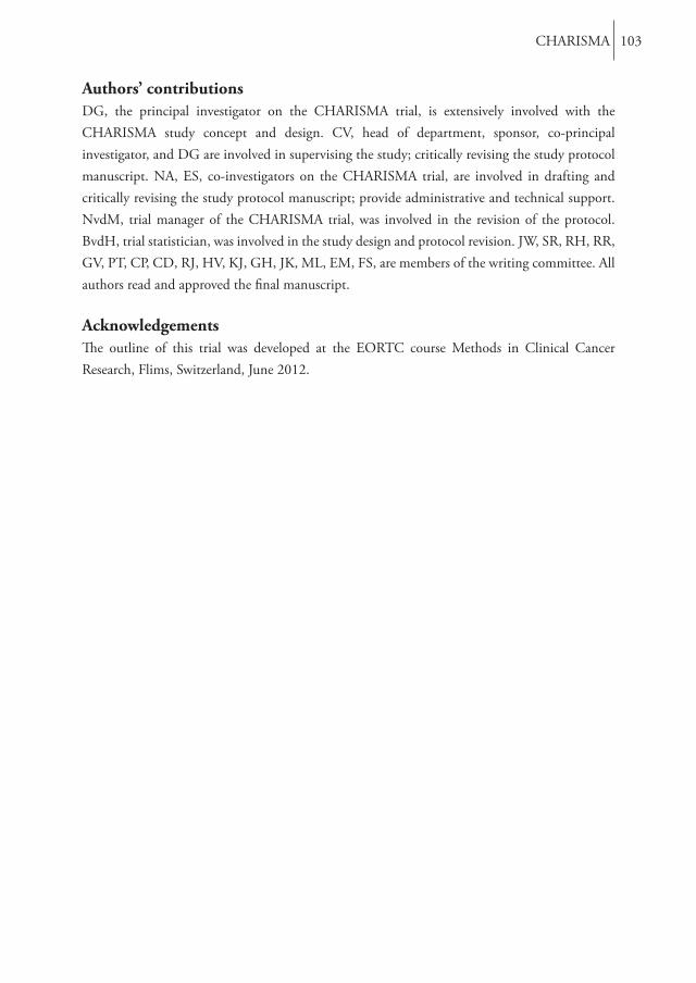
CHARISMA 103
Authors’ contributionsDG, the principal investigator on the CHARISMA trial, is extensively involved with the CHARISMA study concept and design. CV, head of department, sponsor, co-principal investigator, and DG are involved in supervising the study; critically revising the study protocol manuscript. NA, ES, co-investigators on the CHARISMA trial, are involved in drafting and critically revising the study protocol manuscript; provide administrative and technical support. NvdM, trial manager of the CHARISMA trial, was involved in the revision of the protocol. BvdH, trial statistician, was involved in the study design and protocol revision. JW, SR, RH, RR, GV, PT, CP, CD, RJ, HV, KJ, GH, JK, ML, EM, FS, are members of the writing committee. All authors read and approved the final manuscript. AcknowledgementsThe outline of this trial was developed at the EORTC course Methods in Clinical Cancer Research, Flims, Switzerland, June 2012.

Chapter 7104
References
1. Jemal A, Bray F, Center MM, et al. Global cancer statistics. CA Cancer J Clin 2011; 61(2):69-90.
2. van der Pool AE, Damhuis RA, Ijzermans JN, et al. Trends in incidence, treatment and survival of patients with stage IV colorectal cancer: a population-based series. Colorectal Dis 2010; 14(1):56-61.
3. Bengmark S, Hafstrom L. The natural history of primary and secondary malignant tumors of the liver. I. The prognosis for patients with hepatic metastases from colonic and rectal carcinoma by laparotomy. Cancer 1969; 23(1):198-202.
4. van der Pool AE, Lalmahomed ZS, de Wilt JH, et al. Local treatment for recurrent colorectal hepatic metastases after partial hepatectomy. J Gastrointest Surg 2009; 13(5):890-5.
5. Dols LF, Verhoef C, Eskens FA, et al. [Improvement of 5 year survival rate after liver resection for colorectal metastases between 1984-2006]. Ned Tijdschr Geneeskd 2009; 153(11):490-5.
6. Primrose JN. Surgery for colorectal liver metastases. Br J Cancer 2010; 102(9):1313-8.7. Rees M, Tekkis PP, Welsh FK, et al. Evaluation of long-term survival after hepatic
resection for metastatic colorectal cancer: a multifactorial model of 929 patients. Ann Surg 2008; 247(1):125-35.
8. van der Pool AE, de Wilt JH, Lalmahomed ZS, et al. Optimizing the outcome of surgery in patients with rectal cancer and synchronous liver metastases. Br J Surg 2010; 97(3):383-90.
9. Petrelli NJ. Perioperative or adjuvant therapy for resectable colorectal hepatic metastases. J Clin Oncol 2008; 26(30):4862-3.
10. Hurwitz H, Fehrenbacher L, Novotny W, et al. Bevacizumab plus irinotecan, fluorouracil, and leucovorin for metastatic colorectal cancer. N Engl J Med 2004; 350(23):2335-42.
11. Saltz LB, Clarke S, Diaz-Rubio E, et al. Bevacizumab in combination with oxaliplatin-based chemotherapy as first-line therapy in metastatic colorectal cancer: a randomized phase III study. J Clin Oncol 2008; 26(12):2013-9.
12. Van Cutsem E, Kohne CH, Hitre E, et al. Cetuximab and chemotherapy as initial treatment for metastatic colorectal cancer. N Engl J Med 2009; 360(14):1408-17.
13. Douillard JY, Siena S, Cassidy J, et al. Randomized, phase III trial of panitumumab with infusional fluorouracil, leucovorin, and oxaliplatin (FOLFOX4) versus FOLFOX4 alone as first-line treatment in patients with previously untreated metastatic colorectal cancer: the PRIME study. J Clin Oncol 2010; 28(31):4697-705.
14. Araujo R, Gonen M, Allen P, et al. Comparison Between Perioperative and Postoperative Chemotherapy After Potentially Curative Hepatic Resection for Metastatic Colorectal Cancer. Ann Surg Oncol 2013.
15. Nordlinger B, Sorbye H, Glimelius B, et al. Perioperative chemotherapy with FOLFOX4 and surgery versus surgery alone for resectable liver metastases from colorectal cancer (EORTC Intergroup trial 40983): a randomised controlled trial. Lancet 2008; 371(9617):1007-16.
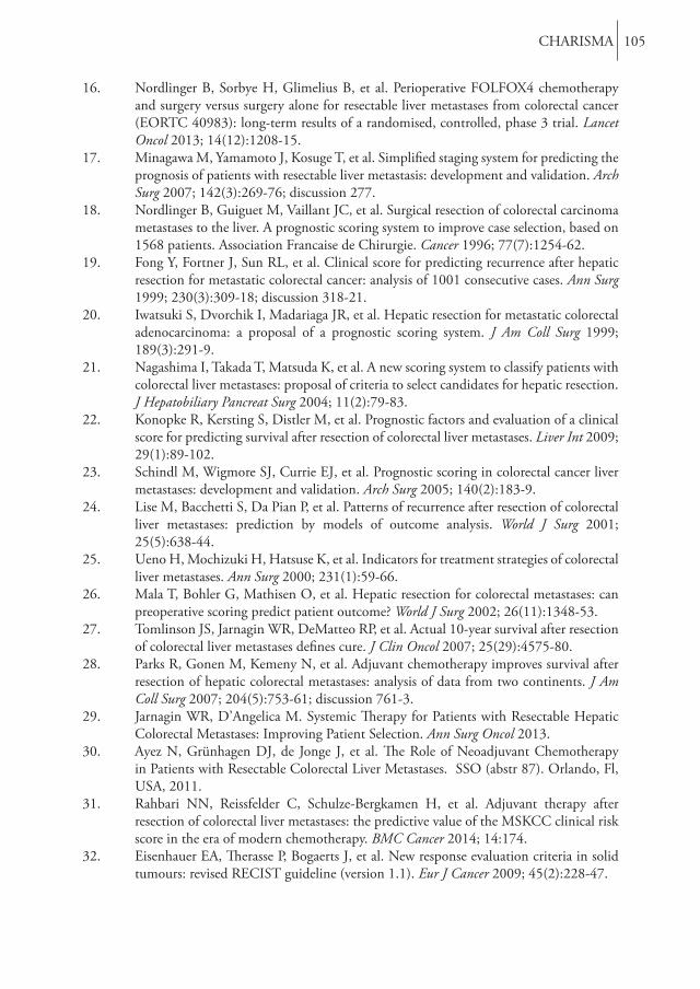
CHARISMA 105
16. Nordlinger B, Sorbye H, Glimelius B, et al. Perioperative FOLFOX4 chemotherapy and surgery versus surgery alone for resectable liver metastases from colorectal cancer (EORTC 40983): long-term results of a randomised, controlled, phase 3 trial. Lancet Oncol 2013; 14(12):1208-15.
17. Minagawa M, Yamamoto J, Kosuge T, et al. Simplified staging system for predicting the prognosis of patients with resectable liver metastasis: development and validation. Arch Surg 2007; 142(3):269-76; discussion 277.
18. Nordlinger B, Guiguet M, Vaillant JC, et al. Surgical resection of colorectal carcinoma metastases to the liver. A prognostic scoring system to improve case selection, based on 1568 patients. Association Francaise de Chirurgie. Cancer 1996; 77(7):1254-62.
19. Fong Y, Fortner J, Sun RL, et al. Clinical score for predicting recurrence after hepatic resection for metastatic colorectal cancer: analysis of 1001 consecutive cases. Ann Surg 1999; 230(3):309-18; discussion 318-21.
20. Iwatsuki S, Dvorchik I, Madariaga JR, et al. Hepatic resection for metastatic colorectal adenocarcinoma: a proposal of a prognostic scoring system. J Am Coll Surg 1999; 189(3):291-9.
21. Nagashima I, Takada T, Matsuda K, et al. A new scoring system to classify patients with colorectal liver metastases: proposal of criteria to select candidates for hepatic resection. J Hepatobiliary Pancreat Surg 2004; 11(2):79-83.
22. Konopke R, Kersting S, Distler M, et al. Prognostic factors and evaluation of a clinical score for predicting survival after resection of colorectal liver metastases. Liver Int 2009; 29(1):89-102.
23. Schindl M, Wigmore SJ, Currie EJ, et al. Prognostic scoring in colorectal cancer liver metastases: development and validation. Arch Surg 2005; 140(2):183-9.
24. Lise M, Bacchetti S, Da Pian P, et al. Patterns of recurrence after resection of colorectal liver metastases: prediction by models of outcome analysis. World J Surg 2001; 25(5):638-44.
25. Ueno H, Mochizuki H, Hatsuse K, et al. Indicators for treatment strategies of colorectal liver metastases. Ann Surg 2000; 231(1):59-66.
26. Mala T, Bohler G, Mathisen O, et al. Hepatic resection for colorectal metastases: can preoperative scoring predict patient outcome? World J Surg 2002; 26(11):1348-53.
27. Tomlinson JS, Jarnagin WR, DeMatteo RP, et al. Actual 10-year survival after resection of colorectal liver metastases defines cure. J Clin Oncol 2007; 25(29):4575-80.
28. Parks R, Gonen M, Kemeny N, et al. Adjuvant chemotherapy improves survival after resection of hepatic colorectal metastases: analysis of data from two continents. J Am Coll Surg 2007; 204(5):753-61; discussion 761-3.
29. Jarnagin WR, D’Angelica M. Systemic Therapy for Patients with Resectable Hepatic Colorectal Metastases: Improving Patient Selection. Ann Surg Oncol 2013.
30. Ayez N, Grünhagen DJ, de Jonge J, et al. The Role of Neoadjuvant Chemotherapy in Patients with Resectable Colorectal Liver Metastases. SSO (abstr 87). Orlando, Fl, USA, 2011.
31. Rahbari NN, Reissfelder C, Schulze-Bergkamen H, et al. Adjuvant therapy after resection of colorectal liver metastases: the predictive value of the MSKCC clinical risk score in the era of modern chemotherapy. BMC Cancer 2014; 14:174.
32. Eisenhauer EA, Therasse P, Bogaerts J, et al. New response evaluation criteria in solid tumours: revised RECIST guideline (version 1.1). Eur J Cancer 2009; 45(2):228-47.
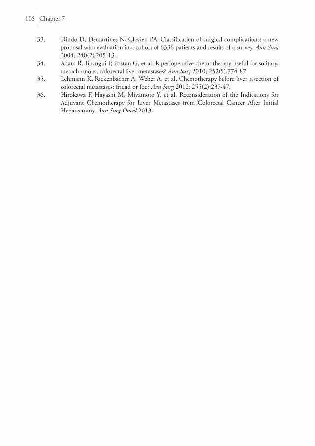
Chapter 7106
33. Dindo D, Demartines N, Clavien PA. Classification of surgical complications: a new proposal with evaluation in a cohort of 6336 patients and results of a survey. Ann Surg 2004; 240(2):205-13.
34. Adam R, Bhangui P, Poston G, et al. Is perioperative chemotherapy useful for solitary, metachronous, colorectal liver metastases? Ann Surg 2010; 252(5):774-87.
35. Lehmann K, Rickenbacher A, Weber A, et al. Chemotherapy before liver resection of colorectal metastases: friend or foe? Ann Surg 2012; 255(2):237-47.
36. Hirokawa F, Hayashi M, Miyamoto Y, et al. Reconsideration of the Indications for Adjuvant Chemotherapy for Liver Metastases from Colorectal Cancer After Initial Hepatectomy. Ann Surg Oncol 2013.
PART 3
Timing


Chapter 8
Long-term results of the ‘liver first’ approach in patients with locally advanced rectal cancer and synchronous
liver metastases
Ninos AyezJacobus W.A. BurgerAnne E. van der Pool
Alexander M.M. EggermontDirk J Grunhagen
Johannes H.W. de WiltCornelis Verhoef
Diseases of the colon & rectum, 2013
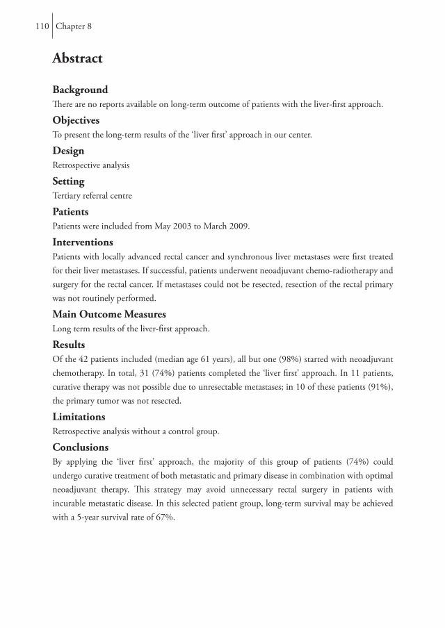
Chapter 8110
Abstract
BackgroundThere are no reports available on long-term outcome of patients with the liver-first approach.
ObjectivesTo present the long-term results of the ‘liver first’ approach in our center.
DesignRetrospective analysis
SettingTertiary referral centre
PatientsPatients were included from May 2003 to March 2009.
InterventionsPatients with locally advanced rectal cancer and synchronous liver metastases were first treated for their liver metastases. If successful, patients underwent neoadjuvant chemo-radiotherapy and surgery for the rectal cancer. If metastases could not be resected, resection of the rectal primary was not routinely performed.
Main Outcome MeasuresLong term results of the liver-first approach.
ResultsOf the 42 patients included (median age 61 years), all but one (98%) started with neoadjuvant chemotherapy. In total, 31 (74%) patients completed the ‘liver first’ approach. In 11 patients, curative therapy was not possible due to unresectable metastases; in 10 of these patients (91%), the primary tumor was not resected.
LimitationsRetrospective analysis without a control group.
ConclusionsBy applying the ‘liver first’ approach, the majority of this group of patients (74%) could undergo curative treatment of both metastatic and primary disease in combination with optimal neoadjuvant therapy. This strategy may avoid unnecessary rectal surgery in patients with incurable metastatic disease. In this selected patient group, long-term survival may be achieved with a 5-year survival rate of 67%.

Long term result of teh ‘liver first‘ approach 111
Introduction
Liver resection is the gold standard for curative therapy of colorectal liver metastases. Treatment of patients with advanced rectal cancer and synchronous liver metastases differs from patients with colon cancer and synchronous liver metastases because rectal cancer often requires long-course neoadjuvant radiotherapy to reduce local recurrence rates.1, 2 Long-course chemo-radiotherapy usually takes about 5 weeks to complete, and 6-10 weeks after the last day of radiotherapy patients undergo rectal surgery. If no complications occur, synchronous liver metastases will traditionally be treated as early as 3 months after rectal surgery. However, complications following rectal surgery are common and often delay adequate therapy. In a prospective randomized controlled trial Sauer et al. demonstrated that up to 50% of patients do not receive optimal treatment after rectal surgery, because of postoperative complications.3
One way to overcome this problem is to start with neoadjuvant radiotherapy (with or without chemotherapy) followed by the combination of rectal and liver surgery. Although this approach has the advantage of a single operation, combining (extended) pelvic surgery with liver surgery might increase morbidity and mortality.4-6 As a consequence, the advantage of the one-stage approach in patients with synchronous liver metastases is far from proven in rectal cancer patients.7 Mentha et al. have described the ‘liver first’ approach in patients with colon and rectal cancer who have advanced synchronous liver metastases.8 However we carry out the ‘liver first’ approach in patients with locally advanced rectal cancer and synchronous liver metastases. This ‘liver first’ approach facilitates optimal treatment of the liver metastases and adequate neoadjuvant treatment for the primary tumor. Initial results from our center have been published6; this study reports on a larger group of patients with a long-term follow-up.
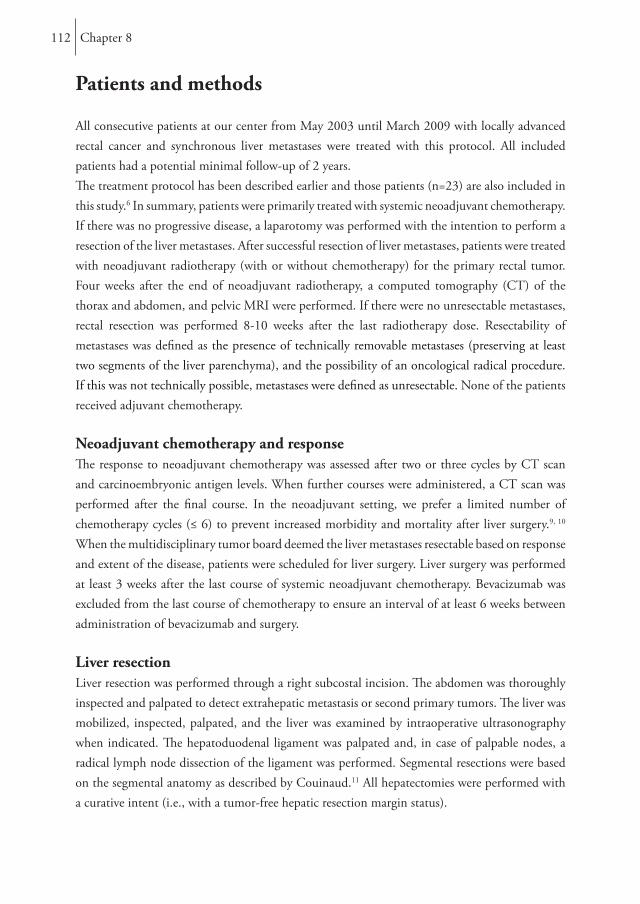
Chapter 8112
Patients and methods
All consecutive patients at our center from May 2003 until March 2009 with locally advanced rectal cancer and synchronous liver metastases were treated with this protocol. All included patients had a potential minimal follow-up of 2 years. The treatment protocol has been described earlier and those patients (n=23) are also included in this study.6 In summary, patients were primarily treated with systemic neoadjuvant chemotherapy. If there was no progressive disease, a laparotomy was performed with the intention to perform a resection of the liver metastases. After successful resection of liver metastases, patients were treated with neoadjuvant radiotherapy (with or without chemotherapy) for the primary rectal tumor. Four weeks after the end of neoadjuvant radiotherapy, a computed tomography (CT) of the thorax and abdomen, and pelvic MRI were performed. If there were no unresectable metastases, rectal resection was performed 8-10 weeks after the last radiotherapy dose. Resectability of metastases was defined as the presence of technically removable metastases (preserving at least two segments of the liver parenchyma), and the possibility of an oncological radical procedure. If this was not technically possible, metastases were defined as unresectable. None of the patients received adjuvant chemotherapy.
Neoadjuvant chemotherapy and responseThe response to neoadjuvant chemotherapy was assessed after two or three cycles by CT scan and carcinoembryonic antigen levels. When further courses were administered, a CT scan was performed after the final course. In the neoadjuvant setting, we prefer a limited number of chemotherapy cycles (≤ 6) to prevent increased morbidity and mortality after liver surgery.9, 10 When the multidisciplinary tumor board deemed the liver metastases resectable based on response and extent of the disease, patients were scheduled for liver surgery. Liver surgery was performed at least 3 weeks after the last course of systemic neoadjuvant chemotherapy. Bevacizumab was excluded from the last course of chemotherapy to ensure an interval of at least 6 weeks between administration of bevacizumab and surgery.
Liver resectionLiver resection was performed through a right subcostal incision. The abdomen was thoroughly inspected and palpated to detect extrahepatic metastasis or second primary tumors. The liver was mobilized, inspected, palpated, and the liver was examined by intraoperative ultrasonography when indicated. The hepatoduodenal ligament was palpated and, in case of palpable nodes, a radical lymph node dissection of the ligament was performed. Segmental resections were based on the segmental anatomy as described by Couinaud.11 All hepatectomies were performed with a curative intent (i.e., with a tumor-free hepatic resection margin status).

Long term result of teh ‘liver first‘ approach 113
In our clinic, the first CT scan is a roadmap for further surgery. This means that in case of a clinically complete response on the CT scan, all known metastases were resected routinely. The pathology findings were defined as follows: complete response (CR) was defined as the absence of vital tumor tissue, partial response (PR) was defined as the presence of vital tumor cells and necrosis, and stable disease (SD) was defined as vital tumor cells and no necrosis.
Chemoradiation and rectal surgeryIf liver resection was successful, patients received neoadjuvant radiotherapy (with or without chemotherapy) for their locally advanced rectal cancer. In our centre locally advanced rectal cancer is defined as a histological proven adenocarcinoma with one of the following characteristics: tumor >5 cm at colonoscopy and MRI (clinically large T3); clinically fixed tumor or with ingrowth in adjacent organ on MRI (T4); N+ tumor (lymph node >8 mm and/or >4 nodes > 5 mm on CT scan or MRI). Regardless of size criteria, any lymph node depicted on MRI with an irregular border or mixed signal intensity was considered a positive node. T4 tumors, but also advanced T3 tumors with a close relation to the circumferential margin, were considered locally advanced rectal cancer. Patients were treated with a long-course radiotherapy, i.e. 45-50 Gy (in fractions of 1.8-2 Gy) with or without chemotherapy (Capecitabine 825 mg/m2 twice a day on radiotherapy days). Radiotherapy was followed by surgery with a delay of 6-10 weeks. Intraoperative radiotherapy was applied if the distance to the circumferential resection margin (CRM) was <2 mm.12 No laparoscopic resections were performed.
Follow-Up Follow-up was performed at the outpatient clinic and consisted of endoscopic surveillance after 1 year, and abdominal CT or ultrasonography and serum carcinoembryonic antigen every 3 months during the first year, every 6 months during the second year and once a year thereafter. Progression-free survival (PFS) was calculated from the start of neoadjuvant chemotherapy until local recurrence, new metastases or date of last follow-up without progression. Overall survival (OS) was calculated from the start of treatment until death or the date of the last follow-up.
StatisticsDescriptive statistics are expressed as median (range). Survival analysis was performed by the Kaplan-Meier method. The SPSS statistical package (version 17.0, Chicago, IL, USA) was used for statistical analysis and a p-value of ≤0.05 was considered statistically significant.
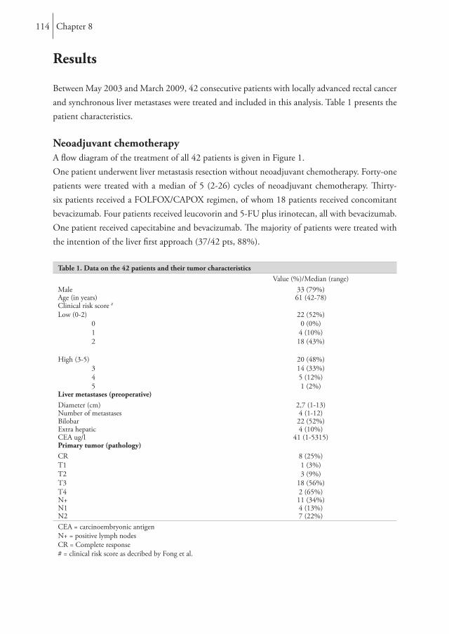
Chapter 8114
Results
Between May 2003 and March 2009, 42 consecutive patients with locally advanced rectal cancer and synchronous liver metastases were treated and included in this analysis. Table 1 presents the patient characteristics.
Neoadjuvant chemotherapyA flow diagram of the treatment of all 42 patients is given in Figure 1. One patient underwent liver metastasis resection without neoadjuvant chemotherapy. Forty-one patients were treated with a median of 5 (2-26) cycles of neoadjuvant chemotherapy. Thirty-six patients received a FOLFOX/CAPOX regimen, of whom 18 patients received concomitant bevacizumab. Four patients received leucovorin and 5-FU plus irinotecan, all with bevacizumab. One patient received capecitabine and bevacizumab. The majority of patients were treated with the intention of the liver first approach (37/42 pts, 88%).
Table 1. Data on the 42 patients and their tumor characteristicsValue (%)/Median (range)
Male 33 (79%)Age (in years) 61 (42-78)Clinical risk score #
Low (0-2) 0 1 2
High (3-5) 3 4 5
22 (52%)0 (0%)4 (10%)18 (43%)
20 (48%)14 (33%)5 (12%)1 (2%)
Liver metastases (preoperative)Diameter (cm) 2,7 (1-13)Number of metastases 4 (1-12)Bilobar 22 (52%)Extra hepatic 4 (10%)CEA ug/l 41 (1-5315)Primary tumor (pathology)CRT1T2T3T4
8 (25%)1 (3%)3 (9%)
18 (56%)2 (65%)
N+ 11 (34%)N1 4 (13%)N2 7 (22%)CEA = carcinoembryonic antigenN+ = positive lymph nodesCR = Complete response# = clinical risk score as decribed by Fong et al.
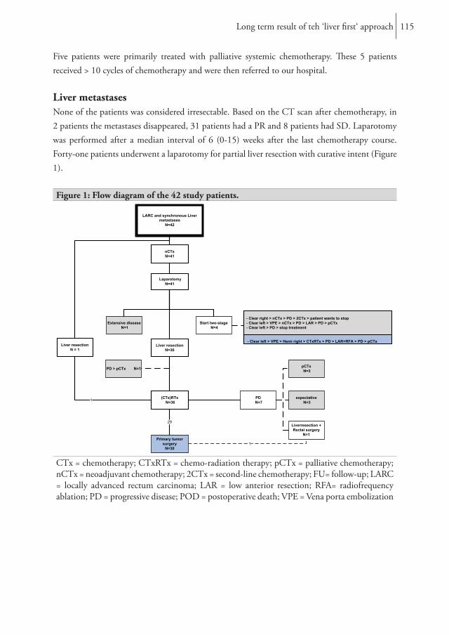
Long term result of teh ‘liver first‘ approach 115
Five patients were primarily treated with palliative systemic chemotherapy. These 5 patients received > 10 cycles of chemotherapy and were then referred to our hospital.
Liver metastasesNone of the patients was considered irresectable. Based on the CT scan after chemotherapy, in 2 patients the metastases disappeared, 31 patients had a PR and 8 patients had SD. Laparotomy was performed after a median interval of 6 (0-15) weeks after the last chemotherapy course. Forty-one patients underwent a laparotomy for partial liver resection with curative intent (Figure 1). Figure 1: Flow diagram of the 42 study patients.
LARC and synchronous Livermetastases
N=42
Liver resectionN = 1
nCTxN=41
LaparotomyN=41
Liver resectionN=36
Start two-stageN=4
Extensive diseaseN=1
(CTx)RTxN=36
PD > pCTx N=1
- Clear right > nCTx > PD > 2CTx > patient wants to stop- Clear left > VPE > nCTx > PD > LAR > PD > pCTx- Clear left > PD > stop treatment
Primary tumorsurgery
N=30
PDN=7
29
1
Liverresection +Rectal surgery
N=1
expectativeN=3
pCTxN=3
1
- Clear left > VPE > Hemi right > CTxRTx > PD > LAR+RFA > PD > pCTx
CTx = chemotherapy; CTxRTx = chemo-radiation therapy; pCTx = palliative chemotherapy; nCTx = neoadjuvant chemotherapy; 2CTx = second-line chemotherapy; FU= follow-up; LARC = locally advanced rectum carcinoma; LAR = low anterior resection; RFA= radiofrequency ablation; PD = progressive disease; POD = postoperative death; VPE = Vena porta embolization
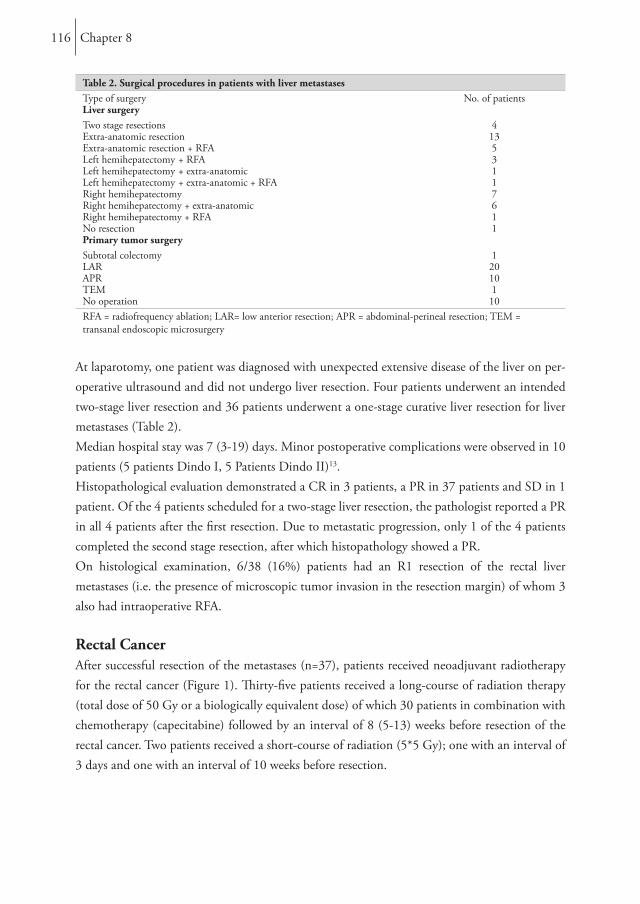
Chapter 8116
Table 2. Surgical procedures in patients with liver metastasesType of surgery No. of patientsLiver surgeryTwo stage resections 4Extra-anatomic resection 13Extra-anatomic resection + RFA 5Left hemihepatectomy + RFA 3Left hemihepatectomy + extra-anatomic 1Left hemihepatectomy + extra-anatomic + RFA 1Right hemihepatectomy 7Right hemihepatectomy + extra-anatomic 6Right hemihepatectomy + RFA 1No resection 1Primary tumor surgerySubtotal colectomy 1LAR 20APR 10TEM 1No operation 10RFA = radiofrequency ablation; LAR= low anterior resection; APR = abdominal-perineal resection; TEM = transanal endoscopic microsurgery
At laparotomy, one patient was diagnosed with unexpected extensive disease of the liver on per-operative ultrasound and did not undergo liver resection. Four patients underwent an intended two-stage liver resection and 36 patients underwent a one-stage curative liver resection for liver metastases (Table 2).Median hospital stay was 7 (3-19) days. Minor postoperative complications were observed in 10 patients (5 patients Dindo I, 5 Patients Dindo II)13. Histopathological evaluation demonstrated a CR in 3 patients, a PR in 37 patients and SD in 1 patient. Of the 4 patients scheduled for a two-stage liver resection, the pathologist reported a PR in all 4 patients after the first resection. Due to metastatic progression, only 1 of the 4 patients completed the second stage resection, after which histopathology showed a PR.On histological examination, 6/38 (16%) patients had an R1 resection of the rectal liver metastases (i.e. the presence of microscopic tumor invasion in the resection margin) of whom 3 also had intraoperative RFA.
Rectal CancerAfter successful resection of the metastases (n=37), patients received neoadjuvant radiotherapy for the rectal cancer (Figure 1). Thirty-five patients received a long-course of radiation therapy (total dose of 50 Gy or a biologically equivalent dose) of which 30 patients in combination with chemotherapy (capecitabine) followed by an interval of 8 (5-13) weeks before resection of the rectal cancer. Two patients received a short-course of radiation (5*5 Gy); one with an interval of 3 days and one with an interval of 10 weeks before resection.
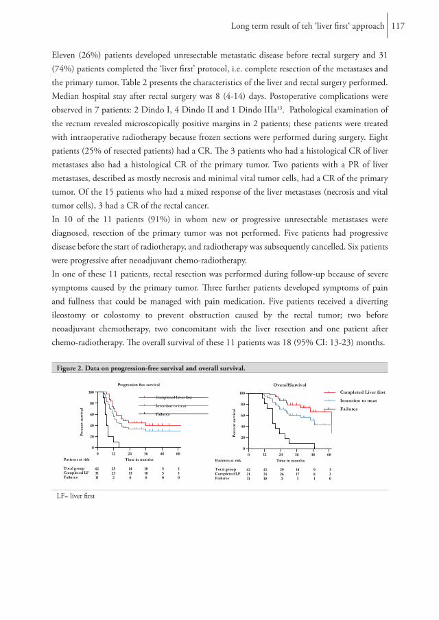
Long term result of teh ‘liver first‘ approach 117
Eleven (26%) patients developed unresectable metastatic disease before rectal surgery and 31 (74%) patients completed the ‘liver first’ protocol, i.e. complete resection of the metastases and the primary tumor. Table 2 presents the characteristics of the liver and rectal surgery performed. Median hospital stay after rectal surgery was 8 (4-14) days. Postoperative complications were observed in 7 patients: 2 Dindo I, 4 Dindo II and 1 Dindo IIIa13. Pathological examination of the rectum revealed microscopically positive margins in 2 patients; these patients were treated with intraoperative radiotherapy because frozen sections were performed during surgery. Eight patients (25% of resected patients) had a CR. The 3 patients who had a histological CR of liver metastases also had a histological CR of the primary tumor. Two patients with a PR of liver metastases, described as mostly necrosis and minimal vital tumor cells, had a CR of the primary tumor. Of the 15 patients who had a mixed response of the liver metastases (necrosis and vital tumor cells), 3 had a CR of the rectal cancer.In 10 of the 11 patients (91%) in whom new or progressive unresectable metastases were diagnosed, resection of the primary tumor was not performed. Five patients had progressive disease before the start of radiotherapy, and radiotherapy was subsequently cancelled. Six patients were progressive after neoadjuvant chemo-radiotherapy.In one of these 11 patients, rectal resection was performed during follow-up because of severe symptoms caused by the primary tumor. Three further patients developed symptoms of pain and fullness that could be managed with pain medication. Five patients received a diverting ileostomy or colostomy to prevent obstruction caused by the rectal tumor; two before neoadjuvant chemotherapy, two concomitant with the liver resection and one patient after chemo-radiotherapy. The overall survival of these 11 patients was 18 (95% CI: 13-23) months.
Figure 2. Data on progression-free survival and overall survival.
LF= liver first

Chapter 8118
Follow-upAll patients had a minimal follow-up of 2 years unless death occurred earlier. Median length of follow-up was 30.5 (10-91) months. The PFS for the total group was 14 months (95% CI: 9-19), with a 1, 3 and 5-year PFS of 62%, 36% and 32%, respectively. Median OS for the total group was 49 (95% CI: 34-64) months, with a 1, 3 and 5-year survival rate of 98%, 60% and 43%, respectively (Figure 2).Median follow-up of the 31 patients who completed the ‘liver-first’ protocol was 39 (16-91) months. Median time to progression was 19 (95% CI: 7-31) months, with a 1, 3 and 5-year PFS of 74%, 45% and 40%, respectively. The median OS in this group was 69 (95% CI: 30-108) months, with a 1, 3 and 5-year OS rate of 100%, 79% and 67%, respectively. (Figure 2)
Discussion
This study reports on the largest series of patients to date with locally advanced rectal cancer and synchronous liver metastases who underwent the ‘liver first’ approach. The results show that in this selected group of patients the majority (74%) could undergo optimal neoadjuvant treatment of both liver metastases and rectal cancer, followed by curative resection of both liver metastases and rectal cancer. Of the 11 patients with unresectable metastatic disease, 10 (91%) did not need rectal surgery.The first report on the ‘liver first’ approach was described by Mentha et al. demonstrating the safety of this procedure.14 Brouquet et al. described the ‘liver first’ approach and demonstrated that it is associated with similar outcomes as the classical approach.15 de Jong et al. described their 5 year experience with the ‘liver first’ approach and found that it was feasible in approximately four-fifth of their patients.16 All of these studies have described the ‘liver first’ approach in patients with colon and rectal cancer who have advanced synchronous liver metastases.A strength of the present study is that all patients had a locally advanced rectal primary with synchronous liver metastases. The common denominator of the locally advanced rectal primary makes this series unique. Another difference between the present study and the current literature is that patients were only included if they had a potential follow up ≥ 2 years. Some consider resection of the primary tumor to be indicated when the primary tumor is symptomatic.17 However, resection does not provide immediate palliative benefit in case of an asymptomatic primary, and is associated with a high mortality (6-10%) and morbidity (20-25%) in patients with metastatic disease.17-20 Recent reviews by Venderbosch et al. and Verhoef et al. suggest a survival benefit in patients who underwent resection of the primary tumor; however, randomized studies are still lacking.21, 22
Long term result of teh ‘liver first‘ approach 119
In a review by Poulsides et al. it was demonstrated that in patients with unresectable metastatic disease, resection of the primary is not warranted, since almost 14 asymptomatic patients need to undergo prophylactic resection of their primary tumor in order to save one patient a subsequent operation for obstruction or perforation.23 In our series of 11 patients with locally advanced rectal cancer and a high metastatic tumor burden, none of these patients underwent a prophylactic resection of the primary tumor. In one patient, secondary resection was necessary at a later stage due to symptoms caused by the primary rectal tumor. Five patients with unresected rectal cancer received a diverting ileostomy or colostomy to prevent obstruction caused by the rectal tumor, even though the primary tumor was still resectable. Two patients received a diverting stoma before neoadjuvant chemotherapy, two concomitant with the liver resection and one patient after chemo-radiotherapy. One may argue that several or all of the stomas could have been avoided had a rectum first approach been chosen. In these specific cases, the disadvantages of a rectal resection (with the associated morbidity and mortality) have to be balanced against those of stoma construction.Despite these strengths, a limitation of our study is that it is a retrospective analysis of selected patients in a single institute. The referral selection bias and the absence of a control group are limitations that need to be considered. A concern regarding the ‘liver first’ approach is non-response to chemotherapy or progression after initial response in patients who present with resectable disease. Another possible concern regarding the liver first approach is that the primary tumor may progress beyond resection. This did not occur in our series, nor has it been described in the mentioned series.8, 14-16 Even in the patients with progressive extra hepatic metastases, the primary tumor did not progress into adjacent structures. In contrast, in case of an excellent response to chemotherapy, vanishing metastases may be a concern since the risk of regrowth is high.24 Regular imaging after 2 or 3 cycles of chemotherapy is warranted to minimize the risk on vanishing metastases and/or progression beyond resection of the primary tumor.In the present series we found 16% R1 resections after neoadjuvant chemotherapy, which might be a concern. In the era of parenchyma-saving surgery, 16% R1 resections is within the range reported in the literature. In several large series the reported R1 was 8-24%.15, 25-28 Two independent studies demonstrated that in the era of modern chemotherapy, patients with an R1 resection and chemotherapy perform equally to patients with an R0 resection.27, 29
Three patients who had a histological CR of liver metastases also had a histological CR of the primary tumor after additional neoadjuvant chemo-radiotherapy. Two patients, in whom the liver metastases were described as mostly necrotic with minimal focal vital cells remaining (PR), also had a CR of the primary tumor. It seems that in patients with a CR or a near CR (near total necrosis) of the liver, these patients are likely to have a CR of the rectal primary tumor as well, albeit after neoadjuvant chemo-radiotherapy.
Chapter 8120
In such cases, resection of the primary tumor may be limited to local excision (transanal excision or transanal endoscopic microsurgery) or even watchful waiting. However, prospective studies are needed to justify such a strategy. In conclusion, in this group of patients, the ‘liver first’ strategy resulted in curative resection of both liver metastases and locally advanced rectal cancer in 74% of selected patients. Moreover, all patients received optimal treatment for both liver metastases and rectal cancer. In patients in whom progressive disease precluded curative rectal surgery, palliative rectal surgery was not necessary in 10 of 11 patients. Thereby, unnecessary morbidity, mortality and delay of palliative chemotherapy could be avoided.

Long term result of teh ‘liver first‘ approach 121
References 1. Gerard JP, Conroy T, Bonnetain F, et al. Preoperative radiotherapy with or without
concurrent fluorouracil and leucovorin in T3-4 rectal cancers: results of FFCD 9203. J Clin Oncol 2006; 24(28):4620-5.
2. Bosset JF, Collette L, Calais G, et al. Chemotherapy with preoperative radiotherapy in rectal cancer. N Engl J Med 2006; 355(11):1114-23.
3. Sauer R, Becker H, Hohenberger W, et al. Preoperative versus postoperative chemoradiotherapy for rectal cancer. N Engl J Med 2004; 351(17):1731-40.
4. Vermaas M, Ferenschild FT, Hofer SO, et al. Primary and secondary reconstruction after surgery of the irradiated pelvis using a gracilis muscle flap transposition. Eur J Surg Oncol 2005; 31(9):1000-5.
5. Vermaas M, Ferenschild FT, Verhoef C, et al. Total pelvic exenteration for primary locally advanced and locally recurrent rectal cancer. Eur J Surg Oncol 2007; 33(4):452-8.
6. Verhoef C, van der Pool AE, Nuyttens JJ, et al. The “liver-first approach” for patients with locally advanced rectal cancer and synchronous liver metastases. Dis Colon Rectum 2009; 52(1):23-30.
7. Adam R. Colorectal cancer with synchronous liver metastases. Br J Surg 2007; 94(2):129-31.
8. Mentha G, Roth AD, Terraz S, et al. ‘Liver first’ approach in the treatment of colorectal cancer with synchronous liver metastases. Dig Surg 2008; 25(6):430-5.
9. Karoui M, Penna C, Amin-Hashem M, et al. Influence of preoperative chemotherapy on the risk of major hepatectomy for colorectal liver metastases. Ann Surg 2006; 243(1):1-7.
10. Allen PJ, Kemeny N, Jarnagin W, et al. Importance of response to neoadjuvant chemotherapy in patients undergoing resection of synchronous colorectal liver metastases. J Gastrointest Surg 2003; 7(1):109-15; discussion 116-7.
11. Couinaud C. Liver anatomy: portal (and suprahepatic) or biliary segmentation. Dig Surg 1999; 16(6):459-67.
12. Ferenschild FT, Vermaas M, Nuyttens JJ, et al. Value of intraoperative radiotherapy in locally advanced rectal cancer. Dis Colon Rectum 2006; 49(9):1257-65.
13. Dindo D, Demartines N, Clavien PA. Classification of surgical complications: a new proposal with evaluation in a cohort of 6336 patients and results of a survey. Ann Surg 2004; 240(2):205-13.
14. Mentha G, Majno PE, Andres A, et al. Neoadjuvant chemotherapy and resection of advanced synchronous liver metastases before treatment of the colorectal primary. Br J Surg 2006; 93(7):872-8.
15. Brouquet A, Mortenson MM, Vauthey JN, et al. Surgical strategies for synchronous colorectal liver metastases in 156 consecutive patients: classic, combined or reverse strategy? J Am Coll Surg 2010; 210(6):934-41.
16. de Jong MC, van Dam RM, Maas M, et al. The liver-first approach for synchronous colorectal liver metastasis: a 5-year single-centre experience. HPB (Oxford) 2011; 13(10):745-52.
Chapter 8122
17. Stillwell AP, Buettner PG, Ho YH. Meta-analysis of survival of patients with stage IV colorectal cancer managed with surgical resection versus chemotherapy alone. World J Surg 2010; 34(4):797-807.
18. Makela J, Haukipuro K, Laitinen S, et al. Palliative operations for colorectal cancer. Dis Colon Rectum 1990; 33(10):846-50.
19. Law WL, Chan WF, Lee YM, et al. Non-curative surgery for colorectal cancer: critical appraisal of outcomes. Int J Colorectal Dis 2004; 19(3):197-202.
20. Rosen SA, Buell JF, Yoshida A, et al. Initial presentation with stage IV colorectal cancer: how aggressive should we be? Arch Surg 2000; 135(5):530-4; discussion 534-5.
21. Venderbosch S, de Wilt JH, Teerenstra S, et al. Prognostic value of resection of primary tumor in patients with stage IV colorectal cancer: retrospective analysis of two randomized studies and a review of the literature. Ann Surg Oncol 2011; 18(12):3252-60.
22. Verhoef C, de Wilt JH, Burger JW, et al. Surgery of the primary in stage IV colorectal cancer with unresectable metastases. Eur J Cancer 2011; 47 Suppl 3:S61-6.
23. Poultsides GA, Paty PB. Reassessing the need for primary tumor surgery in unresectable metastatic colorectal cancer: overview and perspective. Ther Adv Med Oncol 2011; 3(1):35-42.
24. Benoist S, Brouquet A, Penna C, et al. Complete response of colorectal liver metastases after chemotherapy: does it mean cure? J Clin Oncol 2006; 24(24):3939-45.
25. Fong Y, Fortner J, Sun RL, et al. Clinical score for predicting recurrence after hepatic resection for metastatic colorectal cancer: analysis of 1001 consecutive cases. Ann Surg 1999; 230(3):309-18; discussion 318-21.
26. Pawlik TM, Scoggins CR, Zorzi D, et al. Effect of surgical margin status on survival and site of recurrence after hepatic resection for colorectal metastases. Ann Surg 2005; 241(5):715-22, discussion 722-4.
27. de Haas RJ, Wicherts DA, Flores E, et al. R1 resection by necessity for colorectal liver metastases: is it still a contraindication to surgery? Ann Surg 2008; 248(4):626-37.
28. Tanaka K, Nojiri K, Kumamoto T, et al. R1 resection for aggressive or advanced colorectal liver metastases is justified in combination with effective prehepatectomy chemotherapy. Eur J Surg Oncol 2011; 37(4):336-43.
29. Ayez N, Lalmahomed ZS, Eggermont AM, et al. Outcome of microscopic incomplete resection (R1) of colorectal liver metastases in the era of neoadjuvant chemotherapy. Ann Surg Oncol 2012; 19(5):1618-27.
Chapter 9
Is restaging with chest and abdominal CT scan after neoadjuvant chemoradiotherapy for locally advanced
rectal cancer necessary?
Ninos AyezWijnand J. Alberda
Jacobus W.A. BurgerAlexander M. Eggermont
Joost J. M. E. NuyttensRoy S. Dwarkasing
François E. J. A. WillemssenCornelis Verhoef
Annals of surgical oncology, 2012

Chapter 9124
Abstract
BackgroundThere is no evidence regarding restaging of patients with locally advanced rectal cancer after a long course of neoadjuvant radiotherapy with or without chemotherapy. This study evaluated the value of restaging with chest and abdominal computed tomographic (CT) scan after radiotherapy.Methods Between January 2000 and December 2010, all newly diagnosed patients in our tertiary referral hospital, who underwent a long course of radiotherapy for locally advanced rectal cancer, were analyzed. Patients were only included if they had chest and abdominal imaging before and after radiotherapy treatment. ResultsA total of 153 patients who met the inclusion criteria and were treated with curative intent were included. A change in treatment strategy due to new findings on the CT scan after radiotherapy was observed in 18 (12 %) of 153 patients. Twelve patients (8 %) were spared rectal surgery due to progressive metastatic disease.ConclusionRestaging with a chest and abdominal CT scan after radiotherapy for locally advanced rectal cancer is advisable because additional findings may alter the treatment strategy.
Is restaging after neoadjuvant chemotherapy neccesary? 125
IntroductionColorectal cancer is the third most commonly diagnosed cancer in males and the second in females women.1 At the time of diagnosis, approximately 25% of patients already have manifest liver metastases.2, 3 The lungs represent the second most common site of metastases from colorectal cancer. According to non population based studies, lung metastases are present in 10-15% of patients with colorectal cancer.4, 5 A population based study by Mitry et al. reported that lung metastases are present in 2% of patients with CRC.6 Distant metastases have implications on the treatment options. For the screening of liver metastases, the Dutch Association of Comprehensive Cancer Centers (ACCC), the National Institute for Health and Clinical Excellence (NICE) and The American Society of Colon and Rectal Surgeons (ASCRS) recommend a computed tomography (CT) or magnetic resonance imaging (MRI). For the screening of lung metastases, they recommend the use of a chest X-ray or a chest CT-scan.7-9
Locally advanced rectal cancer has a higher risk of developing lung metastases than colon cancer.6,
8, 10 In patients with locally advanced rectal cancer, improved local control can be achieved with a long course of preoperative radiotherapy in combination with neoadjuvant chemotherapy as a radiosensitizer.11 However, no advice is given by the ACCC, NICE or by the ASCRS in any guideline regarding restaging of patients after neoadjuvant treatment of locally advanced rectal cancer (i.e. repeating the imaging, after a long course of neoadjuvant radiotherapy treatment, to ensure that in the intervening time no metastases have developed). This study evaluates the value of restaging patients with locally advanced rectal cancer with a CT-scan.
Patients and Methods
Between January 2000 and December 2010, all newly diagnosed patients who received a long course of radiotherapy for locally advanced rectal cancer in our tertiary referral hospital were analyzed. Patients were included if they had a chest and abdominal CT-scan before and after radiotherapy treatment. An MRI was used for local staging before and after radiotherapy. Neoadjuvant treatment was given with a curative intent. Patient characteristics were collected retrospectively. The database comprised data on age, gender, radiation time and dose, simultaneous chemotherapy, pre- and post-radiotherapy chest and abdominal CT-scan, pathological primary tumor stage, lymph node stage and type of surgery.

Chapter 9126
CT-scanAll CT scans were assessed by radiologists in regular clinical practice. Whenever there was any doubt concerning lesions found on the CT-scans, than these scans were reassessed by a panel of radiologist and discussed in a multidisciplinary meeting.Images were acquired after intravenous injection of 150 cc contrast material at 3.5 mL/sec with a delay of 80 sec. In addition, an arterial phase scan of the liver was acquired at a delay of 30 seconds. Positron emission tomography (PET) scan is not used as standard protocol in our centre.
Locally advanced rectal cancer Locally advanced rectal cancer was defined in our centre as a histological proven adenocarcinoma with one of the following characteristics: tumor >5 cm at colonoscopy and magnetic resonance imaging (MRI) (clinically large T3); clinically fixed tumor or with ingrowth in adjacent organ on MRI (T4); N+ tumor (lymph node >8 mm and/or >4 nodes > 5mm on CT scan or MRI). T4 tumors, but also advanced T3 tumors with a close relation to the circumferential margin (CRM), were considered as locally advanced rectal cancer (Regardless of size criteria, any lymph node depicted on MRI with an irregular border or mixed signal intensity was considered suspicious for metastasis). All patients with locally advanced rectal cancer were discussed in a multidisciplinary team that consists of colorectal surgeons, hepatobiliary surgeons, gastroenterologists, surgical oncologists, medical oncologists, radiation oncologists, radiologists, pathologists and nurse practitioners.
ChemoradiotherapyIn our centre patients with locally advanced rectal cancer have been treated with a long course of neoadjuvant radiotherapy, i.e. 45-50 Gy (in fractions of 1.8-2 Gy) with or without chemotherapy (capecitabine 825 mg/m2 twice a day only on radiotherapy days).12 Reasons for selection of patients who did not receive chemotherapy were dependent on the co-morbidity of patient. Radiotherapy was followed by surgery with a delay of 6-10 weeks. Intraoperative radiotherapy was applied if the CRM was <2 mm.13 No laparoscopic resections were performed. StatisticsDescriptive statistics are expressed as median (interquartile range [IQR]). Pre- and post-CT variables are expressed as binary variables and compared with the Mc Nemar test for paired data. If fewer than 25 cases change values from the first variable to the second variable, the binomial distribution is used to compute the probability.
Statistical package (version 17.0, Chicago, IL, USA) was used for statistical analysis, where a p-value of ≤0.05 was considered statistically significant.
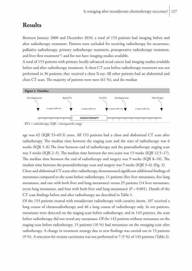
Is restaging after neoadjuvant chemotherapy neccesary? 127
Results
Between January 2000 and December 2010, a total of 153 patients had imaging before and after radiotherapy treatment. Patients were excluded for receiving radiotherapy for recurrence, palliative radiotherapy, primary radiotherapy treatment, postoperative radiotherapy treatment, and liver-first treatment14; and for not have imaging studies available.A total of 153 patients with primary locally advanced rectal cancer had imaging studies available before and after radiotherapy treatment. A chest CT scan before radiotherapy treatment was not performed in 36 patients; they received a chest X-ray. All other patients had an abdominal and chest CT scan. The majority of patients were men (61 %), and the median
Figure 1. Timeline
2/7/2011 - 3/19/2011Radiotherapy
5 weeks (IQR 5-5)
1/1/2011First staging scan
2/7/2011Begin RTx
3/19/2011End RTx
6 weeks (IQR 5-8)
4/9/2011Re-staging scan
3 weeks (IQR 2-5)
5/21/2011Start Surgery
5 weeks (IQR 3-6)
RADIOTHERAPY
RTx = radiotherapy, IQR = interquartile range
age was 62 (IQR 53–69.5) years. All 153 patients had a chest and abdominal CT scan after radiotherapy. The median time between the staging scan and the start of radiotherapy was 6 weeks (IQR 5–8).The time between end of radiotherapy and the postradiotherapy staging scan was 3 weeks (IQR 2–5). The median time between the two scans was 15 weeks (IQR 12.5–17). The median time between the end of radiotherapy and surgery was 9 weeks (IQR 8–10). The median time between the postradiotherapy scan and surgery was 5 weeks (IQR 3–6) (Fig. 1). Chest and abdominal CT scans after radiotherapy demonstrated significant additional findings of metastases compared to the scans before radiotherapy, 11 patients (five liver metastases, five lung metastases, and one with both liver and lung metastases) versus 25 patients (14 liver metastases, seven lung metastases, and four with both liver and lung metastases) (P = 0.001). Details of the CT scan findings before and after radiotherapy are described in Table 1. Of the 153 patients treated with neoadjuvant radiotherapy with curative intent, 107 received a long course of chemoradiotherapy and 46 a long course of radiotherapy only. In ten patients, metastases were detected on the staging scan before radiotherapy, and in 143 patients, the scan before radiotherapy did not reveal any metastases. Of the 143 patients without metastases on the staging scan before radiotherapy, 15 patients (10 %) had metastases on the restaging scan after radiotherapy. A change in treatment strategy due to new findings was carried out in 13 patients (9 %). A resection for rectum carcinoma was not performed in 7 (5 %) of 143 patients (Table 2).
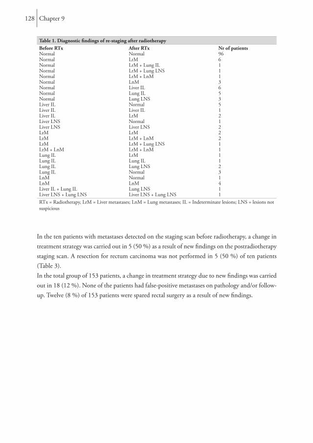
Chapter 9128
Table 1. Diagnostic findings of re-staging after radiotherapyBefore RTx After RTx Nr of patientsNormal Normal 96Normal LrM 6Normal LrM + Lung IL 1Normal LrM + Lung LNS 1Normal LrM + LnM 1Normal LnM 3Normal Liver IL 6Normal Lung IL 5Normal Lung LNS 3Liver IL Normal 5Liver IL Liver IL 1Liver IL LrM 2Liver LNS Normal 1Liver LNS Liver LNS 2LrM LrM 2LrM LrM + LnM 2LrM LrM + Lung LNS 1LrM + LnM LrM + LnM 1Lung IL LrM 1Lung IL Lung IL 1Lung IL Lung LNS 2Lung IL Normal 3LnM Normal 1LnM LnM 4Liver IL + Lung IL Lung LNS 1Liver LNS + Lung LNS Liver LNS + Lung LNS 1RTx = Radiotherapy, LrM = Liver metastases; LnM = Lung metastases; IL = Indeterminate lesions; LNS = lesions not suspicious
In the ten patients with metastases detected on the staging scan before radiotherapy, a change in treatment strategy was carried out in 5 (50 %) as a result of new findings on the postradiotherapy staging scan. A resection for rectum carcinoma was not performed in 5 (50 %) of ten patients (Table 3). In the total group of 153 patients, a change in treatment strategy due to new findings was carried out in 18 (12 %). None of the patients had false-positive metastases on pathology and/or follow-up. Twelve (8 %) of 153 patients were spared rectal surgery as a result of new findings.
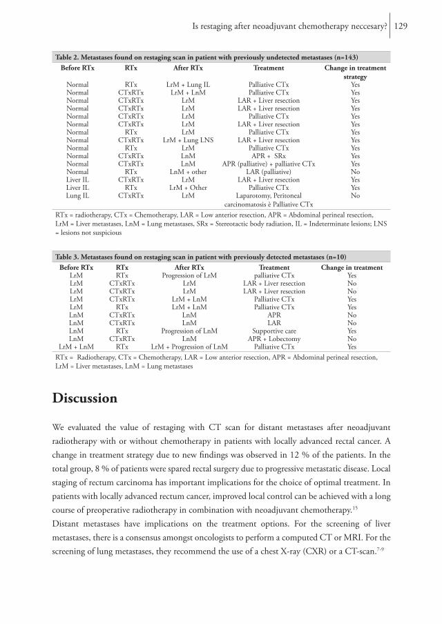
Is restaging after neoadjuvant chemotherapy neccesary? 129
Table 2. Metastases found on restaging scan in patient with previously undetected metastases (n=143)Before RTx RTx After RTx Treatment Change in treatment
strategyNormal RTx LrM + Lung IL Palliative CTx YesNormal CTxRTx LrM + LnM Palliative CTx YesNormal CTxRTx LrM LAR + Liver resection YesNormal CTxRTx LrM LAR + Liver resection YesNormal CTxRTx LrM Palliative CTx YesNormal CTxRTx LrM LAR + Liver resection YesNormal RTx LrM Palliative CTx YesNormal CTxRTx LrM + Lung LNS LAR + Liver resection YesNormal RTx LrM Palliative CTx YesNormal CTxRTx LnM APR + SRx YesNormal CTxRTx LnM APR (palliative) + palliative CTx YesNormal RTx LnM + other LAR (palliative) NoLiver IL CTxRTx LrM LAR + Liver resection YesLiver IL RTx LrM + Other Palliative CTx YesLung IL CTxRTx LrM Laparotomy, Peritoneal
carcinomatosis è Palliative CTxNo
RTx = radiotherapy, CTx = Chemotherapy, LAR = Low anterior resection, APR = Abdominal perineal resection, LrM = Liver metastases, LnM = Lung metastases, SRx = Stereotactic body radiation, IL = Indeterminate lesions; LNS = lesions not suspicious
Table 3. Metastases found on restaging scan in patient with previously detected metastases (n=10)Before RTx RTx After RTx Treatment Change in treatment
LrM RTx Progression of LrM palliative CTx YesLrM CTxRTx LrM LAR + Liver resection NoLrM CTxRTx LrM LAR + Liver resection NoLrM CTxRTx LrM + LnM Palliative CTx YesLrM RTx LrM + LnM Palliative CTx YesLnM CTxRTx LnM APR NoLnM CTxRTx LnM LAR NoLnM RTx Progression of LnM Supportive care YesLnM CTxRTx LnM APR + Lobectomy No
LrM + LnM RTx LrM + Progression of LnM Palliative CTx YesRTx = Radiotherapy, CTx = Chemotherapy, LAR = Low anterior resection, APR = Abdominal perineal resection, LrM = Liver metastases, LnM = Lung metastases
Discussion
We evaluated the value of restaging with CT scan for distant metastases after neoadjuvant radiotherapy with or without chemotherapy in patients with locally advanced rectal cancer. A change in treatment strategy due to new findings was observed in 12 % of the patients. In the total group, 8 % of patients were spared rectal surgery due to progressive metastatic disease. Local staging of rectum carcinoma has important implications for the choice of optimal treatment. In patients with locally advanced rectum cancer, improved local control can be achieved with a long course of preoperative radiotherapy in combination with neoadjuvant chemotherapy.15
Distant metastases have implications on the treatment options. For the screening of liver metastases, there is a consensus amongst oncologists to perform a computed CT or MRI. For the screening of lung metastases, they recommend the use of a chest X-ray (CXR) or a CT-scan.7-9

Chapter 9130
It is known that locally advanced rectal cancer has a higher risk of developing metastases than colon cancer.4, 6, 8, 10 The recommended treatment for locally advanced rectal cancer is a long course of radiotherapy with or without chemotherapy.8 Surgery is usually planned 6-10 weeks after finishing neoadjuvant therapy. During these 3 months, metastases can develop that previously were too small to be detected, or were not present at all. Therefore it seems prudent to restage for distant metastases after radiotherapy and before commencing surgery, since new findings in this relatively long period might alter the treatment options. In case of unresectable metastatic disease, resection of the primary tumor is unnecessary from an oncological point of view.16-19 Through re-staging, patients might therefore be spared an unnecessary extensive pelvic operation. We found a large interval between the staging scan and the beginning of radiotherapy. Most patients were referred to our hospital and this wide interval is a consequence of logistic management. We do not know whether this wide range has an influence on the outcome of our study. Local staging techniques have previously been described for locally advanced rectal cancer.20-25 To our knowledge this is the first study describing re-staging for distant metastases, after radiotherapy and before commencing surgery in patients with locally advanced rectal cancer. Restaging is only necessary if there are consequences for the treatment strategy in case of additional diagnostic findings. Additional findings can result in treatment of metastases, or in case of unresectable metastases, no resection of rectal tumor and optional treatment with palliative chemotherapy. In our series, 12 % of the total group of 153 patients had a change in the treatment due to findings on the postradiotherapy CT scan. A resection for locally advanced rectal cancer was prevented in 67 % of the latter patients as a result of findings on the postradiotherapy CT scan.Several studies have demonstrated the abdominal CT scan to be a reliable diagnostic tool for detecting liver metastases, and CT scan has proven to be better than ultrasound.26-28 There are limited data describing the optimal chest staging strategy for these patients.29 Some authors conclude that the low incidence of pulmonary metastases and minimal consequences for the treatment plan limits the clinical value or routine staging chest CT before operation.29, 30 It has several disadvantages such as costs, radiation exposure, and prolonged uncertainty because of the frequent finding of indeterminate lesions.30 However, these results were not assessed in the selected group of patients with locally advanced rectal cancer. Choi et al. demonstrated that staging before neoadjuvant radiotherapy with a chest CT for patients with locally advanced rectal cancer seems reasonable.5 Moreover, these patients can benefit from resection of pulmonary metastases, since resection can significantly improve survival.31 In our specific patient population, all patients will have two CT-scans with a median interval of 15 weeks. In case of indeterminate lesions, this will help to differentiate between metastases or benign lesions.

Is restaging after neoadjuvant chemotherapy neccesary? 131
We recognize the limitations of this retrospective study in our single centre database; patients were not randomized to have a restaging scan or not, with all inherent biases. Only patients who had complete imaging before and after radiotherapy are included. However, more patients received restaging scans but not all preoperative imaging was available. Not including these patients can cause bias in this study.In conclusion, this study demonstrated that restaging with a CT-scan after radiotherapy is a worthwhile step in the treatment of locally advanced rectal cancer because additional findings may alter this treatment strategy.
Chapter 9132
References
1. Jemal A, Bray F, Center MM, et al. Global cancer statistics. CA Cancer J Clin 2011; 61(2):69-90.
2. Bengmark S, Hafstrom L. The natural history of primary and secondary malignant tumors of the liver. I. The prognosis for patients with hepatic metastases from colonic and rectal carcinoma by laparotomy. Cancer 1969; 23(1):198-202.
3. Poultsides GA, Schulick RD, Pawlik TM. Hepatic resection for colorectal metastases: the impact of surgical margin status on outcome. HPB (Oxford) 2010; 12(1):43-9.
4. Tan KK, Lopes Gde L, Jr., Sim R. How uncommon are isolated lung metastases in colorectal cancer? A review from database of 754 patients over 4 years. J Gastrointest Surg 2009; 13(4):642-8.
5. Choi DJ, Kwak JM, Kim J, et al. Preoperative chest computerized tomography in patients with locally advanced mid or lower rectal cancer: its role in staging and impact on treatment strategy. J Surg Oncol 2010; 102(6):588-92.
6. Mitry E, Fields AL, Bleiberg H, et al. Adjuvant chemotherapy after potentially curative resection of metastases from colorectal cancer: a pooled analysis of two randomized trials. J Clin Oncol 2008; 26(30):4906-11.
7. Tjandra JJ, Kilkenny JW, Buie WD, et al. Practice parameters for the management of rectal cancer (revised). Dis Colon Rectum 2005; 48(3):411-23.
8. The Dutch Association of Comprehensive Cancer Centres. (2008). Available at: www.oncoline.nl. Accessed Accessed 01 April 2012.
9. NICE. National Institute for Health and Clinical Excellence. NICE cancer service guidance. Colorectal cancer. Available at: http://guidance.nice.org.uk/CG131. Accessed Accessed 20 November 2011.
10. van der Pool AE, de Wilt JH, Lalmahomed ZS, et al. Optimizing the outcome of surgery in patients with rectal cancer and synchronous liver metastases. Br J Surg 2010; 97(3):383-90.
11. Sauer R, Becker H, Hohenberger W, et al. Preoperative versus postoperative chemoradiotherapy for rectal cancer. N Engl J Med 2004; 351(17):1731-40.
12. de Bruin AF, Nuyttens JJ, Ferenschild FT, et al. Preoperative chemoradiation with capecitabine in locally advanced rectal cancer. Neth J Med 2008; 66(2):71-6.
13. Ferenschild FT, Vermaas M, Nuyttens JJ, et al. Value of intraoperative radiotherapy in locally advanced rectal cancer. Dis Colon Rectum 2006; 49(9):1257-65.
14. Verhoef C, van der Pool AE, Nuyttens JJ, et al. The “liver-first approach” for patients with locally advanced rectal cancer and synchronous liver metastases. Dis Colon Rectum 2009; 52(1):23-30.
15. Kapiteijn E, Marijnen CA, Nagtegaal ID, et al. Preoperative radiotherapy combined with total mesorectal excision for resectable rectal cancer. N Engl J Med 2001; 345(9):638-46.
16. Makela J, Haukipuro K, Laitinen S, et al. Palliative operations for colorectal cancer. Dis Colon Rectum 1990; 33(10):846-50.
17. Stillwell AP, Buettner PG, Ho YH. Meta-analysis of survival of patients with stage IV colorectal cancer managed with surgical resection versus chemotherapy alone. World J Surg 2010; 34(4):797-807.
Is restaging after neoadjuvant chemotherapy neccesary? 133
18. Law WL, Chan WF, Lee YM, et al. Non-curative surgery for colorectal cancer: critical appraisal of outcomes. Int J Colorectal Dis 2004; 19(3):197-202.
19. Rosen SA, Buell JF, Yoshida A, et al. Initial presentation with stage IV colorectal cancer: how aggressive should we be? Arch Surg 2000; 135(5):530-4; discussion 534-5.
20. Beets-Tan RG, Beets GL. Local staging of rectal cancer: A review of imaging. J Magn Reson Imaging 2011; 33(5):1012-9.
21. Calvo FA, Domper M, Matute R, et al. 18F-FDG positron emission tomography staging and restaging in rectal cancer treated with preoperative chemoradiation. Int J Radiat Oncol Biol Phys 2004; 58(2):528-35.
22. Guillem JG, Puig-La Calle J, Jr., Akhurst T, et al. Prospective assessment of primary rectal cancer response to preoperative radiation and chemotherapy using 18-fluorodeoxyglucose positron emission tomography. Dis Colon Rectum 2000; 43(1):18-24.
23. Chen CC, Lee RC, Lin JK, et al. How accurate is magnetic resonance imaging in restaging rectal cancer in patients receiving preoperative combined chemoradiotherapy? Dis Colon Rectum 2005; 48(4):722-8.
24. Barbaro B, Vitale R, Leccisotti L, et al. Restaging locally advanced rectal cancer with MR imaging after chemoradiation therapy. Radiographics 2010; 30(3):699-716.
25. Dresen RC, Beets GL, Rutten HJ, et al. Locally advanced rectal cancer: MR imaging for restaging after neoadjuvant radiation therapy with concomitant chemotherapy. Part I. Are we able to predict tumor confined to the rectal wall? Radiology 2009; 252(1):71-80.
26. Valls C, Andia E, Sanchez A, et al. Hepatic metastases from colorectal cancer: preoperative detection and assessment of resectability with helical CT. Radiology 2001; 218(1):55-60.
27. Bipat S, van Leeuwen MS, Ijzermans JN, et al. Evidence-base guideline on management of colorectal liver metastases in the Netherlands. Neth J Med 2007; 65(1):5-14.
28. Kinkel K, Lu Y, Both M, et al. Detection of hepatic metastases from cancers of the gastrointestinal tract by using noninvasive imaging methods (US, CT, MR imaging, PET): a meta-analysis. Radiology 2002; 224(3):748-56.
29. Parnaby C, Bailey W, Balasingam A, et al. Pulmonary staging in colorectal cancer: a review. Colorectal Dis 2011.
30. Grossmann I, Avenarius JK, Mastboom WJ, et al. Preoperative staging with chest CT in patients with colorectal carcinoma: not as a routine procedure. Ann Surg Oncol 2010; 17(8):2045-50.
31. Pavoor RS, Shukla PJ, Milsom JW. The Importance of Preoperative Staging with Chest CT Scan in Patients with Colorectal Cancer. Ann Surg Oncol 2010.

Chapter 10
Surgery of the primary tumor in stage IV colorectal cancer with unresectable metastases
Ninos AyezWijnand J. Alberda
Henk M. VerheulJaconus W. A. BurgerJohannes H. de Wilt
Cornelis Verhoef
European oncology & haematology, 2011
Chapter 10136
Abstract
Since patients with incurable metastatic colorectal cancer (CRC) only have a relatively limited life expectancy, and resection of the primary tumor is accompanied by both morbidity and mortality, it is under debate whether resection of the primary tumor has an effect on survival or quality of life. The rationale behind the resection strategy is that prophylactic surgery prevents future complications. With current new chemotherapy regimens, a relatively low number of patients with metastatic CRC require surgery for their primary tumor. Many studies concerning the management of incurable stage IV CRC have been performed and most studies suggest a survival benefit for patients undergoing surgical resection of the primary tumor compared with those who received palliative treatment. However, in stage IV CRC with unresectable metastases, the role of a palliative resection of the primary tumor has never been assessed properly. Because randomized clinical trials are lacking, it is difficult to draw conclusions from the present literature.
Chemotherapy first in patients with unresectable synchronous mCRC 137
Introduction
Colorectal cancer (CRC) is one of the two most commonly diagnosed cancers, with approximately 1.2 million new cases each year and more than 600,000 annual deaths estimated to occur worldwide.1 In addition, roughly one-fifth of patients presents with incurable disseminated disease.2 In the last decade, development of new chemotherapeutic biological agents has significantly improved overall survival (OS) of these patients.3-12 A palliative resection of the primary tumor is frequently performed 13 and there is a clear indication for surgery when patients present with symptoms of the primary tumor. However, if patients present with absence or mild symptoms, the indication for resection is less obvious. Since patients with incurable metastatic CRC (mCRC) only have a relatively limited life expectancy and resection of the primary is accompanied with both morbidity and mortality 14-16, it is under debate whether resection of the primary tumor has an effect on survival or quality of life.17, 18 Many studies concerning the management of incurable stage IV CRC have been performed; however the advantage of a palliative resection of the primary tumor has never been assessed properly.19 Moreover, most studies do not even report whether a resection of the primary has been performed.20
In this paper we aim to evaluate the role of surgery of the primary in stage IV CRC with unresectable metastases.
Treatment of metastatic colorectal cancer (mCRC)
At diagnosis of CRC, approximately 20% of the patients present with synchronous mCRC, and the liver is the predilection site in half these patients.21, 22 The lungs represent the second most common site of metastases from CRC and according to non population based studies lung metastases are present in 10-15% of patients with colorectal cancer.23, 24
When metastases are limited, a possible curative treatment can be obtained by surgical resection, however, only 15-20% of patients is resectable.25 Median 5-year survival for patients undergoing an R0 resection of the metastases is approximately 30% (range 15-67%).26 Despite complete resection and neoadjuvant or adjuvant chemotherapy regimens, recurrences occur in 75% of the patients.27 Extrahepatic disease in combination with liver metastases was generally considered a contraindication for surgery.28 However, resection of both intrahepatic and extrahepatic colorectal metastases should be considered if resection of all metastatic sites can be complete and the disease is controlled by chemotherapy.29 In patients with unresectable metastases, palliative systemic chemotherapy is the treatment of choice.
Chapter 10138
With systemic combination chemotherapy response rates of 40-70% have been reported resulting in a median overall survival rate of approximately 22 months.30-32 Most frequently used combinations are oxaliplatin or irinotecan plus capecitabine or 5-fluorouracil (5-FU) with or without bevacizumab. In case of K-RAS wild type tumors, anti-epidermal growth factor receptor (EGFR) antibodies such as panitumumab and cetuximab are being used.33
Resection of the primary tumor in patients with unresectable synchronous mCRC
Traditional surgical teaching promotes resection of the primary tumor in patients with unresectable metastases, even if the primary is asymptomatic. The rationale behind this strategy is that prophylactic surgery prevents future complications of intestinal obstruction, perforation and haemorrhage.34 However, resection does not provide immediate palliative benefit in case of an asymptomatic primary tumor, and surgery is associated with high mortality (5-13%) and morbidity (23-48%) in patients with metastatic disease.16, 34-37 Some studies tried to selectively apply prophylactic surgery in patients with a low metastatic tumor burden because these patients are presumed to be at risk for obstruction because of long survival. If the metastatic tumor burden is extensive, resection of the primary is unlikely to benefit the patient and is associated with a high risk of postoperative complications. These patients are probably better served by focusing on the disseminated component of their disease and start with systemic treatment early on in their course, reserving surgery for when and if symptoms from the primary tumor are substantial.35, 38 Other studies have shown no association between the incidence of complications and the extent of metastatic disease.39, 40 Due to recent advances in systemic chemotherapy, the risks and benefits of immediate or deferred surgical strategy are under debate.Some clinicians in favor of the surgical approach argue that if the asymptomatic primary cancer is not resected, patients will develop disabling symptoms such as weight loss and nutritional depletion (secondary to “near” obstruction) and anemia due to bleeding of the primary tumor. Arguments supporting surgery include a lower reported operative mortality for elective surgery in patients with stage IV disease (3-6%), compared with the more threatening operative mortality rates for non-elective resections in patients with advanced and symptomatic disease (20-40%).34,
41, 42 Another argument supporting this concept, is that preoperative staging is sometimes unclear and that surgery is considered the last and most effective diagnostic tool for the correct staging of abdominal tumors before treatment.19 In addition, patients are provided with psychological comfort who feel that the “cancer” has been removed.35
Chemotherapy first in patients with unresectable synchronous mCRC 139
Chemotherapy first in patients with unresectable synchronous mCRC
The advocates of a chemotherapy first approach prefer to avoid complications at least in non symptomatic patients. The argument of those who prefer “elective” surgery due to higher mortality if emergent surgery is required, was addressed in several studies, were the risk of death was found to be extremely low.39, 43-45 In fact, Poultsides et al. compared their study population with studies with elective colon resection in the metastatic setting and found that it appears that this deferred approach is associated with at least comparable perioperative mortality.46 Another argument for chemotherapy first, is that chemotherapy will not only treat the metastases but also the primary tumor; many patients will have improvements of their symptoms and therefore evading a possible resection.37, 47 Chau et al. demonstrated that overall, 86% of patients had an improvement in symptoms. Of the patients with symptoms, 71% had diminished pelvic pain/tenesmus, 90% had improvement in diarrhea/constipation, 100% had reduced rectal bleeding, and 93% had weight stabilization or weight gain.Advocates of the deferred surgical approach argue that surgery at diagnosis can delay or even preclude systemic chemotherapy, and that most patients will never develop symptoms and these patients could be spared an unnecessary operation. Additionally, primary CRC surgery may alter the host immune response in such a way that tumor growth is increased in the post operative period.48, 49 An argument against resection is that patients with unresectable metastasis from colorectal cancer who have undergone palliative resection of the primary still face the prospect of further intestinal complications, which may require further surgery (Table 1).34, 50 After resection of the primary tumor, these patients may develop local recurrence or adhesions which can result in obstruction and require subsequent surgery.A decade ago, when patients were treated with single agent 5-FU chemotherapy, approximately 20% of patients with mCRC treated with chemotherapy required palliative surgery for symptoms related to their intact primary CRC.39, 40, 46, 50, 51 In recent years, combinations with modern chemotherapy like FOLFOX, XELOX and FOLFIRI have attained response rates of 50% and disease control rates of 85% in prospective clinical trials.6, 52 With these modern chemotherapy regimens, approximately 7% (range 3-22%) of patients with mCRC required surgical palliation for their intact primary CRC, as stated in an elegant review by Poultsides.43-46 These data suggest that with effective chemotherapy almost 14 asymptomatic patients need to undergo prophylactic resection of their primary tumor in order to save one patient a subsequent operation for obstruction or perforation.46 There are indications that this has led to a decrease over time in the percentage of resection of the primary tumor in case of unresectable metastatic colorectal disease.13

Chapter 10140
Table 1. Study results on stage IV colorectal cancer and unresectable metastases, in which the non-resection arm was treated with chemotherapy
AuthorYears of study
Number of patients
Received chemotherapy
(%)
Secondary palliative surgical
intervention
Palliative Resection of
primary
Scoggins40 1985-1997 resection 66 0 2 (3%) -chemo 23 100 2 (9%) 0
Tebbutt50 1990-1999 resection 280 100 14 (5%) -chemo 82 100 8 (10%) 1 (1%)
Konyalian53 1991-2002 resection 62 58 # -chemo 47 60 17 (36%) 0
Galizia54 1995-2005 resection 42 100 0 -chemo 23 100 6 (26%) 
Ruo51 1996-1999 resection 127 0 6 (5%) -chemo 103 83 30 (29%) 0
Michel45 1996-1999 resection 31 97 0 -chemo 23 100 5 (22%) 3 (13%)
Serela39 1997-2000 resection - - - -chemo 24 88 6 (25%) 4 (17%)
Benoist44 1997-2002 resection 32 94 0 -chemo 27 100 4 (15%) 3 (11%)
Karoui55 1998-2007 resection 85 99 27 (32%) -chemo 123 100 15(12%) 15 (12%)
Aslam56 1998-2007 resection 366 63 ¥ -chemo 281 36 128 (46%) 0
Bajwa57 1999-2005 resection - - - -chemo 67 100 27 (40%) 25 (37%)
Muratore43 2000-2004 resection - - - -chemo 35 100 1 (3%) 0
Poultsides37 2000-2006 resection - - - -chemo 233 100 16 (7%) 8 (3%)
Seo58 2001-2008 resection 144 100 22 (15%) -chemo 83 100 4 (5%) 1 (1%)
# Konyalian53 not described; 12 patients with complications mostly infectious¶ Galizia54 not described; 2 colon perforations, 1 intestinal hemorrhage, 1 bowel obstruction, 2 surgery owing to bowel perforation or stent dislocation¥ Aslam56 not described; 11 full thickness wound dehiscence, 11 intra-abdominal collections, 11 anastomotic leak, 7 intra-abdominal sepsis, 5 hemorrhage, 4 postoperative ileus, 1 splenic tear, 1 inter-loop fistula
Survival
Several studies have been performed to analyze overall survival of patients with stage IV CRC and unresectable metastases to examine whether to resect the primary or not. Recently, Venderbosch et al. performed a retrospective analysis of two phase III studies (CAIRO and CAIRO2)8, 59 and investigated the prognostic and predictive value of resection of the primary tumor in stage IV mCRC patients.60 They demonstrated that resection of the primary tumor was a significantly important prognostic factor for survival in these patients. They also performed a review of the literature and identified 22 nonrandomized studies, most of which showed improved survival for mCRC patients who underwent resection of the primary tumor. These results were confirmed in a systemic review by Anwar et al..48 An overview of these studies is presented in table 2.
Chemotherapy first in patients with unresectable synchronous mCRC 141
However, in all studies presented a selection bias cannot be excluded. Most studies were not randomized, performed in single centers and were retrospective of nature. Patients with a good performance status were more likely to undergo surgery whereas those with extensive disease were more likely to be offered chemotherapy instead. In the absence of randomized controlled trials, the best evidence is obtained from case-matched studies. A case-matched study by Benoist et al. compared 27 patients with asymptomatic colorectal cancer and irresectable synchronous liver metastases who received chemotherapy, with 32 matched patients who were treated by initial resection of the primary tumor. They found no difference in survival between the operative and the non-operative management.Prospective studies on this topic are currently planned. Recently a protocol has been developed in the Netherlands for stage IV colon cancer patients with unresectable metastases.61 In this trial patients will be randomized to either systemic therapy until progression or unacceptable toxicity or to resection of the primary tumor followed by systemic therapy until progression or unacceptable toxicity. The endpoint of the trial is overall survival and the trial is powered to identify a survival benefit of 6 months in the surgery group. Also the National Surgical Adjuvant breast and Bowel Project has started a phase II Trial using 5-fluorouracil, leucovorin, and oxaliplatin chemotherapy plus bevacizumab for patients with unresectable stage IV colon cancer and synchronous asymptomatic primary tumor.62 The primary endpoint is the event rate related to the intact primary tumor requiring surgery. In both trials only patients with colon cancer will be randomized and patients with rectal cancer are excluded. Also a trial from Australia/New Zealand “SUPER” is currently running: “A randomized phase III multicentre trial evaluating the role of palliative surgical resection of the primary tumor in patients with metastatic colorectal cancer”.63 Patients will be randomized to compare chemotherapy followed by surgery to surgery alone. The primary outcome is to determine whether surgical resection of the primary tumor in patients with stage IV colorectal cancer decreases intestinal complications and improves overall survival and quality of life. For patients with rectal cancer and unresectable systemic disease a phase III randomized clinical trial is recently conducted in the Netherlands. In this trial the role of radiotherapy in providing local control will be studied and patients will be randomized to either standard chemotherapy alone or short term course radiotherapy (5x5 Gy) on the primary tumor followed by standard of care chemotherapy. The primary endpoint is the number of patients requiring an unplanned surgical intervention related to symptoms of the primary rectal tumor.
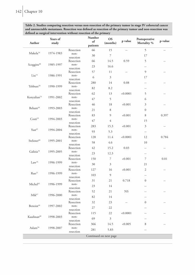
Chapter 10142
Table 2. Studies comparing resection versus non-resection of the primary tumor in stage IV colorectal cancer and unresectable metastases. Resection was defined as resection of the primary tumor and non-resection was defined as surgical intervention without resection of the primary
AuthorYears of study
Number of
patients
OS (months) p value Postoperative
Mortality % p-value
Makela34 1974-1983Resection 66 15 -- 5 --
non-resection 30 7 17
Scoggins40 1985-1997Resection 66 14.5 0.59 5 --
non-resection 23 16.6 --
Liu14 1986-1991Resection 57 11 -- 9 --
non-resection 6 3 17
Tebbutt50 1990-1999Resection 280 14 0.08 -- --
non-resection 82 8.2 --
Konyalian53 1991-2002Resection 62 13 <0.0001 5 --
non-resection 47 5 6
Beham64 1993-2003Resection 46 18 <0.001 3 --
non-resection 21 8 0
Costi19 1994-2003Resection 83 9 <0.001 8 0.397
non-resection 47 4 15
Yun65 1994-2004Resection 283 15.3 <0.001 3 --
non-resection 93 5.3 --
Stelzner66 1995-2001Resection 128 11.4 <0.0001 12 0.784
non-resection 58 4.6 10
Galizia54 1995-2005Resection 42 15.2 0.03 -- --
non-resection 23 12.3 --
Law16 1996-1999Resection 150 7 <0.001 7 0.01
non-resection 30 3 21
Ruo51 1996-1999Resection 127 16 <0.001 2 --
non-resection 103 9 --
Michel45 1996-1999Resection 31 21 0.718 0 --
non-resection 23 14 --
Mik67 1996-2000Resection 52 21 NS -- --
non-resection 82 14 --
Benoist44 1997-2002Resection 32 23 -- 0 --
non-resection 27 22 --
Kaufman68 1998-2003Resection 115 22 <0.0001 -- --
non-resection 69 3 --
Aslam56 1998-2007Resection 366 14.5 <0.005 8 --
non-resection 281 5.83 --
Continued on next page
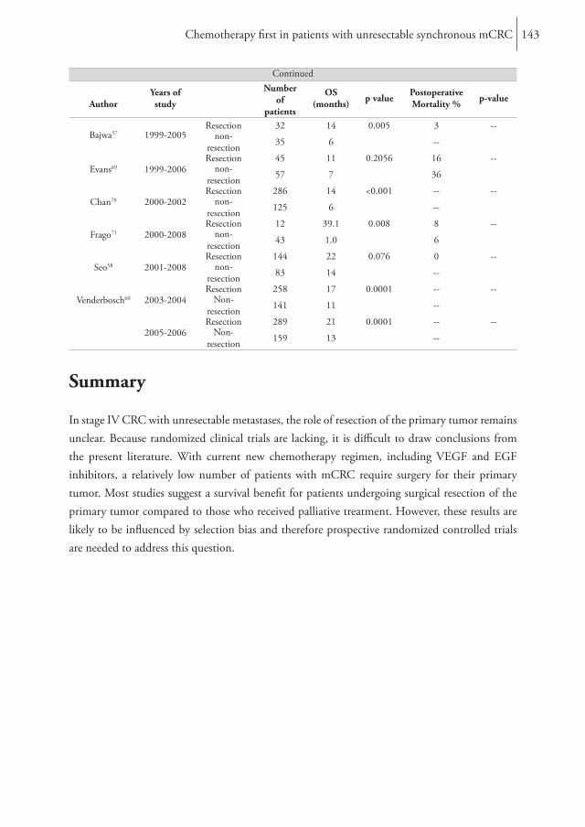
Chemotherapy first in patients with unresectable synchronous mCRC 143
Continued
AuthorYears of study
Number of
patients
OS (months) p value Postoperative
Mortality % p-value
Bajwa57 1999-2005Resection 32 14 0.005 3 --
non-resection 35 6 --
Evans69 1999-2006Resection 45 11 0.2056 16 --
non-resection 57 7 36
Chan70 2000-2002Resection 286 14 <0.001 -- --
non-resection 125 6 --
Frago71 2000-2008Resection 12 39.1 0.008 8 --
non-resection 43 1.0 6
Seo58 2001-2008Resection 144 22 0.076 0 --
non-resection 83 14 --
Venderbosch60 2003-2004Resection 258 17 0.0001 -- --
Non-resection 141 11 --
2005-2006Resection 289 21 0.0001 -- --
Non-resection 159 13 --
Summary
In stage IV CRC with unresectable metastases, the role of resection of the primary tumor remains unclear. Because randomized clinical trials are lacking, it is difficult to draw conclusions from the present literature. With current new chemotherapy regimen, including VEGF and EGF inhibitors, a relatively low number of patients with mCRC require surgery for their primary tumor. Most studies suggest a survival benefit for patients undergoing surgical resection of the primary tumor compared to those who received palliative treatment. However, these results are likely to be influenced by selection bias and therefore prospective randomized controlled trials are needed to address this question.
Chapter 10144
References
1. Jemal A, Bray F, Center MM, et al. Global cancer statistics. CA Cancer J Clin 2011; 61(2):69-90.
2. Mella J, Biffin A, Radcliffe AG, et al. Population-based audit of colorectal cancer management in two UK health regions. Colorectal Cancer Working Group, Royal College of Surgeons of England Clinical Epidemiology and Audit Unit. Br J Surg 1997; 84(12):1731-6.
3. de Gramont A, Figer A, Seymour M, et al. Leucovorin and fluorouracil with or without oxaliplatin as first-line treatment in advanced colorectal cancer. J Clin Oncol 2000; 18(16):2938-47.
4. Douillard JY, Cunningham D, Roth AD, et al. Irinotecan combined with fluorouracil compared with fluorouracil alone as first-line treatment for metastatic colorectal cancer: a multicentre randomised trial. Lancet 2000; 355(9209):1041-7.
5. Saltz LB, Cox JV, Blanke C, et al. Irinotecan plus fluorouracil and leucovorin for metastatic colorectal cancer. Irinotecan Study Group. N Engl J Med 2000; 343(13):905-14.
6. Tournigand C, Andre T, Achille E, et al. FOLFIRI followed by FOLFOX6 or the reverse sequence in advanced colorectal cancer: a randomized GERCOR study. J Clin Oncol 2004; 22(2):229-37.
7. Seymour MT, Maughan TS, Ledermann JA, et al. Different strategies of sequential and combination chemotherapy for patients with poor prognosis advanced colorectal cancer (MRC FOCUS): a randomised controlled trial. Lancet 2007; 370(9582):143-52.
8. Koopman M, Antonini NF, Douma J, et al. Sequential versus combination chemotherapy with capecitabine, irinotecan, and oxaliplatin in advanced colorectal cancer (CAIRO): a phase III randomised controlled trial. Lancet 2007; 370(9582):135-42.
9. Hurwitz H, Fehrenbacher L, Novotny W, et al. Bevacizumab plus irinotecan, fluorouracil, and leucovorin for metastatic colorectal cancer. N Engl J Med 2004; 350(23):2335-42.
10. Saltz LB, Clarke S, Diaz-Rubio E, et al. Bevacizumab in combination with oxaliplatin-based chemotherapy as first-line therapy in metastatic colorectal cancer: a randomized phase III study. J Clin Oncol 2008; 26(12):2013-9.
11. Van Cutsem E, Kohne CH, Hitre E, et al. Cetuximab and chemotherapy as initial treatment for metastatic colorectal cancer. N Engl J Med 2009; 360(14):1408-17.
12. Douillard JY, Siena S, Cassidy J, et al. Randomized, phase III trial of panitumumab with infusional fluorouracil, leucovorin, and oxaliplatin (FOLFOX4) versus FOLFOX4 alone as first-line treatment in patients with previously untreated metastatic colorectal cancer: the PRIME study. J Clin Oncol 2010; 28(31):4697-705.
13. Cook AD, Single R, McCahill LE. Surgical resection of primary tumors in patients who present with stage IV colorectal cancer: an analysis of surveillance, epidemiology, and end results data, 1988 to 2000. Ann Surg Oncol 2005; 12(8):637-45.
14. Liu SK, Church JM, Lavery IC, et al. Operation in patients with incurable colon cancer--is it worthwhile? Dis Colon Rectum 1997; 40(1):11-4.
Chemotherapy first in patients with unresectable synchronous mCRC 145
15. Harris GJ, Senagore AJ, Lavery IC, et al. Factors affecting survival after palliative resection of colorectal carcinoma. Colorectal Dis 2002; 4(1):31-35.
16. Law WL, Chan WF, Lee YM, et al. Non-curative surgery for colorectal cancer: critical appraisal of outcomes. Int J Colorectal Dis 2004; 19(3):197-202.
17. Joffe J, Gordon PH. Palliative resection for colorectal carcinoma. Dis Colon Rectum 1981; 24(5):355-60.
18. Stearns MW, Jr., Binkley GE. Palliative surgery for cancer of the rectum and colon. Cancer 1954; 7(5):1016-9.
19. Costi R, Mazzeo A, Di Mauro D, et al. Palliative resection of colorectal cancer: does it prolong survival? Ann Surg Oncol 2007; 14(9):2567-76.
20. Sorbye H, Kohne CH, Sargent DJ, et al. Patient characteristics and stratification in medical treatment studies for metastatic colorectal cancer: a proposal for standardization of patient characteristic reporting and stratification. Ann Oncol 2007; 18(10):1666-72.
21. Bengmark S, Hafstrom L. The natural history of primary and secondary malignant tumors of the liver. I. The prognosis for patients with hepatic metastases from colonic and rectal carcinoma by laparotomy. Cancer 1969; 23(1):198-202.
22. van der Pool AE, Lalmahomed ZS, Ozbay Y, et al. “Staged” liver resection in synchronous and metachronous colorectal hepatic metastases; differences in clinicopathological features and outcome. Colorectal Dis 2009; 12(10):e229–e235.
23. Tan KK, Lopes Gde L, Jr., Sim R. How uncommon are isolated lung metastases in colorectal cancer? A review from database of 754 patients over 4 years. J Gastrointest Surg 2009; 13(4):642-8.
24. Choi DJ, Kwak JM, Kim J, et al. Preoperative chest computerized tomography in patients with locally advanced mid or lower rectal cancer: its role in staging and impact on treatment strategy. J Surg Oncol 2010; 102(6):588-92.
25. Adam R, Hoti E, Bredt LC. Evolution of neoadjuvant therapy for extended hepatic metastases--have we reached our (non-resectable) limit? J Surg Oncol 2010; 102(8):922-31.
26. Simmonds PC, Primrose JN, Colquitt JL, et al. Surgical resection of hepatic metastases from colorectal cancer: a systematic review of published studies. Br J Cancer 2006; 94(7):982-99.
27. Nordlinger B, Sorbye H, Glimelius B, et al. Perioperative chemotherapy with FOLFOX4 and surgery versus surgery alone for resectable liver metastases from colorectal cancer (EORTC Intergroup trial 40983): a randomised controlled trial. Lancet 2008; 371(9617):1007-16.
28. Hugh TJ, Kinsella AR, Poston GJ. Management strategies for colorectal liver metastases--Part II. Surg Oncol 1997; 6(1):31-48.
29. Adam R, de Haas RJ, Wicherts DA, et al. Concomitant extrahepatic disease in patients with colorectal liver metastases: when is there a place for surgery? Ann Surg 2011; 253(2):349-59.
30. Emmanouilides C, Sfakiotaki G, Androulakis N, et al. Front-line bevacizumab in combination with oxaliplatin, leucovorin and 5-fluorouracil (FOLFOX) in patients with metastatic colorectal cancer: a multicenter phase II study. BMC Cancer 2007; 7:91.
Chapter 10146
31. Van Cutsem E, Rivera F, Berry S, et al. Safety and efficacy of first-line bevacizumab with FOLFOX, XELOX, FOLFIRI and fluoropyrimidines in metastatic colorectal cancer: the BEAT study. Ann Oncol 2009; 20(11):1842-7.
32. Ychou M, Viret F, Kramar A, et al. Tritherapy with fluorouracil/leucovorin, irinotecan and oxaliplatin (FOLFIRINOX): a phase II study in colorectal cancer patients with non-resectable liver metastases. Cancer Chemother Pharmacol 2008; 62(2):195-201.
33. Amado RG, Wolf M, Peeters M, et al. Wild-type KRAS is required for panitumumab efficacy in patients with metastatic colorectal cancer. J Clin Oncol 2008; 26(10):1626-34.
34. Makela J, Haukipuro K, Laitinen S, et al. Palliative operations for colorectal cancer. Dis Colon Rectum 1990; 33(10):846-50.
35. Rosen SA, Buell JF, Yoshida A, et al. Initial presentation with stage IV colorectal cancer: how aggressive should we be? Arch Surg 2000; 135(5):530-4; discussion 534-5.
36. Stillwell AP, Buettner PG, Ho YH. Meta-analysis of survival of patients with stage IV colorectal cancer managed with surgical resection versus chemotherapy alone. World J Surg 2010; 34(4):797-807.
37. Poultsides GA, Servais EL, Saltz LB, et al. Outcome of primary tumor in patients with synchronous stage IV colorectal cancer receiving combination chemotherapy without surgery as initial treatment. J Clin Oncol 2009; 27(20):3379-84.
38. Stillwell AP, Buettner PG, Siu SK, et al. Predictors of postoperative mortality, morbidity, and long-term survival after palliative resection in patients with colorectal cancer. Dis Colon Rectum 2011; 54(5):535-44.
39. Sarela AI, Guthrie JA, Seymour MT, et al. Non-operative management of the primary tumour in patients with incurable stage IV colorectal cancer. Br J Surg 2001; 88(10):1352-6.
40. Scoggins CR, Meszoely IM, Blanke CD, et al. Nonoperative management of primary colorectal cancer in patients with stage IV disease. Ann Surg Oncol 1999; 6(7):651-7.
41. Longo WE, Virgo KS, Johnson FE, et al. Risk factors for morbidity and mortality after colectomy for colon cancer. Dis Colon Rectum 2000; 43(1):83-91.
42. Legendre H, Vanhuyse F, Caroli-Bosc FX, et al. Survival and quality of life after palliative surgery for neoplastic gastrointestinal obstruction. Eur J Surg Oncol 2001; 27(4):364-7.
43. Muratore A, Zorzi D, Bouzari H, et al. Asymptomatic colorectal cancer with un-resectable liver metastases: immediate colorectal resection or up-front systemic chemotherapy? Ann Surg Oncol 2007; 14(2):766-70.
44. Benoist S, Pautrat K, Mitry E, et al. Treatment strategy for patients with colorectal cancer and synchronous irresectable liver metastases. Br J Surg 2005; 92(9):1155-60.
45. Michel P, Roque I, Di Fiore F, et al. Colorectal cancer with non-resectable synchronous metastases: should the primary tumor be resected? Gastroenterol Clin Biol 2004; 28(5):434-7.
46. Poultsides GA, Paty PB. Reassessing the need for primary tumor surgery in unresectable metastatic colorectal cancer: overview and perspective. Ther Adv Med Oncol 2011; 3(1):35-42.
47. Chau I, Brown G, Cunningham D, et al. Neoadjuvant capecitabine and oxaliplatin followed by synchronous chemoradiation and total mesorectal excision in magnetic resonance imaging-defined poor-risk rectal cancer. J Clin Oncol 2006; 24(4):668-74.
Chemotherapy first in patients with unresectable synchronous mCRC 147
48. Anwar S, Peter M, Dent J, et al. Palliative excisional surgery for primary colorectal cancer in patients with incurable metastatic disease. Is there a survival benefit? A systematic review. Colorectal Dis 2011.
49. Allendorf JD, Bessler M, Horvath KD, et al. Increased tumor establishment and growth after open vs laparoscopic surgery in mice may be related to differences in postoperative T-cell function. Surg Endosc 1999; 13(3):233-5.
50. Tebbutt NC, Norman AR, Cunningham D, et al. Intestinal complications after chemotherapy for patients with unresected primary colorectal cancer and synchronous metastases. Gut 2003; 52(4):568-73.
51. Ruo L, Gougoutas C, Paty PB, et al. Elective bowel resection for incurable stage IV colorectal cancer: prognostic variables for asymptomatic patients. J Am Coll Surg 2003; 196(5):722-8.
52. Goldberg RM, Sargent DJ, Morton RF, et al. A randomized controlled trial of fluorouracil plus leucovorin, irinotecan, and oxaliplatin combinations in patients with previously untreated metastatic colorectal cancer. J Clin Oncol 2004; 22(1):23-30.
53. Konyalian VR, Rosing DK, Haukoos JS, et al. The role of primary tumour resection in patients with stage IV colorectal cancer. Colorectal Dis 2007; 9(5):430-7.
54. Galizia G, Lieto E, Orditura M, et al. First-line chemotherapy vs bowel tumor resection plus chemotherapy for patients with unresectable synchronous colorectal hepatic metastases. Arch Surg 2008; 143(4):352-8; discussion 358.
55. Karoui M, Roudot-Thoraval F, Mesli F, et al. Primary colectomy in patients with stage IV colon cancer and unresectable distant metastases improves overall survival: results of a multicentric study. Dis Colon Rectum 2011; 54(8):930-8.
56. Aslam MI, Kelkar A, Sharpe D, et al. Ten years experience of managing the primary tumours in patients with stage IV colorectal cancers. Int J Surg 2010; 8(4):305-13.
57. Bajwa A, Blunt N, Vyas S, et al. Primary tumour resection and survival in the palliative management of metastatic colorectal cancer. Eur J Surg Oncol 2009; 35(2):164-7.
58. Seo GJ, Park JW, Yoo SB, et al. Intestinal complications after palliative treatment for asymptomatic patients with unresectable stage IV colorectal cancer. J Surg Oncol 2010; 102(1):94-9.
59. Tol J, Koopman M, Cats A, et al. Chemotherapy, bevacizumab, and cetuximab in metastatic colorectal cancer. N Engl J Med 2009; 360(6):563-72.
60. Venderbosch S, de Wilt JH, Teerenstra S, et al. Prognostic Value of Resection of Primary Tumor in Patients with Stage IV Colorectal Cancer: Retrospective Analysis of Two Randomized Studies and a Review of the Literature. Ann Surg Oncol 2011.
61. The Dutch Colorecrtal Cancer Group. Available at: http://www.dccg.nl/. 62. National Surgical Adjuvant Breast and Bowel Project [Protocol C-10, A Phase II Trial
of 5-Fluorouracil, Leucovorin, and Oxaliplatin (mFOLFOX6) Chemotherapy Plus Bevacizumab for Patients with Unresectable Stage IV Colon Cancer and a Synchronous Asymptomatic Primary Tumor. web site]. Available at: http://www.nsabp.pitt.edu/C10_Information.asp.
63. Australian New Zealand Clinical Trials Resistry [The SUPER Study: A randomised phase III multicentre trial evaluating the role of palliative surgical resection of the primary tumour in patients with metastatic colorectal cancer. web site]. Available at: http://www.anzctr.org.au/trial_view.aspx?ID=308218.

Chapter 10148
64. Beham A, Rentsch M, Pullmann K, et al. Survival benefit in patients after palliative resection vs non-resection colon cancer surgery. World J Gastroenterol 2006; 12(41):6634-8.
65. Yun HR, Lee WY, Lee WS, et al. The prognostic factors of stage IV colorectal cancer and assessment of proper treatment according to the patient’s status. Int J Colorectal Dis 2007; 22(11):1301-10.
66. Stelzner S, Hellmich G, Koch R, et al. Factors predicting survival in stage IV colorectal carcinoma patients after palliative treatment: a multivariate analysis. J Surg Oncol 2005; 89(4):211-7.
67. Mik M, Dziki L, Galbfach P, et al. Resection of the primary tumour or other palliative procedures in incurable stage IV colorectal cancer patients? Colorectal Dis 2010; 12(7 Online):e61-7.
68. Kaufman MS, Radhakrishnan N, Roy R, et al. Influence of palliative surgical resection on overall survival in patients with advanced colorectal cancer: a retrospective single institutional study. Colorectal Dis 2008; 10(5):498-502.
69. Evans MD, Escofet X, Karandikar SS, et al. Outcomes of resection and non-resection strategies in management of patients with advanced colorectal cancer. World J Surg Oncol 2009; 7:28.
70. Chan TW, Brown C, Ho CC, et al. Primary tumor resection in patients presenting with metastatic colorectal cancer: analysis of a provincial population-based cohort. Am J Clin Oncol 2010; 33(1):52-5.
71. Frago R, Kreisler E, Biondo S, et al. Outcomes in the management of obstructive unresectable stage IV colorectal cancer. Eur J Surg Oncol 2010; 36(12):1187-94.

PART 4
Summery, general discussion, future perspectives and appendices


Chapter 11
Summary and future perspectives
Chapter 11152
Surgery for colorectal metastases has established itself in the last couple of decades and is now mainstay of treatment for stage IV colorectal patients, provided that the metastases are deemed resectable. To further specify the role of surgery in this patient category, numerous aspects still need to be elucidated. In the present thesis, we aimed to address some of these aspects. In Part I, the important technical issue of organ preserving surgery is discussed. Part II focused on the question whether we can predict the outcome of patients after surgery, so as to define which patients are expected to have most benefit of surgery, and possibly to define which patients are likely to benefit from multimodality therapy. Part III is dedicated to the role of surgery within the multimodality treatment that is nowadays standard-of-care especially in patients with synchronously metastasized (locally advanced) colorectal cancer.
Part 1In part one, we analyzed surgical techniques that could lead to more liver parenchyma saving operations. The purpose of chapter 2 was to investigate the influence of a non-anatomical liver resection (NAR) compared with an anatomical resection (AR) on morbidity, mortality, margin positivity, disease-free, and overall survival. The study demonstrated no significant difference in outcome between patients with colorectal liver metastases (CRLM) after AR or NAR. The 5-year disease-free (AR 30% versus NAR 32%) and overall survival (AR 49% vs. NAR 39%) in our study are consistent with the literature. Since there was no significant difference in morbidity, mortality, recurrence rate, or survival according to resection type, we concluded that a NAR can be used as a save procedure to preserve liver parenchyma.
Another more detailed technique of the NAR, is to perform a resection as close to the metastasis as possible and therefore preserve even more liver parenchyma. This width from the metastasis to the resection margin is the called the surgical margin status. Several studies have described risk factors for the outcome of patients with CRLM and the surgical margin status has been described as the major determinant of survival after resection, with R1 resections (incomplete resection) doing worse compared to R0 resections (complete resection). However, the impact of the several proposed cut-off points regarding R0 resections remains controversial. Most studies performed on this subject did not evaluate the specific group of patients who received neoadjuvant chemotherapy before resection of CRLM. In chapter 3 we analyzed whether a resection margin of 0 mm is sufficient in the era of effective neoadjuvant chemotherapy. We found that in patients with neoadjuvant chemotherapy, those with a R1 resection did not fare worst than those with a R0 resection. An explanation might be that neoadjuvant chemotherapy decreases micrometastases in the periphery. We also analyzed whether margin width ≤ 2 mm or > 2 mm influenced survival. This proved to be the case in patients who had a R0 resection without neoadjuvant chemotherapy.
Summary 153
In patients with neoadjuvant chemotherapy this phenomenon could not be demonstrated. A similar trend was found if a tumor-free margin of 0-5 mm versus > 5 mm was chosen. It seems that the width of the resection margin still correlates with survival, but this only applies to patients who did not receive chemotherapy.
Part 2A clinical risk score (CRS) is a predictive tool for patients with CRLM who undergo resection. The predictive value of these CRSs is well-established, but as most CRSs were designed in an era when chemotherapy prior to surgery was seldom used, this raises the question whether CRSs can still adequately predict outcome when neoadjuvant chemotherapy is part of the treatment protocol. In chapter 4 we argue that when the CRS is calculated before starting neoadjuvant chemotherapy it is of no predictive value; however, we demonstrate that the scores are applicable when the score is addressed after administration of neoadjuvant chemotherapy. In this study, chemotherapy downstaged the size and the CEA level. When the pathology report was consulted and complete response was reported, then the number of metastases also decreased significantly. This effect changes the CRS. Patients who had a higher risk score before chemotherapy became patients with a lower risk score after chemotherapy, with an associated improved survival. Bilobar disease showed no statistically significant change after administration of neoadjuvant chemotherapy and extrahepatic disease did not change at all. Although it is impossible to make any definitive conclusions, the data would suggest that response to neoadjuvant therapy, as measured by the variables that comprise the clinical risk scoring systems, may be associated with better survival. Based on our findings, if prediction of prognosis is required, we believe that all the traditional CRSs can be used if they are determined after treatment with neoadjuvant chemotherapy.
A strategy to identify patients with a certain risk which might benefit more from several types of available diagnostic tools or therapies, is to stratify them in risk groups within a CRS. Fluorine-18-deoxyglucose positron emission tomography (FDG-PET), one of the available diagnostic tools, is used for patients with colorectal cancer to demonstrate extrahepatic disease and as a consequence it may improve patient selection for surgical resection of the liver metastases. In chapter 5 we analyzed whether this selection with FDG-PET would result in an improved outcome in surgically treated patients with CRLM if they are stratified by the CRS of Fong. We found that FDG-PET prior to liver resection did not significantly improve disease free survival (DFS) or overall (OS) in patients with both low and high CRS in the present series. It has been demonstrated that patients with a high CRS are expected to have a poor tumor biology and therefore could potentially have more intra- and extrahepatic disease compared to patients with a low CRS.

Chapter 11154
By means of a FDG-PET these metastases might have been detected and resulted in less disease recurrence, which might explain the trend towards a different DFS. In patients with a low CRS there is a minimal risk of occult metastatic disease and the added value of a FDG-PET is therefore limited, if not absent. The observation that patients with a high CRS and selected by FDG-PET do not have a trend towards an improved OS may partially be explained by the fact that currently excellent local and systemic treatment therapies for recurrent disease are available.
Chapter 6 deals with patients who received chemotherapy and stratifies them by the CRS of Fong. Patients with a high CRS who received neoadjuvant chemotherapy had a better OS than patients without chemotherapy. This in contrast to patients with a low CRS who had no advantage of neoadjuvant chemotherapy in terms of OS. Chapter 6 proposes that the Fong CRS may therefore be a powerful tool in selecting patients most likely to benefit from neoadjuvant treatment. The present role of perioperative chemotherapy in the case of resectable metastases was established in a randomized controlled trial (EORTC Intergroup trial 40983), which compared perioperative chemotherapy with surgery alone. Although perioperative chemotherapy improved DFS in these patients, the mature overall survival data of this trial showed no survival difference. We propose that this may be due to the fact that most patients had a relatively low CRS. Also other authors performed a similar study as ours with a different chemotherapy sequence. They used adjuvant chemotherapy instead of neoadjuvant chemotherapy and the results were strikingly similar and therefore support our hypothesis. Although this is a retrospective analysis, it seems that only patients who bare a high risk of developing metastases are the ones who will benefit the most from neoadjuvant chemotherapy.
Chapter 7 describes a randomized multicentre clinical trial (based on the conclusions drawn in chapter 6) which will evaluate the impact of neoadjuvant chemotherapy in patients with high risk resectable colorectal liver metastases without extrahepatic disease. Our hypothesis is that adding neo-adjuvant chemotherapy to surgery will provide an improvement in overall survival in this high-risk patient group.
Part 3In the case of locally advanced rectal cancer with synchronous metastases there are several options in the treatment strategy and preoperative diagnostics. Chapter 8 reports on the largest series of patients to date with locally advanced rectal cancer and synchronous liver metastases who underwent the ‘liver first’ approach. The results show that in this selected group of patients the majority (74%) could undergo optimal neoadjuvant treatment of both liver metastases and rectal cancer, followed by curative resection of both liver metastases and rectal cancer.
Summary 155
Of the 11 patients with unresectable metastatic disease, 10 (91%) did not need rectal surgery. Previous reports have described the ‘liver first’ approach, however this was in patients with colon and rectal cancer. A strength of the present study is that all patients had a locally advanced rectal primary with synchronous liver metastases. Some patients required a diverting stoma and one may argue that a stoma could have been avoided had a rectum first approach been chosen. The disadvantages of a rectal resection (with the associated morbidity and mortality) have to be balanced against those of stoma construction. A concern regarding the ‘liver first’ approach is non-response to chemotherapy or progression in patients who present with resectable disease. Another possible concern regarding the liver first approach is that the primary tumor may progress beyond resection.
In case of locally advanced rectal cancer there is a long period of neoadjuvant treatment. After initial diagnostics and neoadjuvant treatment there is a chance that occult metastases become apparent which may alter the treatment strategy. Chapter 9 evaluated the value of restaging with CT scan for distant metastases after neoadjuvant radiotherapy with or without chemotherapy in patients with locally advanced rectal cancer. A change in treatment strategy due to new findings was observed in 12% of the patients. In the total group, 8% of patients were spared rectal surgery due to progressive metastatic disease. The low incidence of pulmonary metastases might limit the clinical value or routine staging chest CT before operation. However, results of previous studies concerning lung metastases were not assessed in the selected group of patients with locally advanced rectal cancer. Restaging might also have several disadvantages such as costs, radiation exposure, and prolonged uncertainty because of the frequent finding of indeterminate lesions. However, based on this study a change in treatment strategy in 12% of patients is high, and therefore restaging in the group of patient with locally advanced rectal cancer is advised.
There remains a difficult decision in patients who present with or develop unresectable metastatic disease when the primary colorectal cancer is still present.Chapter 10 discusses whether resection of the primary tumor has an advantage in the stage IV colorectal cancer. An advantage of resection might be prevention of intestinal obstruction, perforation or hemorrhage. However when there are no symptoms, surgery does not provide immediate palliative benefit. Moreover surgery is associated with considerable morbidity en mortality. It is discussed that elective surgery for the primary tumor comes with less morbidity. When the primary is not resected, it might lead to disabling symptoms such as weight loss, nutritional depletion and anemia. The advocates of a chemotherapy first approach prefer to avoid complications at least in non symptomatic patients.
Chapter 11156
The argument of those who prefer “elective” surgery due to higher mortality if emergent surgery is required, was addressed in several studies, were the risk of death was found to be extremely low. Another argument for chemotherapy first, is that chemotherapy will not only treat the metastases but also the primary tumor; many patients will have improvements of their symptoms and therefore evading a possible resection. Most studies suggest a survival benefit for patients undergoing surgical resection of the primary tumor compared to those who received palliative treatment. However, these results are likely to be influenced by selection bias and therefore prospective randomized controlled trials are needed to address this question. These trials have been conducted worldwide.

Summary 157
General discussion and future perspectives
Surgical resection can be performed if there will be sufficient residual liver after resection to enable survival of the patient, corresponding to 20-25% of the total functioning liver volume. Patients with pre-existent liver disease such as steatosis, cirrhosis or chemotherapy associated steatohepatitis need at least 30-40% of the liver remnant to survive. In addition, one of the three main hepatic veins must be preserved and the liver remnant has to comprise a portal vein, hepatic artery and a bile duct. Although surgery for CRLM is at present the only treatment with curative intent, cancer relapse is a common phenomenon, with up to 50% recurrences in the first 2 years. Therefore it is wise to preserve as much liver parenchyma as possible, since new resections might be needed. A multidisciplinary and multimodal approach is necessary to achieve optimal individual cancer treatment. Defining the most appropriate sequence or combinations of treatments has become very challenging in this era of numerous available treatments. Optimization through systemic chemotherapy is difficult. The standard neoadjuvant treatment of patients with initially unresectable liver-only metastases currently consists of combination chemotherapy of a fluoropyrimidine plus either oxaliplatin or irinotecan and with triple chemotherapy (fluoropyrimidine+oxaliplatin+irinotecan) shows promising results. The addition of a targeted drug to chemotherapy has been shown to increase response rates, which provides a clear rationale for use in this setting, but a clear preference for either the anti-VEGF antibody bevacizumab or one of the anti-EGFR antibodies cetuximab or panitumumab has not been demonstrated in this setting. In the Netherlands a randomized controlled trial has been conducted to answer this question (CAIRO5 trial). For the remaining patients with multi-organ metastases, it is unknown whether adding maximal tumor debulking to chemotherapy will benefit survival. The ORCHESTRA trial in the Netherlands has been conducted to answer this question. To further improve survival rates, in case a resection is not possible or when the future liver remnant is anticipated to be small, several local therapies have been applied for the treatment of unresectable liver metastases in the past few years including radiofrequency ablation/microwave ablation (RFA), stereotactic ablative radiotherapy (SBRT), portal vein embolization (PVE), two stage hepatectomies and associating liver partition with portal vein ligation (PVL) for staged hepatectomy (ALPPS). A two stage hepatectomy is a strategy in patient with bilobar disease were one hemi liver is cleared of metastases with wedge resections. Then a PVE is used to induce hypertrophy. After 6 to 8 week a new resection will be planned after restaging. A newer technique to further improve hypertrophy is the ALLPS. In addition to the PVE the liver is also transected, leaving only arterial inflow, hepatic venous outflow and biliary drainage intact.
Chapter 11158
Other local therapies comprise irreversible electroporation (IRE), which employs electrical pulses that permeabilize cellular membrane and consequently lead to cell death, and arterial modalities such as selective internal radiation therapy (SIRT), drug eluding beads (DEBS), trans-arterial chemo-embolization and hepatic artery infusion of chemotherapy.The above-mentioned methods are not required in patients with resectable metastases and sufficient remnant liver. It is well established, including our own results from chapter 2, that a non-anatomical resection is comparable to an anatomical resection. It is now standard care to perform a non anatomical resections to preserve liver parenchyma for potential future resections. However, the impasse concerning the surgical margin width remains. Must surgeons pursue a R0 resection? Should this remain the standard goal is this field, or should surgeons seek boundaries in the era of neoadjuvant chemotherapy? Our results, as results from other authors, suggest that an R1 resection should not be a contraindication to surgery with curative intent. There is data that suggests that an R1 resection is associated with a higher risk of developing hepatic recurrences, not only at the surgical site but also elsewhere in the liver. Concerning the recurrences at the surgical site, it might be argued that this is a result of micrometastases that were not treated, either by chemotherapy or by surgery. More relevant is why metastases reoccur elsewhere in the liver or even in other organs. Is it because they were occult or is it because of poor tumor biology? Some studies within this thesis have focused on surgical techniques to improve outcome. Reoccurrence elsewhere in the liver or in other organs cannot be explained by surgical techniques and therefore future studies should be more focused on tumor biological behavior of metastases or primary cancer to explain this phenomenon. Next steps in research should be more focused on understanding why current treatments fail and metastases re-occur. This might help us in identifying patients who will or will not benefit from certain treatment and thereby avoiding unnecessary treatments. In order to predict the likelihood of tumor recurrence and survival after resection for CRLM, several Clinical Risk Scores (CRS) have been developed. These scores are based on clinical findings that are a surrogate for advanced disease. If we can find patients with a high risk for relapse then they might be subjected to more aggressive treatment to improve survival rates. In chapter 6 we tried to identify patient with high risk based on a clinical risk and confirmed this hypothesis. In the future we hope to use the gathered information from the prospective trial described in chapter 7 to select patients who might benefit from neoadjuvant chemotherapy. However, clinical presentation does not inform us about different metastatic pathways. There is a desire for biological markers to inform us why these patients develop such risk and perhaps in the future a more tailor made therapy could be applied. Currently, the only marker routinely used in clinical practice of individualized metastatic colorectal cancer treatment is K-RAS, since treatment with anti-EGFR antibodies was found to be only effective in patients with K-RAS wild-type tumors.
Summary 159
Another prognostic and response predictive marker extensively studied in relation to prognosis and treatment response, but not yet used in routine clinical practice, is microsatellite instability status. CRC patients with microsatellite instable stage II and III tumors have a better prognosis compared to patients with microsatellite stable CRC. For patients with mCRC this relationship could not be proven, mainly due to the low incidence of microsatellite instable mCRC tumors. Circulating tumor cells are cells that circulate in the peripheral blood of cancer patients, originating from either primary or metastatic disease. Recently, the prognostic value of circulating tumor cells in blood was subject of a meta-analysis, showing that detection of these circulating tumor cells in the peripheral blood of patients with resectable colorectal liver metastases is associated with disease progression and poor survival. Obtaining primary tumor or metastatic tumor tissue for analysis preoperatively is often difficult and invasive. An advantage of circulating tumor cells is that they can be obtained by venapunctures. In studies performed at our centre we could not identify patients at risk for early disease recurrence after curative resection of colorectal liver metastases. Further development of this technique might give us insights in metastatic tumor biology and further individualized cancer treatment.
When patient are diagnosed with locally advanced rectal cancer, preoperatively they are expected to undergo a long course of (chemo) radiation therapy. Surgery is usually planned 6-10 weeks after finishing neoadjuvant therapy. If this specific group is diagnosed with synchronous livermetastases the liver first approach is preferred as described in chapter 8. In patient with coloncancer the liver first approach is feasible and non inferior to primary cancer resection first. A randomized controlled trial should be conducted to find out whether the liver first approach has a better survival compared to treating the primary locally advanced rectal cancer first. In our study we also found that patient with a complete response of the metastases after neoadjuvant chemotherapy also tended to have a complete response of their primary tumor. In the future in such cases, resection of the primary tumor may be limited to local excision (transanal excision or transanal endoscopic microsurgery) or even watchful waiting. However, prospective studies are needed to justify such a strategy. During approximately 3 months of preoperative treatment for locally advanced rectal cancer, metastases can develop that previously were too small to be detected, or were not present at all. In chapter 9 we explained why restaging for distant metastases should be performed, since new findings in this relatively long period might alter the treatment options. Recently other researchers from the Netherlands have been exploring their own results and found very similar results as ours. In the future we expect restaging to become standard of care in patients with locally advanced rectal cancer. In this instance the CT-scan was investigated as a modality and seems justifiable because it can be obtained easily and might also be used for restaging of the primary tumor regression.

Chapter 11160
In this case a CT-scan of the thorax-abdomen can be performed for the purpose of restaging for metastases but also for restaging of the primary tumor.In stage IV CRC with unresectable metastases, the role of resection of the primary tumor remains unclear. To answer this question, prospective studies on this topic have been conducted. The CAIRO4 trial has been developed in the Netherlands for stage IV colon cancer patients with unresectable metastases. In this trial patients will be randomized to either systemic therapy or to resection of the primary tumor followed by systemic therapy. Also the National Surgical Adjuvant breast and Bowel Project has started a phase II Trial chemotherapy for patients with unresectable stage IV colon cancer and synchronous asymptomatic primary tumor. Another trial from Australia/New Zealand “SUPER” is currently running: “A randomized phase III multicentre trial evaluating the role of palliative surgical resection of the primary tumor in patients with metastatic colorectal cancer”. Patients will be randomized to compare chemotherapy followed by surgery to surgery alone. With current new chemotherapy regimens, a relatively low number of patients with metastatic CRC require surgery for their primary tumor. Many studies concerning the management of incurable stage IV CRC have been performed and most studies suggest a survival benefit for patients undergoing surgical resection of the primary tumor compared with those who received palliative treatment. Because randomized clinical trials are lacking, it is difficult to draw conclusions from the present literature. And the above mentioned trials have to be awaited.

Chapter 12
Nederlandse samenvatting
Chapter 12162
In de afgelopen decennia heeft de chirurgie zich bewezen als behandeling voor colorectale levermetastasen waarbij chirurgie de voorkeursbehandeling is indien er sprake is van resectabele metastasen. Er moeten echter nog een aantal aspecten worden opgehelderd om de rol van chirurgie bij deze patiënten categorie nader te specificeren. In dit proefschrift worden enkele van deze aspecten besproken. In deel I wordt de belangrijke technische kwestie van parenchym sparende chirurgie besproken. Deel II richt zich op de vraag of we de prognose van patiënten kunnen voorspellen na de operatie, maar ook om te voorspellen welke patiënten naar verwachting het meeste voordeel zullen hebben van de behandeling. Deel III is gewijd aan de rol van chirurgie bij de multimodale behandeling in patiënten met synchroon gemetastaseerde of het lokaal gevorderde colorectaal carcinoom.
Deel 1In deel I worden chirurgische technieken geanalyseerd welke tot weefsel sparende behandelingen kunnen leiden. In hoofdstuk 2 wordt besproken of een niet-anatomische resectie (NAR) van de lever even goed is vergeleken met een anatomische resectie (AR). Er werd gekeken naar morbiditeit, mortaliteit, positieve snijvlakken, ziektevrije overleving en algehele overleving. We zien dat er geen significant verschil bestaat tussen de twee benaderingen. De 5-jaar ziekte vrije overleving (AR 30% versus NAR 32%) en algehele overleving (AR 49% vs. NAR 39%) is gelijk aan de bekende literatuur. We concluderen dan ook aan de hand van onze uitkomsten dat een NAR geen significante verschillen vertoont met een AR als we kijken naar de morbiditeit, mortaliteit, recidief ziekte of algehele overleving.
Meerdere studies hebben risicofactoren beschreven voor de uitkomsten van patiënten met colorectale levermetastasen (CRLM). De resectiemarges tijdens een operatie worden gekenmerkt als een van de belangrijkste risicofactoren voor de overleving, waarbij de R1 resectie een slechtere overleving heeft vergeleken met de R0 resectie. Echter is de invloed van de verschillende afkappunten bij de R0 resecties niet duidelijk. De meeste studies die dit onderwerp bespreken hebben niet de patiënten geëvalueerd die neoadjuvante chemotherapie hebben gekregen voor de resectie van de levermetastasen. In hoofdstuk 3 hebben we gekeken of een resectiemarge van 0 mm genoeg is in het tijdperk met effectievere chemotherapie. We vonden dat de ziektevrije overleving en algehele overleving gelijk was bij patiënten met een R0 of R1 als zij neoadjuvante chemotherapie krijgen. We hebben ook nog bekeken of een marge van ≤ 2 mm of > 2 mm invloed had op de overleving en dit bleek alleen zo te zijn bij patiënten die een R0 resectie ondergingen en geen chemotherapie kregen. Indien zij wel chemotherapie kregen verdween dit effect. Hetzelfde resultaat werd ook gevonden als we een marge van 0-5 mm vergeleken met > 5 mm. Het lijkt er dus op dat de marge van de resectie nog wel invloed heeft op de overleving, maar dit geldt alleen voor patiënten die geen chemotherapie hebben gekregen.

Nederlandse samenvatting 163
Deel 2 Een klinische risico score is een instrument waarmee voorspellingen gedaan kunnen worden over patiënten met CRLM die een resectie ondergaan. Meestal worden ze dus niet gebruikt om te zien of patiënten geopereerd zullen worden, maar met name om iets over de overleving te kunnen zeggen. De voorspellende waarde van de klinische risico scores is bevestigd in meerdere studies. Echter is dit voornamelijk geweest in het tijdperk vóór de neoadjuvante chemotherapie. In hoofdstuk 4 zien we dat indien een risico score voor het starten van chemotherapie gebruikt wordt deze geen voorspellende waarde heeft, maar als men de score na chemotherapie toepast deze wel weer een voorspellende waarde heeft. In dit hoofdstuk laten we zien dat chemotherapie de grootte van de metastasen doet afnemen en ook dat het CEA afnam. Bij een pathologisch complete respons zagen we ook het aantal metastasen afnemen. Dit effect verandert ook de risico score. Patiënten die voor de chemotherapie een hoge risico score hadden werden patiënten met een lagere risico score nadat zij chemotherapie kregen en hiermee gepaard gaande ook een betere overleving hebben gekregen. Er werd geen verandering gezien indien er sprake was van bilobaire of extrahepatische metastasen. Het lijkt erop dat een respons op chemotherapie, gemeten met de variabelen van de risico score, geassocieerd is met een betere overleving. Gebaseerd op deze resultaten is het advies om de risico score toe te passen nadat patiënten neoadjuvante chemotherapie hebben gekregen.Een FDG-PET wordt gebruikt voor patiënten met een colorectaal carcinoom om extrahepatische ziekte aan te tonen. Hiermee kan het een betere selectie leveren van patiënten die in aanmerking zouden kunnen komen voor een resectie van de levermetastasen. In hoofdstuk 5 hebben we bekeken of deze selectie met de FDG-PET scan een verbeterde overleving zou geven in patiënten met CRLM welke dan nog onderverdeeld worden aan de hand van de risico score. Een FDG-PET scan vóór de operatie leverde geen ziektevrije overleving op en ook geen algehele overleving bij patiënten met een lage risico score of hoge risico score. Eerder is al wel aangetoond dat patiënten met een hoge risico score een slechte tumor biologie hebben en daardoor een grote kans op meer intra- en extrahepatische metastasen. Men zou verwachten dat deze metastasen gedetecteerd worden en hierdoor minder recidieven op zouden treden. In onze studie werd wel een trend gezien die hierop duidt, echter was dit niet significant verschillend. In patiënten met een lage risico score is er maar een kleine kans op occulte metastasen en de waarde van de FDG-PET is daarom maar beperkt. We zagen dat patiënten met een hoge risico score en geselecteerd met de FDG-PET geen verbetering hadden in overleving. Dit zou verklaard kunnen worden doordat er tegenwoordig zeer goede locale en systemische therapieën bestaan.

Chapter 12164
In hoofdstuk 6 werden patiënten geanalyseerd die neoadjuvante chemotherapie hebben gekregen en vervolgens ingedeeld werden in een risico score volgens Fong. Patiënten met een hoge risico score en die neoadjuvant chemotherapie kregen, hadden een betere overleving vergeleken met patiënten zonder chemotherapie en een hoge risico score. Bij patiënten met een lage score werd dit verschil niet gevonden. In hoofdstuk 6 stellen we voor dat dit een efficiënte manier zou kunnen zijn om onderscheid te maken tussen wie wél en wie géén baat zou kunnen hebben van de chemotherapie. De huidige rol van perioperatieve chemotherapie werd beoordeeld in een gerandomiseerde studie (EORTC Intergroup trial 40983) waarbij er werd gerandomiseerd tussen perioperatieve chemotherapie en chirurgie. Er was een klein voordeel in ziekte vrije overleving, echter was de algehele overleving niet verschillend. Ook de langetermijn resultaten laten dit zien. Het zou kunnen komen doordat er goede lokale en systemische therapieën bestaan. Wij denken echter dat er te veel patiënten met een lage risico score in de studie zaten. Sommige van onze resultaten zouden verklaard kunnen worden omdat er patiënten al afvallen doordat er progressie optreedt van de metastasen tijdens de chemotherapie. Echter zien we in de literatuur dat deze afvallers maar een zeer klein aantal patiënten betreft. Daarnaast hebben andere auteurs een vergelijkbare studie verricht als die van ons, maar dan met adjuvante chemotherapie. Ook zij vonden deze opvallende bevindingen.
In hoofdstuk 7 beschrijven we een gerandomiseerde studie die gebaseerd is op hoofdstuk 6. Deze studie zal de invloed van neoadjuvante chemotherapie in patiënten met een hoge risico score en resectabele lever metastasen (zonder extrahepatische ziekte) evalueren. Onze hypothese is dat neoadjuvante chemotherapie een overlevingswinst geeft in patiënten met een hoge risico score.
Deel 3Wanneer er sprake is van een lokaal gevorderd rectum carcinoom met synchrone levermetastasen dan zijn er verschillende preoperatieve diagnostische testen en behandelopties. Hoofdstuk 8 beschrijft de grootste serie patiënten tot op heden die een lokaal gevorderd rectumcarcinoom hebben met synchrone lever metastasen en die een zogenaamde ‘liver first’ benadering kregen. In de resultaten zagen we dat 74% een optimale therapie kregen voor zowel de lever als voor het rectum carcinoom. Van de patiënten met niet-resectabele metastasen hoefden er 91% geen rectum operatie te ondergaan. De “liver first’ benadering is al wel eerder beschreven, maar dit was bij een mix van patiënten met colon en rectum carcinoom. Het sterke punt van deze studie is dat alle patiënten een lokaal gevorderd rectumcarcinoom hadden. Sommige patiënten hadden een stoma nodig en men kan zich voorstellen dat indien we het rectum eerst opereren, dit wellicht voorkomen had kunnen worden. Echter moet met de afweging maken tussen een stoma of de complicaties gepaard gaande met grote rectumchirurgie.
Nederlandse samenvatting 165
Een zorg bij de ‘liver first’ benadering is dat er patiënten zijn die niet reageren op chemotherapie maar wel een resectabele tumor hebben. Een andere zorg is dat de primaire tumor zo ver groeit dat deze niet meer te opereren valt. Indien er sprake is van lokaal gevorderd rectumcarcinoom is er een langdurige periode van neoadjuvante behandeling. Na de diagnose en voorafgaande behandeling zou het kunnen zijn dat voorheen niet ontdekte metastasen nu wel zichtbaar worden en daardoor de behandeling strategie veranderen. In hoofdstuk 9 wordt de waarde van opnieuw stadiëren middels een CT scan, na neoadjuvante radiotherapie met of zonder chemotherapie, geëvalueerd in patiënten met een lokaal gevorderd rectumcarcinoom. Er werd een verandering in behandelstrategie gevonden in 12% van de patiënten. In 8% van de patiënten werd rectumchirurgie achterwege gelaten in verband met progressie van de metastasen. De lage incidentie van longmetastasen beperkt de klinische relevantie van routinematig CT-scan onderzoek preoperatief. Echter is deze opvatting gebaseerd op een patiëntengroep die geen lokaal gevorderd rectumcarcinoom hadden. Opnieuw stadiëren zou ook nadelen kunnen hebben zoals het kostenplaatje, blootstelling aan radiatie en nieuwe onzekerheden bij twijfelachtige afwijkingen. Recent hebben Nederlandse onderzoekers deze studie opnieuw verricht bij een homogene groep patiënten, waarbij dezelfde resultaten werden gevonden.
Bij patiënten met metastasen waarbij het colorectale carcinoom nog in situ is, is het nog onduidelijk of er een resectie moet plaats vinden van de primaire tumor.In hoofdstuk 10 bespreken we het effect van resectie van de primaire tumor in het geval van stadium IV colorectaalcarcinoom. Voorbeelden hiervan zijn een obstructie door de tumor, perforatie of een bloeding. Echter geeft resectie geen direct palliatief effect indien er geen symptomen zijn. Verder geeft een operatie ook morbiditeit en mortaliteit. Sommigen zijn van mening dat een electieve ingreep minder gecompliceerd verloopt vergeleken met een spoedoperatie en dat er derhalve wel geopereerd moet worden. Het zou ook kunnen zijn dat als de tumor geen directe problemen geeft deze wel kan leiden tot gewichtsverlies, ondervoeding en bloedarmoede. Anderen geven juist de voorkeur om eerst chemotherapie te geven bij patiënten die geen klachten hebben. Als tegenargument wordt beweerd dat het risico bij een spoedoperatie wel meevalt conform de literatuur. Nog een argument om eerst met chemotherapie te behandelen is dat niet alleen de metastasen behandeld worden maar ook meteen de primaire tumor wordt meebehandeld en hierdoor kunnen eventuele symptomen verdwijnen. De meeste studies over dit onderwerp suggereren dat er een overlevingsvoordeel bestaat indien de primaire tumor wordt verwijderd. Echter zijn de meeste studies beïnvloed door een selectie van patiënten in de studies. Om dit duidelijk te krijgen zijn er wereldwijd nu gerandomiseerde studies gestart die dit proberen aan te tonen dan wel uit te sluiten.
Chapter 12166
Discussie en toekomst perspectieven
Een chirurgische resectie kan worden uitgevoerd als er voldoende rest lever overblijft na resectie, overeenkomend met 20-25% van de totale werkende lever. Patiënten met een reeds bestaande leverziekte zoals steatose, cirrose of chemotherapie geassocieerd steatohepatitis dienen ten minste 30-40% van de lever over te houden om te overleven. Bovendien moet een van de drie levervenen, een poortader en arterie en een galweg behouden blijven. Hoewel chirurgie voor CRLM op dit moment de enige in opzet curatieve behandeling is, is een recidief een veel voorkomend verschijnsel, met ongeveer 50% recidieven in de eerste 2 jaar. Daarom is het verstandig om zoveel mogelijk leverparenchym over te houden na de operatie omdat er mogelijk nog een resectie dient plaats te vinden in de toekomst.
Een multidisciplinaire en multimodale aanpak is noodzakelijk om een optimale individuele behandeling van kanker te bereiken. Het bepalen van de meest geschikte volgorde of combinaties van behandelingen is zeer uitdagend geworden in dit tijdperk van de vele beschikbare behandelingen. Optimalisatie door middel van systemische chemotherapie is hierdoor ook moeilijk geworden. De standaard neoadjuvante behandeling van patiënten met aanvankelijk inoperabele lever metastasen bestaat momenteel uit een combinatie chemotherapie van een fluoropyrimidine samen met ofwel oxaliplatine of irinotecan en met triple chemotherapie (fluoropyrimidinetherapie + oxaliplatine + irinotecan) welke veelbelovende resultaten tonen. De toevoeging van targeted therapy aan chemotherapie heeft aangetoond de respons te verhogen, maar een duidelijke voorkeur voor de anti-VEGF antilichaam bevacizumab of een van de anti-EGFR antilichamen cetuximab of panitumumab is niet aangetoond. In Nederland is een gerandomiseerde gecontroleerde trial opgezet (CAIRO5) om deze vraag te beantwoorden. Voor de overige patiënten met meervoudige orgaan metastasen, is het onbekend of het toevoegen maximale tumor debulking plus chemotherapie de overleving doet toenemen. De ORCHESTRA trial in Nederland is opgezet om deze vraag te beantwoorden.
Om de overlevingskansen te verbeteren, in het geval dat een resectie niet mogelijk is of als de toekomstige restlever te klein zal zijn, zijn er verschillende lokale therapieën beschikbaar voor de behandeling van inoperabele metastasen, waaronder radiofrequente ablatie / microwave ablatie (RFA), stereotactic ablative radiotherapy (SBRT), portal vein embolisatie (PVE), twee stappen hepatectomie en associating liver partition with portal vein ligation (PVL) for staged hepatectomy (ALPPS). Een twee-stappen hepatectomie is een strategie bij patiënten met bilobaire ziekte waarbij metastasen in een hemi lever worden verwijderd middels wig resecties, waarbij vervolgens een PVE gebruikt wordt om hypertrofie te induceren in de restlever.
Nederlandse samenvatting 167
Na 6 tot 8 weken zal een nieuwe resectie worden gepland. Een nieuwere techniek om de hypertrofie te verbeteren is ALLPS. Naast de PVE wordt ook de rest lever doorgenomen, waardoor alleen arteriële instroom, hepatische veneuze uitstroom en galwegdrainage intact blijft. Andere lokale therapieën bestaan irreversible electroporation (IRE), die elektrische pulsen geeft waardoor de celmembraan permeabel wordt en tot celdood leidt, en arteriële modaliteiten zoals “selective internal radiation therapy” (SIRT), drug eluding beads (DEBS), transarteriële chemo- embolisatie en de arteriële chemotherapie.
De bovengenoemde methoden zijn niet vereist bij patiënten met resectabele metastasen en voldoende restlever. Het is aangetoond, met inbegrip van onze eigen resultaten uit hoofdstuk 2, dat een niet-anatomische resectie vergelijkbaar is met een anatomische resectie. Het is nu standaard zorg om een niet-anatomische resecties uit te voeren om op deze wijze leverparenchym voor potentieel nieuwe operatie te behouden. Echter, de discussie met betrekking tot de chirurgische marge blijft bestaan. Moeten chirurgen streven naar een R0 resectie en moet dit de norm blijven op dit gebied, of moet chirurgen juist de grenzen opzoeken in het tijdperk van de neoadjuvante chemotherapie? De resultaten in dit proefschrift, maar ook de resultaten van andere auteurs, suggereren dat een R1 resectie geen contra-indicatie is voor een in opzet curatieve operatie. Er zijn gegevens die suggereert dat een R1 resectie is geassocieerd met een hoger risico op het ontwikkelen van hepatische recidieven, niet alleen in het operatiegebied maar ook elders in de lever. Wat betreft de recidieven in het operatiegebied, zou men kunnen stellen dat dit een gevolg is van micrometastasen die niet behandeld werden, hetzij door chemotherapie of door een operatie.Wat veel belangrijker is, is de vraag waarom metastasen terugkomen elders in de lever of zelfs in andere organen. Is dat omdat ze er al waren en nog niet zichtbaar waren voor ons of is het vanwege de slechte tumor biologie? Onze studies in dit proefschrift hebben zich onder andere gericht op de chirurgische technieken om uitkomsten van de operatie te verbeteren. Een recidief elders in de lever of in andere organen kan niet verklaard worden door chirurgische technieken. Toekomstige studies moeten zich meer richten op het biologisch gedrag van metastasen of van de primaire ziekte om dit verschijnsel te verklaren. In de toekomst dienen we uit te zoeken waarom de huidige behandelingen falen en waarom er metastasen recidiveren. Dit kan ons helpen bij het identificeren van patiënten die wel of niet zullen profiteren van een bepaalde behandeling en daarmee kunnen onnodige behandelingen worden voorkomen.
Chapter 12168
Om de kans op een recidief en de overleving na resectie van CRLM te voorspellen, zijn er diverse Clinical Risk Scores (CRS) ontwikkeld. Deze scores zijn gebaseerd op klinische bevindingen die een surrogaat voor gevorderde ziekte zijn. Als we patiënten kunnen vinden met een hoog risico op een recidief dan zouden zij een agressieve behandeling kunnen krijgen om de overlevingskansen te verbeteren. In hoofdstuk 6 hebben we geprobeerd om de patiënten te identificeren met een hoog risico op basis van een klinische risicofactoren en bevestigden hiermee de bovenstaande hypothese. In de toekomst hopen we, met de verzamelde informatie van de prospectieve studie beschreven in hoofdstuk 7, om patiënten te kunnen selecteren die kunnen profiteren van neoadjuvante chemotherapie. Echter, de klinische presentatie van deze patiënten informeert ons niet over de verschillende paden van metastasering. Er is behoefte aan biologische markers om in te zien waarom deze patiënten een bepaalde risico hebben en in de toekomst een meer op maat gemaakte therapie kan worden toegepast.
Momenteel is K-RAS de enige marker die routinematig klinisch wordt gebruikt in de behandeling van gemetastaseerde colorectale kanker, aangezien behandeling met anti-EGFR antilichamen alleen effectief bleek bij patiënten met K-RAS wildtype tumoren. Een andere prognostische en respons voorspellende marker die uitgebreid is bestudeerd, maar nog niet gebruikt wordt in de dagelijkse klinische praktijk, is microsatelliet instabiliteit status. CRC patiënten stadium II en III met microsatelliet instabiele tumoren hebben een betere prognose dan patiënten met microsatelliet stabiel CRC. Voor patiënten met gemetastaseerd CRC kan deze relatie niet worden bewezen. Dit is vooral te wijten aan de lage incidentie van microsatelliet instabiliteit in de metastasen. Circulerende tumorcellen zijn cellen die circuleren in het perifere bloed van kankerpatiënten en zijn afkomstig van primaire of metastatische ziekte. Onlangs werd de prognostische waarde van circulerende tumorcellen in bloed in een meta-analyse beschreven. Er blijkt dat detectie van deze circulerende tumorcellen in perifeer bloed van patiënten met resectabele colorectale levermetastasen ziekteprogressie kan voorspellen maar ook de overleving. Het preoperatief verkrijgen van weefsel uit de primaire tumor of uit de metastase voor analyse is vaak moeilijk en invasief. Een voordeel van circulerende tumorcellen is dat deze verkregen kunnen worden door simpele venapuncties. Verdere ontwikkeling van deze techniek zou ons inzicht kunnen geven in de metastatische tumor biologie en het verder individualiseren van behandeling van kanker.
De “National Surgical Adjuvant breast and Bowel Project” is een studie gestart bij patiënten met niet resectabele metastasen en een niet symptomatische coloncarcinoom. De “SUPER” trial uit Australie/Nieuw Zeeland is een fase 3 studie die patiënten randomiseert tussen resectie en chemotherapie of resectie alleen. Met de huidige chemotherapie zijn er maar een paar patiënten met metastasen die chirurgie nodig zullen hebben voor hun primaire tumor.

Nederlandse samenvatting 169
Er zijn veel studies verschenen over patiënten met niet resectabele metastasen en veel van deze studies laten een overlevingswinst zien als primaire tumor verwijderd wordt in vergelijking met alleen palliatieve chemotherapie. Omdat er het geen gerandomiseerde studies zijn is het moeilijk om hier conclusies aan te verbinden. Hopelijk zullen in de toekomst de bovengenoemde studies hier een antwoord op geven.


Dankwoord.
Mijn laatste stelling heb ik niet zomaar gekozen. Zonder hulp zou dit proefschrift niet tot stand zijn gekomen. Een aantal personen wil ik in het bijzonder bedanken. Allereerst mijn promotor, prof.dr. C. Verhoef, beste Kees, mijn dankbaarheid voor je begeleiding gedurende mijn promotietraject is zo groot dat dit dankwoord daar geen eer aandoet. De periode in de Daniel den Hoed zal ik nooit vergeten. Ik heb het dan niet over de potten banaantjes, de kroepia’s en de barbecues op het dak, maar over je vertrouwen in mij en de hulp die ik kreeg om mijn promotie succesvol af te ronden. Een klop op de deur, “doorwerken”, was soms genoeg om een artikel af te schrijven. Jouw gave om ieder percentage te onthouden zorgde ervoor dat ik me nog meer wilde inzetten. Je steun om in opleiding te komen bij de chirurgie zal ik nooit vergeten, ik zal je daar altijd dankbaar voor zijn. Je wist ook altijd tijd te maken om nog even te vragen hoe het thuis allemaal ging, dat heb ik altijd aan je gewaardeerd. Bedankt! Bedankt! Bedankt!
Mijn promotor, prof.dr. A.M.M. Eggermont, beste Lex, ik weet nog dat ik mocht komen solliciteren, onverwachts, met een spijkerbroek met gaten en sportschoenen. Het gekleurde overhemd (ken je die nog) heb ik inmiddels verbrand. Bedankt voor het vertrouwen dat je in mij had en dat ik mocht starten bij jullie. Het is nu gelukkig afgerond. Mijn copromotor, beste Dirk, heel veel dank voor de begeleiding en je kritische blik gedurende mijn promotietraject. Je was altijd bereikbaar voor adviezen en commentaar. Inmiddels zal ik in de kliniek ook van je leren. Ik hoop in de toekomst nog meer van je te kunnen leren. Graag wil ik de leescommissie, prof.dr. Tilanus, prof.dr. de Wilt en prof.dr. van Gulik bedanken voor het beoordelen van dit proefschrift. Tevens wil ik de overige leden bedanken voor de bereidheid om als opponent deel te nemen in de grote commissie. Daarnaast dank ik alle co-auteurs van mijn proefschrift voor hun interesse en de tijd die ze erin hebben gestoken. Aan alle chirurgen uit de Daniel den Hoed wil ik mijn dank betuigen voor de ontzettend fijne periode bij jullie. Er was een zeer aangename sfeer waar ik met plezier heb gewerkt.Iedereen met wie ik heb gewerkt (dr. van Geel, dr. Menke, dr. Tilanus-Linthorst, Joris, Joost, Pim, Linetta en alle arts-assistenten) ontzettend bedankt voor de leuke tijd. Alexander van Akkooi, bedankt dat je mij hebt voorgesteld in de Daniel den Hoed.
172
Corine Leenheer, bedankt voor je hulp. Niet alleen met de administratie maar nog meer voor onze privégesprekken. Mijn collega onderzoekers, Jan en Stijn. Wat heb ik een geluk gehad om samen met jullie onderzoek te doen. We wilden samen promoveren, Stijn die kon niet wachten en Jan nu mogen wij gelukkig samen. Jongens, ik hoop dat we nog lang vrienden blijven.Erik, mijn opvolger, succes! Bedankt voor het afmaken van ons artikel.Anne van der Pool, mijn voorganger, bedankt voor al het voorwerk dat je hebt verricht. Mijn paranimfen, Raul Plomp en 'Alex' mijn lieve broertje. Bedankt dat jullie mij willen bijstaan bij de verdediging. Papa en mama, bedankt voor de vrijheid die ik heb gekregen in mijn leven. Jullie hebben mij altijd gesteund in alles. Ik ben blij met het liefdevol gezin dat wij hebben. Ik draag dit proefschrift op aan jullie. Tot slot, mijn koekie, Sandra. Met jou is het leven makkelijk. Jij zorgt zo goed voor ons twee. Al vier jaar hoorde je mij iedere dag zeggen dat ik mijn proefschrift moest afronden. Het is zover, bedankt voor je geduld en het nakijken van het proefschrift. Ik hou ontzettend veel van je en ik hoop dat wij samen nog een lang en gelukkig leven zullen hebben.
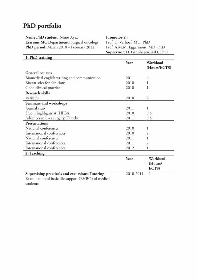
PhD portfolio
Name PhD student: Ninos AyezErasmus MC Department: Surgical oncologyPhD period: March 2010 – February 2012
Promotor(s): Prof. C. Verhoef, MD, PhDProf. A.M.M. Eggermont, MD, PhDSupervisor: D. Grúnhagen, MD, PhD
1. PhD trainingYear Workload
(Hours/ECTS)General courses Biomedical english writing and communicationBiostatistics for cliniciansGood clinical practice
201120102010
411
Research skillsstatistics 2010 2Seminars and workshopsJournal clubDutch highlights at IHPBAAdvances in liver surgery, Utrecht
201120102011
10.50.5
PresentationsNational conferencesInternational conferencesNational conferencesInternational conferencesInternational conferences
20102010201120112012
12121
2. TeachingYear Workload
(Hours/ECTS)
Supervising practicals and excursions, TutoringExamination of basic life support (EHBO) of medical students
2010-2011 1


Curriculum Vitae
176
Ninos werd geboren in Syrie. Op 8-jarige leeftijd kwam hij met zijn ouders naar Nederland. Hij groeide op in Dordrecht en verhuisde vervolgens naar Enschede alwaar hij in 2003 zijn VWO diploma behaalde aan Het Stedelijk Lyceum Zuid. In dat zelfde jaar begon hij ook aan de studie geneeskunde aan de Erasmus Universiteit te Rotterdam. Zijn afstudeeronderzoek deed hij op de afdeling Heelkunde van het Erasmus MC te rotterdam.In 2009 behaalde hij cum laude het artsexamen. Aansluitend werkte hij als assistent geneeskunde niet in opleiding op de afdeling Heelkunde van het Maasstad ziekenhuis te Rotterdam.
In 2010 startte hij met een promotieonderzoek op de afdeling Heelkunde van het Erasmus MC te Rotterdam op de locatie Daniel den Hoed kliniek, onder supervisie van prof.dr. C. Verhoef, met dit proefschrift als resultaat. In juli 2012 startte hij met zijn opleiding Heelkunde van het Erasmus MC (opleiders: Prof.dr. IJzermans en dr. van Wijnhoven) waarvan een groot gedeelte in het Maasstad ziekenhuis te Rotterdam (opleiders: Dr. van der Harst en drs. Klaassen).
Curriculum Vitae 177
Publicatie lijst
1. Angioplasty versus stenting for iliac artery lesions.Bekken J, Jongsma H, Ayez N, Hoogewerf CJ, Van Weel V, Fioole B. Cochrane Database Syst Rev. 2015 May 29
2. The use of neo-adjuvant chemotherapy in patients with resectable colorectal liver metastases: Clinical risk score as possible discriminator.Ayez N, van der Stok EP, Grünhagen DJ, Rothbarth J, van Meerten E, Eggermont AM, Verhoef C. Eur J Surg Oncol. 2015 Jul
3. Neo-adjuvant chemotherapy followed by surgery versus surgery alone in high-risk patients with resectable colorectal liver metastases: the CHARISMA randomized multicenter clinical trial.Ayez N, van der Stok EP, de Wilt H, Radema SA, van Hillegersberg R, Roumen RM, Vreugdenhil G, Tanis PJ, Punt CJ, Dejong CH, Jansen RL, Verheul HM, de Jong KP, Hospers GA, Klaase JM, Legdeur MC, van Meerten E, Eskens FA, van der Meer N, van der Holt B, Verhoef C, Grünhagen DJ. BMC Cancer. 2015 Mar 26
4. Prognostic value of circulating tumour cells for early recurrence after resection of colorectal liver metastases.Lalmahomed ZS, Mostert B, Onstenk W, Kraan J, Ayez N, Gratama JW, Grünhagen D, Verhoef C, Sleijfer S. Br J Cancer. 2015 Feb 3
5. Safety and efficacy of a novel, dry powder fibrin sealant for hemostasis in hepatic resection.Ruitenbeek K, Ayez N, Verhoef C, de Wilt JH, Bottema J, Rijken AM, van Rij M, Koopman J, Zuckerman LA, Frohna P, Porte RJ. Dig Surg. 2014
6. Surgery of the primary tumour in stage IV colorectal cancer with unresectable metastasesNinos Ayez, Wijnand J Alberda, Henk M Verheul, Jacobus W Burger, Johannes H de Wilt, Cornelis Verhoef. European Oncology & Haematology, 2011
7. Long-term results of the ‘liver first’ approach in patients with locally advanced rectal cancer and synchronous liver metastases.Ayez N, Burger JW, van der Pool AE, Eggermont AM, Grunhagen DJ, de Wilt JH, Verhoef C. Dis Colon Rectum. 2013 Mar
178
8. Is restaging with chest and abdominal CT scan after neoadjuvant chemoradiotherapy for locally advanced rectal cancer necessary?Ayez N, Alberda WJ, Burger JW, Eggermont AM, Nuyttens JJ, Dwarkasing RS, Willemssen FE, Verhoef C. Ann Surg Oncol. 2013 Jan
9. Preoperative FDG-PET-scan in patients with resectable colorectal liver metastases does not improve overall survival: a retrospective analyses stratified by clinical risk score.Ayez N, de Ridder J, Wiering B, Oyen WJ, de Wilt JH, Verhoef C. Dig Surg. 2013
10. Fibrin sealant for prevention of resection surface-related complications after liver resection: a randomized controlled trial.de Boer MT, Klaase JM, Verhoef C, van Dam RM, van Gulik TM, Molenaar IQ, Bosscha K, Dejong CH, Van der Jagt EJ, Porte RJ; FRESCO Trial Group (Ayez N)Ann Surg. 2012 Aug
11. The basilic vein and the cephalic vein perform equally in upper arm arteriovenous fistulae.Ayez N, van Houten VA, de Smet AA, van Well AM, Akkersdijk GP, van de Ven PJ, Fioole B. Eur J Vasc Endovasc Surg. 2012 Aug
12. Outcome of microscopic incomplete resection (R1) of colorectal liver metastases in the era of neoadjuvant chemotherapy.Ayez N, Lalmahomed ZS, Eggermont AM, Ijzermans JN, de Jonge J, van Montfort K, Verhoef C. Ann Surg Oncol. 2012 May
13. Body composition and outcome in patients undergoing resection of colorectal liver metastases.van Vledder MG, Levolger S, Ayez N, Verhoef C, Tran TC, Ijzermans JN.Br J Surg. 2012 Apr
14. Is the clinical risk score for patients with colorectal liver metastases still useable in the era of effective neoadjuvant chemotherapy?Ayez N, Lalmahomed ZS, van der Pool AE, Vergouwe Y, van Montfort K, de Jonge J, Eggermont AM, IJzermans JN, Verhoef C. Ann Surg Oncol. 2011 Oct
15. Secondary interventions in patients with autologous arteriovenous fistulas strongly improve patency rates.Ayez N, Fioole B, Aarts RA, van den Dorpel MA, Akkersdijk GP, Dinkelman MK, de Smet AA. J Vasc Surg. 2011 Oct

Curriculum Vitae 179
16. Influence of margins on overall survival after hepatic resection for colorectal metastasis: a meta-analysis.Kok NF, Grünhagen DJ, Ayez N, Verhoef C. Ann Surg. 2011 Aug
17. Anatomical versus nonanatomical resection of colorectal liver metastases: is there a difference in surgical and oncological outcome?Lalmahomed ZS, Ayez N, van der Pool AE, Verheij J, IJzermans JN, Verhoef C.World J Surg. 2011 Mar