surface roughness of the restored enamel after orthodontic ... ·...
TRANSCRIPT

ORIGINAL ARTICLE
Surface roughness of the restored enamel afterorthodontic treatment
Torun Ozer,a Guvenc Basxaran,a and Jalen Devecıoglu Kamab
Diyarbakir, Turkey
Introduction: After fixed appliance treatment, one concern is to restore the enamel surface as closely to itsoriginal state as possible. A variety of cleanup processes are available, but all are time-consuming and carrysome risk of enamel damage. The purpose of this study was to examine tooth surfaces restored with differentcleanup protocols. Methods: Ninety-nine premolars extracted for orthodontic purposes were used. The 2 ma-terials tested were Sof-Lex disks (3 M ESPE AG, Seefeld, Germany) and fiberglass burs (Stain Buster, Carbo-tech, Ganges, France). These were used alone and in combination with high- and low-speed handpieces, withwhich they were also compared. Eight groups were ultimately tested. All groups were compared with intactenamel, which served as the control group. From each group, 10 samples were examined with profilometryand 1 with scanning electron microscopy. Adhesive remnant index scores were recorded to ensure equal dis-tributions for the groups. The time required for the cleanup processes and profilometry test results were alsorecorded. Results: The fastest procedure was performed with high-speed handpieces, followed by low-speed handpieces. Sof-Lex disks and fiberglass burs required more time than carbide burs but did not resultin significantly longer times for the cleanup procedure when combined with tungsten carbide-driven low- orhigh-speed handpieces or when used alone with low-speed handpieces. Although Sof-Lex disks were themost successful for restoring the enamel, it was not necessary to restore the enamel to its original surface con-dition. Generally, all enamel surface-roughness parameters were increased when compared with the values ofintact enamel. The average roughness and maximum roughness depth measurements with Sof-Lex diskswere statistically similar to measurements of intact enamel. Conclusions: No cleanup procedure used inthis study restored the enamel to its original roughness. The most successful was Sof-Lex disks, whichrestored the enamel closer to its original roughness. (Am J Orthod Dentofacial Orthop 2010;137:368-74)
After fixed appliance treatment, the main concernis to restore the enamel surface as closely to itsoriginal state as possible. At bracket removal,
bond failure can occur at the adhesive-enamel interface(adhesive failure) or the adhesive-bracket (cohesivefailure) interface. Cohesive failure is safer than adhesivefailure,1 but, with adhesive failures, less adhesive is lefton the enamel, and less time is spent on cleanup.2 Thepreferred site of failure is controversial. In both cases,cleanup process requires a considerable amount of time.
Adhesive enamel interface failures can lead toenamel loss and depend largely on the bracket materialand the method of debonding. Previous studies reportedundesirable enamel fractures with ceramic brackets.3,4
From the Department of Orthodontics, Faculty of Dentistry, Dicle University,
Diyarbakir, Turkey.aAssociate professor.bProfessor.
The authors report no commercial, proprietary, or financial interest in the prod-
ucts or companies described in this article.
Reprint requests to: Torun Ozer, Dicle University, Dental Faculty, Orthodontics,
Diyarbakir, Turkey; e-mail, [email protected].
Submitted, September 2007; revised and accepted, February 2008.
0889-5406/$36.00
Copyright � 2010 by the American Association of Orthodontists.
doi:10.1016/j.ajodo.2008.02.025
368
Because metal brackets are used routinely, the risk is re-duced. A small amount of enamel fracture might stilloccur because of the micromechanical bond betweenthe composite resin bonding agent and the acid-etchedenamel surface. Therefore, some enamel loss is inevita-ble when bond failure occurs at the adhesive-enamelinterface.5
Residual adhesive on the enamel surface after de-bonding can be removed in various ways, but studieshave shown that some recommended modalities dam-age enamel surfaces.6-8 Campbell9 preferred to use car-bide burs in a high-speed handpiece, followed byEnhance rubber points and cups, a water slurry of finepumice, and, finally, brown and green cups in a low-speed handpiece. Zachrisson and Artun8 suggestedthat enamel loss can be minimized with a tungsten car-bide bur in a low-speed handpiece. Ultrasonic debond-ing units,10 electrothermal debonding devices,11 andlasers12 have also been used to degrade bonding resins.
No universally approved protocol has been estab-lished for this potentially litigious treatment stage.The alterations in the enamel surface caused by rotaryinstruments can be irreversible. However, new commer-cial products might reduce enamel damage. In previous
American Journal of Orthodontics and Dentofacial Orthopedics Ozer, Basxaran, and Kama 369Volume 137, Number 3
studies, assessment of the effectiveness of rotary instru-ments was limited to inspecting the surface under scan-ning electron microscopy (SEM) to see the morphologyof the enamel surface.6-12 However, such investigationsare subjective and cannot be used alone to judge the re-liability of a cleanup protocol.13 Alternative techniquesshould be used, such as profilometry (surface roughnessparameters). Profilometry provides quantitative results,whereas SEM affords a more subjective inspection.Nevertheless, SEM evaluations permit a better under-standing of what happens to the treated enamel. Thesemodalities can be combined to better assess the reliabil-ity of a protocol. In this study, we studied tooth surfacestreated with different cleanup protocols using both pro-filometry and SEM.
MATERIAL AND METHODS
Ninety-nine premolars extracted for orthodonticpurposes, with a maximum storage period of 1 month,were used. They had no carious lesions or microcracksand were kept in distilled water that was changed weeklyto prevent bacterial growth.14 The teeth were embeddedhorizontally in self-cure acrylic resin so that at least2 mm of buccal enamel was exposed. The buccalenamel surfaces of the teeth were pumiced, washed for30 seconds, and dried for 10 seconds with a moisture-free air spray. The 90 teeth to be debonded and undergoprofilometry testing were assigned to 1 of 9 groupsrandomly. The remaining 9 teeth to be prepared forSEM analysis were not embedded in acrylic resin. Tenspecimens for profilometry testing and 1 for SEMevaluation were obtained from each group. Eleven teethwere not bonded to permit comparison with the originalenamel surface. After the teeth had been debondedwith hand pliers by gently squeezing the bracket wings,the adhesive remnant index (ARI) scores were alsorecorded. In this study, Sof-Lex disks (3M ESPE AG,Seefeld, Germany) were compared with fiberglass burs(Stain Buster, Carbotech, Ganges, France). These mate-rials can be used separately or combined with a tungstencarbide bur driven on a high- or low-speed handpiece.
The groups studied were as follows: group A, tung-sten carbide bur with a high-speed handpiece (aerator)(ATC); group B, tungsten carbide bur with a low-speedhandpiece (micromotor) (MTC); group C, tungsten car-bide bur with a high-speed handpiece (aerator) and Sof-Lex disks (ATC 1 SL); group D, tungsten carbide burwith a low-speed handpiece (micromotor) and Sof-Lex disks (MTC 1 SL); group E, Sof-Lex disks alone(SL); group F, tungsten carbide bur with a high-speedhandpiece (aerator) and fiberglass bur (ATC 1 FB);group G, tungsten carbide bur with a low-speed hand-
piece (micromotor) and fiberglass bur (MTC 1 FB);group H, fiberglass bur (FB); and group I, intact enamel.
The following test procedure was used. After thebuccal enamel surfaces of the teeth had been pumiced,they were kept in distilled water and bonded on thesame day. The enamel was etched with 37% gel phos-phoric acid for 15 seconds, rinsed with a water and airspray for 15 seconds, and dried for another 15 seconds.The etched enamels had a uniform dull, frosty appear-ance. After the etching, stainless steel standard edge-wise premolar brackets (GAC, Central Islip, NewYork, NY) were bonded. A thin, uniform coating of ad-hesive agent was applied to the etched surfaces. Afterapplying the bonding material (Transbond XT, 3M Uni-tek, Monrovia, Calif), the bracket was placed on thetooth surface, adjusted to its final position, and pressedfirmly in place. Excess sealant and adhesive wereremoved from the periphery of the bracket base tokeep each bond area uniform. Each side of the tooth(mesial, distal, occlusal, and gingival) was light-curedfor 10 seconds, for a total of 40 seconds.
After storing the specimens in water at 37�C for24 hours, thermocycling was performed for 500 cyclesat 5� to 55�C with a dwell time of 30 seconds. The teethwere debonded wioth hand pliers. After debonding, theteeth and brackets were examined under 10 times mag-nification to evaluate the amount of resin remaining onthe tooth. The ARI of Artun and Bergland15 was used todefine the quantity of resin remaining on the tooth sur-faces. The ARI scores ranged from 0 to 3: 0, no adhesiveremaining on the tooth; 1, less than half of the enamelbonding site covered with adhesive; 2, more than halfof the enamel bonding site covered with adhesive; and3, the enamel bonding site entirely covered with adhe-sive. The first author (T.O.) performed all tests, includ-ing bonding, debonding, and adhesive removal. Visualinspection of the enamel surfaces was useful in the resinremoval process. The remaining adhesive was cleanedup by using the technique for the respective group.The times required for all groups were also recorded.The cleaned enamel surfaces were subjected to a testwith a profilometer (Surtronic, Taylor & Hobson,Leicester, United Kingdom) (Fig 1) operating undera 0.5-mm maximum length. The profilometer has a tipthat is placed on the enamel surface and scans the sur-face to measure the surface roughness. Two recordingswere made in contact with the enamel for each speci-men, and the mean values were recorded. Three surfaceroughness measurements were recorded. Averageroughness (Ra) indicates the overall roughness of theenamel surface. It is the arithmetic mean of all absolutedistances of the surface roughness from the center linewithin the measuring length. Root mean square

Fig 1. The profilometer used in the study.
Table I. Adhesive remnant index (ARI) score distribu-tion for the groups
Group 0 1 2 3
ATC 1 1 6 2
MTC 1 0 7 2
ATC 1 SL 1 0 9 0
MTC 1 SL 0 3 6 1
SL 0 1 8 1
ATC 1 FB 0 0 9 1
MTC 1 FB 1 2 6 1
FB 2 0 7 1
There were no statistically significant differences between the groups.
Table II. Required times in seconds for cleanup
Group Mean SD
ATC A 6.22 1.08
MTC B 13.02 2.96
ATC 1 SL C 25.76 4.03
MTC 1 SL C 30.82 5.68
SL C 24.63 6.22
ATC 1 FB C 26.19 3.78
MTC 1 FB C 31.64 4.57
FB C 23.62 4.24
There were no statistically significant differences with the same letters
370 Ozer, Basxaran, and Kama American Journal of Orthodontics and Dentofacial Orthopedics
March 2010
roughness (Rq) describes the height distribution relativeto the mean line. Maximum roughness depth (Rt) re-flects isolated features on the enamel surface.
The ARI scores were evaluated with the chi-squaretest. The time required for cleanup and the surfaceroughness parameters for each group were analyzed sta-tistically by using analysis of variance (ANOVA). TheTukey multiple comparison test was used to comparedifferences within the groups.
at the 0.05 significance level.
RESULTS
Table I shows the distribution of the ARI scores ofthe groups. No statistically significant difference wasfound between the groups. The distributions were simi-lar; this is essential for accurate comparison.
Table II shows the times required for cleanup. Be-cause the micromotor is a lower-speed handpiece thanthe aerator, the procedures involving the micromotortook longer. The fastest procedure was performed by us-ing ATC. Sof-Lex disks and fiberglass burs took signif-icantly longer for the cleanup procedures, eithercombined with a tungsten carbide bur driven bya high- or low-speed handpiece or with low-speed hand-pieces alone.
Table III lists the enamel surface roughness param-eters. Generally, all parameters increased when com-pared with the values for intact enamel. This meansthat no cleanup procedure used in the study restoredthe enamel to its original roughness.
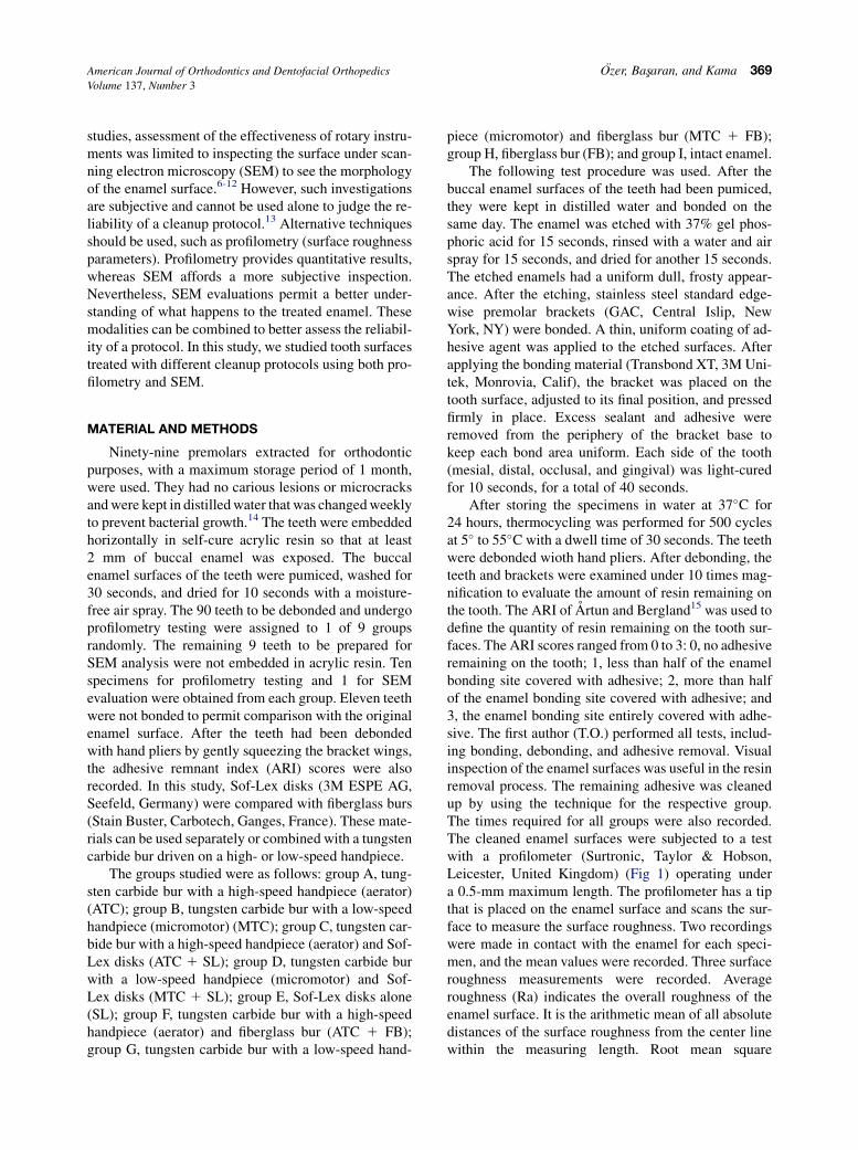
Figures 2 through 10 show images of groups Athrough I, respectively. The images are not intendedfor objective comparison but to better demonstratewhat happens to the enamel. All SEM evaluationswere performed under 750 times magnification for stan-dardization purposes. The enamel in Figure 2 hasa scratched surface with deep grooves. Figure 3 repre-
sents the worst case, with a punctured, scratched, andscarred surface with many protuberances and grooves.Figure 4 shows a smooth, scratched surface. Figures 5and 7 also show scarred surfaces, whereas Figure 6shows a more even surface than the others. Figures 8and 9 are similar and show some scarring, but thiswas shallower than that in Figures 2, 5, and 7. Figure 6shows the most even roughened surface, followed byFigures 4, 8, 9, 7, 5, 2, and 3, in that order.
DISCUSSION
The concern over debonding and inducing enamelsurface alterations arises from the importance of the up-permost layer of the enamel, the hardness and highermineral and fluoride content of which are particularlyimportant. The loss of surface enamel and associatedexposure of enamel prism endings to the oral environ-ment can result in decreased resistance of the enamelto the organic acids in plaque and make it more proneto decalcification.16 The microscopic appearanceshowed enamel scarring. Nonetheless, enamel scarringafter debonding is inevitable. However, the advantagesof bonding orthodontic attachments unquestionablyoutweigh the disadvantages. The clinician must accept

Table III. Profilometry test results in mm for each cleanup protocol
Ra Rq Rt
Group Mean 6 SD Maximum Minimum Mean 6 SD Maximum Minimum Mean 6 SD Maximum Minimum
ATC A 1.75 6 1.02 2.86 0.69 A 1.92 6 1.28 3.23 0.63 A 8.23 6 2.77 11.47 5.03
MTC A 2.03 6 0.68 2.77 1.12 A 2.14 6 1.17 3.71 0.82 A 7.54 6 3.42 11.43 3.94
ATC 1 SL B 0.74 6 0.34 1.25 0.39 B 0.96 6 0.57 1.65 0.35 B 4.56 6 2.13 6.82 2.07
MTC 1 SL B 0.82 6 0.23 1.18 0.58 B 1.34 6 0.74 2.12 0.52 A 6.93 6 3.76 10.73 3.05
SL C 0.50 6 0.22 1.04 0.26 B 1.46 6 0.28 1.83 0.37 C 2.42 6 1.89 4.41 0.39
ATC 1 FB B 1.23 6 0.56 1.83 0.62 B 1.67 6 0.77 2.49 0.68 B 5.54 6 2.41 7.98 1.96
MTC 1 FB B 1.36 6 0.38 1.84 0.91 B 1.75 6 0.64 2.44 1.03 B 5.34 6 3.72 9.34 1.58
FB B 1.04 6 0.79 1.86 0.22 B 1.28 6 0.68 2.31 0.45 B 4.67 6 1.17 5.92 3.27
Intact enamel C 0.40 6 0.19 0.81 0.19 C 0.62 6 0.34 1.04 0.23 C 2.04 6 1.16 3.34 0.76
There were no statistically significant differences with the same letters at the 0.05 significance level.
Fig 2. SEM examination of the enamel surface restoredby using a tungsten carbide bur with a high-speed hand-piece (ATC).
Fig 3. SEM examination of the enamel surface restoredby using a tungsten carbide bur with a low-speed hand-piece (micromotor) (MTC).
American Journal of Orthodontics and Dentofacial Orthopedics Ozer, Basxaran, and Kama 371Volume 137, Number 3
some enamel scarring after orthodontic therapy and beaware of the best methods for restoring the enamel toits original condition.9,17-21
We tested different materials and procedures. Tung-sten carbide burs in low- and high-speed handpieceswere used alone and with various polishing materials,and all combinations were compared with intactenamel. The time spent removing adhesives from theenamel and the resultant enamel after the cleanup de-pends largely on the amount of remnant adhesive. Inthis study, the ARI scores for the experimental groupswere broadly similar. SEM evaluation of an enamel sur-face shows its topography only; it is not quantitative andcannot be used for comparative purposes. Profilometricexamination of a surface permits an objective determi-nation, and profilometry was the main testing instru-ment used in this study, although the SEM evaluation
helped us to make a visual comparison of the cleanupprocedures. Another problem commonly associatedwith cleanup is heat. Water and air cooling, used withhigh- and low-speed instruments, were not tested inour study. Retief and Denys7 reported that adequateair cooling can be used instead of water cooling, al-though other researchers insisted on water-coolingwith high-speed handpieces.9,17 In this study, watercooling was preferred with high-speed handpieces,whereas air cooling was used with low-speed handpie-ces at 10,000 rpm.
Less time was spent with ATC than with the othersystems, but the SEM images and surface roughnessmeasurements demonstrated that ATC is more harmfulto the enamel compared with other combined methods.MTC proved to be unsuitable for adhesive removal.ATC and MTC cannot be used alone in the debonding
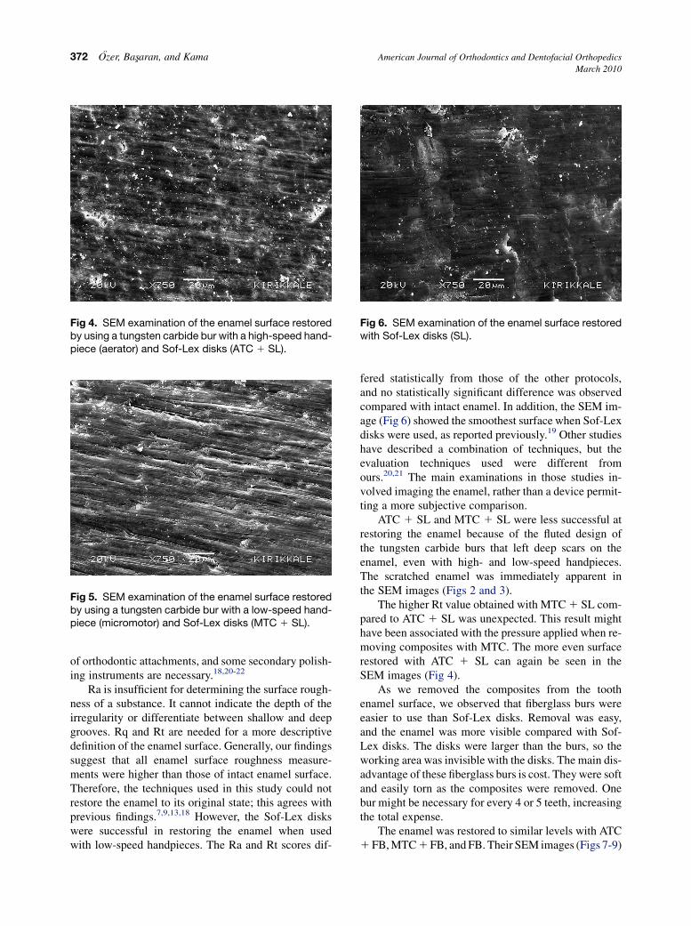
Fig 4. SEM examination of the enamel surface restoredby using a tungsten carbide bur with a high-speed hand-piece (aerator) and Sof-Lex disks (ATC 1 SL).
Fig 5. SEM examination of the enamel surface restoredby using a tungsten carbide bur with a low-speed hand-piece (micromotor) and Sof-Lex disks (MTC 1 SL).
Fig 6. SEM examination of the enamel surface restoredwith Sof-Lex disks (SL).
372 Ozer, Basxaran, and Kama American Journal of Orthodontics and Dentofacial Orthopedics
March 2010
of orthodontic attachments, and some secondary polish-ing instruments are necessary.18,20-22
Ra is insufficient for determining the surface rough-ness of a substance. It cannot indicate the depth of theirregularity or differentiate between shallow and deepgrooves. Rq and Rt are needed for a more descriptivedefinition of the enamel surface. Generally, our findingssuggest that all enamel surface roughness measure-ments were higher than those of intact enamel surface.Therefore, the techniques used in this study could notrestore the enamel to its original state; this agrees withprevious findings.7,9,13,18 However, the Sof-Lex diskswere successful in restoring the enamel when usedwith low-speed handpieces. The Ra and Rt scores dif-
fered statistically from those of the other protocols,and no statistically significant difference was observedcompared with intact enamel. In addition, the SEM im-age (Fig 6) showed the smoothest surface when Sof-Lexdisks were used, as reported previously.19 Other studieshave described a combination of techniques, but theevaluation techniques used were different fromours.20,21 The main examinations in those studies in-volved imaging the enamel, rather than a device permit-ting a more subjective comparison.
ATC 1 SL and MTC 1 SL were less successful atrestoring the enamel because of the fluted design ofthe tungsten carbide burs that left deep scars on theenamel, even with high- and low-speed handpieces.The scratched enamel was immediately apparent inthe SEM images (Figs 2 and 3).
The higher Rt value obtained with MTC 1 SL com-pared to ATC 1 SL was unexpected. This result mighthave been associated with the pressure applied when re-moving composites with MTC. The more even surfacerestored with ATC 1 SL can again be seen in theSEM images (Fig 4).
As we removed the composites from the toothenamel surface, we observed that fiberglass burs wereeasier to use than Sof-Lex disks. Removal was easy,and the enamel was more visible compared with Sof-Lex disks. The disks were larger than the burs, so theworking area was invisible with the disks. The main dis-advantage of these fiberglass burs is cost. They were softand easily torn as the composites were removed. Onebur might be necessary for every 4 or 5 teeth, increasingthe total expense.
The enamel was restored to similar levels with ATC1 FB, MTC 1 FB, and FB. Their SEM images (Figs 7-9)
Fig 8. SEM examination of the enamel surface restoredby using a tungsten carbide bur with a low-speed hand-piece (micromotor) and a fiberglass bur (MTC 1 FB).
Fig 9. SEM examination of the enamel surface restoredby using a fiberglass bur (FB).
Fig 10. SEM examination of intact enamel.
Fig 7. SEM examination of the enamel surface restoredby using a tungsten carbide bur with a high-speed hand-piece (aerator) and a fiberglass bur (ATC 1 FB).
American Journal of Orthodontics and Dentofacial Orthopedics Ozer, Basxaran, and Kama 373Volume 137, Number 3
and surface roughness measurements were also similar.Instead of FB, ATC 1 FB or MTC 1 FB might be pref-erable to reduce costs.
No statistically significant differences were ob-served between the times spent with Sof-Lex disksand fiberglass burs, although the procedure was fasterwith ATC and MTC. This would be of no great clinicalimportance, however, because Sof-Lex disks, whenused alone, provide more even enamel surfaces thanthe other techniques.
CONCLUSIONS
Our goal was to restore the enamel to its original stateafter orthodontic treatment. The methods tested in this
study could not restore the original enamel surface, butthey were close to the values of intact enamel. Sof-Lexdisk performance was superior to that of other combinedprotocols or fiberglass burs. Sof-Lex disks restored theenamel the closest to the original enamel surface.
REFERENCES
1. Bishara SE, VonWald L, Laffoon JF, Warren JJ. Effect of a self-
etch primer/adhesive on shear bond strength of orthodontic
brackets. Am J Orthod Dentofacial Orthop 2001;119:621-4.
2. Bishara SE, Gordon VV, VonWald L, Jacobsen JR. Shear bond
strength of composite, glass ionomer, and acidic primer adhesive
sytems. Am J Orthod Dentofacial Orthop 1999;115:24-8.
3. Joseph VP, Rossouw PE. The shear bond strengths of stainless
steel orthodontic brackets bonded to teeth with orthodontic com-
posite resin and various fissure sealants. Am J Orthod Dentofacial
Orthop 1990;98:66-71.

374 Ozer, Basxaran, and Kama American Journal of Orthodontics and Dentofacial Orthopedics
March 2010
4. Jeroudi MT. Enamel fracture caused by ceramic brackets. Am J
Orthod Dentofacial Orthop 1991;99:97-9.
5. Redd TB, Shivapura PK. Debonding ceramic brackets: effects on
enamel. J Clin Orthod 1991;25:475-81.
6. Gwinnett AJ, Gorelick L. Microscopic evaluation of enamel after
debonding. Am J Orthod 1977;71:651-65.
7. Retief DH, Denys FR. Finishing of enamel surfaces after de-
bonding of orthodontic attachments. Angle Orthod 1979;49:
1-10.
8. Zachrisson BU, Artun J. Enamel surface appearance after various
debonding techniques. Am J Orthod 1979;75:121-37.
9. Campbell P. Enamel surfaces after orthodontic bracket debond-
ing. Angle Orthod 1995;65:103-10.
10. Bishara SE, Trulove TS. Comparisons of different debonding
techniques for ceramic brackets: an in-vitro study. Part 1. Back-
ground and methods. Am J Orthod Dentofacial Orthop 1990;98:
145-53.
11. Brouns E, Schopf P, Kojancic B. Electrothermal debracketing
of ceramic brackets: an in vitro study. Eur J Orthod 1993;15:
115-23.
12. Strobl K, Bahns TL, Willham L, Bishara SE, Stwalley WC. Laser-
aided debonding of orthodontic ceramic brackets. Am J Orthod
Dentofacial Orthop 1992;101:152-8.
13. Eliades T, Gioka C, Eliades G, Makou M. Enamel surface rough-
ness following debonding using two resin grinding methods. Eur J
Orthod 2004;26:333-8.
14. Von Fraunhofer JA, Allen DJ, Orbell GM. Laser etching of enamel
for direct bonding. Angle Orthod 1993;63:73-6.
15. Artun J, Bergland S. Clinical trials with crystal growth condition-
ing as an alternative to acid-etch enamel pretreatment. Am J
Orthod 1984;85:333-40.
16. Øgaard B. Oral microbiological changes, long term enamel alter-
ations due to decalcification and caries prophylactic aspects. In:
Brantley WA, Eliades T, editors. Orthodontic materials:
scientific and clinical aspects. Stuttgart, Germany: Thieme;
2001. p. 124-39.
17. Rouleau BD Jr, Marshall GW Jr, Cooley RO. Enamel surface eval-
uations after clinical treatment and removal of orthodontic
brackets. Am J Orthod 1982;81:423-6.
18. Eliades T, Kakaboura A, Eliades G, Brantley TG. Comparison of
enamel colour changes associated with orthodontic bonding using
two different adhesive. Eur J Orthod 2001;23:85-90.
19. Zarinnia K, Eid NM, Kehoe MJ. The effect of different debonding
techniques on the enamel surface: an in vitro qualitative study.
Am J Orthod Dentofacial Orthop 1995;108:284-93.
20. Hong YH, Lew KKK. Quantitative and qualitative assessment of
enamel surface following five composite removal methods after
bracket debonding. Eur J Orthod 1995;17:121-8.
21. Bishara SE, Trulove TS. Comparisons of different debonding
techniques for ceramic brackets: an in vitro study. Part II. Findings
and clinical implications. Am J Orthod Dentofacial Orthop 1990;
98:263-73.
22. Krell KV, Courey JM, Bishara SE. Orthodontic bracket removal
using conventional and ultrasonic debonding techniques, enamel
loss, and time requirements. Am J Orthod Dentofacial Orthop
1993;103:258-66.