surface-induced dissociation of homotetramers with d2 ... · chemistry & biology article...
TRANSCRIPT

Article
Surface-Induced Dissociation of Homotetramers
with D2 Symmetry Yields their Assembly Pathwaysand Characterizes the Effect of Ligand BindingGraphical Abstract
Highlights
d Surface collision of D2 tetramers yields dimers that further
fragment to monomers
d Collision energy needed for dissociation tracks with dimer-
dimer interface area
d Location of ligand binding sites affects tetramer dissociation
behavior
Quintyn et al., 2015, Chemistry & Biology 22, 583–592May 21, 2015 ª2015 Elsevier Ltd All rights reservedhttp://dx.doi.org/10.1016/j.chembiol.2015.03.019
Authors
Royston S. Quintyn, Jing Yan, Vicki H.
Wysocki
In Brief
Gas-phase dissociation pathways
observed for homotetramers upon
collision with a surface depend on the
interface area and thus give insight into
their solution-phase assembly pathways.
Quintyn et al. apply the approach to
protein complexes with ligands bound
and confirm that changes in
conformational flexibility and tetramer
stability depend on ligand binding
location.

Chemistry & Biology
Article
Surface-Induced Dissociation of Homotetramers withD2 Symmetry Yields their Assembly Pathways andCharacterizes the Effect of Ligand BindingRoyston S. Quintyn,1,2 Jing Yan,1,2 and Vicki H. Wysocki1,*1Department of Chemistry and Biochemistry, Ohio State University, Columbus, OH 43210, USA2Co-first author
*Correspondence: [email protected]://dx.doi.org/10.1016/j.chembiol.2015.03.019
SUMMARY
Understanding of protein complex assembly andthe effect of ligand binding on their native topologiesis integral to discerning how alterations in their archi-tecture can affect function. Probing the disassemblypathway may offer insight into the mechanismsthrough which various subunits self-assemble intocomplexes. Here, a gas-phase dissociation method,surface-induced dissociation (SID) coupled with ionmobility (IM), was utilized to determine whetherdisassembly pathways are consistent with the as-sembly of three homotetramers and to probe the ef-fects of ligand binding on conformational flexibilityand tetramer stability. The results indicate that thesmaller interface in the complex is initially cleavedupon dissociation, conserving the larger interface,and suggest that assembly of a D2 homotetramerfrom its constituent monomers occurs via a C2
dimer intermediate. In addition, we demonstratethat ligand-mediated changes in tetramer SID disso-ciation behavior are dependent on where and howthe ligand binds.
INTRODUCTION
The majority of proteins exist in vivo as oligomers with varying
quaternary structural attributes, rather than as individual chains
(Shen and Chou, 2009). Approximately 50%–70% of oligomers
with known quaternary structure are homomers, which are
formed by the assembly of identical protein subunits (Goodsell
and Olson, 2000; Levy et al., 2006). These homomers often
adopt highly symmetrical structures, with most having either cy-
clic (Cn) or dihedral (Dn) symmetry (Levy et al., 2008). Homome-
rization and the evolutionary selection of symmetry is driven by
functional, genetic, and physicochemical needs (Goodsell and
Olson, 2000). For example, self-assembly of the p53 tumor sup-
pressor protein into a homotetramer with D2 symmetry enables it
to bind variably spaced DNA target sequences, essential for p53
transactivation and tumor suppressor functions (Okorokov et al.,
2006). The misfolding and subsequent misassembly of homo-
mers into structures that deviate from their native functional
Chemistry & Biology 22,
conformational state is amajor contributing factor in a wide array
of human diseases (Kelly, 1997; Ng et al., 2012; Wetzel, 1996).
Consequently, developing an understanding of how different
subunits interact to form functional protein complexes is key to
gaining insight into how these homomers carry out their normal
biological functions and how alterations in their architecture
lead to malfunction.
In recent years, native mass spectrometry (MS) coupled with
ion mobility (IM) has emerged as a powerful tool for probing
the assembly of protein complexes, as it is capable of simulta-
neously identifying the various subcomplexes produced after
solution disruption of subunit interfaces within oligomers (Her-
nandez and Robinson, 2007; Levy et al., 2008). The perturbation
of subunit interfaces can occur in solution by changing the ionic
strength (Kapur et al., 2002; Kershaw et al., 2002), pH (Light-
Wahl et al., 1994), or adding organic solvents (Gupta et al., 2004).
The dissociation of oligomers can also occur in the gas phase;
collision-induced dissociation (CID) is the most popular method
available in commercial mass spectrometers. In CID, however,
the precursor ion undergoes multiple low-energy collisions with
an inert low-mass gas, which typically produces fragmentation
patterns that are not representative of the oligomer substructure
(an unfolded monomer and (n � 1) multimer), and thus provides
limited information on the assembly of these oligomers (Benesch
et al., 2006). Alternatively, there is substantial evidence illus-
trating that fragmentation is more reflective of multimer architec-
ture in surface-induced dissociation (SID) (Blackwell et al., 2011;
Zhou and Wysocki, 2014), where ions undergo a collision with
a surface target often composed of a fluorinated alkanethiol
self-assembled monolayer (Jones et al., 2006). SID is a fast,
high-energy deposition process (Blackwell et al., 2011), and
thus allows for better characterization of the oligomer assembly,
because faster structurally informative dissociation pathways
can outcompete the slower monomer unfolding (rearrangement)
dissociation pathway observed in CID (Zhou et al., 2013).
Although previous studies utilizing solution disruption followed
by MS detection demonstrated that the assembly/evolutionary
pathway of an oligomer is the reverse of the dissociation process
(Levy et al., 2008), the utilization of SID as a direct means of
discerning the assembly pathway of oligomers has not been sys-
tematically investigated.
A statistical distribution of the various types of quaternary
structures that existed in the Swiss-Prot databank in April,
2008, revealed that the majority of oligomers exist either as
dimers or tetramers (Shen and Chou, 2009). Moreover, dihedral
583–592, May 21, 2015 ª2015 Elsevier Ltd All rights reserved 583
homomers are over ten times more abundant than cyclic homo-
mers with the same numbers of subunits (Levy et al., 2008). A
search of the PDB reveals that the majority of homomers with
crystal structures are either dimers or have dihedral symmetry
(Marsh and Teichmann, 2014). Because our research focuses
on self-assembly, we chose homotetramers with D2 symmetry
as model systems for our study. In the present study, we utilize
three D2 homotetramers (transthyretin, streptavidin, and neutra-
vidin, the deglycosylated form of avidin) as model systems. We
determine whether the assembly pathway inferred from SID
dissociation (disassembly) is similar to that previously described
for D2 tetramers where monomers / C2 dimers / D2 tetramer
(Villar et al., 2009).
One of the many functional benefits associated with the self-
assembly of monomers into oligomers is that it can occasionally
facilitate the formation of active sites and ligand binding sites at
homomeric interfaces (Marsh and Teichmann, 2014). This is the
case for transthyretin (TTR), whose function as a thyroid hor-
mone (T4, T3) transporter is dependent on the assembly into
the tetramer, because the ligand binding sites are located at
the dimer-dimer interface (Nencetti and Orlandini, 2012). The
main biological function of both streptavidin and avidin is as an
antibiotic, through mediation by their interaction with biotin
(Green, 1990; Weber et al., 1989). While only a single residue
(Trp 120 in streptavidin and Trp 110 in avidin) from onemonomer
makes contact with biotin bound in an adjacent monomer
through the dimer-dimer interface in the streptavidin tetramer,
previous studies demonstrate that mutation of this residue re-
sults in a significant decrease in biotin affinity (Sano and Cantor,
1995).
The presence of low-stability regions within a protein complex
appears to provide a mechanism for eliciting high ligand binding
affinity (Luque et al., 2002), with binding often triggering changes
in protein structure and conformational flexibility that correlate to
changes in thermal stability (Celej et al., 2003). By using CID
coupled with IM-MS, Hyung et al. (2009) demonstrated that
monitoring the unfolding and dissociation of the TTR tetramer al-
lows for the investigation of the stability differences between
wild-type and disease-associated variants of TTR not normally
observed by MS alone. Studies conducted in the Wysocki labo-
ratory have demonstrated that protein subunits produced by SID
of ligand bound complexes such as tryptophan synthase (Quin-
tyn et al., 2013) and C-reactive protein (our unpublished data)
retain their ligands, indicating retention of protein fold or at least
retention of the ligand binding pocket. Hence, the second part of
this research focuses on the application of SID to characterize
the changes in stability and conformational flexibility induced
by binding of T4 (TTR) and biotin (streptavidin and neutravidin).
RESULTS
Predicting Assembly Pathways of Homotetramers withD2 Symmetry by Surface-Induced DissociationPrevious studies reported in the literature showed that the
largest interfaces are formed first during homomer assembly,
and thus the assembly of homomers can be predicted by using
a model in which the interface strength is assumed to be propor-
tional to the interface area calculated from crystal structures
(Chen et al., 2013; Levy et al., 2008). Considering that SID yields
584 Chemistry & Biology 22, 583–592, May 21, 2015 ª2015 Elsevier
fragmentation more reflective of multimer architecture, we were
interested in determining whether this interface model could
be applied to the SID dissociation of the D2 homotetramers. To
test this model, we first calculated the interface areas from the
crystal structures of three homotetramers, streptavidin, neutravi-
din, and TTR (Figures 1A, 1D, and 1G), using PISA analysis (Kris-
sinel and Henrick, 2007). As shown in Table 1, SID of the three
tetramers can conceivably yield primary noncovalent cleavage
of different interfaces to produce a variety of products, with
dimer formation favored over other products.
The SID dissociation of the charge-reduced +11 streptavidin
tetramer at 330 eV (Figure 1B) yields primarily dimer. However,
it is apparent from Table 1 that there are three possible types
of dimers that can be produced from the cleavage of different
interfaces by SID. Based on the large differences in the total
interface area cleaved, cleavage to form I-IV and II-III dimers is
expected. Using the drift times obtained from IM, it was deter-
mined that the experimental collision cross section (CCS) of
the observed dimer is consistent with the calculated CCS of
the dimer expected from cleavage of the lowest total interface
area (1176 A2), assuming no collapse before IM measurement
(Figure 2A). Once we established that low-energy SID results in
cleavage of a dimer-dimer interface, we subsequently employed
higher SID collision energies to determine whether we could
observe fragments that represent cleavage of larger total inter-
face area. The increase in the intensity of themonomers at higher
SID collision energies (Figure 1B, 770 eV and 1650 eV) may sug-
gest subsequent secondary cleavage of themonomer-monomer
interface (1551 A2) within the initially formed dimer. However, the
presence of monomer and trimer fragments at the onset for
monomer fragmentation also suggests cleavage of all three
interface types to yield monomer and its complementary trimer
(requiring cleavage of a total interface area of 2139 A2). The
fact that this pathway is observed after dimer-dimer cleavage
is consistent with cleavage of the lowest total interface area
(1176 A2) first to form dimer at lower energy followed by cleavage
of the next lowest total interface area (2139 A2). In addition,
another alternative pathway may be the direct dissociation of
tetramer to four monomers but that would require cleavage of
four interfaces with a total interface area of 4278 A2. Using IM,
it is possible to deduce the relative intensities of the different
subunits present at each collision energy, e.g. the IM-MS plot
shown in Figure S1A illustrates that the relative abundance of
each individual subunit present can be determined by summing
the intensities of highlighted regions separately. Therefore, to
resolve the pathways responsible for the increase in the mono-
mer intensity at higher energies, we plotted the relative abun-
dance of the various subunits produced over a range of SID
collision energies. These plots are typically referred to as en-
ergy-resolved mass spectrometry (ERMS) plots. From the SID-
ERMS plot generated for streptavidin (Figure 3A), not only is it
evident that the streptavidin tetramer does not fragment directly
to four monomers, because monomer onset is accompanied by
trimer onset, but it is clear that monomer intensity increases as
the dimer intensity decreases, which suggests secondary cleav-
age of the monomer-monomer interface within the dimer at
higher SID collision energies. Furthermore, the various charge
states of the SID products were monitored over a range of colli-
sion energies to determine the pathway responsible for the
Ltd All rights reserved
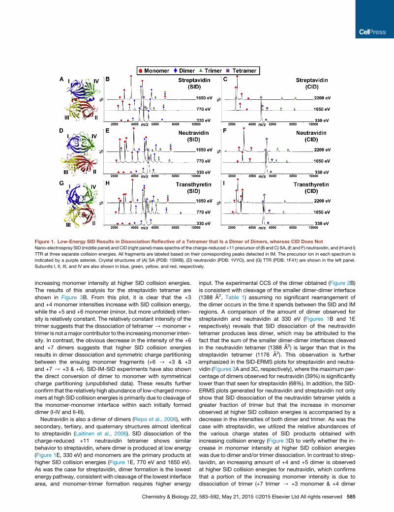
Figure 1. Low-Energy SID Results in Dissociation Reflective of a Tetramer that Is a Dimer of Dimers, whereas CID Does Not
Nano-electrospray SID (middle panel) and CID (right panel) mass spectra of the charge-reduced +11 precursor of (B and C) SA, (E and F) neutravidin, and (H and I)
TTR at three separate collision energies. All fragments are labeled based on their corresponding peaks detected in IM. The precursor ion in each spectrum is
indicated by a purple asterisk. Crystal structures of (A) SA (PDB: 1SWB), (D) neutravidin (PDB: 1VYO), and (G) TTR (PDB: 1F41) are shown in the left panel.
Subunits I, II, III, and IV are also shown in blue, green, yellow, and red, respectively.
increasing monomer intensity at higher SID collision energies.
The results of this analysis for the streptavidin tetramer are
shown in Figure 3B. From this plot, it is clear that the +3
and +4 monomer intensities increase with SID collision energy,
while the +5 and +6 monomer (minor, but more unfolded) inten-
sity is relatively constant. The relatively constant intensity of the
trimer suggests that the dissociation of tetramer / monomer +
trimer is not amajor contributor to the increasingmonomer inten-
sity. In contrast, the obvious decrease in the intensity of the +6
and +7 dimers suggests that higher SID collision energies
results in dimer dissociation and symmetric charge partitioning
between the ensuing monomer fragments (+6 / +3 & +3
and +7 / +3 & +4). SID-IM-SID experiments have also shown
the direct conversion of dimer to monomer with symmetrical
charge partitioning (unpublished data). These results further
confirm that the relatively high abundance of low-chargedmono-
mers at high SID collision energies is primarily due to cleavage of
the monomer-monomer interface within each initially formed
dimer (I-IV and II-III).
Neutravidin is also a dimer of dimers (Repo et al., 2006), with
secondary, tertiary, and quaternary structures almost identical
to streptavidin (Laitinen et al., 2006). SID dissociation of the
charge-reduced +11 neutravidin tetramer shows similar
behavior to streptavidin, where dimer is produced at low energy
(Figure 1E, 330 eV) and monomers are the primary products at
higher SID collision energies (Figure 1E, 770 eV and 1650 eV).
As was the case for streptavidin, dimer formation is the lowest
energy pathway, consistent with cleavage of the lowest interface
area, and monomer-trimer formation requires higher energy
Chemistry & Biology 22,
input. The experimental CCS of the dimer obtained (Figure 2B)
is consistent with cleavage of the smaller dimer-dimer interface
(1388 A2, Table 1) assuming no significant rearrangement of
the dimer occurs in the time it spends between the SID and IM
regions. A comparison of the amount of dimer observed for
streptavidin and neutravidin at 330 eV (Figures 1B and 1E
respectively) reveals that SID dissociation of the neutravidin
tetramer produces less dimer, which may be attributed to the
fact that the sum of the smaller dimer-dimer interfaces cleaved
in the neutravidin tetramer (1388 A2) is larger than that in the
streptavidin tetramer (1176 A2). This observation is further
emphasized in the SID-ERMS plots for streptavidin and neutra-
vidin (Figures 3A and 3C, respectively), where the maximum per-
centage of dimers observed for neutravidin (39%) is significantly
lower than that seen for streptavidin (68%). In addition, the SID-
ERMS plots generated for neutravidin and streptavidin not only
show that SID dissociation of the neutravidin tetramer yields a
greater fraction of trimer but that the increase in monomer
observed at higher SID collision energies is accompanied by a
decrease in the intensities of both dimer and trimer. As was the
case with streptavidin, we utilized the relative abundances of
the various charge states of SID products obtained with
increasing collision energy (Figure 3D) to verify whether the in-
crease in monomer intensity at higher SID collision energies
was due to dimer and/or trimer dissociation. In contrast to strep-
tavidin, an increasing amount of +4 and +5 dimer is observed
at higher SID collision energies for neutravidin, which confirms
that a portion of the increasing monomer intensity is due to
dissociation of trimer (+7 trimer / +3 monomer & +4 dimer
583–592, May 21, 2015 ª2015 Elsevier Ltd All rights reserved 585

Table 1. Interface Areas between Subunits and Dissociation
Fragments
Individual interface area (A2)
SA NA TTR
415 550 320
173 144 379
1551 1889 874
Fragments from tetramer
dissociation
Interface area cleaveda (A2)
SA NA TTR
1176 1388 1398
2139 2583 1573
3448 4066 2506
3932 4878 2388
4278 5166 3146
aInterface area cleaved is the sum of the interface areas involved in the
cleavage of tetramer to produce fragments. For example, the interface
area cleaved to produce transthyretin monomer and trimer is 874 A2 +
379 A2 + 320 A2. Note that in production of monomer and trimer, I, II,
III, or IV can be dissociated from tetramer with the same amount of inter-
face area cleaved (i.e. there are four equivalent pathways available).
and +8 trimer / +3 monomer & +5 dimer). However, as these
were only minor changes, it is not the main source of monomers.
Instead, the increase inmonomer at higher SID collision energies
is primarily due to dissociation of the +6 and +7 dimer (+6 / +3
& +3 and +7/ +3 & +4). Hence, like streptavidin, higher energy
SID of the neutravidin tetramer results in secondary cleavage of
the monomer-monomer interface (1889 A2) within each I-IV/II-III
dimer to produce monomers.
We subsequently extended our SID analysis to TTR, which is
also a dimer of dimers (Blake et al., 1978). While PISA analysis
(shown in Table 1) illustrated that the largest interface in TTR is
also the I-IV and II-III interface (874 A2 each), the other two inter-
faces in the TTR tetramer (I-III and II-IV, 379 A2 each; I-II and III-
IV, 320 A2 each) are more similar than observed for streptavidin
and neutravidin. Again, as was the case for streptavidin and
586 Chemistry & Biology 22, 583–592, May 21, 2015 ª2015 Elsevier
neutravidin, SID dissociation of the charge-reduced +11 TTR
tetramer yields primarily dimer and monomers at low and higher
SID collision energies, respectively (Figure 1H). Figure 2C illus-
trates that the experimental CCS calculated for the dimers ob-
tained from SID shows good correlation with the theoretical
CCS for the dimers expected from cleavage of the lowest total
interface area (1,398 A2). An examination of the SID-ERMS plot
generated for TTR (Figure 3E) reveals that SID dissociation of
TTR yields the lowest percentage of dimer (34%) and the highest
percentage of trimer (19%) among the three homotetramers
studied. We propose that this is due to the stronger contacts
that exist across the dimer-dimer interface in TTR and the rela-
tively similar total interface area needed to produce dimer
(1398 A2) versus monomer/trimer (1573 A2). Even with the higher
percentage of trimer observed for TTR, an analysis of the relative
abundances of the various charge states of SID products ob-
tained with increasing collision energy (Figure 3F) reveals that
the major contributing factor to the increased monomer intensity
observed at higher SID collision energies is the dissociation of
the +6 and +7 dimers, which correlates with cleavage of the
monomer-monomer interface within each dimer (I-IV and II-III).
Collision-Induced Dissociation of Homotetramers withD2 SymmetryAs mentioned previously, CID is the most popular dissociation
method available in commercial mass spectrometers. Conse-
quently, we dissociated the three homotetramers over a range
of collision energies to determine whether we could gain any in-
formation on the assembly of these homotetramers. From the
CID spectra shown in Figures 1C, 1F, and 1I for streptavidin,
neutravidin, and TTR, respectively, it is clear that the lab-frame
energy required to dissociate the homotetramers via CID is
much higher than that needed in SID. This is not surprising as
SID is known to be a higher energy deposition process due to
the more massive surface target (Blackwell et al., 2011; Zhou
and Wysocki, 2014). CID of the homotetramer primarily yields
highly charged monomer and its complementary low-charged
trimer, even at the highest possible voltage difference within
the instrument, e.g. CID collision energy (2200 eV) for the +11
tetramer. It should be noted that the contrasting intensities
observed for monomer and trimer are typically seen in quadru-
pole time-of-flight (Q-TOF) instruments, and reflect in part the
differing transmission and detection efficiencies of the frag-
ments. The high charge on the monomer is consistent with an
unfolded monomer plus a trimer rather than direct production
of four monomers. Moreover, the CID-ERMS plots for streptavi-
din, neutravidin, and TTR (Figures S2A–S2C) offer limited insight
into the assembly of the tetramers because these plots not only
show no significant dimer present but also increasing intensities
of monomer and trimer with increasing CID collision energy.
While the CID dissociation pathway is not consistent with the
D2 homotetramer assembly pathway, we analyzed the CID-
ERMS plots obtained for streptavidin, neutravidin, and TTR to
ascertain whether there was any additional information we could
learn from the CID dissociation pathway. We observed a lower
energy onset of monomer ejection for streptavidin (Figure S2A)
compared with its homologue neutravidin (Figure S2B). This
result matches well with the smaller trimer-monomer interface
of streptavidin (shown in Table 1) and agrees with results from
Ltd All rights reserved

Figure 2. Dimers Produced from SID Disso-
ciation Have C2 Symmetry
Plots showing that the CCS of the dimer fragments
produced from SID of the +11 precursor of (A)
streptavidin (B) neutravidin, and (C) TTR correlate
well with the expected CCS of C2 dimers as
calculated from the clipped structures, assuming
no rearrangement prior to IM. It should be noted
that the CCS of the products were similar over
all the collision energies sampled for all SID
experiments.
stability studies on thermally induced unfolding of neutravidin
and streptavidin in the presence of high concentrations of guani-
dinium hydrochloride, which demonstrated that the midpoint
denaturation temperature (Tm) of neutravidin was 10�C higher
than that of streptavidin (Gonzalez et al., 1999). The proposed
model for CID of a protein complex is described as the contin-
uous unfolding of a monomer and charge migration until the
interaction between the monomer and (n � 1)-mer are disrupted
(Benesch et al., 2006). Hence, CID may provide information
related to the ease of monomer unfolding and its interaction
with its neighboring subunits, as was the case with streptavidin
and neutravidin. However, the products observed in CID suggest
that it is unable to give direct information on the relative strengths
of the dimer-dimer interfaces in the tetramer, due to the unfold-
ing behavior typically associated with CID and the lack of signif-
icant dimer products.
Characterizing Effect of Ligand BindingThe increase in thermal stability of protein complexes upon
ligand binding is due to the effect of binding on the unfolding
equilibrium (Celej et al., 2003). Consequently, in this part of the
study, we utilized SID to determine the effects that ligand binding
has on both the stability and unfolding of the homotetramer. We
first added biotin to both streptavidin and neutravidin and
each +11 tetramer with four biotin molecules was dissociated
via SID over a range of collision energies. The calculated masses
of streptavidin and neutravidin show an increase in mass equiv-
alent to four biotin molecules, for both holo-streptavidin and
holo-neutravidin tetramers (see Table S1). The low-energy (330
eV) SID spectra for the streptavidin and neutravidin holo-
tetramer are shown in Figures 4A and 4C, respectively. SID of
holo-streptavidin and holo-neutravidin tetramers yields amixture
of dimers with a range of 0–2 biotin molecules bound (it should
be noted that the breadth of the peaks in Figure 4A is compro-
mised by the fact that streptavidin also has variable N-terminal
methionine inclusions). To determine whether SID could give
any information on the relationship between stability and unfold-
ing, the experimental CCS of the +11 tetramer (both apo and holo
forms) following 330 eV surface collision were calculated using
drift times obtained from SID-IM-MS experiments and com-
pared. The results of this analysis are shown in Figures 4B and
4D for streptavidin and neutravidin, respectively, with apo shown
on the left and holo on the right. The CCS plots for the SID of the
apo forms of streptavidin and neutravidin show both folded and
unfolded tetramer, whereas similar plots for the SID of holo-
streptavidin and holo-neutravidin only show the presence of
folded tetramer. These results illustrate that binding of biotin
Chemistry & Biology 22,
leads to the stabilization of the native conformation of streptavi-
din and neutravidin, changing the energy required for unfolding in
tetramer dissociation.
Next, we added thyroxine (T4) to TTR and dissociated the +11
tetramer over a range of SID collision energies. The low-energy
SID spectrum of TTR with two T4 reveals a triplet of peaks for
the TTR tetramer corresponding to zero, one, and two T4 mole-
cules bound (Figure 4E). In addition, the calculated masses of
the dimers produced from SID dissociation of apo and holo-
TTR tetramers are 27,803 Da and 27,759 Da, respectively, which
is close to the theoretical mass of 27,555 Da. Considering that
the mass of T4 is 777 Da, the extra mass observed for the dimers
is too low to be explained by retention of T4 by dimers from holo-
TTR tetramer and is likely due to variable salt and solvent ad-
ducts as is typical in native mass spectrometry. As was the
case for streptavidin and neutravidin, the effect of ligand binding
on the stability and unfolding of the tetramer was also deter-
mined by comparing the CCS of both apo- and holo-TTR
tetramer after activation via SID (Figure 4F). From the CCS plots
obtained, it is evident that binding of two T4 results in only the
folded TTR tetramer being present. We also discovered that
the binding of only one T4 molecule leads to a lower amount of
unfolded TTR tetramer relative to that seen for apo-TTR (results
not shown), which suggests that the effect of stabilization of the
native TTR tetramer is greater when both T4 binding sites are
occupied.
To determine whether ligand binding has an effect on the inter-
action between subunits, we compared the relative intensities of
fragments obtained from SID dissociation of holo and apo forms
of the homotetramers over a range of similar collision energies
(Figure S3). The comparison of the relative intensities of frag-
ments obtained from SID dissociation of apo versus holo forms
of streptavidin and neutravidin (Figures S3A and S3B, respec-
tively) reveal that the binding of biotin results in a decrease of
undissociated tetramer by less than 10% at energies where
complete biotin loss has not yet occurred. This suggests that
the binding of biotin leads to slightly more dissociation of holo-
streptavidin and neutravidin tetramers. In contrast, the results
for TTR (Figure S3C) reveal that binding of T4 to TTR results in
an increase of undissociated tetramer by as much as 15%,
accompanied by a decrease in the relative abundance of mono-
mer and dimer fragments. This suggests that binding of T4 to the
TTR tetramer results in the tetramer being more resistant to
dissociation. Moreover, because the two T4 binding sites are
located in a channel that runs through the dimer-dimer interface
of the TTR tetramer (Wojtczak et al., 1996), the absence of T4 in
the dimers is further evidence that SID results in cleavage of the
583–592, May 21, 2015 ª2015 Elsevier Ltd All rights reserved 587

Figure 3. SID Dissociation Pathway Occurs via Tetramer / Dimer / Monomer
Energy-resolved plots showing the relative abundance of SID fragments produced from the charge-reduced +11 precursor of (A) streptavidin, (C) neutravidin, and
(E) TTR. The SID collision energy where the maximum relative intensity of dimer occurs is labeled as ED,max and the SID collision energy where half the maximum
amount of dimer occurs is labeled as ED,1/2max. The errors bars represent the SD of three independentmeasurements. Plots showing the relative abundance of the
various charged SID fragments produced from the charge-reduced +11 precursor of (B) streptavidin, (D) neutravidin, and (F) TTR, as a function of collision energy
are also shown.
lowest total interface area (1398 A2) to produce I-IV and II-III
dimers.
DISCUSSION
Our results reveal that SID dissociation of a D2 homotetramer
initially results in cleavage of the smaller dimer-dimer interfaces
yielding C2 dimers, a structurally informative pathway that is not
seen by the common CID activation method. SID dissociation
at higher energies results in primary cleavage to dimers and
secondary cleavage of the larger monomer-monomer interface
within the C2 dimer to produce monomers. Moreover, a semi-
quantitative comparison between the SID collision energies
required for cleavage of the dimer-dimer interface in streptavi-
din, neutravidin, and TTR tests the validity of the assumption
that the interface strength is proportional to the interface area
588 Chemistry & Biology 22, 583–592, May 21, 2015 ª2015 Elsevier
calculated from crystal structures. Because the appearance of
dimers from tetramers occurs over a range of SID collision en-
ergies, we first determined the collision energy at which dimer in-
tensity maximizes (represented as ED,max in Figures 3A, 3C, and
3E), and then compared that with the collision energy at which
half themaximum amount of dimer occurs (ED,1/2max). The similar
ED,1/2max values determined for neutravidin (360 eV) and TTR
(370 eV) agree well with the similar dimer-dimer interface areas
calculated for neutravidin (1388 A2) and TTR (1398 A2). In addi-
tion, the lower ED,1/2max value determined for streptavidin (210
eV) correlates well with the smaller dimer-dimer interface for
streptavidin (1176 A2). However, the ratio of ED,1/2max for strepta-
vidin/neutravidin (1:1.7) is lower than the ratio of their correspon-
sive dimer-dimer interface areas (1:1.2), which suggests that the
neutravidin interface is stronger than revealed by interface area
alone. A PISA analysis of the crystal structures of the three
Ltd All rights reserved

Figure 4. Ligand Binding Results in Stabilization of Native Homotetramer
Nano-electrospray SIDmass spectra of the charge reduced +11 precursor of (A) streptavidin-biotin (C) neutravidin-biotin, and (E) TTR-T4 complexes at a collision
energy of 330 eV. TTR-T4 tetrameric complex shows 1:1 (complex/ligand) and 1:2 stoichiometry (complex/ligand), whereas streptavidin-biotin and neutravidin-
biotin tetrameric complexes show 1:4 stoichiometry. The precursor ion in each spectrum is indicated by a purple asterisk. Zoomed-in regions of the binding
pockets of streptavidin-biotin (PDB: 3RY2), neutravidin-biotin (PDB: 1AVD), and TTR-T4 (PDB: 2ROX) complexes are shown in the inset of the right panel. The
ligand is shown as gray spheres. Subunits I, II, III, and IV are also shown in blue, green, yellow, and red, respectively. CCS plots showing apo- (left) versus holo-
(right) forms of (B) streptavidin, (D) neutravidin, and (F) TTR at a SID collision energy of 330 eV are also shown. The native forms of the homotetramers (based on
crystal structure) are indicated by the dotted lines.
homotetramers reveals that the number of possible H bonds
formed at the dimer-dimer interface of neutravidin is equal to
that of transthyretin but is 2.5 times that of streptavidin. (None
of the complexes have salt bridges at the dimer-dimer interfaces,
otherwise those would have also been considered.) This sug-
gests that the energy required to break the dimer-dimer interface
is not only related to the total interface area but also the number
of interactions at the interface with the number of H bonds play-
ing a greater role than the total relative interface area. Together,
the above results show that by monitoring the SID products as a
function of increasing collision energy, we are able to gain a
fundamental mechanistic insight into the assembly of homote-
tramers because the SID dissociation pathway (D2 tetramer /
C2 dimer / monomer) observed for streptavidin, neutravidin,
and TTR is simply the reverse of their assembly pathway. In addi-
tion, the SID collision energies associated with the appearance
of dimers may be used as a means of characterizing different
dimer-dimer interface areas andH-bonding sites that exist within
D2 homotetramers.
The flexibility induced by D2 symmetry of homotetramers is
believed to play a critical role in their biological function and
Chemistry & Biology 22,
thus has direct implications in understanding structure-function
relationships (Schulze et al., 2013; Spyrakis et al., 2011). Further-
more, protein flexibility allows increased affinity to be achieved
between a drug and its target by having a direct effect on the
location of binding sites, binding orientation, and binding kinetics
(Teague, 2003). While most structure-based drug design and
molecular modeling studies are limited to the use of static struc-
tures (Cozzini et al., 2008), IM-MS can be used to study flexible
biomolecules. By coupling SID with IM-MS, we observed unfold-
ing of all three apo-tetramers upon SID activation, thereby indi-
cating the presence of flexible regions within each tetramer.
Ligand binding results in a decrease in conformational flexibility
of the tetramers, which correlates to the folded state of the holo-
tetramer being present (Figure 4). This decrease in flexibility also
had an effect on the dissociation behavior, with SID of biotin-
bound streptavidin and neutravidin tetramers yielding less than
10% lower intensities of undissociated holo-tetramer and a
higher fraction of dimer compared with apo-tetramer. The bind-
ing of biotin has been previously shown to occur in a noncooper-
ative fashion leading to four individual ligand binding sites with
most of the protein-biotin interactions occurring within each
583–592, May 21, 2015 ª2015 Elsevier Ltd All rights reserved 589

monomer (Deng et al., 2013; Hendrickson et al., 1989). There-
fore, we hypothesize that the lower number of active degrees
of freedom for the more rigid holo-tetramer may result in a higher
proportion of the internal energy contributing to the dissociation
of tetramer to dimer, whereas the internal energy of the more
flexible apo-tetramer is shared between intra-complexmonomer
unfolding and dissociation of the tetramer to dimer.
Although binding of T4 to TTR also results in decreased flex-
ibility, the SID dissociation behavior observed for holo-TTR is
quite different to that seen for streptavidin and neutravidin
with intact TTR tetramer intensities approximately 15% higher
for holo- versus apo-TTR. These differences may be due to
the different locations of the biotin-binding pockets of strepta-
vidin and neutravidin and the T4 binding sites in TTR (see crys-
tal structures in Figure 4). The biotin-binding site in both strep-
tavidin and neutravidin is a pocket located at the end of the
b-barrel of each monomer (Celej et al., 2004) and previous
studies have shown that the biotin in one monomer only inter-
acts with a single residue (Trp 120 in streptavidin and Trp 110 in
avidin) in an adjacent monomer through the dimer-dimer inter-
face (Sano and Cantor, 1995). In contrast, not only are the two
T4 binding sites located at the dimer-dimer interface of the TTR
tetramer but T4 binding results in formation of interactions be-
tween T4 and several residues present in the binding pockets.
Therefore, T4 binding results in stabilization of the dimer-dimer
interface which correlates to a larger amount of undissociated
holo-tetramer than seen for the apo-tetramer at similar SID
collision energies. This work suggests that SID-IM may be a
useful tool for studying the effect of ligand binding by moni-
toring changes to the CCS and dissociation behavior of the
protein complex.
SIGNIFICANCE
Previous studies highlight that analysis of the interfaces
within oligomers is critical to gaining insight into their as-
sembly pathways (Perica et al., 2012) and that the solution
disassembly pathway mimics the assembly pathway (Levy
et al., 2008). While several strategies exist through which
the dissociation patterns of homomers can be studied by
mass spectrometry, they usually require multiple steps
that involve solution perturbation and subsequent gas-
phase dissociation and detection. Here, we employ direct
surface-induced dissociation (SID) to predict the self-as-
sembly of three D2 homotetramers and demonstrate that
the SID dissociation pathway (D2 tetramer / C2 dimer /
C1monomer) is simply the reverse of the assembly pathway.
Moreover, we illustrate that the SID collision energy associ-
ated with the D2 tetramer/C2 dimer transition shows good
correlation with the dimer-dimer interface area and the in-
tersubunit H-bonding interactions, and thus has potential
to be used as an indicator of the relative strength of subunit
interfaces. In addition, by monitoring the ligand-mediated
changes to the CCS and relative abundance of undissoci-
ated tetramer utilizing SID-MS and ion mobility, we deter-
mined that ligand binding leads to reduced conformational
flexibility. However, this does not always translate to
increased stability of the tetramer because the dissociation
of the complex is highly dependent on the location of the
590 Chemistry & Biology 22, 583–592, May 21, 2015 ª2015 Elsevier
binding site and the binding interactions, with increased
propensity for dissociation possible if ligand increases
intra-monomer rigidity and reduced propensity for dissocia-
tion possible if ligand involves direct bonding at a relevant
interface. Current work in progress extends the approach
described here to complexes with a variety of sizes, symme-
tries, and interface types.
EXPERIMENTAL PROCEDURES
Protein Preparation
TTR was purchased from Sigma-Aldrich, streptavidin and neutravidin from
Thermo Scientific Pierce Biotechnology. Protein samples were diluted to
10 mM and buffer exchanged into 100 mM ammonium acetate (pH 7.0)
with 6-kDa cut-off size exclusion chromatography spin columns (Bio-Rad).
A solution of 100 mM triethylammonium acetate (TEAA) was added to
the protein samples in a 1:4 (TEAA/ammonium acetate) ratio to produce
charge-reduced protein. TEAA and ammonium acetate were purchased
from Sigma-Aldrich.
Nanoelectrospray Ionization Mass Spectrometry Analysis
Nanoelectrospray ionization mass spectrometry (ESI MS) analysis was con-
ducted by utilizing a modified quadrupole ion mobility time-of-flight (Q-IM-
TOF) instrument (Synapt G2-S, Waters) with a customized SID device installed
before the IM chamber (SID-IM) as previously described (Zhou et al., 2012).
The direct current (DC) voltages on the lenses of our customized SID device
are tuned differently depending on the experiments being conducted. In MS
and CID experiments, similar voltages are applied to all the lenses in our
customized SID device to allow for the transmission of ions without undergoing
any surface collisions. The acceleration voltage in CID is defined by the Trap
CE setting on the tune page in the instrument software and it is the potential
difference between the DC offsets of the quadrupole and the trap traveling
wave ion guide that acts as the collision cell in these experiments. In SID ex-
periments, the voltages on the front deflector lenses are tuned to steer the
ions onto the surface for collision and the rear deflector lenses are tuned to
collect and steer the products to downstream optics. The acceleration voltage
in SID is defined by the potential difference between the DC offset of the trap
traveling wave ion guide and the surface, and can be adjusted using the Trap
bias setting on the tune page in the instrument software. The collision energy,
which is used as a means of comparing the relative strengths of the interfaces
in this study, is calculated bymultiplying the acceleration voltage by the charge
state of the precursor. All experiments were conducted using a capillary
voltage of 1.0–1.2 kV, cone voltage of 20 V, 2.4 mbar gas pressure in the IM
cell, a gas flow rate of 120ml/min into the helium cell, and a TOF analyzer pres-
sure of �63 10�7 mbar. Wave conditions in the IM cell were as follows: wave
velocity, 300 m/s; wave height, 20 V.
IM Experiments
CCS calibration curves were generated following a published protocol (Bush
et al., 2010) using four protein complexes as standards: transthyretin, avidin,
concanavalin A, and serum amyloid P. The IM wave conditions chosen
(wave velocity, 300 m/s; wave height, 20 V) yielded an R2 value of 0.998 for
the calibration plot. Voltages in the instrument were tuned to ensure that acti-
vation between ion optics was minimized without compromising the transmis-
sion of ions.
Theoretical CCS Calculation
Crystal structures of TTR (PDB: 1F41), streptavidin (PDB: 1SWB), and neutra-
vidin (PDB: 1VYO) were obtained from the Protein Data Bank. Using PyMOL,
the crystal structures for fragments were obtained by removing the subunits
not present. Hydrogen atoms were then added to the crystal structures using
the BABEL software and the theoretical CCS values of the native complex
were calculated using the Projection Approximation (PA) model (Mack, 1925)
implemented in the open source software MOBCAL. The CCS values obtained
were corrected as previously described (Hall et al., 2012) because the PA
model typically underestimates CCS by approximately 14% (Shvartsburg
Ltd All rights reserved

and Jarrold, 1996). The MOBCAL calculations were performed on the High
Performance Computing servers at Ohio State University.
SUPPLEMENTAL INFORMATION
Supplemental Information includes one table and three figures and can
be found with this article online at http://dx.doi.org/10.1016/j.chembiol.2015.
03.019.
AUTHOR CONTRIBUTIONS
R.S.Q. and J.Y. performed the experiments and analyzed the data. R.S.Q. and
J.Y. designed the figures and tables. R.S.Q. wrote the manuscript with input
from J.Y. and V.H.W.
ACKNOWLEDGMENTS
We are grateful for financial support from the National Science Foundation
(NSF DBI 0923551 to V.H.W.). An allocation of computer time from the
Ohio Supercomputing Center at Ohio State University is also gratefully
acknowledged.
Received: November 30, 2014
Revised: March 23, 2015
Accepted: March 26, 2015
Published: April 30, 2015
REFERENCES
Benesch, J.L.P., Aquilina, J.A., Ruotolo, B.T., Sobott, F., and Robinson, C.V.
(2006). Tandem mass spectrometry reveals the quaternary organization of
macromolecular assemblies. Chem. Biol. 13, 597–605.
Blackwell, A.E., Dodds, E.D., Bandarian, V., and Wysocki, V.H. (2011).
Revealing the quaternary structure of a heterogeneous noncovalent protein
complex through surface-induced dissociation. Anal. Chem. 83, 2862–2865.
Blake, C.C.F., Geisow, M.J., Oatley, S.J., Rerat, B., and Rerat, C. (1978).
Structure of prealbumin: secondary, tertiary and quaternary interactions deter-
mined by Fourier refinement at 1.8 A. J. Mol. Biol. 121, 339–356.
Bush,M.F.,Hall, Z.,Giles,K.,Hoyes,J.,Robinson,C.V., andRuotolo,B.T. (2010).
Collision crosssectionsof proteinsand their complexes: a calibration framework
and database for gas-phase structural biology. Anal. Chem. 82, 9557–9565.
Celej, M.S., Montich, G.G., and Fidelio, G.D. (2003). Protein stability induced
by ligand binding correlates with changes in protein flexibility. Protein Sci.
12, 1496–1506.
Celej, M.S., Montich, G.G., and Fidelio, G.D. (2004). Conformational flexibility
of avidin: the influence of biotin binding. Biochem. Biophysical Res. Commun.
325, 922–927.
Chen, J., Sawyer, N., and Regan, L. (2013). Protein-protein interactions: gen-
eral trends in the relationship between binding affinity and interfacial buried
surface area. Protein Sci. 22, 510–515.
Cozzini, P., Kellogg, G.E., Spyrakis, F., Abraham, D.J., Costantino, G.,
Emerson, A., Fanelli, F., Gohlke, H., Kuhn, L.A., Morris, G.M., et al. (2008).
Target flexibility: an emerging consideration in drug discovery and design.
J. Med. Chem. 51, 6237–6255.
Deng, L., Kitova, E., and Klassen, J. (2013). Dissociation kinetics of the strep-
tavidin–biotin interaction measured using direct electrospray ionization mass
spectrometry analysis. J. Am. Soc. Mass Spectrom. 24, 49–56.
Gonzalez, M.N., Argarana, C.E., and Fidelio, G.D. (1999). Extremely high ther-
mal stability of streptavidin and avidin upon biotin binding. Biomol. Eng. 16,
67–72.
Goodsell, D.S., and Olson, A.J. (2000). Structural symmetry and protein func-
tion. Annu. Rev. Biophys. Biomol. Struct. 29, 105–153.
Green, N.M. (1990). [5] Avidin and streptavidin. In Methods Enzymol, Vol. 184,
W. Meir and A.B. Edward, eds. (Academic Press), pp. 51–67.
Chemistry & Biology 22,
Gupta, R., Hamdan, S.M., Dixon, N.E., Sheil, M.M., and Beck, J.L. (2004).
Application of electrospray ionization mass spectrometry to study the hydro-
phobic interaction between the ε and q subunits of DNA polymerase III.
Protein Sci. 13, 2878–2887.
Hall, Z., Politis, A., Bush, M.F., Smith, L.J., and Robinson, C.V. (2012). Charge-
state dependent compaction and dissociation of protein complexes: insights
from ionmobility andmolecular dynamics. J. Am. Chem. Soc. 134, 3429–3438.
Hendrickson, W.A., Pahler, A., Smith, J.L., Satow, Y., Merritt, E.A., and
Phizackerley, R.P. (1989). Crystal structure of core streptavidin determined
from multiwavelength anomalous diffraction of synchrotron radiation. Proc.
Natl. Acad. Sci. USA 86, 2190–2194.
Hernandez, H., and Robinson, C.V. (2007). Determining the stoichiometry and
interactions of macromolecular assemblies from mass spectrometry. Nat.
Protoc. 2, 715–726.
Hyung, S.-J., Robinson, C.V., and Ruotolo, B.T. (2009). Gas-phase unfolding
and disassembly reveals stability differences in ligand-bound multiprotein
complexes. Chem. Biol. 16, 382–390.
Jones, C.M., Beardsley, R.L., Galhena, A.S., Dagan, S., Cheng, G., and
Wysocki, V.H. (2006). Symmetrical gas-phase dissociation of noncovalent
protein complexes via surface collisions. J. Am. Chem. Soc. 128, 15044–
15045.
Kapur, A., Beck, J.L., Brown, S.E., Dixon, N.E., and Sheil, M.M. (2002). Use of
electrospray ionization mass spectrometry to study binding interactions be-
tween a replication terminator protein and DNA. Protein Sci. 11, 147–157.
Kelly, J.W. (1997). Amyloid fibril formation and protein misassembly: a struc-
tural quest for insights into amyloid and prion diseases. Structure 5, 595–600.
Kershaw, N.J., McNaughton, H.J., Hewitson, K.S., Hernandez, H., Griffin, J.,
Hughes, C., Greaves, P., Barton, B., Robinson, C.V., and Schofield, C.J.
(2002). ORF6 from the clavulanic acid gene cluster of Streptomyces clavulige-
rus has ornithine acetyltransferase activity. Eur. J. Biochem. 269, 2052–2059.
Krissinel, E., and Henrick, K. (2007). Inference of macromolecular assemblies
from crystalline state. J. Mol. Biol. 372, 774–797, ’Protein interfaces, surfaces
and assemblies’ service PISA at the European Bioinformatics Institute. http://
www.ebi.ac.uk/pdbe/prot_int/pistart.html.
Laitinen, O.H., Hytonen, V.P., Nordlund, H.R., and Kulomaa, M.S. (2006).
Genetically engineered avidins and streptavidins. Cell Mol Life Sci 63, 2992–
3017.
Levy, E.D., Pereira-Leal, J.B., Chothia, C., and Teichmann, S.A. (2006). 3D
complex: a structural classification of protein complexes. PLoS Comput.
Biol. 2, e155.
Levy, E.D., Erba, E.B., Robinson, C.V., and Teichmann, S.A. (2008). Assembly
reflects evolution of protein complexes. Nature 453, 1262–1265.
Light-Wahl, K.J., Schwartz, B.L., and Smith, R.D. (1994). Observation of the
noncovalent quaternary associations of proteins by electrospray ionization
mass spectrometry. J. Am. Chem. Soc. 116, 5271–5278.
Luque, I., Leavitt, S.A., and Freire, E. (2002). The linkage between protein
folding and functional cooperativity: two sides of the same coin? Annu. Rev.
Biophys. Biomol. Struct. 31, 235–256.
Mack, E. (1925). Average cross-sectional areas of molecules by gaseous diffu-
sion methods. J. Am. Chem. Soc. 47, 2468–2482.
Marsh, J.A., and Teichmann, S.A. (2014). Structure, dynamics, assembly, and
evolution of protein complexes. Annu. Rev. Biochem. http://dx.doi.org/10.
1146/annurev-biochem-060614-034142.
Nencetti, S., and Orlandini, E. (2012). TTR fibril formation inhibitors: is there a
SAR? Curr. Med. Chem. 19, 2356–2379.
Ng, D.P., Poulsen, B.E., and Deber, C.M. (2012). Membrane protein misas-
sembly in disease. Biochim. Biophys. Acta 1818, 1115–1122.
Okorokov, A.L., Sherman, M.B., Plisson, C., Grinkevich, V., Sigmundsson, K.,
Selivanova, G., Milner, J., and Orlova, E.V. (2006). The structure of p53 tumour
suppressor protein reveals the basis for its functional plasticity. EMBO J. 25,
5191–5200.
Perica, T., Chothia, C., and Teichmann, S.A. (2012). Evolution of oligomeric
state through geometric coupling of protein interfaces. Proc. Natl. Acad. Sci.
USA 109, 8127–8132.
583–592, May 21, 2015 ª2015 Elsevier Ltd All rights reserved 591
Quintyn, R., Zhou, M., Dagan, S., Finke, J., and Wysocki, V. (2013). Ligand
binding and unfolding of tryptophan synthase revealed by ion mobility-tandem
mass spectrometry employing collision and surface induced dissociation. Int.
J. Ion Mobil. Spec. 16, 133–143.
Repo, S., Paldanius, T.A., Hytonen, V.P., Nyholm, T.K.M., Halling, K.K.,
Huuskonen, J., Pentikainen, O.T., Rissanen, K., Slotte, J.P., Airenne, T.T.,
et al. (2006). Binding properties of HABA-Type Azo derivatives to avidin and
avidin-related protein 4. Chem. Biol. 13, 1029–1039.
Sano, T., and Cantor, C.R. (1995). Intersubunit contacts made by tryptophan
120 with biotin are essential for both strong biotin binding and biotin-induced
tighter subunit association of streptavidin. Proc. Natl. Acad. Sci. USA 92,
3180–3184.
Schulze, B., Sljoka, A., and Whiteley, W. (2013). How does symmetry impact
the flexibility of proteins? Philos. Trans. A Math. Phys. Eng. Sci. 372,
20120041.
Shen, H.-B., and Chou, K.-C. (2009). QuatIdent: a web server for identifying
protein quaternary structural attribute by fusing functional domain and
sequential evolution information. J. Proteome Res. 8, 1577–1584.
Shvartsburg, A.A., and Jarrold, M.F. (1996). An exact hard-spheres scattering
model for the mobilities of polyatomic ions. Chem. Phys. Lett. 261, 86–91.
Spyrakis, F., BidonChanal, A., Barril, X., and Luque, F.J. (2011). Protein flexi-
bility and ligand recognition: challenges for molecular modeling. Curr. Top.
Med. Chem. 11, 192–210.
592 Chemistry & Biology 22, 583–592, May 21, 2015 ª2015 Elsevier
Teague, S.J. (2003). Implications of protein flexibility for drug discovery. Nat.
Rev. Drug Discov. 2, 527–541.
Villar, G., Wilber, A.W., Williamson, A.J., Thiara, P., Doye, J.P.K., Louis, A.A.,
Jochum, M.N., Lewis, A.C.F., and Levy, E.D. (2009). Self-assembly and evolu-
tion of homomeric protein complexes. Phys. Rev. Lett. 102, 118106.
Weber, P.C., Ohlendorf, D.H., Wendoloski, J.J., and Salemme, F.R. (1989).
Structural origins of high-affinity biotin binding to streptavidin. Science 243,
85–88.
Wetzel, R. (1996). For protein misassembly, it’s the ‘‘I’’ decade. Cell 86,
699–702.
Wojtczak, A., Cody, V., Luft, J.R., and Pangborn, W. (1996). Structures of hu-
man transthyretin complexed with thyroxine at 2.0 A resolution and 3’,5’-
dinitro-N-acetyl-l-thyronine at 2.2 A resolution. Acta Crystallogr. D Biol.
Crystallogr. 52, 758–765.
Zhou, M., and Wysocki, V.H. (2014). Surface induced dissociation: dissecting
noncovalent protein complexes in the gas phase. Acc. Chem. Res. 47, 1010–
1018.
Zhou, M., Dagan, S., and Wysocki, V.H. (2012). Protein subunits released by
surface collisions of noncovalent complexes: nativelike compact structures re-
vealed by ion mobility mass spectrometry. Angew. Chem. Int. Ed. Engl. 51,
4336–4339.
Zhou, M., Jones, C.M., and Wysocki, V.H. (2013). Dissecting the large nonco-
valent protein complex GroEL with surface-induced dissociation and ion
mobility-mass spectrometry. Anal. Chem. 85, 8262–8267.
Ltd All rights reserved