surface coverage dependent renaturation of oxidized cytochrome c adsorbed to a fused silica surface
TRANSCRIPT
Surface Coverage Dependent Renaturation of Oxidized
Cytochrome c Adsorbed to a Fused Silica Surface
S. A. Mistry, S. A. Hocker, V. S. Fahrenbach, T. L. Benz, G. C. Campanello
G. C. Hoops, T. A. Hopkins, M.-C. Su
Department of Chemistry, Butler University, Indianapolis, IN 46208

Cytochrome c• In most eukaryotic cells
• Water soluble peripheral protein
• Resides in intermembrane space of mitochondria
• Positively charged at pH 7 ~since pI at 10.7
• Located near negatively charged phospholipid bilayer surface
• Heme consists of Iron
• Two states: oxidized Fe(III) and reduced Fe(II)
Heme Tryptophan Tyrosine

Cyt c Function
Voet, Donald; Voet, Judith; Pratt, Charlotte. Fundamentals of Biochemistry Upgrade Ed. 2001:501
Plays a major role in the electron transport chain in the inner membrane of mitochondria
Shuttles electrons between complexes III & IV

Solution Absorption
20 µM [Cyt c] in 10mM pH 4.7 Succinate Buffer
Cox, M., Nelson, D. Principles of Biochemistry 2000:194
Soret Band
300 400 500 600
0.0
0.2
0.4
0.6
0.8
1.0
1.2
Abs
orba
nce
Wavelength (nm)
• Soret peak at 408 nm
• Used to measure unfolding
• Conformation is considered
“native” in solution under
physiological conditions (pH≈7)

Solution Alcohol Denatured
•At 20 μM, native cyt c has soret location at 408nm
• n-propanol denatured cyt c at 400nm
•Protein degree of denaturation is not dependent on [cyt c]400
0.0
0.2
0.4
0.6
0.8
1.0
1.2
Solution Absorption
Abs
orba
nce
Wavelength (nm)
20µM [cyt c], 0% n-propanol
20µM [cyt c], 60% n-propanol
• [Succinate Buffer] = 10 mM • [NaCl] = 150 mM• pH = 4.7
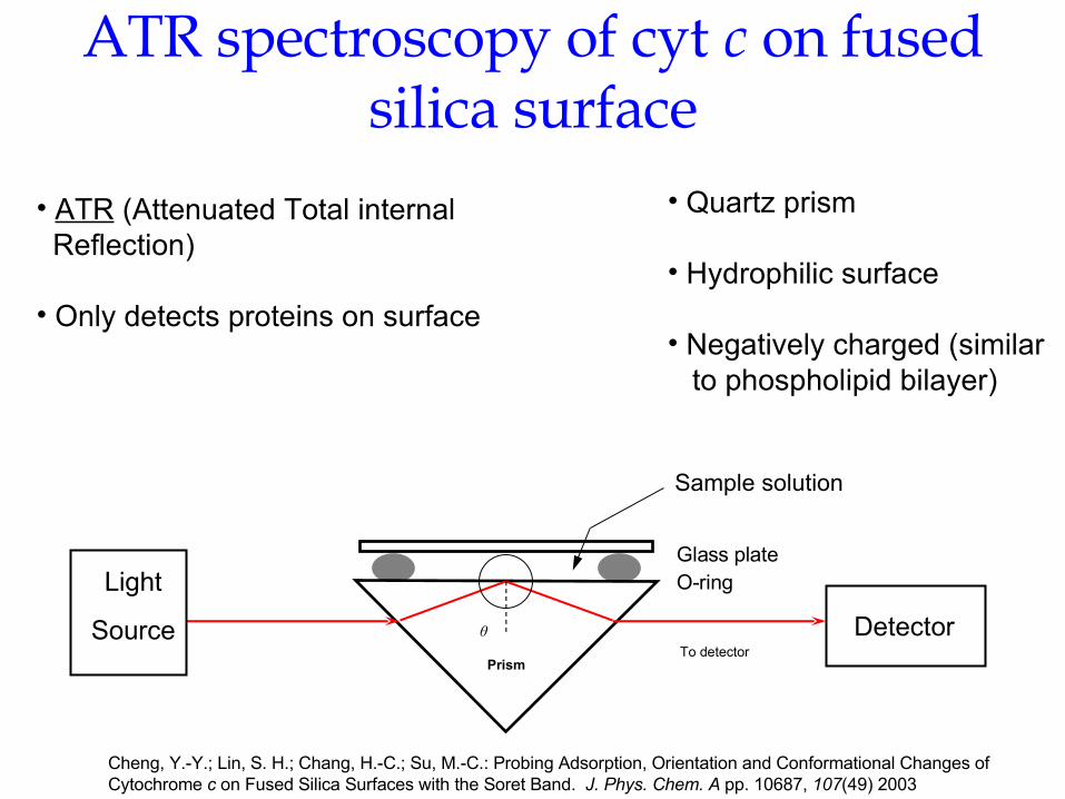
ATR spectroscopy of cyt c on fused silica surface
Cheng, Y.-Y.; Lin, S. H.; Chang, H.-C.; Su, M.-C.: Probing Adsorption, Orientation and Conformational Changes ofCytochrome c on Fused Silica Surfaces with the Soret Band. J. Phys. Chem. A pp. 10687, 107(49) 2003
• ATR (Attenuated Total internal Reflection)
• Only detects proteins on surface
DetectorPrism
θ
Glass plateO-ring
Sample solution
To detector
Light
Source
• Quartz prism
• Hydrophilic surface
• Negatively charged (similar to phospholipid bilayer)

Alcohol denatured Isotherm
The adsorption isotherm shows
that the surface coverage of
cyt c reaches a saturation level
at 15-20 µM bulk concentration0 20 40 60
0.002
0.004
0.006
0.008
0.010
0.012
0.014
Adsorption Isotherm
Ab
sorb
anc
e
[cyt c]
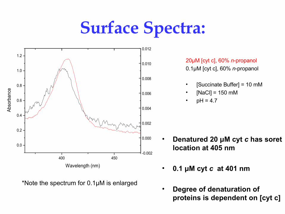
Surface Spectra:
• Denatured 20 μM cyt c has soret location at 405 nm
• 0.1 μM cyt c at 401 nm
• Degree of denaturation of proteins is dependent on [cyt c]
*Note the spectrum for 0.1µM is enlarged
400 450
0.0
0.2
0.4
0.6
0.8
1.0
1.2
-0.002
0.000
0.002
0.004
0.006
0.008
0.010
0.012
Abs
orba
nce
Wavelength (nm)
20µM [cyt c], 60% n-propanol
0.1µM [cyt c], 60% n-propanol
• [Succinate Buffer] = 10 mM • [NaCl] = 150 mM• pH = 4.7

Surface adsorbed vs. Solution:
[Succinate Buffer] = 10 mM pH = 4.7[NaCl] = 150 mM
*Note the spectrum for 0.1 µM is enlarged
•At lower bulk [cyt c], surface adsorbed proteins are more denatured than at higher [cyt c]
•Denatured proteins in the solution are renatured at the surface
400 450
0.0
0.2
0.4
0.6
0.8
1.0
1.2
-0.002
0.000
0.002
0.004
0.006
0.008
0.010
0.012Solution:
20 0% n-propanol 20 60% n-propanol
Abs
orba
nce
Wavelength (nm)
Surface: 20 , 60% n-propanol
Solution and Surface spectra
0.1 , 60% n-propanol

Surface adsorbed vs. Solution:
0 5 10 15 20 25 30 35 40 45 50 55 60399
400
401
402
403
404
405
406
Surface Solution
La
md
a M
ax
(nm
)
[ Cyt c ] (M)
Solution and Surface Absorbance
[Succinate Buffer] = 10 mM 60% n-propanol[NaCl] = 150 mM pH = 4.7
Concentration dependence:
•In solution cyt c proteins are “completely” denatured
•At all [cyt c] the surface adsorbed proteins are less denatured than in solution
•The degree of denaturation of surface adsorbed proteins depends on concentration

Protein-Protein Interactions:
• At low [cyt c] the proteins adsorb to the surface with little change to their state of denaturation
• As the [cyt c] increases the proteins are renaturing on the surface due to increasing protein - protein interactions
• When the surface is saturated (>15-20 µM) the protein-protein interactions remain constant and protein renaturation reaches a limit

ConclusionProtein-protein interactions are an important
factor in the conformation of cyt c
adsorbed to fused silica surface
Future Direction• Increase signals of low concentration spectra via multiple
reflection ATR absorbance spectroscopy using a multi pass cell
• Surface Fluorescence to indicate conformational changes

Special Thanks To:
Dr. Todd A. Hopkins
Dr. Geoffrey C. Hoops
Dr. Meng-Chih Su
Scott A. Hocker
Victoria Fahrenbach
Tara Benz
Greg Campanello
Butler University Department of Chemistry
Collaborators:Y.-Y. Cheng, S. H. Lin, and H.-C. ChangInstitute of Atomic and Molecular Sciences,Academia Sinica