surface chemistry of gold nanoparticles mediates their ... · pdf filesurface chemistry of...
TRANSCRIPT
OH AND PARK VOL. 8 ’ NO. 6 ’ 6232–6241 ’ 2014
www.acsnano.org
6232
May 16, 2014
C 2014 American Chemical Society
Surface Chemistry of GoldNanoparticles Mediates TheirExocytosis in MacrophagesNuri Oh†,‡ and Ji-Ho Park†,‡,§,*
†Department of Bio and Brain Engineering, ‡Institute for Optical Science and Technology, and §Institute for the Nanocentury, Korea Advanced Institute of Scienceand Technology (KAIST), Daejeon 305-701, Republic of Korea
Synthetic nanoparticles engineered forsystemic administration into the bodyhave great potential for detection and
treatment of complex human diseases.1,2
Despite significant effort to improve theirtargeting capability, significant quantitiesof nanoparticles in circulation are eventuallycleared by the mononuclear phagocyticsystem (MPS),3�10 part of the immune sys-tem consisting of phagocytic cells. Amongvarious MPS cell types, the Kupffer cells ofthe liver and red pulp macrophages of thespleen play major roles in clearing, proces-sing, and degrading the nanoparticles. Op-sonin proteins in the blood rapidly adsorb tothe nanoparticle surface, forming a proteincorona.11�14 Opsonin-coated nanoparticlesare then recognized and taken up by MPScells, and finally trapped in the lysosomesfor a relatively long period, depending ontheir physicochemical properties.4,5,7,8 Suchlong retention times increase the likelihoodof nanoparticle-mediated chronic toxicity intheMPS via inflammation or immunologicalresponses.4,15,16 Thus, assessing the fate of
nanoparticles in the resident macrophagesof theMPS is important to clarify their in vivopotential toxicity.Much progress has been made in under-
standing the size, shape, and surface chem-istry effects of non-biodegradable nano-particles on macrophage endocytosis.17�21
Nanoparticles smaller than 100 nm report-edly enter macrophages by macropinocyto-sis, or scavenger receptors.20,22�24 However,relatively little attention has been given tohow these nanoparticles leave the macro-phages, i.e., themechanism behind systemicexcretion.5,25�27 Various nanoparticle for-mulations, such as polymeric nanoparti-cles,28�30 gold nanoparticles,31�35 carbonnanotubes36,37 metal oxide nanoparticles,38,39
quantumdots,40�42 mesoporous silica nano-particles,43,44 and nanodiamonds,45 havebeen tested to characterize their exocytosisin many types of cells, primarily cancer cells;these studies yielded several important find-ings: nanoparticle exocytosis is an energy-dependent process and is influenced bynanoparticle size, surface chemistry, shape,
* Address correspondence [email protected].
Received for review March 26, 2014and accepted May 16, 2014.
Published online10.1021/nn501668a
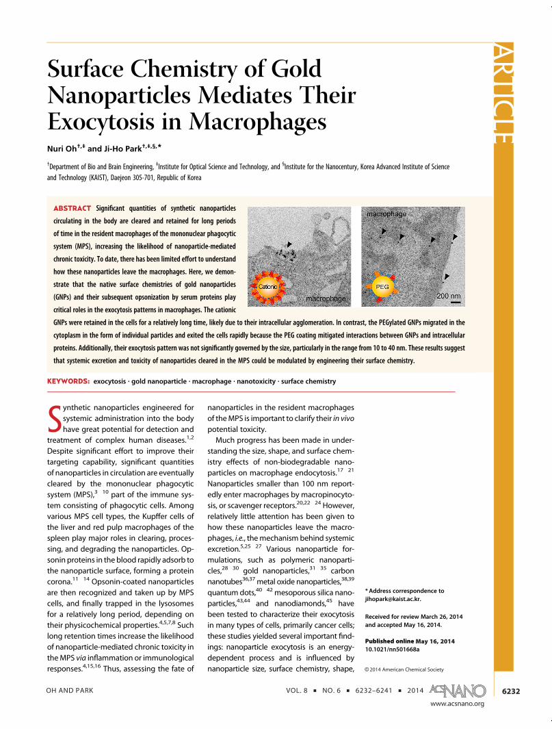
ABSTRACT Significant quantities of synthetic nanoparticles
circulating in the body are cleared and retained for long periods
of time in the resident macrophages of the mononuclear phagocytic
system (MPS), increasing the likelihood of nanoparticle-mediated
chronic toxicity. To date, there has been limited effort to understand
how these nanoparticles leave the macrophages. Here, we demon-
strate that the native surface chemistries of gold nanoparticles
(GNPs) and their subsequent opsonization by serum proteins play
critical roles in the exocytosis patterns in macrophages. The cationic
GNPs were retained in the cells for a relatively long time, likely due to their intracellular agglomeration. In contrast, the PEGylated GNPs migrated in the
cytoplasm in the form of individual particles and exited the cells rapidly because the PEG coating mitigated interactions between GNPs and intracellular
proteins. Additionally, their exocytosis pattern was not significantly governed by the size, particularly in the range from 10 to 40 nm. These results suggest
that systemic excretion and toxicity of nanoparticles cleared in the MPS could be modulated by engineering their surface chemistry.
KEYWORDS: exocytosis . gold nanoparticle . macrophage . nanotoxicity . surface chemistry
ARTIC
LE

OH AND PARK VOL. 8 ’ NO. 6 ’ 6232–6241 ’ 2014
www.acsnano.org
6233
and cell type. However, to our knowledge, onlytwo brief reports have investigated the exocytosis ofsurface-tailored nanoparticles in macrophages.15,38
Fischer et al. examined the exocytosis of surface-functionalized quantum dots (QD) in primary murineKupffer cells for ∼400 min; however, this time wastoo short to observe complete excretion of the intra-cellular QDs.15 Serda et al. also examined the surfacechemistry-dependent exocytosis of magnetic nano-particles (MNP) delivered into murine macrophagesby porous silicon microcarriers; the native surfaceproperties of MNPs embedded in the microcarrierswere secured during cellular uptake.38 This outcomeis different from that observed in in vivo situations, inwhich the nanoparticles are usually coated with serumproteins prior to cellular uptake. Thus, more systematicinvestigation is essential to better clarify their exo-cytosis process in MPS-associated macrophages.Cellular uptake and biodistribution of gold-based
nanomaterials have been extensively investigated interms of their biosafety, due to their tremendouspotential in medical applications (e.g., gold salts ap-proved for rheumatoid arthritis therapies46).18,31,47�50
However, lack of knowledge about how the MPS ridsitself of these nanoparticles has impeded their clinicaltranslation. Particularly in the liver, these nanoparticlesmust escapemacrophages andmigrate to hepatocytesto be excreted through the hepatobiliary system. Theirlong-term retention in macrophages has been shownto induce inflammation and apoptosis in the liver.4
Although several studies have demonstrated hepato-biliary clearance of systemically injected gold-basednanomaterials over the long-term,6,51,52 none havereported complete excretion of the nanomaterial fromthe MPS within a reasonable period of time (excretionpercentage:∼50%of poly(ethylene glycol) (PEG)ylatedgold nanorods at two months post-injection,6 and∼10% of anionic gold nanoparticles at six monthspost-injection51). We believe that the potential forin vivo toxicity from gold-based nanomaterials couldbe reduced if they are designed to efficiently facilitateexocytosis of the MPS macrophages. Thus, we system-atically assessed the effect of size and surface chem-istry of monodisperse gold nanoparticles (GNPs) onexocytosis patterns in macrophages in vitro. Thisexocytosis mechanism in macrophages is discussedhere in an effort to elucidate it and thereby improveefficiency.
RESULTS AND DISCUSSION
To engineer the design parameters for efficient exo-cytosis in macrophages, GNPs with various sizes andsurface chemistries were prepared (Figure 1). We wereparticularly interested in studying the cellular fate ofGNPs smaller than 50 nm, because smaller sizes arefavorable for systemic injection. Citrate-coated GNPs(average sizes: ∼10, 20, and 40 nm) were synthesized
using a previously established method,53 and used asanionic GNPs. Zwitterionic and PEGylated GNPs werethen prepared by coating the citrated GNPs withcysteine andmethoxy-PEG (mPEG)-sulfhydryl (2k) mol-ecules, respectively. Cysteamine-coatedGNPs (averagesizes: ∼10, 20, and 40 nm) were also synthesized forcationic GNPs using methods specified in previousstudies.54,55 Subsequently, all GNP formulations werecoated with serum proteins prior to macrophage treat-ment, to mimic the opsonization process in bloodcirculation (Figure 1a). Transmission electron micro-scopy (TEM) revealed that all GNP formulations werefairly spherical with uniform diameters (Figure 1b andSupporting Information Figure S1). The average hydro-dynamic diameters (HD) were slightly larger thanthe diameters observed using TEM, because TEMimages did not reveal the organic-molecule coating(Figure 1c). The zeta potential results prior to serumcoating well reflected the characterizations of theorganic coatings. After serum coating, the HDs of allGNP formulations increased (regardless of their initialsize) by ∼10 nm for PEGylated GNPs, ∼15 nm forcationic GNPs, and ∼20 nm for other GNPs. Gel elec-trophoresis and thermogravimetric analysis (TGA)results, with proteins eluted from the GNPs, also sup-ported the surface chemistry dependency on serumprotein adsorption (Supporting Information Figure S2),which is in accordance with previous findings.56,57
Zeta potential measurements revealed an anionicsurface charge ranging from �40 to �15 mV for allGNP formulations, regardless of their original charge,reflecting the charges of the adsorbed proteins.56 Theprotein coating can also cause a slight shift in theabsorption spectra to longer wavelengths by ∼5 nm.The PEGylated surface exhibited the lowest proteinadsorption, as described in previous reports.58,59
Aggregated or agglomerated forms of nanoparticlesinfluence their endocytosis patterns.49 In this study, wefocused on the exocytosis of GNPs taken up by macro-phages in the formof individual particles. To determinewhether the serum-coated GNPswere stable under thegiven test conditions, their HD and absorption spectrawere monitored for 6 h (the incubation time for theirendocytosis in the following experiments) in serum-free media at 37 �C using a dynamic light scattering(DLS) instrument and an ultraviolet�visible (UV/vis)spectrometer, respectively. The shift and broadeningof the absorption spectrum indicate their aggregation/agglomeration.49 The HDs and absorption spectra ofthe GNP formulations did not change significantly(Supporting Information Figures S3 and S4), suggest-ing that no aggregation or agglomeration occurredduring the incubation period. Thus, our experimentalsetup allowed single serum-coated GNPs to interactwith macrophages in vitro.Systemically administered nanoparticles interact
with circulatingmonocytes and eventually accumulate
ARTIC
LE

OH AND PARK VOL. 8 ’ NO. 6 ’ 6232–6241 ’ 2014
www.acsnano.org
6234
in the resident macrophages of the MPS. Thus, we firstevaluated the endocytosis of various GNP formulationsin bothmonocytes andmacrophages, before assessingtheir subsequent exocytosis. Human macrophage-likeU937 cells were selected as the MPS macrophagemodel, because these are capable of efficient phago-cytosis of foreign materials.60�64 Undifferentiated(monocytes) or phorbol-12-myristate-13-acetate (PMA)-differentiated U937 cells (macrophages) were treatedwith each formulation of serum-coated GNPs for 6 hin serum-supplemented media at 37 �C to saturatetheir endocytosis.19,20,28 The quantities of GNPs taken
up by cells 6-h post-treatment were measured usinginductively coupled plasmamass spectrometry (ICP-MS).Dark field microscopy (DFM) was used to visualize thecellular uptake with the strong scattering property.The scattering signal can be also used to estimatethe aggregated state of GNPs in the cells; aggregatedGNPs appear as orange-red and single GNPs appear asgreen-yellow.65 Only a small amount of GNP formula-tionswas observed in themonocytes, although cationicGNPs exhibited a relatively higher cellular uptake(Figure 2a,c). ICP-MS results revealed that in the macro-phages, cellular uptake of cationic GNPs was higher
Figure 1. Preparation and characterization of serum-coated gold nanoparticles. (a) Coating procedure of gold nanoparticles(GNPs). Citrate-coated GNPs with averages sizes of ∼10, 20, and 40 nm were first synthesized and used as anionic GNPs.Zwitterionic and PEGylated GNPs were then prepared by coating the citrated ones with cysteine and mPEG-sulfhydryl (2k)molecules, respectively. Cysteamine-coated GNPs were also synthesized for cationic ones. Subsequently, all GNP formula-tions were further coated with serum proteins prior to treating macrophages. (b) Transmission electron microscopic imagesof citrate- (left) and cysteamine-coated (right) GNPs with average diameters of ∼10 (top), 20 (middle), and 40 nm (bottom).Scale bar is 20 nm. (c) Characterization of surface-functionalizedGNPs before and after serum coating. The number after theG(GNP identifier) designates the size. The letters A, C, Z, and P indicate anionic, cationic, zwitterionic, and PEGylated surface,respectively. The physical diameters (TEM diameter (TD)) were determined from the images obtained with TEM (n = 10). Thehydrodynamic diameters (HD) and zeta potentials (ZP) were determined based on dynamic light scattering (DLS) measure-ments (n=3). The peak represents themaximumpeakof absorption spectrumofGNPs. All data of TD, HD, andZP aremeans(SD (n = 3).
ARTIC
LE

OH AND PARK VOL. 8 ’ NO. 6 ’ 6232–6241 ’ 2014
www.acsnano.org
6235
than that of other GNPs, regardless of their size(Figure 2b). Our results also demonstrated that thePMA-differentiated U937 cells preferentially interna-lized the 20 nm-diameter GNPs, compared with the10 and 40 nm GNPs. Moreover, DFM images displayedsignificantly more orange-red dots in the cells treatedwith cationic GNPs, compared with those having otherGNPs; this indicated the presence of aggregatedGNPs, presumably in the endosomes and lysosomes(Figure 2d), supporting the ICP-MS results. In contrast,PEGylated GNPs were inefficient in entering themacro-phages; the internalized ones appeared as yellowishdots in DFM images, indicating their intracellular reten-tion in the form of individual particles. Collectively,these results suggest that the native surface propertiesof GNPs, which would be associated with type andquantity of adsorbed serum proteins via hydrophobicor electrostatic interactions, primarily determine theirendocytotic fate.Most of the nanoparticles taken up by macro-
phages undergo translocation from endosomes tolysosomes,66,67 where they are further processed fordigestion. However, to date, few studies have investi-gated the exocytotic fate of non-biodegradable nano-particles. We next examined how the size and surfacechemistry of GNPsmediated serumprotein adsorption,subsequent endocytosis, and eventual exocytosis inmacrophages. PMA-differentiated U937 cells weretreated with each formulation of GNPs for 6 h inserum-supplemented media at 37 �C, and were then
extensively washed to remove anyweakly boundGNPson the plasma membrane; the samples were furtherincubated for 48 h in GNP-free serum-supplementedmedia. To measure the quantity of GNPs exocytosedfrom the cells, supernatants were collected over a 48-hpost-incubation period, and the amount of Au ionsin the supernatants was analyzed using ICP-MS. Theresulting exocytosis pattern represented the percen-tage of GNPs that remained in the macrophagesat each time point after incubation in the GNP-freemedia. Our results demonstrated that, regardless ofsize, cationic GNPs remained in cells longer than anyother formulations, while PEGylated GNPs exhibitedthe highest rate of exocytosis (Figure 3a�c). Previousfinding also revealed that cationic (polyethylenimine-coated) nanoparticles (diameter: ∼130 nm) were exo-cytosed more slowly in cancer cells, compared withanionic (phosphate-modified) ones.44 For internalizedcationic GNPs with diameters of ∼10, 20, and 40 nm,25.01, 20.32, and 37.30%, respectively, were extracel-lularly released within 48 h. In contrast, 83.34, 71.65,and 81.08% of intracellular PEGylated GNPs with dia-meters of∼10, 20, and 40 nm, respectively, underwentexocytosis within 48 h. The anionic and zwitterionicGNPs exhibited medium exocytosis rates betweenthose of PEGylated and cationic ones. Additionally, itwas unlikely that their exocytosis patterns were influ-enced by the size range tested here (10�40 nm)(Supporting Information Figure S5), indicating thatsurface properties of the GNPs are a more dominant
Figure 2. Endocytosis of serum-coated gold nanoparticles in monocytes and macrophages. (a and b) Amount of goldnanoparticles (GNPs) taken up by monocytes (a) and macrophages (b). The amount of intracellular GNPs was determined byICP-MS after the cells were treated with GNPs for 6 h. (c and d) Dark fieldmicroscopic images of serum-coated GNPs taken upby monocytes (c) and macrophage (c). The intracellular GNPs were imaged using a dark field microscope after the cells weretreated with GNPs for 6 h. Scale bars indicate 10 μm. Data represent means( SD (n = 3, *p < 0.05, and **p < 0.01, analyzed bytwo-way analysis of variance, followed by Bonferroni test).
ARTIC
LE

OH AND PARK VOL. 8 ’ NO. 6 ’ 6232–6241 ’ 2014
www.acsnano.org
6236
factor in determining cellular excretion of GNPs smallerthan 40 nm. This differs somewhat from a previousreport that the exocytosis rate of transferrin-coatedGNPs (with diameters of 14�100 nm in cancer cells)is proportional to their size.31 Specific ligand�receptorinteractions play critical roles in mediating the exocy-tosis of transferrin-coated GNPs, whereas nonspecificinteractions between coated serum proteins and plas-ma membranes seem to be mainly responsible for theexocytosis of serum-coated GNPs here.Exocytosis patterns were also visualized using both
DFM and multiphoton excitation microscopy. Particu-larly, intrinsic multiphoton luminescence of GNPs al-lowed clear visualization of their intracellular fate.68,69
The DFM and multiphoton microscopic images at 48-hpost-incubation revealed more orange-red dots, indi-cating the formation of aggregated GNPs and strongerluminescence signals were observed in cells treatedwith cationic GNPs, respectively, compared with thosetreated with other formulations (Figure 3d�i andSupporting Information Figure S6), supporting the ICP-MS results. Importantly, the multiphoton microscopicimages revealed that the luminescence signals of intra-cellular GNPs in the perinuclear regions decreased overtime, suggesting that they were indeed exocytosedfrom the cytoplasm, presumably after passing through
lysosomes. Lastly, the GNP treatment did not induce anysignificant cytotoxicity within the tested exocytosistime period (Supporting Information Figure S7). To-gether, these results suggest that the surface propertiesof GNPs, prior to serum coating, also play a primary rolein determining their exocytosis fate in macrophages.Lastly, we further investigated the intracellular fates
of 20 nm cationic and PEGylated GNPs in macro-phages, due to the dramatic difference in their exocy-tosis rates. We first observed their exocytosis processesusing TEM: the GNP-treated macrophages were incu-bated in GNP-free media for 24 or 48 h, fixed, sliced,and visualized using TEM. Immediately after the 6-htreatment, most of the cationic GNPs appeared ag-glomerated in the endosomes, while PEGylated GNPswere observed in the cytoplasm, in the form of indivi-dual particles (Figures 4a�d). During exocytosis, largeagglomerates of cationic GNPs enclosed in the vesiclesappeared to migrate slowly in the cytoplasm, andwere then excreted from the cell by lysosomal fusionwith the plasma membrane. In contrast, individualPEGylated GNPs, either trapped in the vesicles or inthe cytosol, were more likely to move rapidly towardthe plasmamembrane, without agglomeration, and exitthe cell, presumably via diffusion. Gel electrophoresisresults with proteins eluted from the exocytosed GNPs
Figure 3. Exocytosis of serum-coated gold nanoparticles in macrophages. (a�c) Exocytosis rate of serum-coated goldnanoparticles (GNPs) with sizes of 10 (a), 20 (b), and 40 nm (c) inmacrophages. Macrophageswere treatedwith serum-coatedGNPs for 6 h, washed, and further incubated in the GNP-free media for 6, 24, or 48 h. At each time post-incubation, thesupernatant of cells were collected and the amount of GNPs exocytosed from the cells during the incubated period wasdetermined by ICP-MS. (d�f) Dark field microscopic images of serum-coated GNPs with sizes of 10 (d), 20 (e), and 40 nm (f),resident in macrophages at 0 and 48 h postincubation. (g�i) Multiphoton microscopic images of serum-coated GNPs withsizes of 10 (g), 20 (h), and 40 nm (i), resident in macrophages at 0 and 48 h post-incubation. Excitation wavelength at 800 nmwas used to image multiphoton luminescence of GNPs, while emission wavelengths ranging from 500 to 650 nm werecollected. Scale bars indicate 10 μm. Data represent means ( SD (n = 3, *p < 0.05 and **p < 0.01 between the data at 48 h,analyzed by two-way analysis of variance, followed by Bonferroni test).
ARTIC
LE

OH AND PARK VOL. 8 ’ NO. 6 ’ 6232–6241 ’ 2014
www.acsnano.org
6237
revealed that the PEGylated GNPs were likely to interactwith fewer intracellular proteins during the exocytosisprocess, comparedwith cationic GNPs (Figure 4e). It waspreviously reported that the binding stability betweennanoparticles and intracellular proteins could enhancethe intracellular retention of nanoparticles.70 Thus, theseresults suggest that the PEG coating may mitigateinteractions between GNPs and intracellular proteins,thereby facilitating exocytosis. TEM images of GNPsexcreted to the media, 48-h post-incubation, exhibitedmany cationic GNP agglomerates in the extracellularsolution, whereas the exocytosed PEGylated GNPs werein the form of individual particles (Supporting Informa-tion Figure S8). The broadened absorption spectrumof exocytosed cationic GNPs also indicated their ag-glomeration, supporting the TEM results (SupportingInformation Figure S9). Additionally, no exocytosed
membrane vesicles enclosing the GNPs were observedin the TEM images of either formulation, which isdifferent from previous observations.38,71
We also investigated whether the initial intracellularconcentration of GNPs would influence their exocyto-sis rate (8006153 particles/cell for cationic GNPs versus898585 particles/cell for PEGylated GNPs; Figure 2b).The initial concentration of cationic GNPs in the cellstreatedat gold ion concentrationsof 15μMAuwas similarto that of PEGylated GNPs in the cells treated at 150 μMAu (Supporting Information Figure S10a). Neither did cat-ionic GNPs (15 and 150 μM Au) exhibit a significantdifference in exocytosis rate, although the initial intra-cellular concentration in the cells treated at 150 μMAuwas ∼8� higher than in that in cells treated at 15 μMAu (Supporting Information Figure S10b). Thus, theexocytosis of PEGylated GNPs was faster than that of
Figure 4. Exocytosis processes of 20 nm cationic and PEGylated gold nanoparticles. (a and b) Transmission electronmicroscopic (TEM) images of exocytosis process of serum-coated cationic (a) or PEGylated (b) gold nanoparticles (GNPs) inmacrophages. Macrophages were treated with serum-coated GNPs, washed, and further incubated in the GNP-freemedia for24 or 48 h. At each time post-incubation, the cells were collected, fixed, and examined by TEM to visualize the exocytosisprocess of intracellular GNPs. Themagnified images on the right side come from the squares in the corresponding images onthe left side. (c and d) TEM images showing the exocytosis of serum-coated cationic (c) or PEGylated GNPs (d). GNPs from theplasmamembrane of macrophages. (e) Coomassie blue-stained gel staining of GNPs exocytosed frommacrophages. At 48 hpost-incubation; the exocytosed GNPswere collected from the supernatant by centrifugation. The proteins were eluted fromthe nanoparticles in sample loading buffer and separated by gel electrophoresis. Untreated cell lysates and 0.5% FBS wereused as controls. The number after the G (GNP identifier) designates the size. The letters A, C, Z, and P indicate anionic,cationic, zwitterionic, and PEGylated surface, respectively. Arrow heads and C indicate GNPs and cytoplasm, respectively.
ARTIC
LE

OH AND PARK VOL. 8 ’ NO. 6 ’ 6232–6241 ’ 2014
www.acsnano.org
6238
cationic GNPs, even at similar initial intracellular GNPconcentrations. Additionally, it was found that theexocytosis rate of PEGylated GNPs was similar to thatof PEGylated gold nanorods (GNR) with aspect ratio of∼4.6 (Supporting Information Figure S11), indicatingthat the exocytosis pattern of PEGylated GNPs is notsignificantly influenced by their shape. These resultsconfirm that the exocytosis pattern of GNPs is indeeddependent on their native surface properties.
CONCLUSIONS
In summary, we demonstrated that the nativesurface chemistries of GNPs and their subsequentopsonization by serum proteins play a critical role inthe exocytosis patterns in macrophages. The cationicGNPs were retained in the cells for a relatively longtime, while the PEGylated GNPs exhibited the highest
exocytosis rate. Intracellular agglomeration of cationicGNPs seemed to delay their exocytosis, whereasPEGylated GNPs, migrating in the cytoplasm in theform of individual particles, were excreted rapidly.We also observed that the exocytosis pattern of GNPswas not significantly governed by their size, particu-larly in the range from 10 to 40 nm. Although in vivo
systematic work is still required, our findings suggestthat exocytosis of GNPs in the macrophages and theirsubsequent systemic excretion could be modulatedby engineering their surface chemistries. Currently, weare investigating the detailed intracellular molecularmechanisms governing the complex exocytosis pro-cess of GNPs in macrophages. We believe that theresults of the present study demonstrate promisingpossibilities for the clinical translation of gold-basednanomaterials.
MATERIALS AND METHODS
Preparation of Gold Nanoparticles. Citrate-capped gold nano-particles (GNPs) with averages sizes of∼10, 20, and 40 nmweresynthesized using previously established method53 and wereused as anionic GNPs. Briefly, 20 mL of 1.0 mM HAuCl4 3 3H2O(Sigma) was brought to a boil in a 100 mL bottle and 2.0, 1.6, or1.0mL of 38.8mM trisodiumcitrate (Sigma) was added for 10, 20,or 40 nm GNPs, respectively. The color of mixture solutionchanged from yellow to dark red over 5 min. After a boilingand stirring period of 15 min, the GNP solution was keptunder room temperature. Zwitterionic and PEGylated GNPswere prepared by coating the citrated GNPs with cysteineand mPEG-sulfhydryl (2k) molecules, respectively. Briefly, forzwitterionic and PEGylated GNPs, 1 mL of 150 μM Au citrated-capped GNPs was mixed with 70 μL of 0.001 M L-cysteine(Sigma) and 25 mg of mPEG(2K)-SH (Sunbio, South Korea),respectively. The mixture was vortexed under room tempera-ture overnight. On the other hand, cysteamine-coated GNPswith averages sizes of ∼10, 20, and 40 nm were directlysynthesized for cationic GNPs using different methods fromthose used for citrated-coated GNPs.54,55 For cystamine-coatedGNPs with a diameter of 10 nm, 400 μL of 84.5 μM cysteamine(Sigma) was added to 40 mL of 1.4 mM HAuCl4 solution. After astirring period of 20 min, 1 mL of 1 mM NaBH4 solution wasadded dropwise for 2 min. The mixture was stirred for 12 h atroom temperature. For larger cystamine-coated GNPs, 400 μL of213 mM cysteamine was added to 40 mL of 1.42 mM HAuCl4solution. After a stirring period of 20 min at room temperature,10 or 30 μL of 10 mM NaBH4 solution was added to the mixturefor cysteamine-coated GNPs with diameters of 20 or 40 nm,respectively. The mixture was vigorously stirred for 25 min atroom temperature. To remove free organic chemicals, thesolution was dialyzed against ultrapure water. For PEGylatedgold nanorods (GNR)with aspect ratio of∼4.6, the seed solutionwas prepared bymixing 5mL of 0.2M cetyltrimethylammoniumbromide (CTAB, Sigma), 5 mL of 0.5 mM HAuCl4 (Sigma), and0.6 mL of 0.01 M NaBH4 (Sigma) and aged for 2 h. To grow theseed into the rod shape, 12 μL of seed solution was mixed with5 mL of 0.2 M CTAB, 5 mL of 1 mM HAuCl4, 250 μL of 4 mMAgNO3, and 70 μL of 78.84 mM ascorbic acid at 25 �C. Thesynthesized GNRs were washed by centrifugation at 10 000 rpmfor 10 min, resuspended in ultrapure water, and coated with3 mg/mL of mPEG(5K)-SH. For serum coating, all GNP formula-tions were incubated with 10% fetal bovine serum (FBS) solution(in ultrapurewater) for 1 h at roomtemperature and thenwashedto remove unbound serum proteins by repeated centrifugationat 12000 rpm for 30 min. The serum-coated GNPs were finallyresuspended in 1 mL of phosphate-buffered saline (PBS).
Characterization of Gold Nanoparticles. The absorption spectrawere characterized with UV/vis spectrometry (MolecularDevices). The size and morphology were observed with a fieldemission-transmission electron microscope (FE-TEM, JEOL). Thehydrodynamic size and zeta potential were measured at 25 �Cusing a dynamic light scattering (DLS) instrument (MalvernInstruments). To test colloidal stability of serum-coated GNPsin the physiological condition, the hydrodynamic size andabsorption spectrum were monitored hourly for 6 h in serum-free media at 37 �C using DLS instrument and UV/vis spectro-meter, respectively. To identify the serum proteins adsorbed onthe GNP surface, the serum proteins were eluted from the GNPsin the sample loading buffer (80 mM SDS, 75 μM SDS, 1.25%β-mercaptoethanol, 10% glycerol, and 62.5mM Tris-HCl, pH 6.8)by heating to 95 �C for 5 min and separated by running SDSpolyacrylamide gel electrophoresis (SDS-PAGE) at 200 V for30 min. Finally, the eluted serum proteins were visualized bystaining the gel with Coomassie brilliant blue R-250. A total of0.5% FBS was used as a control. To quantify the serum proteinsadsorbed on the surface of GNPs, the serum-coated GNPs wereanalyzed by thermogravimetric analysis (TGA, TG 209 F3,NETZSCH). Protein analysis was performed from room tempera-ture to 800 �C at a heating rate of 10 �C/min under nitrogenatmosphere with a gas flow rate of 30 mL/min.
Cell Culture. Monocyte-like undifferentiated U937 cells weremaintained in RPMI 1640 medium supplemented with 10%fetal bovine serum and 100 μg/mL penicillin and streptomycinat 37 �C. For differentiation into macrophages, nonadherentmonocyte-like undifferentiated U937 cells were exposed to40 nM phorbol-12-myristate-13-acetate (PMA) for 72 h. Differ-entiated U937 cells were maintained by replacement of PMA-containing media every 2�3 days.
Endocytosis Study. Monocyte-like undifferentiated and PMA-differentiatedU937 cellswere seededat 5.0� 105 cells/(mL/well)in 6-well tissue culture plates with ∼90% confluence. The cellswere treated with each formulation of serum-coated GNPs at anAu concentration of 150 μM for 6 h in serum-supplementedmedia at 37 �C to allow sufficient cellular uptake of GNPs, andintensively washed to remove free and any weakly bound GNPson the plasma membrane. To measure the quantities of GNPstaken up by cells 6 h post-treatment, the GNP-treated cells werecollected by centrifugation at 1000 rpm for 5 min and incubatedin aqua regia for 24 h to dissolve GNPs and organic materials.The amount of Au ions in the cells was then analyzed byinductively coupled plasma mass spectrometry (ICP-MS, AgilentTechnologies) and converted to the number of GNPs per cell.The time-dependent cellular uptake of GNPs was also visualizedwith their scattering property using dark field microscopy.
ARTIC
LE

OH AND PARK VOL. 8 ’ NO. 6 ’ 6232–6241 ’ 2014
www.acsnano.org
6239
The microscopy system consisted of an inverted microscope(Ti�U, Nikon) equipped with a dark-field condenser (N.A.0.8�0.95), a halogen lamp (100 W), and a true-color digitalcamera (DS-Ri1-U3, Nikon). Dark field microscopic images ofmonocyte-like undifferentiated and PMA-differentiated U937cells treated with each GNP formulation were obtained with a40� objective (N.A. 0.6).
Exocytosis Study. PMA-differentiated U937 cells were treatedwith each formulation of GNPs at an Au ion concentration of150 μM for 6 h in serum-supplemented media at 37 �C,intensively washed to remove free and any weakly bound GNPson the plasma membrane, and further incubated over 48 h inGNP-free serum-supplemented media to induce their exo-cytosis. To measure the quantities of GNPs exocytosed fromthe cells, the GNPs in the supernatants at 0, 6, 24, or 48 h post-incubation were collected by centrifugation at 14 000 rpm for1 h and dissolved in aqua regia for 24 h. The amount of Au ionsin the supernatants was then analyzed by ICP-MS. The exo-cytosis pattern was represented by the percentage of GNPsremaining in the macrophages at each time point after incuba-tion in the GNP-free media. The exocytosis of intracellular GNPsin the PMA-differentiated U937 cells was also visualized at eachtime point using both dark field and multiphoton excitationmicroscopy. Time-dependent migration of 20 nm cationic andPEGylated GNPs in the cells was observed by TEM (Tecnai G2,FEI). The GNP-treated cells were incubated for 24 or 48 h withGNP-free serum-supplemented media. The cells were pelletedby centrifugation at 1000 rpm for 5 min and fixed with 2.5%glutaraldyhyde (Sigma) for 1 day. The pellet was then incubatedin 1% osmium tetroxide (OsO4, Sigma) for 1 h, dehydrated in aseries of alcohol, and substituted with 100% prophylene oxide.The cells were then embedded in the solution composed ofprophylene oxide and Epon812 for 24 h, and solidified at 70 �C.Finally, the cells are sliced to a thickness of 70 nm and imagedby TEM. The physicochemical properties of exocytosed GNPs at48 h post-incubation were examined by TEM, UV�vis spectro-metry, and gel electrophoresis. The GNP-treated cells wereincubated for 48 h with GNP-free serum-supplemented media.The GNPs in the supernatants were collected by centrifugation at14000 rpm for 1 h and resuspended in ultrapure water. For TEM,the GNPs were placed on the grid and negatively stained on adrop of 2% phosphotungstic acid (pH 8.0, Sigma) for 30 s for TEMimaging. To identify the serum proteins adsorbed on the surfaceof exocytosed GNPs, the proteins were eluted from the GNPsin the sample loading buffer (80 mM SDS, 75 μM SDS, 1.25%β-mercaptoethanol, 10% glycerol, and 62.5 mM Tris-HCl, pH 6.8)by heating to 95 �C for 5 min and separated by running SDSpolyacrylamide gel electrophoresis (SDS-PAGE) at 200 V for30 min. Finally, the eluted serum proteins were visualized bystaining the gel with Coomassie brilliant blue R-250. Untreated celllysates and 0.5% FBS were used as controls. For untreated cellsamples, PMA-differentiatedU937cellswere lysed in100μLof RIPAbuffer (0.5%sodiumdeoxycholate, 0.1%SDS, 1%NP40, 5mMEDTAin TBS, pH 8.0) for 10 min at 4 �C. The cell lysates were centrifugedat 13000 rpm for 15 min at 4 �C, and the protein concentrationsof cell lysates were determined with the BCA protein assay.
Cytotoxicity Assay. PMA-differentiatedU937 cells were treatedwith each formulation of GNPs at an Au ion concentrationof 150 μM for 6 h in serum-supplemented media at 37 �C,intensively washed to remove free and any weakly bound GNPson the plasma membrane, and further incubated for 6, 24, or48 h in GNP-free serum-supplemented media. The cell viabilitywas examined by the colormetric thiazoly blue tetrazoliumbromide (MTT) assay (Sigma).
Statistics. Statistical analyses of the data were performedusing one- or two-way analysis of variance (ANOVA), followedby Tukey's or Bonferroni test for multiple comparisons, respec-tively. The p-values less than 0.05 were considered statisticallysignificant. All experiments were performed at least three times.All error bars indicate standard deviation (SD).
Conflict of Interest: The authors declare no competingfinancial interest.
Acknowledgment. Thisworkwas supported by Basic ScienceResearchProgramand theBio&Medical TechnologyDevelopment
Program of the National Research Foundation (NRF) fundedby the Ministry of Science, ICT & Future Planning (Grant Nos.NRF-2012R1A1A1011058 and NRF-2012M3A9C6050125), andthe National R&D Program for Cancer Control, Ministry for Healthand Welfare (Grant No. 1220070), Republic of Korea. The authorsthank the Korea Basic Science Institute, Daejeon, Republic ofKorea, for assistance with TEM analysis.
Supporting Information Available: Additional TEM data; sur-face chemistry-dependent absorptionof serumproteinsonGNPs;stability of serum-coated GNPs in the media; time-dependentchange of absorption spectra of serum-coated GNPs; dark fieldmicroscopic images; cell viability of macrophages data; adsorp-tion spectra; comparison of exocytosis efficiency; exocytosis ofserum-coated PEGylated GNRs in macrophages. This material isavailable free of charge via the Internet at http://pubs.acs.org.
REFERENCES AND NOTES1. Kim, B. Y. S.; Rutka, J. T.; Chan, W. C. W. Nanomedicine.
N. Engl. J. Med. 2010, 363, 2434–2443.2. Cheng, Z.; Al Zaki, A.; Hui, J. Z.; Muzykantov, V. R.; Tsourkas,
A. Multifunctional Nanoparticles: Cost versus Benefit ofAdding Targeting and Imaging Capabilities. Science 2012,338, 903–910.
3. Choi, H. S.; Liu,W.; Misra, P.; Tanaka, E.; Zimmer, J. P.; Itty Ipe,B.; Bawendi, M. G.; Frangioni, J. V. Renal Clearance ofQuantum Dots. Nat. Biotechnol. 2007, 25, 1165–1170.
4. Cho, W.-S.; Cho, M.; Jeong, J.; Choi, M.; Cho, H.-Y.; Han, B. S.;Kim, S. H.; Kim, H. O.; Lim, Y. T.; Chung, B. H.; et al. AcuteToxicity and Pharmacokinetics of 13 nm-Sized PEG-CoatedGold Nanoparticles. Toxicol. Appl. Pharmacol. 2009, 236,16–24.
5. Liu, Z.; Davis, C.; Cai,W.; He, L.; Chen, X.; Dai, H. Circulation andLong-Term Fate of Functionalized, Biocompatible Single-Walled Carbon Nanotubes in Mice Probed by Raman Spec-troscopy. Proc. Natl. Acad. Sci. U.S.A. 2008, 105, 1410–1415.
6. von Maltzahn, G.; Park, J.-H.; Agrawal, A.; Bandaru, N. K.;Das, S. K.; Sailor, M. J.; Bhatia, S. N. Computationally GuidedPhotothermal Tumor Therapy Using Long-CirculatingGold Nanorod Antennas. Cancer Res. 2009, 69, 3892–3900.
7. Gu, L.; Fang, R. H.; Sailor, M. J.; Park, J.-H. In Vivo Clearanceand Toxicity of Monodisperse Iron Oxide Nanocrystals.ACS Nano 2012, 6, 4947–4954.
8. Thakor, A. S.; Luong, R.; Paulmurugan, R.; Lin, F. I.; Kempen,P.; Zavaleta, C.; Chu, P.; Massoud, T. F.; Sinclair, R.; Gambhir,S. S. The Fate and Toxicity of Raman-Active Silica-GoldNanoparticles in Mice. Sci. Transl. Med. 2011, 3, 79ra33.
9. Liu, Z.; Cai, W.; He, L.; Nakayama, N.; Chen, K.; Sun, X.; Chen,X.; Dai, H. In Vivo Biodistribution and Highly EfficientTumour Targeting of Carbon Nanotubes in Mice. Nat.Nanotechnol. 2007, 2, 47–52.
10. Park, J.-H.; von Maltzahn, G.; Zhang, L.; Schwartz, M. P.;Ruoslahti, E.; Bhatia, S. N.; Sailor, M. J. Magnetic Iron OxideNanoworms for Tumor Targeting and Imaging. Adv. Mater.2008, 20, 1630–1635.
11. Cedervall, T.; Lynch, I.; Lindman, S.; Berggård, T.; Thulin, E.;Nilsson, H.; Dawson, K. A.; Linse, S. Understanding theNanoparticle�Protein Corona Using Methods to QuantifyExchange Rates andAffinities of Proteins for Nanoparticles.Proc. Natl. Acad. Sci. U.S.A. 2007, 104, 2050–2055.
12. Lundqvist, M.; Stigler, J.; Elia, G.; Lynch, I.; Cedervall, T.;Dawson, K. A. Nanoparticle Size and Surface PropertiesDetermine the Protein Corona with Possible Implicationsfor Biological Impacts. Proc. Natl. Acad. Sci. U.S.A. 2008,105, 14265–14270.
13. Salvati, A.; Pitek, A. S.; Monopoli, M. P.; Prapainop, K.;Bombelli, F. B.; Hristov, D. R.; Kelly, P. M.; Aberg, C.; Mahon,E.; Dawson, K. A. Transferrin-Functionalized NanoparticlesLose Their Targeting Capabilities When a BiomoleculeCorona Adsorbs on the Surface. Nat. Nanotechnol. 2013,8, 137–143.
14. Tenzer, S.; Docter, D.; Kuharev, J.; Musyanovych, A.; Fetz, V.;Hecht, R.; Schlenk, F.; Fischer, D.; Kiouptsi, K.; Reinhardt, C.;et al. Rapid Formation of Plasma Protein Corona Critically
ARTIC
LE

OH AND PARK VOL. 8 ’ NO. 6 ’ 6232–6241 ’ 2014
www.acsnano.org
6240
Affects Nanoparticle Pathophysiology. Nat. Nanotechnol.2013, 8, 772–781.
15. Fischer, H. C.; Hauck, T. S.; Gomez-Aristizabal, A.; Chan,W. C. Exploring Primary Liver Macrophages for StudyingQuantum Dot Interactions with Biological Systems. Adv.Mater. 2010, 22, 2520–2524.
16. Park, E.-J.; Park, K. Oxidative Stress and Pro-InflammatoryResponses Induced by Silica Nanoparticles In Vivo andIn Vitro. Toxicol. Lett. 2009, 184, 18–25.
17. Yu, S. S.; Lau, C. M.; Thomas, S. N.; Jerome, W. G.; Maron, D. J.;Dickerson, J. H.; Hubbell, J. A.; Giorgio, T. D. Size- andCharge-Dependent Non-Specific Uptake of PEGylated Nanoparti-cles by Macrophages. Int. J. Nanomed. 2012, 7, 799–813.
18. Walkey, C. D.; Olsen, J. B.; Guo, H.; Emili, A.; Chan, W. C.Nanoparticle Size and Surface Chemistry DetermineSerum Protein Adsorption and Macrophage Uptake.J. Am. Chem. Soc. 2012, 134, 2139–2147.
19. Arnida; Janát-Amsbury, M. M.; Ray, A.; Peterson, C. M.;Ghandehari, H. Geometry and Surface Characteristicsof Gold Nanoparticles Influence Their Biodistribution andUptake by Macrophages. Eur. J. Pharm. Biopharm. 2011,77, 417–423.
20. Lunov, O.; Syrovets, T.; Loos, C.; Beil, J.; Delacher, M.;Tron, K.; Nienhaus, G. U.; Musyanovych, A.; Mailänder, V.;Landfester, K.; et al. Differential Uptake of FunctionalizedPolystyrene Nanoparticles by Human Macrophages and aMonocytic Cell Line. ACS Nano 2011, 5, 1657–1669.
21. Clift, M. J. D.; Rothen-Rutishauser, B.; Brown, D. M.; Duffin,R.; Donaldson, K.; Proudfoot, L.; Guy, K.; Stone, V. TheImpact of Different Nanoparticle Surface Chemistry andSize on Uptake and Toxicity in a Murine Macrophage CellLine. Toxicol. Appl. Pharmacol. 2008, 232, 418–427.
22. Dykman, L. A.; Khlebtsov, N. G. Uptake of Engineered GoldNanoparticles into Mammalian Cells. Chem. Rev. 2014,114, 1258–1288.
23. Dobrovolskaia, M. A.; McNeil, S. E. Immunological Proper-ties of Engineered Nanomaterials.Nat. Nanotechnol. 2007,2, 469–478.
24. Wang, H.; Wu, L.; Reinhard, B. M. Scavenger ReceptorMediated Endocytosis of Silver Nanoparticles into J774A.1Macrophages Is Heterogeneous. ACS Nano 2012, 6, 7122–7132.
25. Sakhtianchi, R.; Minchin, R. F.; Lee, K.-B.; Alkilany, A. M.;Serpooshan, V.; Mahmoudi, M. Exocytosis of Nanoparticlesfrom Cells: Role in Cellular Retention and Toxicity. Adv.Colloid Interface Sci. 2013, 201�202, 18–29.
26. Furumoto, K.; Ogawara, K.-i.; Yoshida, M.; Takakura, Y.;Hashida, M.; Higaki, K.; Kimura, T. Biliary Excretion ofPolystyrene Microspheres Depends on the Type of Receptor-Mediated Uptake in Rat Liver. Biochim. Biophys. Acta, Gen.Subj. 2001, 1526, 221–226.
27. Souris, J. S.; Lee, C.-H.; Cheng, S.-H.; Chen, C.-T.; Yang, C.-S.;Ho, J.-a. A.; Mou, C.-Y.; Lo, L.-W. Surface Charge-MediatedRapid Hepatobiliary Excretion of Mesoporous Silica Nano-particles. Biomaterials 2010, 31, 5564–5574.
28. Panyam, J.; Labhasetwar, V. Dynamics of Endocytosis andExocytosis of Poly(D,L-lactide-co-glycolide) Nanoparticlesin Vascular Smooth Muscle Cells. Pharm. Res. 2003, 20,212–220.
29. Dombu,C. Y.; Kroubi,M.; Zibouche, R.;Matran, R.; Betbeder,D.Characterization of Endocytosis and Exocytosis of CationicNanoparticles in Airway Epithelium Cells. Nanotechnology2010, 21, 355102.
30. Park, J. S.; Han, T. H.; Lee, K. Y.; Han, S. S.; Hwang, J. J.; Moon,D. H.; Kim, S. Y.; Cho, Y. W. N-acetyl histidine-ConjugatedGlycol Chitosan Self-Assembled Nanoparticles for Intracy-toplasmic Delivery of Drugs: Endocytosis, Exocytosis andDrug Release. J. Controlled Release 2006, 115, 37–45.
31. Chithrani, B. D.; Chan, W. C. Elucidating the Mechanismof Cellular Uptake and Removal of Protein-Coated GoldNanoparticles of Different Sizes and Shapes. Nano Lett.2007, 7, 1542–1550.
32. Bartczak, D.; Nitti, S.; Millar, T. M.; Kanaras, A. G. Exocytosisof Peptide Functionalized Gold Nanoparticles in Endothe-lial Cells. Nanoscale 2012, 4, 4470–4472.
33. Chen, R.; Huang, G.; Ke, P. C. Calcium-Enhanced Exocytosisof Gold Nanoparticles. Appl. Phys. Lett. 2010, 97, 093706.
34. Cho, E. C.; Zhang, Y.; Cai, X.; Moran, C. M.; Wang, L. V.; Xia, Y.Quantitative Analysis of the Fate of Gold Nanocages InVitro and In Vivo after Uptake by U87-MG Tumor Cells.Angew. Chem., Int. Ed. 2013, 52, 1152–1155.
35. Kim, C. S.; Le, N. D. B.; Xing, Y.; Yan, B.; Tonga, G. Y.; Kim, C.;Vachet, R. W.; Rotello, V. M. The Role of Surface Function-ality in Nanoparticle Exocytosis. Adv. Healthcare Mater.2014, DOI: 10.1002/adhm.201400001.
36. Jin, H.; Heller, D. A.; Sharma, R.; Strano,M. S. Size-DependentCellular Uptake and Expulsion of Single-Walled CarbonNanotubes: Single Particle Tracking and a Generic UptakeModel for Nanoparticles. ACS Nano 2009, 3, 149–158.
37. Jin, H.; Heller, D. A.; Strano, M. S. Single-Particle Trackingof Endocytosis and Exocytosis of Single-Walled CarbonNanotubes in NIH-3T3 Cells. Nano Lett. 2008, 8, 1577–1585.
38. Serda, R. E.; Mack, A.; van de Ven, A. L.; Ferrati, S.; Dunner, K.,Jr.; Godin, B.; Chiappini, C.; Landry, M.; Brousseau, L.; Liu, X.;et al. Logic-Embedded Vectors for Intracellular Partition-ing, Endosomal Escape, and Exocytosis of Nanoparticles.Small 2010, 6, 2691–2700.
39. Wang, Y.; Wu, Q.; Sui, K.; Chen, X.-X.; Fang, J.; Hu, X.; Wu, M.;Liu, Y. A Quantitative Study of Exocytosis of TitaniumDioxide Nanoparticles from Neural Stem Cells. Nanoscale2013, 5, 4737–4743.
40. Ruan, G.; Agrawal, A.; Marcus, A. I.; Nie, S. Imaging andTracking of Tat Peptide-Conjugated Quantum Dots inLiving Cells: New Insights into Nanoparticle Uptake, Intra-cellular Transport, and Vesicle Shedding. J. Am. Chem. Soc.2007, 129, 14759–14766.
41. Jiang, X.; Rocker, C.; Hafner, M.; Brandholt, S.; Dorlich, R. M.;Nienhaus, G. U. Endo- and Exocytosis of ZwitterionicQuantum Dot Nanoparticles by Live HeLa Cells. ACS Nano2010, 4, 6787–6797.
42. Ohta, S.; Inasawa, S.; Yamaguchi, Y. Real Time Observationand Kinetic Modeling of the Cellular Uptake and Removalof Silicon Quantum Dots. Biomaterials 2012, 33, 4639–4645.
43. Slowing, I. I.; Vivero-Escoto, J. L.; Zhao, Y.; Kandel, K.;Peeraphatdit, C.; Trewyn, B. G.; Lin, V. S. Y. Exocytosis ofMesoporous Silica Nanoparticles from Mammalian Cells:From Asymmetric Cell-to-Cell Transfer to Protein Harvest-ing. Small 2011, 7, 1526–1532.
44. Yanes, R. E.; Tarn, D.; Hwang, A. A.; Ferris, D. P.; Sherman,S. P.; Thomas, C. R.; Lu, J.; Pyle, A. D.; Zink, J. I.; Tamanoi, F.Involvement of Lysosomal Exocytosis in the Excretion ofMesoporous Silica Nanoparticles and Enhancement of theDrug Delivery Effect by Exocytosis Inhibition. Small 2013,9, 697–704.
45. Fang, C.-Y.; Vaijayanthimala, V.; Cheng, C.-A.; Yeh, S.-H.;Chang, C.-F.; Li, C.-L.; Chang, H.-C. The Exocytosis ofFluorescent Nanodiamond and Its Use as a Long-TermCell Tracker. Small 2011, 7, 3363–3370.
46. Pincus, T.; Ferraccioli, G.; Sokka, T.; Larsen, A.; Rau, R.; Kushner,I.; Wolfe, F. Evidence from Clinical Trials and Long-TermObservational Studies that Disease-Modifying Anti-Rheumatic Drugs Slow Radiographic Progression in Rheu-matoid Arthritis: Updating a 1983 Review. Rheumatology2002, 41, 1346–1356.
47. Chithrani, B. D.; Ghazani, A. A.; Chan, W. C. Determining theSize and Shape Dependence of Gold Nanoparticle Uptakeinto Mammalian Cells. Nano Lett. 2006, 6, 662–668.
48. Jiang, W.; Kim, B. Y.; Rutka, J. T.; Chan, W. C. Nanoparticle-Mediated Cellular Response is Size-Dependent.Nat. Nano-technol. 2008, 3, 145–150.
49. Albanese, A.; Chan, W. C. Effect of Gold NanoparticleAggregation on Cell Uptake and Toxicity. ACS Nano 2011,5, 5478–5489.
50. Marchesano, V.; Hernandez, Y.; Salvenmoser, W.;Ambrosone, A.; Tino, A.; Hobmayer, B.; M. de la Fuente,J.; Tortiglione, C. Imaging Inward and Outward Traffickingof Gold Nanoparticles in Whole Animals. ACS Nano 2013,7, 2431–2442.
ARTIC
LE

OH AND PARK VOL. 8 ’ NO. 6 ’ 6232–6241 ’ 2014
www.acsnano.org
6241
51. Sadauskas, E.; Danscher, G.; Stoltenberg, M.; Vogel, U.;Larsen, A.; Wallin, H. Protracted Elimination of Gold Nano-particles from Mouse Liver. Nanomed. Nanotechnol. Biol.Med. 2009, 5, 162–169.
52. Goel, R.; Shah, N.; Visaria, R.; Paciotti, G. F.; Bischof, J. C.Biodistribution of TNF-R-Coated Gold Nanoparticles in anin vivo Model System. Nanomedicine 2009, 4, 401–410.
53. Grabar, K. C.; Freeman, R. G.; Hommer, M. B.; Natan, M. J.Preparation and Characterization of Au Colloid Mono-layers. Anal. Chem. 1995, 67, 735–743.
54. Lee, S. H.; Bae, K. H.; Kim, S. H.; Lee, K. R.; Park, T. G. Amine-Functionalized Gold Nanoparticles as Non-Cytotoxic andEfficient Intracellular siRNA Delivery Carriers. Int. J. Pharm.2008, 364, 94–101.
55. Jv, Y.; Li, B.; Cao, R. Positively-Charged Gold Nanoparticlesas Peroxidiase Mimic and Their Application in HydrogenPeroxide and Glucose Detection. Chem. Commun. 2010,46, 8017–8019.
56. Ehrenberg, M. S.; Friedman, A. E.; Finkelstein, J. N.;Oberdörster, G.; McGrath, J. L. The Influence of ProteinAdsorption on Nanoparticle Association with CulturedEndothelial Cells. Biomaterials 2009, 30, 603–610.
57. Thomas, S. N.; van der Vlies, A. J.; O'Neil, C. P.; Reddy, S. T.;Yu, S. S.; Giorgio, T. D.; Swartz, M. A.; Hubbell, J. A.EngineeringComplementActivationonPolypropyleneSulfideVaccine Nanoparticles. Biomaterials 2011, 32, 2194–2203.
58. Gref, R.; Lück, M.; Quellec, P.; Marchand, M.; Dellacherie, E.;Harnisch, S.; Blunk, T.; Müller, R. H. 'Stealth' Corona-CoreNanoparticles Surface Modified by Polyethylene Glycol(PEG): Influences of the Corona (PEG Chain Length andSurface Density) and of the Core Composition on Phago-cytic Uptake and Plasma Protein Adsorption. Colloids Surf.,B 2000, 18, 301–313.
59. Choi, H. S.; Ashitate, Y.; Lee, J. H.; Kim, S. H.; Matsui, A.; Insin,N.; Bawendi, M. G.; Semmler-Behnke, M.; Frangioni, J. V.;Tsuda, A. Rapid Translocation of Nanoparticles from theLung Airspaces to the Body. Nat. Biotechnol. 2010, 28,1300–1303.
60. Weissleder, R.; Kelly, K.; Sun, E. Y.; Shtatland, T.; Josephson,L. Cell-Specific Targeting of Nanoparticles by MultivalentAttachment of Small Molecules. Nat. Biotechnol. 2005, 23,1418–1423.
61. Wang, X.-b.; Gao, H.-y.; Hou, B.-l.; Huang, J.; Xi, R.-g.; Wu, L.-j.Nanoparticle Realgar Powders Induce Apoptosis in U937Cells through CaspaseMAPK andMitochondrial Pathways.Arch. Pharm. Res. 2007, 30, 653–658.
62. Zhu,M.-T.;Wang, B.;Wang, Y.; Yuan, L.;Wang,H.-J.;Wang,M.;Ouyang, H.; Chai, Z.-F.; Feng, W.-Y.; Zhao, Y.-L. EndothelialDysfunctionand Inflammation Inducedby IronOxideNano-particle Exposure: Risk Factors for Early Atherosclerosis.Toxicol. Lett. 2011, 203, 162–171.
63. Ting, S. R. S.; Whitelock, J. M.; Tomic, R.; Gunawan, C.; Teoh,W. Y.; Amal, R.; Lord, M. S. Cellular Uptake and Activityof Heparin Functionalised Cerium Oxide Nanoparticles inMonocytes. Biomaterials 2013, 34, 4377–4386.
64. Lord, M. S.; Jung, M.; Teoh, W. Y.; Gunawan, C.; Vassie, J. A.;Amal, R.; Whitelock, J. M. Cellular Uptake and ReactiveOxygen Species Modulation of CeriumOxide Nanoparticlesin Human Monocyte Cell Line U937. Biomaterials 2012, 33,7915–7924.
65. Aaron, J.; Travis, K.; Harrison, N.; Sokolov, K. DynamicImaging of Molecular Assemblies in Live Cells Based onNanoparticle Plasmon Resonance Coupling. Nano Lett.2009, 9, 3612–3618.
66. Moghimi, S. M.; Hunter, A. C.; Murray, J. C. Long-Circulatingand Target-Specific Nanoparticles: Theory to Practice.Pharmacol. Rev. 2001, 53, 283–318.
67. Sadauskas, E.; Wallin, H.; Stoltenberg, M.; Vogel, U.;Doering, P.; Larsen, A.; Danscher, G. Kupffer Cells are Centralin the Removal of Nanoparticles from the Organism. Part.Fibre Toxicol. 2007, 4, 10.
68. Farrer, R. A.; Butterfield, F. L.; Chen, V. W.; Fourkas, J. T.Highly Efficient Multiphoton-Absorption-Induced Lumi-nescence from Gold Nanoparticles. Nano Lett. 2005, 5,1139–1142.
69. Dowling, M. B.; Li, L.; Park, J.; Kumi, G.; Nan, A.; Ghandehari,H.; Fourkas, J. T.; DeShong, P. Multiphoton-Absorption-Induced-Luminescence (MAIL) Imagingof Tumor-TargetedGold Nanoparticles. Bioconjugate Chem. 2010, 21, 1968–1977.
70. Wu, L.-C.; Chu, L.-W.; Lo, L.-W.; Liao, Y.-C.; Wang, Y.-C.; Yang,C.-S. Programmable Cellular Retention of Nanoparticles byReplacing the Synergistic Anion of Transferrin. ACS Nano2012, 7, 365–375.
71. Chu, Z.; Huang, Y.; Tao, Q.; Li, Q. Cellular Uptake, Evolution,and Excretion of Silica Nanoparticles in Human Cells.Nanoscale 2011, 3, 3291–3299.
ARTIC
LE