supporting information for supramolecular … supporting information for supramolecular assembly of...
TRANSCRIPT

S-1
Supporting Information for
Supramolecular Assembly of Fluorogenic Glyco-Dots from
Perylenediimide-based Glycoclusters for Targeted Imaging of
Cancer Cells
Ding-Kun Ji,a,d Ying Liu,a,b,d Lei Dong,c Nicolas Galanos,c Yi Zang,b Jia Li,b,* Sébastien Vidal,c,*
Xiao-Peng He,a,*
a Key Laboratory for Advanced Materials & Institute of Fine Chemicals, School of Chemistry
and Molecular Engineering, East China University of Science and Technology, 130 Meilong
Rd., Shanghai 200237, PR China. E-mail: [email protected] b National Center for Drug Screening, State Key Laboratory of Drug Research, Shanghai
Institute of Materia Medica, Chinese Academy of Sciences, 189 Guo Shoujing Rd., Shanghai
201203, PR China, Email: [email protected] c Institut de Chimie et Biochimie Moléculaires et Supramoléculaires (UMR 5246), Laboratoire
de Chimie Organique 2 – Glycochimie, CNRS and Université Claude Bernard Lyon 1, 43
Boulevard du 11 Novembre 1918, F-69622 Villeurbanne, France. Email:
[email protected] d Equal contribution
Contents List:
S1. Additional figures
S2. Experimental section
S3. Syntheses of the PDI-based glycoclusters
Electronic Supplementary Material (ESI) for ChemComm.This journal is © The Royal Society of Chemistry 2017
S-2
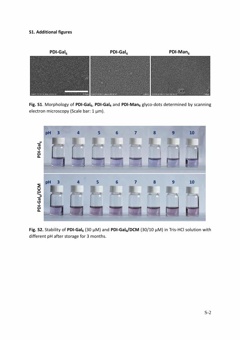
S1. Additional figures
PDI-Gal6 PDI-Gal4 PDI-Man6
Fig. S1. Morphology of PDI-Gal6, PDI-Gal4 and PDI-Man6 glyco-dots determined by scanning
electron microscopy (Scale bar: 1 μm).
PD
I-G
al6
PD
I-G
al6/
DC
M
pH 4 5 6 7 8 9 103
pH 4 5 6 7 8 9 103
Fig. S2. Stability of PDI-Gal6 (30 μM) and PDI-Gal6/DCM (30/10 μM) in Tris-HCl solution with
different pH after storage for 3 months.

S-3
500 600 700 8000.0
0.4
0.8
1.2
0.1 0.2 0.3 0.4
0.01
0.02
0.03
0 5 10 15 20
1
2
3
4
5
F 0/F
1/(
F 0-F
)
[PDI-Gal6](μM) 1/[PDI-Gal6]
R2=0.996 R2=0.998
a b
No
rmal
ized
I f
Wavelength (nm)
c
PDI-Gal6
Fig. S3 (a) Fluorescence titration of DCM (5 μM) in the presence of increasing PDI-Gal6 (0-30
μM). (b) Stern-Volmer plots and (c) Lineweaver−Burk plots for DCM (5 μM) with increasing
PDI-Gal6, where F0 and F are the fluorescence intensity of DCM in the absence and presence
of PDI-Gal6, respectively. The data for DCM were recoded with an excitation and emission
wavelength of 460 and 620 nm, respectively. The dynamic and static quenching can be
described by the Stern−Volmer’s equation (eq 1) and Lineweaver−Burk equation (eq 2),
respectively.
F0 /F = 1 + KSVcq (1)
1/(F0-F) = 1/F0 + KLB/(F0cq) (2)
where F0 and F are the fluorescence intensities of the fluorophores in the absence and the
presence of a quencher (PDI-Gal6), respectively, cq is the concentration of the quencher, KSV
is the dynamic quenching constant, and KLB is the static quenching constant.

S-4
560 640 7200.0
0.4
0.8
1.2
Wavelength (nm)
No
rmal
ized
I f
DCM
DCM@PDI-Gal6
HSA
FBS
BSA
PNA
ConA
SBA
Fig. S4. Fluorescence spectra of PDI-Gal6/DCM (5/30 μM) in the absence and presence of
different serum proteins including human serum albumin (HSA) (7.5 μM), fetal bovine serum
(FBS) (14 μL, from cesarean section fetal bovine) and bovine serum albumin (BSA) (7.5 μM),
and different lectins including soybean agglutinin (SBA, 7.5 μM), concanavalin A (Con A, 7.5
μM) and peanut agglutinin (PNA, 7.5 μM). All fluorescence measurements were tested in
Tris-HCl buffer (0.02 M, pH 7.4) with an excitation wavelength of 460 nm.

S-5
0.0000
0.0004
0.0008
0.4
0.8
1.2
0.0
0.4
0.8
1.2
Rel
ativ
e m
RN
A le
vel
Rel
ativ
e m
RN
A le
vel
Hep-G2 HeLa A549 MB-231 Hela A549
a b
Fig. S5. Relative mRNA level of (a) asialoglycoprotein receptor (ASGPr) in Hep-G2, HeLa and
A549 cells, and (b) mannose receptor (MR) in MDA-MB-231, HeLa and A549 cells
determined by real-time quantitative polymerase chain reaction.

S-6
Hep
-G2
MD
A-M
B-2
31
Free D-Gal (mM)
0 1 2 5 10
Free D-Man (mM)
0 1 2 5 10
a
0.0
0.4
0.8
1.2
1.6
0.0
0.4
0.8
1.2
1.6b
No
rmal
ize
dI F
No
rmal
ize
dI F
c
*** ***
PDI-Gal6/DCMFree D-Gal (mM)
+-
+1
+2
+5
+10
PDI-Man6/DCMFree D-Gal (mM)
+-
+1
+2
+5
+10
Fig. S6. Fluorescence imaging (a) and quantification (b,c) of different cancer cells with
PDI-Gal6/DCM or PDI-Man6/DCM in the presence of increasing free D-galactose or
D-mannose (***P < 0.001). Excitation channel: 460–490 nm, emission: 580–650 nm; the cell
nucleus was stained by Hoechst 33342; scale bar: 100 μm.
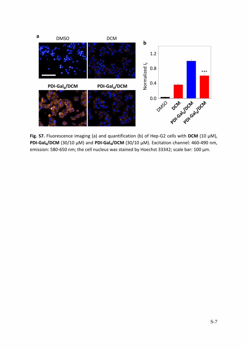
S-7
DCM
PDI-Gal6/DCM PDI-Gal4/DCM
DMSO
0.0
0.4
0.8
1.2
No
rmal
ized
I F
ab
***
Fig. S7. Fluorescence imaging (a) and quantification (b) of Hep-G2 cells with DCM (10 μM),
PDI-Gal6/DCM (30/10 μM) and PDI-Gal4/DCM (30/10 μM). Excitation channel: 460-490 nm,
emission: 580-650 nm; the cell nucleus was stained by Hoechst 33342; scale bar: 100 μm.

S-8
0
50
100
150
0
50
100
150
0
50
100
150
Cel
l via
bili
ty (
%)
Cel
l via
bili
ty (
%)
Cel
l via
bili
ty (
%)
Hep-G2
PDI-Gal6 (0-100 μM)
293T
L02
Hep-G2
293T
L02
Hep-G2
293T
L02
PDI-Gal4 (0-100 μM) PDI-Man6 (0-100 μM)
Fig. S8. Cell viability of Hep-G2 (human liver cancer) cells, 293T (human embryonic kidney)
cells and L02 (human normal liver) cells with increasing PDI-Gal6, PDI-Gal4 or PDI-Man6
determined by MTS cell proliferation assay.
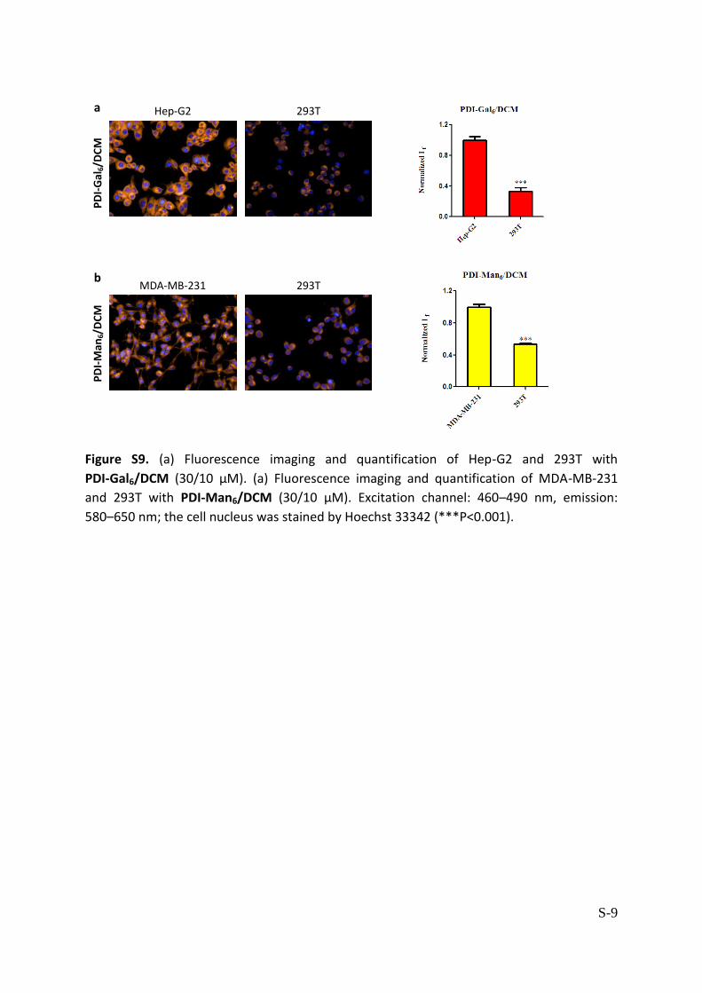
S-9
Hep-G2 293T
293TMDA-MB-231
PD
I-G
al6/D
CM
PD
I-M
an6/D
CM
a
b
Figure S9. (a) Fluorescence imaging and quantification of Hep-G2 and 293T with
PDI-Gal6/DCM (30/10 μM). (a) Fluorescence imaging and quantification of MDA-MB-231
and 293T with PDI-Man6/DCM (30/10 μM). Excitation channel: 460–490 nm, emission:
580–650 nm; the cell nucleus was stained by Hoechst 33342 (***P<0.001).

S-10
S2. Experimental section
General. UV-absorbance spectra were carried out on a Varian Cary 500 spectrophotometer.
Fluorescence spectra were recorded on a Varian Cary Eclipse fluorescence
spectrophotometer. Zeta potential was determined by a Horiba LB-550 DLS Nano-Analyzer.
Ultrapure water was obtained from a Milli-Q integral Pure/Ultrapure Water Production unit.
Supramolecular self-assembly of glyco-dots. DCM (1 mM, DMSO, 100 µL) was added to a
solution of glyco-PDIs (3 mM, Tris-HCl, 0.02 M, pH 7.4, 100 µL). The resulting mixture was
sonicated for 10 min, and then stirred at room temperature for 12 h in the dark to produce
the supramolecular glyco-dots, which can be used as is.
Scanning electron microscopy (SEM). A droplet of glycocluster (60 μM), which had been
sonicated for 15 min in ultrapure water, was cast onto a freshly cleaved mica surface,
followed by drying at room temperature. Then, SEM images of the materials were obtained
by S-3400N (HITACHI, Japan).
High-resolution transmission electron microscope (HRTEM). A droplet of glycocluster (60
μM) with or without DCM (20 μM) was dropped onto carbon copper grids for HRTEM
characterizations. JEOL 2100 equipped with a Gatan Orius charged-coupled device camera
and Tridiem energy filter operating at 200 kV was used for TEM images, and the data were
processed using Image J software.
Cell culture. Hep-G2 Hela and 293T cells were cultured in Dulbecco’s Modified Eagle’s
Medium (Invitrogen, Carlsbad, CA, USA) supplemented with 10% fetal bovine serum (Gibco,
Gland Island, NY, USA). A549 cells were cultured in F12 supplemented with 10% FBS in a
humidified atmosphere of 5% CO2 and 95% air at 37 ̊C and split when the cells reached 85%
confluency. L02 cells were cultured in Roswell Park Memorial Institute (1640) supplemented
with 10% FBS. MDA-MB-231 cells were cultured in 1640 supplemented with 5% FBS in a
humidified atmosphere of 5% CO2 and 95% air at 37 ̊C and split when the cells reached 90%
confluency.
Generation of shASGPR1 and control shRNA-HepG2 stable cell lines infected with
lentivirus. pCAGVSVG (a plasmid encoding envelope protein) and PAX2 (packaging plasmid)
were kind gifts from Dr. J. Wong (East China Normal University, China). The shRNA plasmids
encoding ASGPR1-specific shRNA or scramble shRNA were purchased from Santa Cruz
Biotechnology Inc. (Santa Cruz, CA, USA). Lentiviral particles were generated according to
the manufacturer’s instructions. Briefly, 293T cells were seeded in a six-well tissue culture
plate and were grown to a 80-90% confluency in antibiotic-free normal growth medium
supplemented with FBS. Then, shRNA plasmid (shRNA of ASGPR1 or control, 3 µg) was
cotransfected with pCAG-VSVG (1.8 µg) and PAX2 (2.7 µg) into 293T cells using 15 µL of
lipofectamine 2000 (Invitrogen, Carlsbad, CA, USA). After 6 h, the medium was changed to

S-11
fresh DMEM with 10% FBS. After 72 h, the lentivirus-cotaining supernatant were collected,
filtered, and then employed for analysis.
Hep-G2 cells were plated in a 12-well plate 24 h prior to viral infection. The cells at
approximately 50% confluency were infected with the lentiviral particles prepared as
described above. The plates were incubated overnight and the medium was then changed
to fresh complete medium. Two days after infection, the cells were splited at 1:5 and
incubated for another 24 h in complete medium. Then puromycin (4 μg mL-1) was added to
select the stable clones expressing the shRNA. Medium was replaced with fresh
puromycin-containing medium every 3-4 days until resistant colonies can be identified.
Several colonies were picked, expanded, and then assayed for stable shRNA expression by
evaluating level of ASGPR1 mRNA via real-time quantitative polymerase chain reaction
(qPCR).
Generation of shMR and control shRNA-MDA-MB-231 stable cell lines infected with
lentivirus. MDA-MB-231 cells were plated in a 6-well plate 24 h prior to viral infection. The
cells at approximately 50% confluency were infected with the lentiviral particles containing
shRNA targeting human MR or control, which were purchased from Genomeditech
(Genomeditech, Shanghai, China). The plates were incubated for 6-8 h according to the
manufacturer’s instructions and the medium was then changed to fresh complete medium.
Two days after infection, the cells were splited at 1:3 and incubated for another 24 h in
complete medium. Then puromycin (4 μg mL-1) was added to select the stable clones
expressing the shRNA. Medium was replaced with fresh puromycin-containing medium
every 3-4 days until resistant colonies can be identified. Several colonies were picked,
expanded, and then assayed for stable shRNA expression by evaluating level of MR mRNA
via real-time qPCR.
Fluorescence imaging of cells. Cells (Hep-G2/MDA-MB-231, 25000/well; HeLa, 12000/well;
A549, 15000/well) were seeded on a black 96-well microplate with optically clear bottom
(Greiner bio-one, Germany) overnight. Then, the cells were incubated with DCM in the
absence and presence of glyco-dots for 15 min. For the competition assay, Hep-G2 and
MDA-MB-231 cells were preincubated with free D-galactose and D-mannose for 2 h,
followed by incubation with PDI-Gal6/DCM and PDI-Man6/DCM for 15 min, respectively.
Then, the cells were gently washed with PBS (phosphate buffered saline) three times, and
stained with Hoechst 33342 (5 μg mL-1) at 37 °C in a humidified atmosphere of 5% CO2 in air
for 5 min. Then, cells were washed with PBS three times. The fluorescence images were
recorded using an Operetta high content imaging system and quantified by the Columbus
image data analysis system (Perkinelmer, US).
Real-time quantitative PCR. Total RNA was isolated from cells using TRIzol Reagent
(Invitrogen) according to the manufacturer’s protocol. Complementary DNA generated
using a PrimeScript® RT reagent kit (TaKaRa, Dalian, China) was analyzed by quantitative
PCR using SYBR® Premix Ex TaqTM. Real-time PCR was performed using a 7300 Real-Time

S-12
PCR system (Applied Biosystems, CA, USA). GAPDH was detected as the housekeeping gene.
Primers for qPCR were as follows:
GAPDH forward, 5’-ATCACTGCCACCCAGAAGAC-3’
and reverse, 5’-ATGAGGTCCACCACCCTGTT-3’
ASGPR1 forward, 5’-CTGGACAATGAGGAGAGTGAC-3’
and reverse, 5’-TTGAAGCCCGTCTCGTAGTC-3’
Mannose Receptor forward,5’-GCAGCTCTGGGAACTTGGAT-3’
and reverse, 5’-TTGCCTGGTGTCCAGTAGGA-3’
Cell viability assay. Cells were plated overnight on 96-well plates at 8000 cells per well in
growth medium. After seeding, cells were treated with glycoclusters at different
concentrations for 48 h. After 48 h exposure, a MTS/PMS (20:1, Promega Corp) solution (10
μL per well) was added to each well, followed by a gentle shake. After 2-4 h incubation at
37 °C under 5% CO2, the absorbance of the mixture solutions was measured at 490 nm as a
reference with an M5 microplate reader (Molecular Device, USA). The optical density of the
result in MTS assay was directly proportional to the number of viable cells.

S-13
S3. Syntheses of the PDI-based glycoclusters
General procedures
All reagents for synthesis commercially available (highest purity available for reagent grade
compounds) were used without further purification. Solvents were distilled over CaH2
(CH2Cl2), Mg/I2 (MeOH), Na/benzophenone (THF) or purchased dry. Reactions under
microwave activation were performed on a Biotage Initiator system. Thin-layer
chromatography (TLC) was carried out on aluminum sheets coated with silica gel 60 F254
(Merck). TLC plates were inspected by UV light (λ = 254 nm, 365 nm) and developed by
treatment with a mixture of 10% H2SO4 in EtOH/H2O (95:5 v/v) followed by heating. Silica
gel column chromatography was performed with silica gel Si 60 (40–63 μm). Optical rotation
was measured using a Perkin Elmer polarimeter. NMR spectra were recorded at 293 K,
unless stated otherwise. Chemical shifts are referenced relative to deuterated solvent
residual peaks. The following abbreviations are used to explain the observed multiplicities: s,
singlet; d, doublet; t, triplet; q, quadruplet; m, multiplet; p, pseudo and b, broad. Complete
signal assignments were based on 1D and 2D NMR correlations COSY and HSQC. High
resolution (HR-ESI-QToF) mass spectra were recorded using a Bruker MicroToF-Q II XL
spectrometer. The glycoclusters tested in bioassays were purified using automated
purification systems with medium pressure chromatography on reverse C18 silica gel. Their
purity was verified by 1H and 13C NMR techniques, indicating ca. 95% purity.
General procedure for 1,3-dipolar cycloadditions (Method A)
The alkyne-functionalized perylenediimide 5 or 6 (1 eq.), CuI (0.5 eq,), DIPEA (1.5 eq. per
alkyne function) and azido-derivative 7 (1.5 eq. per alkyne function) in DMF were introduced
into a Biotage Initiator 2-5 mL vial. The vial was sealed with a septum cap and heated at
110°C for 15 min under microwave irradiation (solvent absorption level : High). The crude
mixture was concentrated and co-evaporated with toluene 6 times then purified by flash
silica gel column chromatography to afford the desired cycloadducts.
General procedure for the Zemplén deacetylation (Method B)
To a suspension of acetylated glycocluster (1 eq.) in distilled MeOH was added MeONa (0.2
eq.). The mixture was stirred at r.t. for 16 hours, neutralized with Amberlite IR-120 resin (H+
form), filtrated and concentrated in vacuo to afford the corresponding hydroxylated
glycoclusters.

S-14
N,N’-Bis-{1-[1-(2,3,4,6-tetra-O-acetyl- -D-galactopyranosyloxy)-3,6-dioxaoct-8-yl]-1,2,3-tri
azol-4-ylmethyl}-1,6,7,12-tetra-(4-{1-[1-(2,3,4,6-tetra-O-acetyl- -D-galactopyranosyloxy)-3,
6-dioxaoct-8-yl]-1,2,3-triazol-4-ylmethyloxy}phenoxy)perylene-3,4,9,10-tetracarboxylic
diimide (2-Gal): Obtained as a purple foam following Method A: 1 (10 mg, 0.010 mmol, 1
eq.), compound Ac4Gal-TEG-N3 (45 mg, 0.089 mmol, 9.4 eq.), CuI (1 mg, 0.005 mmol, 0.5
eq.) and DIPEA (12 µL, 0.066 mmol, 7 eq.). Purified by silica gel flash chromatography
(CH2Cl2/MeOH 99/1 to 95/5).
Yield = 77% (30 mg), Rf = 0.45 (CH2Cl2/MeOH 95/5). 1H NMR (500 MHz, CDCl3) δ (ppm): 8.09 (s, 4H, perylene-H), 7.98 (s, 4H, H-triaz), 7.75 (s, 2H,
H-triaz), 6.91 (d, J = 9.9 Hz, 16H, H-ar), 5.43 (bs, 4H, NCH2-triaz), 5.38 (d, J = 3.1 Hz, 4H, H-4),
5.36 (d, J = 3.1 Hz, 2H, H-4’), 5.23 (bs, 8H, OCH2-triaz), 5.21-5.13 (M, 6H, H-2, H-2’), 5.02 (dd,
J = 10.4, 3.2 Hz, 4H, H-3), 5.00 (dd, J = 10.4, 3.3 Hz, 2H, H-3’), 4.64 (s, 8H, NCH2CH2), 4.55 (d, J
= 8.0 Hz, 4H, H-1), 4.53 (d, J = 8.1 Hz, 2H, H-1’), 4.47 (bs, 4H, NCH2CH2), 4.21-4.06 (m, 12H,
H-6, H-6’), 4.02-3.87, 3.87-3.79 (2m, 18H, OCH2, H-5, H-5’), 3.79-3.39 (m, 48H, 4×OCH2), 2.13,
2.12, 2.04, 2.02, 2.01, 2.01, 1.97, 1.96 (8s, 72H, 24×COCH3). 13C NMR (125 MHz, CDCl3) δ (ppm): 170.5, 170.4, 170.3, 169.58, 169.55 (4×COCH3), 163.1
(ArCON), 156.63, 155.58, 149.0 (3×CIV-ar-O), 143.8, 143.3 (2×CIV-triaz), 133.0 (CIV-ar), 124.3
(6C, CH-triaz), 122.4 (CIV-ar), 121.6 (8C, CH-ar), 120.2, 119.4 (2×CIV-ar), 119.3 (CH-perylene),
116.3 (8C, CH-ar), 101.51 (C-1), 101.47 (C-1’), 71.0 (C-3, C-3’), 70.82 (C-5), 70.80, 70.78,
70.74, 70.72, 70.66, 70.38, 70.34, 69.61, 69.56 (C-5’, 4×OCH2), 69.32, 69.29 (GalOCH2), 69.96
(C-2), 68.95 (C-2’), 67.2 (C-4, C-4’), 62.7 (OCH2-triaz), 61.4 (C-6), 60.5 (C-6’), 50.5, 50.2
(NCH2CH2), 35.4 (NCH2-triaz), 20.93, 20.91, 20.82, 20.81, 20.7 (4×COCH3).
HR-ESI-QTof (positive mode) m/z: calcd for C186H227N20O84 [M+3H]3+ 1361.4696, found
1361.4659.
S-15

S-16
N,N’-Bis-{1-[1-(2,3,4,6-tetra-O-acetyl- -D-mannopyranosyloxy)-3,6-dioxaoct-8-yl]-1,2,3-tria
zol-4-ylmethyl}-1,6,7,12-tetra-(4-{1-[1-(2,3,4,6-tetra-O-acetyl- -D-mannopyranosyloxy)-3,6
-dioxaoct-8-yl]-1,2,3-triazol-4-ylmethyloxy}phenoxy)perylene-3,4,9,10-tetracarboxylic
diimide (2-Man): Obtained as a purple foam following Method A: 1 (50 mg, 0.048 mmol, 1
eq.), compound Ac4Man-TEG-N3 (216 mg, 0.428 mmol, 9 eq.), CuI (4.5 mg, 0.024 mmol, 0.5
eq.) and DIPEA (74 µL, 0.428 mmol, 9 eq.). Purified by silica gel flash chromatography
(CH2Cl2/MeOH 99/1 to 95/5).
Yield = 67% (130 mg), Rf = 0.45 (CH2Cl2/MeOH 95/5). 1H NMR (400 MHz, CDCl3) δ (ppm): 8.03 (s, 4H, perylene-H), 7.82 (s, 4H, H-triaz), 7.66 (s, 2H,
H-triaz), 6.85 (d, J = 6.3 Hz, 16H, H-ar), 5.34 (bs, 4H, NCH2-triaz), 5.31-5.15 (m, 18H, H-2, H-2’,
H-3, H-3’, H-4, H-4’), 5.12 (bs, 8H, OCH2-triaz), 4.82 (d, J = 1.0 Hz, 4H, H-1), 4.79 (2d, J = 1.1
Hz, 2H, H-1’), 4.55 (t, J = 4.9 Hz, 8H, NCH2CH2), 4.40 (t, J = 4.9 Hz, 4H, NCH2CH2), 4.25-4.15 (m,
6H, H-6a, H-6a’), 4.08-3.95 (m, 12H, H-5, H-5’, H-6b, H-6b’), 3.87 (t, J = 5.0 Hz, 8H, OCH2),
3.80-3.47 (m, 52H, OCH2), 2.08, 2.06, 2.02, 2.00, 1.97, 1.95, 1.92, 1.90 (8s, 72H, 24×COCH3). 13C NMR (100 MHz, CDCl3) δ (ppm): 170.56, 170.54, 170.0, 169.92, 169.86, 169.81, 169.6 (7s,
COCH3), 162.9 (ArCON), 156.4, 155.4, 148.8 (3×CIV-ar-O), 143.6, 143.1 (2×CIV-triaz), 132.7
(CIV-ar), 124.1 (6C, CH-triaz), 122.2 (CIV-ar), 121.4 (8C, CH-ar), 119.9 (CIV-ar), 119.1
(CH-perylene, CIV-ar), 116.1 (8C, CH-ar), 97.59, 97.55 (2s, C-1, C-1’), 70.60, 70.54, 70.45,
70.40, 69.9 (3×OCH2), 69.45, 69.42, 69.37 (OCH2, C-2, C-2’), 69.0 (C-3, C-3’), 68.4, 68.3 (C-5,
C-5’), 67.3, 67.2 (OCH2), 66.0 (C-4, C-4’), 62.4 (OCH2-triaz), 62.32, 62.29 (C-6, C-6’), 50.3, 50.0
(NCH2CH2), 35.2 (NCH2-triaz), 20.9-20.8, 20.7-20.6 (2m, 4×COCH3).
HR-ESI-QTof (positive mode) m/z: calcd for C186H224N20Na2O84 [M+2Na]2+ 2063.6828, found
2063.6744.

S-17

S-18
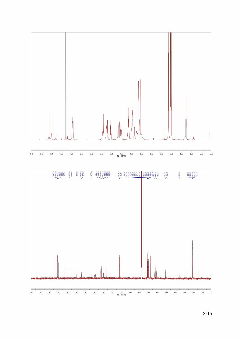
N,N’-Bis-{1-[1- -D-galactopyranosyloxy)-3,6-dioxaoct-8-yl]-1,2,3-triazol-4-ylmethyl}-1,6,7,
12-tetra-(4-{1-[1- -D-galactopyranosyloxy)-3,6-dioxaoct-8-yl]-1,2,3-triazol-4-ylmethyloxy}
phenoxy)perylene-3,4,9,10-tetracarboxylic diimide (PDI-Gal6): Obtained from 2-Gal (112
mg, 0.027 mmol) as a deep purple foam following Method B.
Yield = 88% (75 mg).
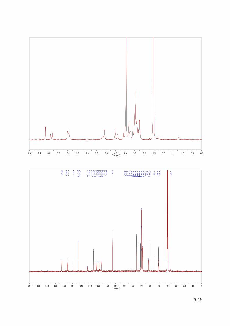
1H NMR (300 MHz, DMSO-d6 2O) δ (ppm): 8.16 (s, 4H, H-triaz), 7.91 (s, 2H, H-triaz), 7.81
(s, 4H, perylene-H), 6.96 (d, J = 15.9 Hz, 16H, CH-ar), 5.08 (s, 12H, OCH2-triaz, NCH2-triaz),
4.51, 4.38 (2s, 12H, NCH2CH2), 4.11-4.04 (m, 6H, H-1), 3.88-3.17 (m, 96H, H-2, H-3, H-4, H-5,
H-6, 5×OCH2).
13C NMR (100 MHz, DMSO-d6 2O) δ (ppm): 162.5 (ArCON), 156.2, 155.5, 148.4
(3×CIV-ar-O), 142.8, 142.7 (2×CIV-triaz), 132.5 (CIV-ar), 125.4, 124.0 (2×CH-triaz), 122.4 (CIV-ar),
121.6 (CH-ar), 119.2, 118.8 (2×CIV-ar), 118.1 (CH-perylene), 116.4 (CH-ar), 103.7 (C-1), 75.4
(C-3), 75.3 (C-2), 73.5 (C-5), 70.7, 70.0, 69.9, 69.8, 69.7, 69.0, 68.8 (OCH2), 68.5-68.2 (m, C-4,
OCH2), 68.1 (OCH2), 61.7 (OCH2-triaz), 60.6 (C-6), 49.8, 49.7 (NCH2CH2), 35.7 (NCH2-triaz).
HR-ESI-QTof (positive mode) m/z: calcd for C138H176N20Na2O60 [M+2Na]2+ 1559.5580, found
1559.5608.
S-19

S-20
N,N’-Bis-{1-[1- -D-mannopyranosyloxy)-3,6-dioxaoct-8-yl]-1,2,3-triazol-4-ylmethyl}-1,6,7,
12-tetra-(4-{1-[1- -D-mannopyranosyloxy)-3,6-dioxaoct-8-yl]-1,2,3-triazol-4-ylmethyloxy}
phenoxy)perylene-3,4,9,10-tetracarboxylic diimide (PDI-Man6): Obtained from 2-Man (119
mg, 0.029 mmol) as a deep purple foam following Method B.
Yield = 87% (78 mg).
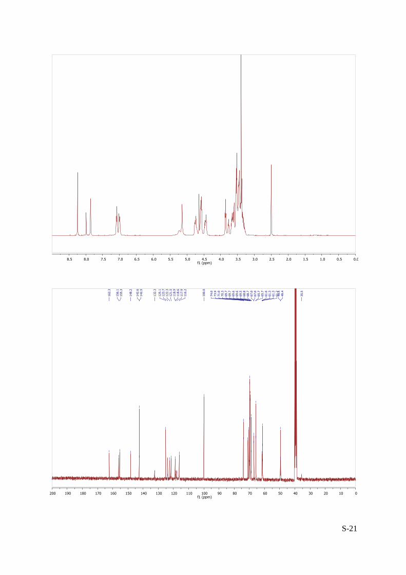
1H NMR (400 MHz, DMSO-d6 2O) δ (ppm): 8.24 (s, 4H, H-triaz), 7.99 (s, 2H, H-triaz), 7.86
(s, 4H, perylene-H), 7.04 (dd, J = 33.3, 8.7 Hz, 16H, CH-ar), 5.14 (bs, 12H, OCH2-triaz,
NCH2-triaz), 4.64 (d, J = 1.2 Hz, 4H, H-1), 4.62-4.54 (m, 10H, H-1’, NCH2CH2), 4.50-4.39 (m, 4H,
NCH2CH2), 3.85 (t, J = 5.1 Hz, 8H, OCH2), 3.77 (t, J = 5.1 Hz, 4H, OCH2), 3.71-3.26 (m, 84H, H-2,
H-2’, H-3, H-3’, H-4, H-4’, H-5, H-5’, H-6, H-6’, 4×OCH2).
13C NMR (100 MHz, DMSO-d6 + 2O) δ (ppm): 162.3 (ArCON), 156.1, 155.4, 148.2
(3×CIV-ar-O), 142.6, 142.5 (2×CIV-triaz), 132.3 (CIV-ar), 125.1, 123.8 (2s, 2×CH-triaz), 122.3
(CIV-ar), 121.5 (CH-ar), 118.9, 118.6 (2s, 2×CIV-ar), 117.9 (CH-perylene), 116.2 (CH-ar), 100.0
(C-1, C-1’), 74.0 (C-4, C-4’), 71.0 (C-3, C-3’), 70.3 (C-2, C-2’), 69.74, 69.72, 69.60, 69.56, 69.50,
68.8, 68.7 (4×OCH2), 67.0 (C-5, C-5’), 65.74, 65.70 (OCH2), 61.6 (OCH2-triaz), 61.3 (C-6, C-6’),
49.6, 49.4 (2s, NCH2CH2) 35.5 (NCH2-triaz).
HR-ESI-QTof (positive mode) m/z: calcd for C138H178N20O60 [M+2H]2+ 1537.5741, found
1537.5681.
S-21

S-22
Nia, A. S. et al. Tetrahedron 2012, 68, 722-729.
1,6,7,12-Tetrachloroperylene-3,4,9,10-tetracarboxylic dianhydride (4)
Iodine (1.62 g, 6.4 mmol, 0.25 eq.) was added to a solution of 3,4,9,10-perylene
tetracarboxylic dianhydride 3 (10 g, 25.5 mmol, 1 eq.) in chlorosulfonic acid (100 mL) at
room temperature. The reacting mixture was heated to 70°C for 5 h. Afterwards, the
mixture was poured slowly into an ice-water mixture (1 L). The insoluble product was
collected by filtration and washed with water until pH reached 7. The crude product was
dried at 100°C in oven for 3 days and purified by soxhlet extraction using dichloromethane
as solvent. The solvent was evaporated off to afford 4 as a red solid.
Yield = 51% (6.96 g).
1H NMR (300 MHz, CDCl3) δ (ppm): 8.75 (s, 4H, perylene-H).

S-23
N,N’-Bis-(t-butoxycarbonylmethyl)-1,6,7,12-tetrachloroperylene-3,4,9,10-tetracarboxylic
diimide (5): Triethylamine (188 µL, 1.36 mmol, 3.6 eq.) was added to a suspension of 4 (200
mg, 0.377 mmol, 1 eq.) and glycine t-butyl ester hydrochloride (190 mg, 1.13 mmol, 3 eq.) in
isopropanol (10 mL). The mixture was stirred at 90°C for 22 h. The mixture was then poured
into water (100 mL) and the red solid was filtered off then washed with water (2×10mL)
until neutral pH. The red residue was collected with CH2Cl2 (50 mL) and the solvent was
evaporated off. The crude solid was purified by silica gel flash chromatography
(CH2Cl2/EtOAc 97/3) to afford 5 as a red solid.
Yield = 87% (247 mg); Rf = 0.30 (CH2Cl2/EtOAc 95/5).
1H NMR (400 MHz, CDCl3) δ (ppm): 8.65 (s, 4H, perylene-H), 4.85 (s, 4H, NCH2), 1.50 (s, 18H,
CMe3).
13C NMR (100 MHz, CDCl3) δ (ppm): 166.6 (COO), 162.0 (NCO), 135.5 (CCl), 133.3 (CH-ar),
131.5 (CIV-ar), 128.9 (CIV-ar), 123.5 (CIV-ar), 122.9 (CIV-ar), 82.8 (CMe3), 42.4 (NCH2), 28.1
(CMe3).
HR-ESI-QTof (positive mode) m/z: calcd for C36H26Cl4N2NaO8 [M+Na]+ 777.0335, found
777.0320.

S-24

S-25
N,N’-Bis-(t-butoxycarbonylmethyl)-1,6,7,12-tetra-(4-propargyloxyphenoxy)perylene-3,4,9,
10-tetracarboxylic diimide (6): Potassium carbonate (239 mg, 1.73 mmol, 8 eq.) was added
to a suspension of 5 (163 mg, 0.216 mmol, 1 eq.) and p-propargyloxyphenol (192 mg, 1.30
mmol, 6 eq.) in N-methyl-2-pyrrolidone (15 mL). The reaction was stirred at r.t. and turned
immediately from dark red to black. After 30 min, the reaction mixture was stirred at 90°C
for 15 h. The mixture was then poured into 1N HCl (150 mL) and the deep purple solid was
filtered off then washed with water (2×20mL) until neutral pH. The deep purple residue was
collected with CH2Cl2 (50 mL) then evaporated and purified by silica gel flash
chromatography (CH2Cl2/EtOAc 97/3) to afford 6 as a deep purple solid.
Yield = 23% (61 mg); Rf = 0.70 (CH2Cl2/EtOAc 99/1).
1H NMR (400 MHz, CDCl3) δ (ppm): 8.13 (s, 4H, perylene-H), 6.88 (s, 16H, CH-ar), 4.75 (bs,
4H, NCH2), 4.66 (d, J = 2.4 Hz, 8H, OCH2), 2.57 (t, J = 2.4 Hz, 4H, C≡CH), 1.46 (s, 18H, CMe3).
13C NMR (100 MHz, CDCl3) δ (ppm): 167.1 (COO), 163.1 (NCO), 156.4, 154.6, 149.4
(3×CIV-ar-O), 133.0, 122.1 (2×CIV-ar), 121.3 (CH-ar), 120.4, 119.6 (2×CIV-ar), 119.5
(CH-perylene), 116.5 (CH-ar), 82.5 (CMe3), 78.5 (C≡CH), 75.9 (C≡CH), 56.5 (OCH2), 42.2
(NCH2), 28.2 (CMe3).
HR-ESI-QTof (positive mode) m/z: calcd for C72H54N2NaO16 [M+Na]+ 1225.3366, found
1225.3354.

S-26

S-27
N,N’-Bis-(t-butoxycarbonylmethyl)-1,6,7,12-tetra-(4-{1-[1-(2,3,4,6-tetra-O-acetyl- -D-galac
topyranosyloxy)-3,6-dioxaoct-8-yl]-1,2,3-triazol-4-ylmethyloxy}phenoxy)perylene-3,4,9,10
-tetracarboxylic diimide (7-Gal) : Obtained as a purple foam following Method A: 6 (47 mg,
0.039 mmol, 1 eq.), Ac4Gal-TEG-N3 (99 mg, 0.196 mmol, 5 eq.), CuI (4 mg, 0.020 mmol, 0.5
eq.) and DIPEA (34 µL, 0.196 mmol, 5 eq.). Purified by silica gel flash chromatography
(CH2Cl2/MeOH 99/1 to 96/4).
Yield = 52% (66 mg), Rf = 0.45 (CH2Cl2/MeOH 95/5).
1H NMR (300 MHz, CDCl3) δ (ppm): 8.11 (s, 4H, H-triaz), 7.86 (s, 4H, perylene-H), 6.92 (s, 16H,
CH-ar), 5.39 (d, J = 3.4 Hz, 4H, H-4), 5.25-5.15 (m, 12H, H-2, OCH2-triaz), 5.02 (dd, J = 10.4,
3.4 Hz, 4H, H-3), 4.76 (bs, 4H, NCH2CO), 4.60 (t, J = 5.2 Hz, 8H, NCH2CH2), 4.55 (d, J = 7.9 Hz,
4H, H-1), 4.13 (m, 8H, H-6), 4.02-3.88 (m, 12H, H-5, OCH2), 3.78-3.54 (m, 32H, 4×OCH2), 2.14,
2.04, 2.03, 1.98 (4s, 48H, COCH3), 1.46 (s, 18H, CMe3).
13C NMR (100 MHz, CDCl3) δ (ppm): 170.5, 170.3, 170.2, 169.5 (4×COCH3), 167.0 (COO),
163.1 (ArCON), 156.5, 155.5, 149.0 (3×CIV-ar-O), 132.9 (CIV-ar), 124.2 (CIV-triaz), 122.1 (CIV-ar),
121.5 (CH-ar), 120.3, 119.4 (2×CIV-ar), 119.3 (CH-perylene), 116.2 (CH-ar), 101.4 (C-1), 82.4
(CMe3), 70.9 (C-3), 70.7 (C-5, OCH2), 70.6, 70.3, 69.5, 69.2 (4×OCH2), 68.9 (C-2), 67.1 (C-4),
62.5 (OCH2-triaz), 61.3 (C-6), 50.4 (NCH2CH2), 42.2 (NCH2CO), 28.1 (CMe3), 20.9, 20.8, 20.7
(4×COCH3).
HR-ESI-QTof (positive mode) m/z: calcd for C152H180N14O64 [M+2H]2+ 1612.5625, found
1612.5634.

S-28

S-29
N,N’-Bis-(carboxymethyl)-1,6,7,12-tetra-(4-{1-[1-(2,3,4,6-tetra-O-acetyl- -D-galactopyrano
syloxy)-3,6-dioxaoct-8-yl]-1,2,3-triazol-4-ylmethyloxy}phenoxy)perylene-3,4,9,10-tetracar
boxylic diimide (7-Gal-CO2H): Obtained from 7-Gal (47 mg, 0.039 mmol, 1 eq.) as a purple
foam following Method C.
Rf = 0.30 (CH2Cl2/MeOH 95/5). 1H NMR (300 MHz, CDCl3) δ (ppm): 8.04 (s, 4H, H-triaz), 7.84 (s, 4H, perylene-H), 6.86 (s, 16H,
CH-ar), 5.38 (d, J = 3.2 Hz, 4H, H-4), 5.19 (dd, J = 10.5, 7.9 Hz, 4H, H-2), 5.15 (bs, 8H,
OCH2-triaz), 5.02 (dd, J = 10.4, 3.2 Hz, 4H, H-3), 4.93-4.83 (bs, 4H, NCH2CO), 4.63-4.57 (m, 8H,
NCH2CH2), 4.54 (d, J = 7.9 Hz, 4H, H-1), 4.14 (m, 8H, H-6), 4.00-3.84 (m, 12H, H-5, OCH2),
3.77-3.54 (m, 32H, 4×OCH2), 2.13, 2.03, 2.02, 1.97 (4s, 48H, COCH3).
HR-ESI-QTof (positive mode) m/z: calcd for C144H164N14O64 [M+2H]2+ 1556.4999, found
1556.4916.

S-30

S-31
N,N’-Bis-(carboxymethyl)-1,6,7,12-tetra-(4-{1-[1-( -D-galactopyranosyloxy)-3,6-dioxaoct-8-
yl]-1,2,3-triazol-4-ylmethyloxy}phenoxy)perylene-3,4,9,10-tetracarboxylic diimide
(PDI-Gal4): Obtained from 7-Gal-CO2H (121 mg, 0.038 mmol) as a deep purple foam
following Method B.
Yield = 90% (82 mg). After two steps
1H NMR (400 MHz, DMSO-d6 + D2O) δ (ppm): 8.24 (s, 4H, H-triaz), 7.75 (s, 4H, perylene-H),
7.01 (bs, 8H, CH-ar), 6.88 (bs, 8H, CH-ar), 5.12 (s, 12H, OCH2-triaz, NCH2-triaz), 4.56 (s, 12H,
NCH2CH2), 4.07 (d, J = 7.0 Hz, 4H, H-1), 3.84 (bs, 12H, OCH2), 3.61(s, 4H, H-4), 3.70-3.34 (m,
52H, OCH2), 3.34-3.20 (m, 12H, H-2, H-3, H-5).
13C NMR (100 MHz, DMSO-d6 2O) δ (ppm): 170.0 (COO), 162.2 (ArCON), 155.9, 155.0,
148.5 (3×CIV-ar-O), 142.5 (CIV-triaz), 132.2 (CIV-ar), 125.16 (CH-triaz), 122.29 (CIV-ar), 121.3
(CH-ar), 118.9, 118.4 (2×CIV-ar), 117.9 (CH-perylene), 116.1 (CH-ar), 103.6 (C-1), 75.2 (C-3),
73.49 (C-2), 70.53 (C-5), 69.8, 69.7, 69.6, 68.7 (4×OCH2), 68.0 (C-4), 67.74 (OCH2), 61.5
(OCH2-triaz), 60.3 (C-6), 49.5 (NCH2CH2), 43.9 (NCH2CO).
HR-ESI-QTof (positive mode) m/z: calcd for C112H132N14O48 [M+2H]2+ 1220.4154, found
1220.4117.

S-32

S-33