supplementary information - royal society of chemistry
TRANSCRIPT

Supplementary Information
Novel microwell with roof capable of buoyant spheroid cultureDaehan Kim,a Kideok Kimab and Joong Yull Park*ac
a School of Mechanical Engineering, College of Engineering, Chung-Ang University, 84 Heukseok-ro, Dongjak-gu, Seoul 06974, Republic of Korea
b Cell-smith Inc., 195 Ogeum-ro, Songpa-gu, Seoul 05643, Republic of Korea
c Department of Intelligent Energy and Industry, Graduate School, Chung-Ang University, 84 Heukseok-ro, Dongjak-gu, Seoul 06974, Republic of Korea
*Author to whom any correspondence should be addressed.
Figure S1. Images of the sigma-well holder and incline stand. (a) The sigma-well holder serves to hold and seal the mold made of PDMS. A glass slide (7.6 x 5.2 cm) covers the holder preventing unwanted leakage of PDMS solution. (b) The sigma-well incline stand serves to tilt the sigma-well holder containing the mold on an inclined surface at an angle. Both the sigma-well holder and incline stand were made with a 3D printer. Scale bars are 3 cm.
Electronic Supplementary Material (ESI) for Lab on a Chip.This journal is © The Royal Society of Chemistry 2021

Figure S2. Photographs of volcanic mountain-like mold and sigma-well in the various depth of volcanic conduit (cavity) conditions. (a) the depth of the volcanic conduit was changed by applying 3 (i), 2.5 (ii), and 1.5 (iii) mg of PDMS solution; the depth of volcanic conduit was 1.35, 0.9, and 0.37 mm, respectively. Scale bars are 1 mm. (b) The trapped air volume is proportional to the depth of the volcanic conduit, and this leads us to have and larger sigma-wells with deeper volcanic conduit cases (i), and smaller sigma-wells with shallower volcanic conduit cases (ii). Scale bars are 1 mm.
Figure S3. Photographs of the sigma-well to show its reproducibility and throughput. (a) Photograph of the sigma-well full array. Scale bar is 5 cm. (b) Part of usable sigma-well. Scale bar is 1 cm. (c) Part of unusable sigma-well. Scale bars are 1 cm. Air not trapped properly; therefore, (i) cavities are not created or (ii) adjacent cavities are connected.
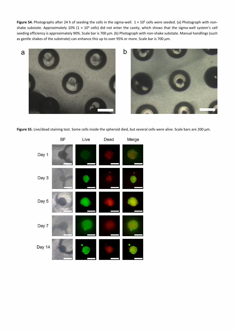
Figure S4. Photographs after 24 h of seeding the cells in the sigma-well. 1 × 10⁵ cells were seeded. (a) Photograph with non-shake substate. Approximately 10% (1 × 10⁴ cells) did not enter the cavity, which shows that the sigma-well system’s cell seeding efficiency is approximately 90%. Scale bar is 700 µm. (b) Photograph with non-shake substate. Manual handlings (such as gentle shakes of the substrate) can enhance this up to over 95% or more. Scale bar is 700 µm.
Figure S5. Live/dead staining test. Some cells inside the spheroid died, but several cells were alive. Scale bars are 200 µm.

Figure S6. Microscopic images of spheroids fixed with formaldehyde solution. (a) Bright field image. Most of the cells lost the shape of the spheroid and were degregated. (b) Fluorescence image merged with the bright field image. Some cells remain in the center of the sigma-well cavity. Scale bars are 700 µm.
Figure S7. HCS LipidTOX™ Green neutral lipid staining test with MRC-5 cells. This test was performed to confirm the accuracy of the neutral lipid staining specific to differentiated adipocytes. (a) Bright field image of 2D cultured MRC-5 cells and (b) its fluorescence image. Unlike adipocyte-differentiated cells, as expected, no MRC-5 cells were stained through the lipid staining. Scale bars are 700 µm.

Figure S8. Bead trap experiment in the sigma-well. (a) Schematic images of sigma-well specimen for photographs. To facilitate bead trapping and photographing, a part of the sigma well is cut and a PDMS block is attached to make a specimen. (b) Top view photographs of the sigma-well with a bead trapped. Glass bead, which is denser than water, is placed at the bottom of the sigma-well by gravity. Scale bars are 1 cm. (c) Side view photograph of the sigma-well with a bead trapped. Sigma-well is flipped to simulate a situation where spheroids float owing to buoyancy. The glass bead is lowered by gravity and trapped at the roof of the sigma-well. Through this experiment, it was physically proved that floating spheroids could be cultured using the sigma-well. Scale bar is 1 cm.
Figure S9. Simulation results on the mean cytokine mass fraction at 300 μm from the center of spheroid of sigma-well and conventional microwell.

Figure S10. A diffusion experiment using a glass capillary tube. Trypan blue was loaded in a 500 μm diameter capillary tube filled with deionized (DI) water. Dye intensity (in x direction) was measured using imageJ software via time-lapse pictures taken at 1 h intervals. A simulation under the same condition as in this experiment was also performed. As a result of comparing the diffusion of the experiment and simulation, it was found that the diffusion tendency was similar. This further demonstrates the reliability of the simulation of cytokine diffusion in the sigma-well and the conventional microwell.