supplementary information - nature · supplementary information ... eugenol was silyl etherated...
TRANSCRIPT

1
Surface initiated self-healing of polymers in aqueous media
Supplementary information (SI)
To prepare the underwater self-healing polymers, silane-protected eugenol acrylates (1 in figure
S1) and silane protected methacrylates (2 in figure S1) were synthesized based on the previously
reported silane protection of eugenol 1, epoxidation 2, 3 and acrylation 4 of alkene group, followed
by UV radical polymerization (figure S1).
Figure S1. Synthetic scheme of self-healing polymers
Synthesis of acrylated (1) and methacrylated (2) silyl protected eugenol
Silyl etherated eugenol. Eugenol was silyl etherated with triethylsilane based on previous report
from our group 1. 1H NMR (600MHz, CDCl3): = 6.77 (d, 1H, Ar-H), 6.69 (d, 1H, Ar-H), 6.64 (q,
1H, Ar-H), 5.98 (m, 1H, -CH=CH2), 5.06 (m, 2H, -CH=CH2), 3.29 (d, 2H, -CH2-CH=CH2), 1.03
(t, 18H, -Si-CH2-CH3), 0.78 (q, 12H, -Si-CH2-CH3). EI-MS, m/z = 401.23 [M+Na+]
Surface-initiated self-healing of polymers in aqueous media
SUPPLEMENTARY INFORMATIONDOI: 10.1038/NMAT4037
NATURE MATERIALS | www.nature.com/naturematerials 1
© 2014 Macmillan Publishers Limited. All rights reserved.

2
Epoxidized silyl eugenol. Epoxidation of silyl etherated eugenol was based on previously
reported method by Ahn et al 2. Briefly, silyl etherated eugenol (30g, 79.22 mmol) was stirred at
0 ˚C to room temperature for 18h with mCPBA (27.34 g, 158.44 mmol) in 300 mL
dichloromethane. Subsequently, the solvent was removed, then the crude material was extracted
with 300 mL ethyl acetate and washed with 2 × 200 mL saturated sodium bisulfite solution, then
washed with saturated sodium bicarbonate until pH indicates neutral. After purification by
column chromatography (silica; hexane/ethyl acetate 99 : 1 to 80 : 20 gradient), a clear oil in 60 %
yield. 1H NMR (600MHz, CDCl3): = 6.77 (d, 1H, Ar-H), 6.69 (d, 1H, Ar-H), 6.64 (q, 1H, Ar-H),
3.11 (m, 1H, -CH2CH-O-CH2), 2.81 (q, 1H, -CH2CH-O-CH2), 2.78 (t, 1H, -CH2CH-O-CH2),
2.69 (q, 1H, -CH2CH-O-CH2), 2.52 (q, 1H, -CH2CH-O-CH2), 1.03 (t, 18H, -Si-CH2CH3), 0.78
(q, 12H, -Si-CH2CH3). 13C NMR (600 MHz, CDCl3): = 146.61, 145.48, 130.13, 121.78, 121.30,
120.32, 52.52, 46.70, 37.98, 6.63, 5.08, 5.04 ppm. EI-MS, m/z = 417.23 [M+Na+]
Acrylated silyl eugenol (1). Epoxy silyl eugenol (13.5g, 34.2 mmol) was agitated with acrylic
acid (3.94g, 54.72 mmol), hydroquinone (37.66 mg, 342 µmol), and AMC-2 (catalyst for acid-
epoxy reaction, AMPAC Fine Chemical, CA, USA) at 90˚C for 4 hours. The crude material was
extracted with 200 mL ethyl acetate and washed with saturated sodium bicarbonate until pH
indicates neutral. After purification by column chromatography (silica; hexane/ethyl acetate 99 :
1 to 80 : 20 gradient), a clear oil was obtained in 50 % yield. 1H NMR (600MHz, CDCl3): = 6.77
(d, 1H, Ar-H), 6.69 (d, 1H, Ar-H), 6.64 (q, 1H, Ar-H), 6.16 (d, 1H, -CH2CH=CH2), 6.17 (q, 1H,
-CH2CH=CH2), 5.86 (d, 1H, -CH2CH=CH2), 4.24 (q, 1H, -CH(OH)CH2OOC-), 4.08(q, 1H, -
CH(OH)CH2OOC-), 2.72 (m, 2H, -CH2CH(OH)-), 2.20(d, 1H, -CH(OH)-), 1.03 (t, 18H, -Si-
CH2CH3), 0.78 (q, 12H, -Si-CH2CH3). 13C NMR (600 MHz, CDCl3): = 166.19, 146.76, 145.65,
2 NATURE MATERIALS | www.nature.com/naturematerials
SUPPLEMENTARY INFORMATION DOI: 10.1038/NMAT4037
© 2014 Macmillan Publishers Limited. All rights reserved.

3
131.19, 128.04, 122.15., 121.66, 120.47, 70.77, 67.56, 39.31, 6.62, 5.07 ppm. EI-MS, m/z =
489.25 [M+Na+]
Methacrylated silyl eugenol (2). Method was same as acrylation above but replacing acrylic
acid with methacrylic acid. After purification by column chromatography (silica; hexane/ethyl
acetate 99 : 1 to 80 : 20 gradient), a clear oil was obtained in 54 % yield. 1H NMR (600MHz,
CDCl3): = 6.77 (d, 1H, Ar-H), 6.69 (d, 1H, Ar-H), 6.64 (q, 1H, Ar-H), 6.13 (d, 1H, -
CH2C(CH3)=CH2), 5.58 (d, 1H, -CH2C(CH3)=CH2), 4.22 (q, 1H, -CH(OH)CH2OOC-), 4.10(q,
1H, -CH(OH)CH2OOC-), 2.72 (m, 2H, -CH2CH(OH)CH2OOC-), 2.20(d, 1H, -CH2CH(OH)
CH2OOC-), 1.95 (d, 3H, -CH2-C(CH3)=CH2), 1.03 (t, 18H, -Si-CH2CH3), 0.78 (q, 12H, -Si-
CH2CH3). 13C NMR (600 MHz, CDCl3): = 166.19, 146.76, 145.65, 131.19, 128.04, 122.15.,
121.66, 120.47, 70.77, 67.56, 39.31, 6.62, 5.07 ppm. EI-MS, m/z = 503.26 [M+Na+]
UV Polymerization. UV radical polymerization was carried out with the Fusion UV system
(Gaithersburg, MD, USA) consisted of a 300 watt/inch (2.54 cm) H lamp and LC6B benchtop
conveyor belt. The lamp distance from the conveyor belt was 10 cm. This UV system recorded
UVA Band (320-390 nm) and 615–660 mJ/cm2 of UV radiation dose with EIT Power Puck 1
Radiometer (Sterling, VA, USA) at a belt speed of 20 feet (6.1 m)/min at focus. Belt speed was
set up at 20ft/min for the polymerization process in this study, and UV dose was controlled by
number of scans. We prepared the polymers with a photointiator (Irgacure 819, Bis(2,4,6-
trimethylbenzoyl)-phenylphosphineoxide), which was kindly provided from BASF (Florham
Park, NJ , USA). 1 wt. % (0.01 g) of Irgacure was added to neat 1 or 2 (1 g). The mixture was
coated on a substrate in 25 µm thick. Soft polymer sample was produced from 1 with 4 scans
(UV radiation dose: 2460-2640 mJ/cm2) and semi-rigid polymer was with 8 scans (UV radiation
NATURE MATERIALS | www.nature.com/naturematerials 3
SUPPLEMENTARY INFORMATIONDOI: 10.1038/NMAT4037
© 2014 Macmillan Publishers Limited. All rights reserved.

4
dose: 4920-5280 mJ/cm2). Rigid polymer sample was produced from 2 was with 8 scans (UV
radiation dose: 4920-5280 mJ/cm2).
Tensile strength. The tensile strength was measured with a Bionix 200 tensile tester (MTS
Systems, Cary, North Carolina). The polymer rods were prepared at diameter 5 mm by casting
and kneading from the UV polymerized polymer films on a Teflon liner. The tensile strength
results showed difference between the semi rigid polymer with acrylate backbone (Young’s
modulus at 1.3 MPa) (left in figure S2) and the rigid polymer with methacrylate backbone
(Young’s modulus at 0.35 GPa) (right in figure S2). We bisected each polymer rod with a clean
razor blade, then soaked the pieces in different pH buffers (pH 3, 7, and 10); the silyl-protecting
groups of the catechols at the polymer surface were conveniently removed at low pH (pH 3
buffer) according to the previously reported method 1 to expose superficial catechol moieties.
However, we do not believe that the pH 3 buffer completely removes the silyl groups at the
interface, thus the study for quantification and optimization of silyl deprotection will be done in a
future study, and it is now under exploration. Subsequently, the tensile strength of each sample
was measured to study the self-healing effect of surface catechol moieties using the Instron.
Polymer self-healing was also investigated in de-aerated buffer at pH 3 to eliminate the
possibility of O2-coupled catechol oxidation (DI water was refluxed under vacuum for 24h and
nitrogen purging). Although most catechols do not undergo auto-oxidation5 at pH 3, tethered
catecholic functionalities on polyacrylate surfaces may be in a unique environment. The results
obtained with untreated or deaerated buffer, however, were essentially indistinguishable (Fig.
S3). To determine whether polyacrylates functionalized with neither catechol- nor triethylsilyl-
blocked catechols show self-healing, we prepared poly(n-butyl-acrylates) using same UV
4 NATURE MATERIALS | www.nature.com/naturematerials
SUPPLEMENTARY INFORMATION DOI: 10.1038/NMAT4037
© 2014 Macmillan Publishers Limited. All rights reserved.

5
polymerization described on page 3. Accordingly, the poly(n-butyl-acrylates) with Young’s
modulus ~350 MPa showed no self-healing in water (pH 3, 7, 10) and exhibited an average
breaking stress of 5.8 MPa.
Figure S2. Tensile test results of the semi-rigid polyacrylates (left) and the rigid
polymethacrylates (right). Original means pristine.
Figure S3. Tensile test results of semi-rigid and rigid polymer rods in pH buffer compared with
degassed pH 3 buffer
0 1 2 3 4 5 6 7 8
Semi-‐rigid pris5ne (bulk) Semi-‐rigid pris5ne (bulk) in the degassed Rigid pris5ne (bulk)
Rigid pris5ne (bulk) in the degassed
Stress (Mpa)
NATURE MATERIALS | www.nature.com/naturematerials 5
SUPPLEMENTARY INFORMATIONDOI: 10.1038/NMAT4037
© 2014 Macmillan Publishers Limited. All rights reserved.

6
NEXAFS were performed at the NIST/Dow soft X-ray materials characterization facility,
beamline U7A at the National Synchrotron Light Source (NSLS) of Brookhaven National
Laboratory. Carbon K-edge partial electron yield data were collected at a grid bias of -150 V.
GIWAXS scans were obtained at the Advanced Light Source at the Lawrence Berkeley National
Laboratory. An X-ray beam was impinged onto the sample at a grazing angle above and below
the critical angle of the polymer film, but below the critical angle of the silicon substrate.
Figure S4. Synchrotron GIWAXS of silyl protected (blocked catechol) and catechol (exposed
catechol) polymer films.
XRR. X-ray reflectivity (XRR) was performed on drop cast polymer film samples on glass
substrates. A Rigaku Smartlab x-ray diffractometer was used to perform the XRR measurement
in high resolution mode in which a 4 bounce Ge(220) monochromator provided a beam (size =
0.2mm) divergence of ~0.005 deg. A solid state scintillator detector with two sets of
programmable receiving slits (size=0.3mm) were used to collect the diffraction data
6 NATURE MATERIALS | www.nature.com/naturematerials
SUPPLEMENTARY INFORMATION DOI: 10.1038/NMAT4037
© 2014 Macmillan Publishers Limited. All rights reserved.

7
Figure S5. XRR, after pH treatment (exposed catechol, top) and before pH treatment (blocked
catechol, bottom)
Confocal Raman spectroscopy. Confocal Raman spectroscopy was conducted with Rennishaw
Raman microscope at the University of California Irvine. The depth of each spectrum was 0.1
µm, and the data was collected from top 10.0-0.1 µm to bottom 0.1-10.0 µm, where depth zero
was presumed as an interface between catechol-exposed polymer films (25 µm thick) on glass
surfaces.
NATURE MATERIALS | www.nature.com/naturematerials 7
SUPPLEMENTARY INFORMATIONDOI: 10.1038/NMAT4037
© 2014 Macmillan Publishers Limited. All rights reserved.

8
Figure S6. Confocal Raman spectroscopy; depth zero is the interface between catechol-exposed
polymer samples
XPS, Kratos Axis Ultra (Kratos Analytical, Manchester UK), was conducted with
survey scans at a pass energy of 160 eV, and high-resolution scans at a pass energy of 20 eV.
8 NATURE MATERIALS | www.nature.com/naturematerials
SUPPLEMENTARY INFORMATION DOI: 10.1038/NMAT4037
© 2014 Macmillan Publishers Limited. All rights reserved.
9
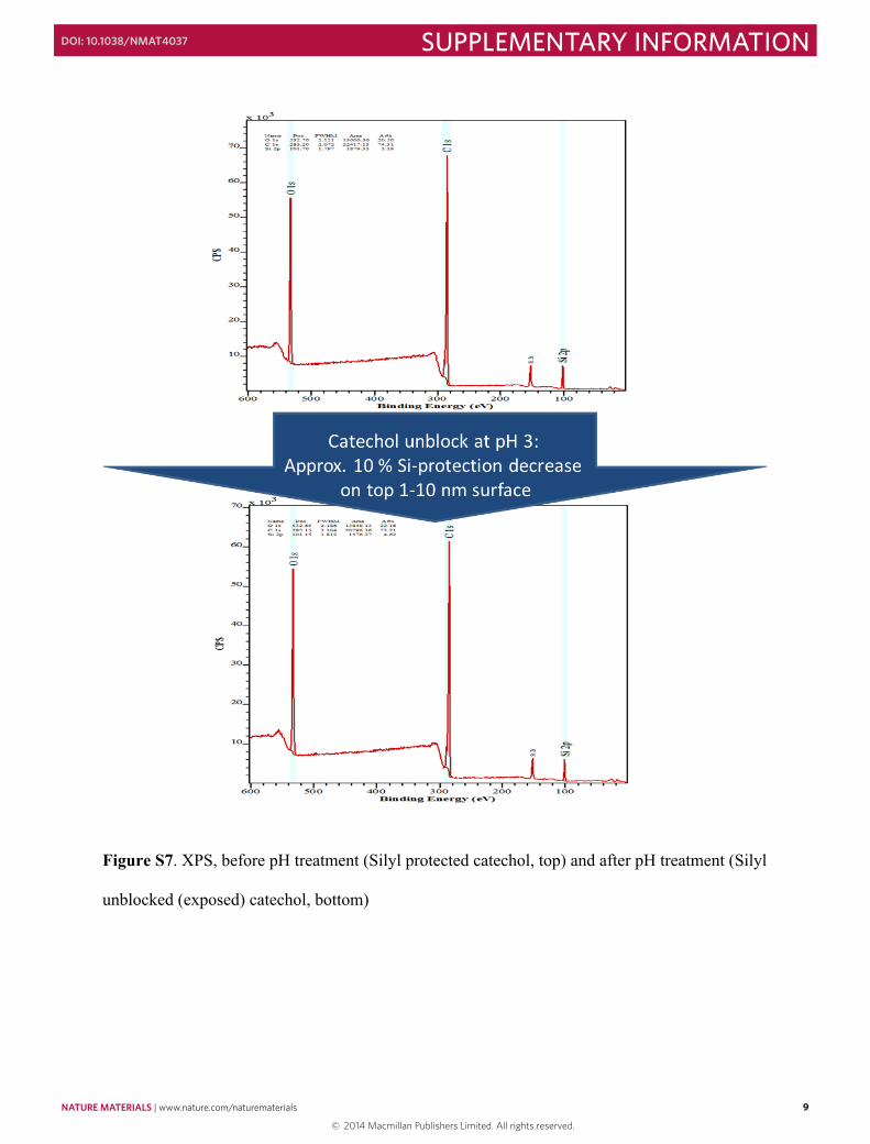
Figure S7. XPS, before pH treatment (Silyl protected catechol, top) and after pH treatment (Silyl
unblocked (exposed) catechol, bottom)
NATURE MATERIALS | www.nature.com/naturematerials 9
SUPPLEMENTARY INFORMATIONDOI: 10.1038/NMAT4037
© 2014 Macmillan Publishers Limited. All rights reserved.

10
Contact angle measurements were performed using a custom-built contact angle goniometer. A 1
sealed contact angle chamber was built of glass, and Teflon. A syringe needle was inserted 2
through a hole at the top Teflon cap of the chamber. The syringe was controlled from the outside 3
by a motorized syringe device (KDS LEGATO270, Kd Scientific). A video camera was used to 4
record the image of the drop. The air inside the chamber was saturated with water vapor for 30 5
min before experiments, maintaining the humidity around ~85%. The water droplet was infused 6
for 2 min with the constant volumetric flow rate of 5 l/min and was in rest for 60 min (Fig. S8). 7
8
Figure S8. Steady state contact angle experiment of water droplet on polymer surface (soft, 9
catechol exposed). 10
The contact angle decreased with time (from 138 to 119°) and three phase contact line 11
consequently crept out (horizontal arrows in panel D). The creeping of contact line is an 12
indication of molecular overturning of polymer, inducing less hydrophobic polymer. In addition, 13
the initial hydrophobicity returned when the surface was dried, suggesting a reversible 14
rearrangement of the polymeric surfaces. 15
10 NATURE MATERIALS | www.nature.com/naturematerials
SUPPLEMENTARY INFORMATION DOI: 10.1038/NMAT4037
© 2014 Macmillan Publishers Limited. All rights reserved.

11
Adhesion/cohesion measurements
Surface Preparation
Two glass discs (one spherical with R=2 cm, and one flat) were thoroughly cleaned with
chloroform and ethanol. The spherical disc was firstly mounted into a custom made ‘cup’ (see
Fig. 3A) prior to polymer deposition. The monomers (silane-protected eugenol acrylates or silane
protected methacrylates) were spread on to the glass discs and UV cured for predetermined times
to achieve soft, semi-rigid, or rigid polymeric surfaces (see UV polymerization section for
details).
Surface modification
For the contact time dependence experiment (Fig. 3), no additional modification was performed
prior to mounting the surfaces into the SFA. For the experiment to check the effect of catechol
oxidation (Fig. 4), catechol moieties at the surface were unblocked (exposed) by soaking in pH 3
buffer for 30 min. Oxidation of catechols to hydroquinone was derived by soaking in 0.01 mM to
100 mM of periodate solution in pH 3 buffer for 10 min.
SFA experiments
The surfaces prepared as above are mounted in SFA 20006 attached with semiconductive strain
gauges at double cantilever springs for load measurement7. Both surfaces are always immersed
in desired buffer (pH 3 sodium aceate or pH 7 phosphate) using a miniaturized ‘cup’ which
contains buffer reservoir (Fig. 3A). Loading and unloading were performed using course
micrometer which gives a maximum displacement of 0.5 cm at the velocity of ~2 mm/s.
10
Contact angle measurements were performed using a custom-built contact angle goniometer. A 1
sealed contact angle chamber was built of glass, and Teflon. A syringe needle was inserted 2
through a hole at the top Teflon cap of the chamber. The syringe was controlled from the outside 3
by a motorized syringe device (KDS LEGATO270, Kd Scientific). A video camera was used to 4
record the image of the drop. The air inside the chamber was saturated with water vapor for 30 5
min before experiments, maintaining the humidity around ~85%. The water droplet was infused 6
for 2 min with the constant volumetric flow rate of 5 l/min and was in rest for 60 min (Fig. S8). 7
8
Figure S8. Steady state contact angle experiment of water droplet on polymer surface (soft, 9
catechol exposed). 10
The contact angle decreased with time (from 138 to 119°) and three phase contact line 11
consequently crept out (horizontal arrows in panel D). The creeping of contact line is an 12
indication of molecular overturning of polymer, inducing less hydrophobic polymer. In addition, 13
the initial hydrophobicity returned when the surface was dried, suggesting a reversible 14
rearrangement of the polymeric surfaces. 15
NATURE MATERIALS | www.nature.com/naturematerials 11
SUPPLEMENTARY INFORMATIONDOI: 10.1038/NMAT4037
© 2014 Macmillan Publishers Limited. All rights reserved.
12
Depending on the stiffness of double cantilever spring (up to ~3000 N/m), this system can
measure up to adhesion force of ~15 N. Applied load is determined by change of the normal
force signal to positive direction and adhesion (pull out) force was measured by the negative
normal force signal at which it jumped to zero normal force (Fig. 3B).
For the first set of experiment (Fig. 3), after loading, the system was equilibrated for a certain
amount of contact time tc (5 – 3600 sec) followed by unloading, to investigate the relation
between the polymer rigidity, contact time and adhesion force. After unloading, the lower
surface was dismounted from SFA to check the damage. For the second set of experiment (Fig.
4), a fixed tc=5 sec was applied, while different surfaces were investigated to study the effects of
hydrogen bonding on adhesion force. Applied load L was set to 250 mN for all cases.
Distinct optical properties between exposed catechols appeared under the microscope
using a polarized light source at near edge of the interface (figure S9), suggesting a mesogen
liquid crystal-like structure formed by the hydrogen bonding between catechol moieties. The
hydrogen bonding between catechol moieties also leads surface self-remanding of the polymer
after scratch (figure S10).
12 NATURE MATERIALS | www.nature.com/naturematerials
SUPPLEMENTARY INFORMATION DOI: 10.1038/NMAT4037
© 2014 Macmillan Publishers Limited. All rights reserved.

13
Figure S9. Polarized microscope image at near interface between blocked catechol polymers
(left) and between unblocked (exposed) catechol polymers (right)
Resealing experiments
The surfaces prepared as above (soft) are scratched with the edge of cover glass (Fisher brand,
22×22 – 1.5) to produce a scar with same thickness. The surfaces are placed under buffers of pH
3 (Fig S10A) and pH 7 (Fig S10B) and monitored under optical microscope (Nikon Eclipse
E800) in order to check the resealability.
NATURE MATERIALS | www.nature.com/naturematerials 13
SUPPLEMENTARY INFORMATIONDOI: 10.1038/NMAT4037
© 2014 Macmillan Publishers Limited. All rights reserved.

14
1
2
Figure S10. Polymer resealing experiment. Catechol (A) exposed and (B) blocked polymeric 3
surfaces were dented with cover glass and were monitored under optical microscope in pH 3 and 4
pH 7 solutions, respectively. (A) Catechol exposed polymer was resealed in 10 min, while (B) 5
catechol blocked polymer was not resealed completely up to 5 hrs. 6
7
References 8
S1. Heo J, Kang T, Jang SG, Hwang DS, Spruell JM, Killops KL, et al. Improved Performance of Protected 9 Catecholic Polysiloxanes for Bioinspired Wet Adhesion to Surface Oxides. J Am Chem Soc 2012, 134(49): 10 20139-20145. 11
12
14 NATURE MATERIALS | www.nature.com/naturematerials
SUPPLEMENTARY INFORMATION DOI: 10.1038/NMAT4037
© 2014 Macmillan Publishers Limited. All rights reserved.

15
S2. Ahn BK, Kraft S, Sun XS. Chemical pathways of epoxidized and hydroxylated fatty acid methyl esters and triglycerides with phosphoric acid. J Mater Chem 2011, 21(26): 9498-9505.
S3. Ahn BK, Kraft S, Wang D, Sun XS. Thermally Stable, Transparent, Pressure-Sensitive Adhesives from
Epoxidized and Dihydroxyl Soybean Oil. Biomacromolecules 2011, 12(5): 1839-1843. S4. Bunker SP, Wool RP. Synthesis and characterization of monomers and polymers for adhesives from methyl
oleate. J Polym Sci Pol Chem 2002, 40(4): 451-458. S5. Lee BP, Messersmith PB, Israelachvili JN, Waite JH. Mussel-Inspired Adhesives and Coatings. In: Clarke
DR, Fratzl P (eds). Annual Review of Materials Research, Vol 41, vol. 41. Annual Reviews: Palo Alto, 2011, pp 99-132.
S6. Israelachvili J, Min Y, Akbulut M, Alig A, Carver G, Greene W, et al. Recent advances in the surface
forces apparatus (SFA) technique. Rep Prog Phys 2010, 73(3): 16. S7. Lee DW, Banquy X, Israelachvili JN. Stick-slip friction and wear of articular joints. Proc Natl Acad Sci U
S A 2013, 110(7): E567-E574.
NATURE MATERIALS | www.nature.com/naturematerials 15
SUPPLEMENTARY INFORMATIONDOI: 10.1038/NMAT4037
© 2014 Macmillan Publishers Limited. All rights reserved.