supplemental tables and figures the … · (a-c) qrt-pcr reveals no significant reduction of bgn,...
TRANSCRIPT

Supplemental Tables and Figures
The metalloproteinase-proteoglycans ADAMTS7 and ADAMTS12 provide an innate,
tendon-specific protective mechanism against heterotopic ossification
Timothy Mead et al

Supplemental Table 1.
Antibodies used for immunofluorescence and western blots.
Antibody Product # Source Dilution
anti-biglycan LF-159 Dr. Larry Fisher, NIH 1:200
anti-fibromodulin LF-150 Dr. Larry Fisher, NIH 1:200
anti- decorin LF-114 Dr. Larry Fisher, NIH 1:200
anti-aggrecan AB1031 EMD-Millipore 1:500
anti-collagen X ab58632 Abcam 1:1000
anti-pSMAD159 #13820 Cell Signaling 1:200
anti-GAPDH MAB374 EMD-Millipore 1:5000
anti-ADAMTS7 AB45044 Abcam 1:100
anti-ADAMTS12 24934-1-AP Proteintech 1:50

Supplemental Table 2.
Quantitative Real-Time PCR primer pairs
Gapdh Forward: 5'-TGGAGAAACCTGCCAAGTATGA-3'
Reverse: 5'-CTGTTGAAGTCGCAGGAGACA-3'
Adamts7 Forward: 5'-GGAGTGAGGACCCAGATAAGTA-3'
Reverse: 5'-CGTGCATAGGTGAAGGTAGTG-3'
Adamts12 Forward: 5'-CCAAAGGTGCGAGGGATATAAG-3'
Reverse: 5'-ACCCTCCGTTGAGGTAGTATT-3'
Scx Forward: 5'-GCACCTTCTGCCTCAGCAAC-3'
Reverse: 5'-TTCTGTCACGGTCTTTGCTCA-3'
Mkx Forward: 5'-ACAATCCACACACAGGGCCG-3'
Reverse: 5'-GGTCTGCCGCCAGCTTTTATC-3'
Tnm Forward: 5'-CTACAGCAATGGCGAGAAGAAGAAG-3'
Reverse: 5'-GACCTACAAAGTAGATGCCAGTGTATC-3'
Col1a1 Forward: 5'-GTCCGAGGTCCTAATGGAGATGC-3'
Reverse: 5'-GGTCCAGGGAATCCGATGT-3'
Col3a1 Forward: 5'-GAGGAATGGGTGGCTATCCT-3'
Reverse: 5'-GGTATCCAGGAGAACCAGGAG-3'
Acan Forward: 5'-CTGTCTATCTGCACGCCAACC-3'
Reverse: 5'-CCTCTTCACCACCCACTCCGA-3'
Col10a1 Forward: 5'-CCAGGACACAATACTTCATCCCATACC-3'
Reverse: 5'-CCAGGAATGCCTTGTTCTCCTCTTAC-3'
Runx2 Forward: 5'-CCACAGAGCTATTAAAGTGACAGTG-3'

Reverse: 5'-AACAAACTAGGTTTAGAGTCATCAAGC-3'
Sp7 Forward: 5'-CTCTCCATCTGCCTGACTCC-3'
Reverse: 5'-CCAAATTTGCTGCAGGCT-3'
Bgn Forward: 5'-ATTGCCCTACCCAGAACTTGAC-3'
Reverse: 5'-GCAGAGTATGAACCCTTTCCTG-3’
Fmod Forward: 5'-CAAGGCAACAGGATCAATGAG-3'
Reverse: 5'-CTGCAGCTTGGAGAAGTTCA-3'
Dcn Forward: 5'-GACTCCACGACAATGAGATCACC-3'
Reverse: 5'-GTTGCCATCCAGATGCAGTTC-3'
ADAMTS7 Forward: 5'-CTTCTGCGAGGACATGGATAAT-3'
Reverse: 5'-CCCACTGAGACACCACTTATTC-3'
ADAMTS12 Forward: 5'-TGGGAAACAGTGGCAAGATAG-3'
Reverse: 5'-TGCTCAAGGATTGGGAAGTG-3'
BGN Forward: 5'-AACTAGTCAGCCTGCGCCT-3'
Reverse: 5'-GTCCCAGAAGCCTCTCTGCT-3'
FMOD Forward: 5'-AGCAGCCTCCTTGAGCTAGA-3'
Reverse: 5'-CAGAAGCTGCTGATGGAGAA-3'
DCN Forward: 5'-AATGCCATCTTCGAGTGGTC-3'
Reverse: 5'-AGCAATGCGGATGTAGGAGA-3'
GAPDH Forward: 5'-AGCCTCAAGATCATCAGCAATG-3'
Reverse: 5'-CTTCCACGATACCAAAGTTGTCAT-3'

Supplemental Figure 1. Adamts7 and Adamts12 are not expressed in growth plate cartilage or forelimb tendons. (A) 18.5 day-old embryonic tibia shows lack of Adamts7 (β-gal staining, blue nuclei, eosin counterstain is red) and Adamts12 expression (RNA in situ hybridization (red signal) in the growth plate cartilage (C), but both are expressed in the perichondrial groove of Ranvier (arrows). The data are representative of n=5. (B) 18.5 day-old embryo forelimbs show sparse Adamts7 (β-gal staining, blue nuclei, eosin counterstain is red) and Adamts12 expression (RNA in situ hybridization (red signal)) respectively in triceps and supraspinatus tendons. The data are representative of n=6. R; radius; H, humerus; U, ulna; T, triceps; A, acromion. (C) 18.5 day-old mouse embryo humeral head shows Adamts7 and Adamts12 expression in the perichondrial groove of Ranvier (arrows). The data are representative of n=4. (D) RT-PCR of

18.5 day-old embryo distal femoral cartilage reveals expression of chondrocyte markers Sox9, Col2a1 and Col10a1, but neither Adamts7 nor Adamts12. n=3. Scale bars: 100µm.


Supplemental Figure 2. Adamts7-/-;Adamts12-/- mice have normal skeletal patterning and growth. (A) Alizarin red-Alcian blue stained skeletal preparations of 3 week-old wild type, Adamts7-/-, Adamts12-/- and Adamts7-/-;Adamts12-/- mice reveals no skeletal patterning, structural or growth abnormalities. Data are representative of n=5. At 6 months of age, Alizarin red-Alcian blue stained skeletal preparations show that Adamts7-/-;Adamts12-/- mice have normal skeletal dimensions and maturity. Data are representative of n=6. (B) No difference was observed in the femoral, tibial, humeral and ulnar length in 6 month-old mice. n=6. (C) Comparable body weights of wild type and Adamts7-/-;Adamts12-/- mice at 3 weeks and 12 months of age. n=12 at 3 weeks, n=9 at 12 months. (D) Representative H&E-stained sections of newborn femur and tibia showing no change in growth plate dimensions. HZ, hypertrophic zone; PZ, pre-hypertrophic zone; Total, total growth plate. Femur: n=10, 8, respectively. Tibia: n=5, 9, respectively. Scale bars: 1cm in A; 200µm in D. Error bars represent ± SEM. Significance was determined by the Student t-test.

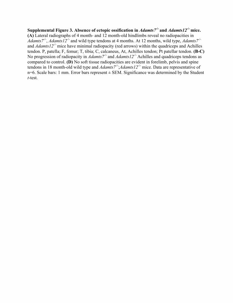
Supplemental Figure 3. Absence of ectopic ossification in Adamts7-/- and Adamts12-/- mice. (A) Lateral radiographs of 4 month- and 12 month-old hindlimbs reveal no radiopacities in Adamts7-/-, Adamts12-/- and wild type tendons at 4 months. At 12 months, wild type, Adamts7-/-
and Adamts12-/- mice have minimal radiopacity (red arrows) within the quadriceps and Achilles tendon. P, patella; F, femur; T, tibia; C, calcaneus, At, Achilles tendon; Pt patellar tendon. (B-C) No progression of radiopacity in Adamts7-/- and Adamts12-/- Achilles and quadriceps tendons as compared to control. (D) No soft tissue radiopacities are evident in forelimb, pelvis and spine tendons in 18 month-old wild type and Adamts7-/-;Adamts12-/- mice. Data are representative of n=6. Scale bars: 1 mm. Error bars represent ± SEM. Significance was determined by the Student t-test.

Supplemental Figure 4. Heterotopic ossification, no arthritic change and normal subchondral bone in Adamts7-/-;Adamts12-/- hindlimbs. (A) Alizarin red-alcian blue stained skeleton preparations of 6 month-old wild type and Adamts7-/-;Adamts12-/- hindlimbs reveal ectopic ossification in Achilles tendon (At), medial collateral ligament (MCL) and quadriceps tendon (qt). Data are representative of n=8. (B) Safranin O stained 18 month-old Adamts7-/-
;Adamts12-/- hindlimbs shows normal articular cartilage on the femoral and tibial surfaces. M, meniscus; F, femur; T, tibia; P, patella. Images are representative of n=5. (C-D) No change in subchondral bone thickness (C) or volume (D) of 18 month-old wild type and Adamts7-/-
;Adamts12-/- femur and tibia. n=10. Scale bars: 1 mm in A; 200µm in B. Error bars represent ± SEM. Significance was determined by the Student t-test.


Supplemental Figure 5. Reduction of small leucine-rich proteoglycans in Adamts7-/-
;Adamts12-/- patellar tendons. (A-B) Reduced staining of biglycan, decorin and fibromodulin (green) in 10 day-old patellar tendons near the insertion site (A) and mid-substance (B). Sections were counterstained with DAPI (blue). Data are representative of n=3. (C) Reduced biglycan, decorin and fibromodulin (green) staining in 3-month-old (3M) Adamts7-/-;Adamts12-/- patellar tendons. Data are representative of n=3. (D) No change in cartilage oligomeric matrix protein (Comp) (green) staining in 3-month-old (3M) Adamts7-/-;Adamts12-/- quadriceps tendons. Sections were counterstained with DAPI (blue). Data are representative of n=3. Scale bars: 100µm.
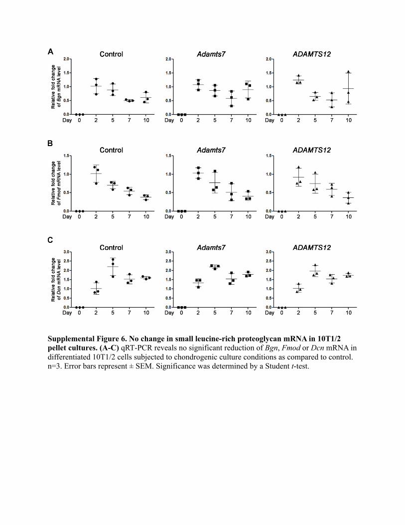
Supplemental Figure 6. No change in small leucine-rich proteoglycan mRNA in 10T1/2 pellet cultures. (A-C) qRT-PCR reveals no significant reduction of Bgn, Fmod or Dcn mRNA in differentiated 10T1/2 cells subjected to chondrogenic culture conditions as compared to control. n=3. Error bars represent ± SEM. Significance was determined by a Student t-test.
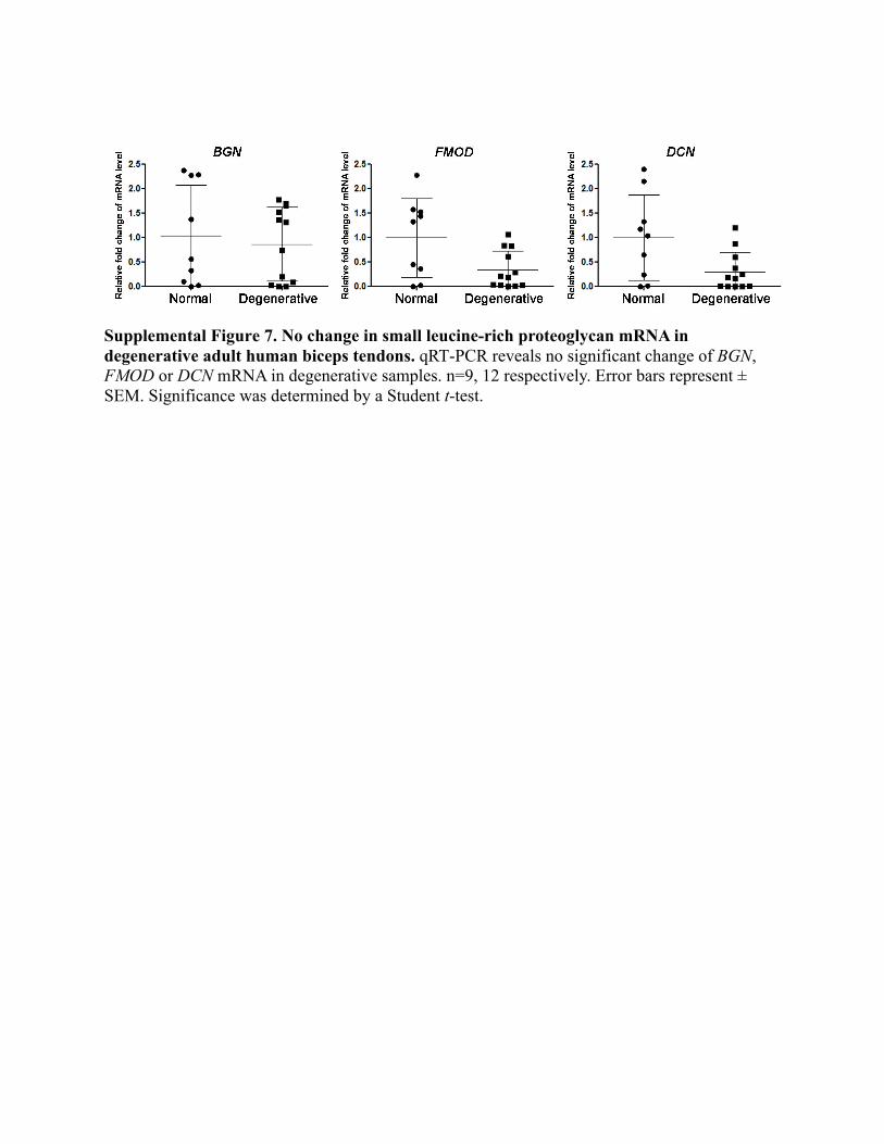
Supplemental Figure 7. No change in small leucine-rich proteoglycan mRNA in degenerative adult human biceps tendons. qRT-PCR reveals no significant change of BGN, FMOD or DCN mRNA in degenerative samples. n=9, 12 respectively. Error bars represent ± SEM. Significance was determined by a Student t-test.