summary of acute spinal cord compression
TRANSCRIPT

Summary of Acute Spinal Cord Compression n engl j med 376;14 April 6, 2017
Alexander E. Ropper Introduction
• The disorders that account for most instances of acute spinal cord compression: o Trauma o Tumor o Epidural abscess o Epidural hematoma.
• These disorders affect spinal cord function but also affect stability of the spinal column: o damage vertebrae, intervertebral disks, ligaments, facet joints
• Stability is defined by the retention of normal spinal alignment under physiologic conditions (loads) such as standing, walking, bending, or lifting.
• Spinal instability permits subluxation of vertebrae (spondylolisthesis), which narrows the spinal canal. • Instability that poses a risk of cord damage generally requires surgical fixation of the spine, and bony
fusion of adjacent vertebrae (spinal fusion) may be necessary for durable stabilization. Clinical Features
• Relatively symmetric weakness or paralysis of the limbs • Urinary retention on incontinence • Circumferential loss of sensation, a “sensory level” • Hyperreflexia and Babinski signs (characteristic of intrinsic diseases of the spinal cord) may not be
evident in cases of acute severe cord compression, especially due to trauma. • Limbs may be flaccid and areflexic, accompanied by systemic hypotension (spinal shock). • Localized back or neck pain often present. • Cauda equina (compression of nerve roots below level L1-L2) causes flaccid paraparesis and early
incontinence, BLE pain, areflexia. • Two exam points commonly omitted:
o Determine the sensory level even it is above the clavicles o Spinal percussion can identify level of a fracture or metastatic lesion
TRAUMATIC SPINAL CORD COMPRESSION
• Results from combinations of: o Fractured and retropulsed bone fragments o Disk herniation o Subluxation of vertebral bodies o Minor trauma superimposed on chronic degenerative spondylosis with narrow canal.
• 20% of spinal injuries (especially cervical) affect more than one level. • Cervical spine especially vulnerable to displacement because:
o It lacks rib cage support o Facet joints are smaller and flatter (axially oriented) so easier to sublux o Cranium acts as a load on the fulcrum of the neck
Assessment
• Injury level determined by lowest cord segment with normal motor and sensory
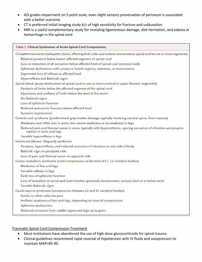
• AIS grades impairment on 5 point scale, even slight sensory preservation of perineum is associated with a better outcome.
• CT is preferred initial imaging study b/c of high sensitivity for fracture and subluxation. • MRI is a useful complementary study for revealing ligamentous damage, disk herniation, and edema or
hemorrhage in the spinal cord
Traumatic Spinal Cord Compression Treatment
• Most institutions have abandoned the use of high-dose glucocorticoids for spinal trauma • Clinical guidelines recommend rapid reversal of hypotension with IV fluids and vasopressors to
maintain MAP>85-90.

• Surgery o Neurologic outcome better if surgical decompression was performed within 24hrs after injury. o Surgically stabilizing the spine allows for early mobilization and rehabilitation. o Surgical stabilization can involve screws in vertebrae, rods or plates between vertebrae, fusion
of vertebrae by decorticating surfaces of adjacent bones and adding autologous or cadaveric bone graft or synthetic material
o Complications: infection, hardware failure, pseudoarthrosis (failure of fusion).
NEOPLASTIC EPIDURAL SPINAL CORD COMPRESSION
• Spinal metastases cause spinal cord compression when they extend from the bone into the epidural space.
• Aching back pain and tenderness on percussion over the affected site are typical and may precede neurologic features by several weeks.
• Pain may be worse when the patient is supine and causes awakening from sleep. • The spinal cord syndrome evolves over a period of hours or days • Includes hyperreflexia and Babinski signs but is infrequently characterized by sphincter dysfunction
alone. • With bony destruction and pathologic vertebral compression fracture, the spinal column becomes
unstable, leading to more severe back pain. • Common primary cancers that metastasize:
o Prostate, Breast, Kidney, non-Hodgkin’s lymphoma, Myeloma • Imaging of the entire spine reveals multiple levels of compression in up to a third of cases. • Survival in patients with multiple spinal metastases and cord compression is generally less than 6
months, but a retained ability to walk before treatment is associated with longer survival. Assessment

• Compression of the spinal cord by epidural tumor is detected by imaging, foremost MRI, preferably with the administration of gadolinium. MRI has been reported to be 100% in detecting spinal cord compression.
• Imaging of the entire spine is recommended to catch all lesions. • Tumor can be detected in many cases without the use of gadolinium, so MRI should not be withheld
if the patient has a risk factor, such as allergy, for administration of the agent. • CT Myelography is an alternative in patients who cannot undergo MRI. • CT without myeloraphy will show vertebral infiltration from tumor but won’t detect cord compression.
Treatment • Vertebral infiltration with tumor that does not compress the cord is treated with radiation if the spinal
column is stable. • Steroids reduce impairment and pain. • Dexamethasone 100mg commonly used but 10mg IV followed by 4mg q6hrs is another common
regimen. • Radiotherapy- lymphoma and myeloma highly sensitive to radiation therapy. • Non–small-cell lung cancer and renal, thyroid, and gastrointestinal cancers, as well as sarcoma and
melanoma, are relatively radioresistant and are generally treated with surgery • An influential trial comparing surgical decompression followed by radiother-apy with radiotherapy
alone for radioresistant tumors at a single level showed that surgery pre-served ambulation for a longer time, even in some patients who were unable to walk in the 48 hours before treatment.
• In summary, spinal surgery is the most rapid method for relief of acute spinal cord compression and is necessary if there is spinal instability. Radiation is usually administered after surgical decompression. If radiotherapy can be delivered expeditiously, it can be used to treat cord compression due to hematologic tumors. Patients who are not expected to survive longer than the time required for recovery from the operation (generally 2 to 3 months) are treated with palliative radiotherapy.
SPINAL EPIDURAL ABSCESS
• The thoracic spine is most often affected, and abscesses usually oc-cupy several contiguous or noncontiguous levels of the spine.
• Bacterial infection at a site distant from the spine is found in only half of affected patients, and one fourth have no primary infection, even at autopsy.
• Diabetes, in particular, but also cancer, immunosuppression, renal failure, and intravenous drug and alcohol abuse are underlying conditions.
Assessment • MRI with gadolinium • LP should be avoided • Leukocytosis and elevated Sed rate and CRP seen
Treatment • Surgical evacuation more successful than abx alone. • Decompression by means of laminectomy (see pic) • more effective if performed before severe weakness occurs, • Longitudinally long extensive abscess treated with
decompression at maximal collection with irrigation above and below that level. • paralysis for 48 hours is a poor prognostic sign • antibiotics may be adequate for an abscess that is not causing weakness, however surgery is often
required later. SPINAL EPIDURAL HEMATOMA

• Can occur after spinal surgery • Can occur spontaneously with the use of anticoagulant or antiplatelet meds and in patients with
coagulopathies. • Surgical evacuation usually necessary if there is cord compression.