“suministro local de fármacos mediante nanopartículas … · repuesta sistema reticulo...
TRANSCRIPT

““SuministroSuministro local de local de ffáármacosrmacos mediantemediantenanopartnanopartíículas culas magnmagnééticasticas””
M.R. Ibarra1,2
R. Fernández-Pacheco1, C. Marquina2 , J.G Valdivia1,3 M. Gutierrez1,3, A. Viloria4, M.T. García4, A. Fernandez4, J. A. Jalón4 M. Arruebo1
1Instituto de Nanociencia de Aragón INA, Edificio Interfacultades II, Zaragoza 2Instituto de Ciencia de Materiales de Aragón (CSIC/Universidad de Zaragoza) 3Hospital Clínico Universitario, Universidad de Zaragoza (Spain)4Hospital Clínico Veterinario, Universidad de Zaragoza (Spain)
http//:ina.unizar.es
CONSOLIDER-Ingenio 2010

INDICE
Nanopartículas magnéticas y su aplicaciones en biomedicina
-Aplicaciones
-Preparación
-Caracterización
Experimentación “in-vitro”
-Biocompatibilidad
-Adsorción/desorción de fármacos
Experimentación “in-vivo”
Conclusiones

NANOPARTÍCULAS MAGNÉTICAS
PRINCIPALES APLICACIONESPRINCIPALES APLICACIONES
SEPARACIÓN MAGNÉTICA
AGENTES CONTRASTE
MRI
BIOSENSORESINMUNO-MAGNÉTICOS
SUMINISTRO LOCAL
FÁRMACOS
HIPERTERMIA
NANODIAGNÓSTICO
NANOTERÁPIA

Suministro local de fármacos mediante
partículas magnéticas
Tumor sólido
Implantar un iman
Modular el campo magnético aplicado
Inyectar las nanopartículas magnéticas cargadas con el fármaco
Nuevos desarrollos en el INAColocar un imán permanente cerca del tumor

Ventajas de la utilización de Ventajas de la utilización de nanopartículas magnéticasnanopartículas magnéticas
Se puede controlar el tamaño
Se puede funcionalizar su superficie
Respuesta frente a campos magnéticos
Versátiles para su utilización “in-vivo”, “in-vitro” y “ex-vivo”
El tamaño puede modular su repuesta frente a campos magnéticos en organismos vivos

Repuesta nanopartículas Repuesta nanopartículas magnéticasmagnéticas
ESTÁTICA
DINÁMICA
-Suministro de fármacos (Terapia)
-Detección y cuantificación de biomoléculas(Inmunoensayos)
-Inhomogeneidadesmagnéticas para contraste (MRI)
-Absorción resonante de radiación electromagnética (Hipertermina, desorción de fármacos)
-Resonancia magnética
m
B

Repuesta sistema Repuesta sistema reticuloreticulo--endotelialendotelial
Inyección intravenosaInyección intravenosa
Torrente sanguíneo
Médula ósea
Bazo
Nódulos linfáticos
Inflamación
Tumor
Hígado
OPSONIZACIÓNLos agentes extraños son rodeados por proteínas plasmáticas para poder ser detectadas por los receptores fagocíticos del RES
¿Cómo evitarla?Hidrofilizando la superficie de las nanopartículas (PEG)
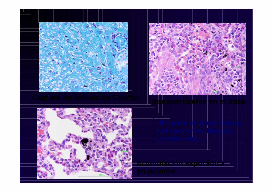
Captura en células de Kupffer Nanopartículas en el bazo
Acumulación esporádica en pulmón
Útil para el tratamiento del cancer de hígado, leishmania…..

Comportamiento magnético de la materia
Diamagnetismo
Toda la materia
Paramagnetismo
Atomos aislados
Ferromagnetismo
Algunos metales
Superparamagnetismo
Nanopartículas
100 µm 20 nm
Q.A. Pankhurst et al. J. Phys. D.:Appl.
Phys. 36 (2003) R167
Dominios magnéticosPartículas monodominio

G. Goya et al. INA (2006)

Las nanopartículas tipo “core-shell” reunen las
especificaciones para aplicaciones
biomédicas:
FeCarbon
-Son pequeñas
-Tienen una fuerte repuesta magnética
-Biocompatibles
-Capacidad de adsorber fármacos rápidamente
-Desorción lenta

BIOFERROFLUIDOSBIOFERROFLUIDOS
Fe2O3Nanoparticle
GraphiteEncapsulation
Fe2O3Maghemite
HRTEM
CGraphite
Plasma Krästchmer-Hoffman
-Biocompatibilidad
-Adsorción de fármacos
-Conjugación con proteinas

Fe
SiO2 FeFe
SiO2
Silica
Fe2O3-δδδδ
Fe
NanopartNanopartíículas de hierro encapsuladas en culas de hierro encapsuladas en ssíílicalica
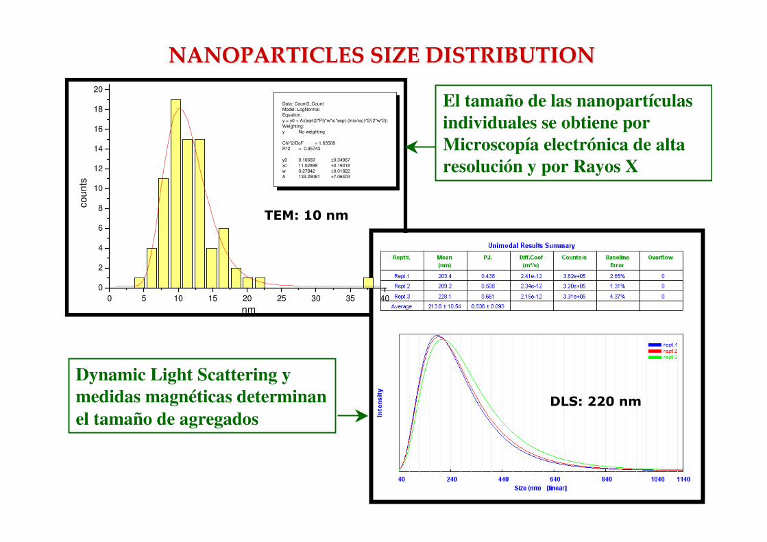
Dynamic Light Scattering y medidas magnéticas determinanel tamaño de agregados
0 5 10 15 20 25 30 35 400
2
4
6
8
10
12
14
16
18
20
Data: Count3_CountModel: LogNormal Equation: y = y0 + A/(sqrt(2*PI)*w*x)*exp(-(ln(x/xc))^2/(2*w^2)) Weighting:y No weighting Chi^2/DoF = 1.63508R^2 = 0.95743 y0 0.16669 ±0.34967xc 11.02898 ±0.19318w 0.27842 ±0.01822A 133.29081 ±7.06403
coun
ts
nm
TEM: 10 nm
NANOPARTICLES SIZE DISTRIBUTIONNANOPARTICLES SIZE DISTRIBUTION
El tamaño de las nanopartículas individuales se obtiene porMicroscopía electrónica de altaresolución y por Rayos X
DLS: 220 nm

Los ferrofluidos obtenidos tienen una fuerte repuesta magnética

INDICE
Nanopartículas magnéticas y su aplicaciones en biomedicina
-Aplicaciones
-Preparación
-Caracterización
Experimentación “in-vitro”
-Biocompatibilidad
-Adsorción/desorción de fármacos
Experimentación “in-vivo”
Conclusiones

TestTest de de biocompatibilidadbiocompatibilidadhematolhematolóógicagica
“In vivo” conejos de raza neozelandesa
• Se tomaron muestras de sangre antes y después de injectar lasnanopartículas.
• 1 ml de Nanoparticulas a una concentración de 12.5 mg/ml de Gelafundineno modifican la viscosidad de la sangre ni del plasma.
• La agregación eritrocitaria se presenta pequeñas variaciones sin importancia
“In vitro” en sangre humana
• Se han medido los parámetros hematológicos en sangre humana con distintas concentraciones de nanopartículas (5 ml de sangre y 0, 0.06, 0.12, 0.24 and 0.5 ml of ferrofluid). Se obtuvieron valores normales en todos lostest, con variaciones inferiores al 10% respecto de controles normales.

NanopartNanopartíículas en culas en torentetorente sangusanguííneoneoTest de biocompatibilidad
•Ausencia de “Blue Perls stain”: buen encapsulamiento por capas grafíticas
•Ausencia de partículas en monocitos y granulocitos

VS230 VS 23 VS5,7 AE5M AE5M1 AE10M AE10M1BASAL 4,44 6,7 7,2 3,4 10 8,3 22,5C.0,06 4,36 5 7,6 3,4 8,9 7,6 20,6C.0,12 4,45 7 8 3 8,4 7,6 18C.0,24 4,33 5,4 6,9 2,9 7 7,1 18,6C.0,5 4,1 6,2 6,4 2,9 6,9 8,8 20,1
0
5
10
15
20
25
VS230 VS5,7 AE5M1 AE10M1
BASALC.0,06C.0,12C.0,24C.0,5
RESULTS IN HUMAN BLOODRESULTS IN HUMAN BLOOD
AE = Erythrocyte aggregation(M: stasis, M1 minutes under low shear rate)
VS = Blood viscosity Shear rate (s-1)
C= Particles concentration (mg/ml)

Estudio de adsorción y Estudio de adsorción y desorcióndesorción de fármacosde fármacos
Doxorubicin (11x 13 Å)
300 400 500 600 700 800
1
2
3
Abs
nm
498 nm296 nm
Fe2O3Nanoparticle
GraphiteEncapsulation

Absorbance spectra of Doxorubicin. Kinetics of adsorptionAbsorbance spectra of Doxorubicin. Kinetics of adsorption
The adsorbent particles were sedimented with a 3 KOe permanent magnet, and the optical density of the
supernatant measured with a UV spectrophotometer (method proposed by Kuznetsov, A. et al, (1999) J. Mag.
Mag. Mat. 194, 22).
200 400 600 8000,2
0,4
0,6
0,8
1,0
1,2
1,4
1,6
1,8
2,0
2,2
Ab
s
λλλλ (nm)
INICIAL
-20 0 20 40 60 80 100 120 140 160 180 200
0,0
0,2
0,4
0,6
0,8
1,0
t (min)
adso
rbed
do
xoru
bic
in (
a.u
.)

Absorbance spectra of Doxorubicin after Absorbance spectra of Doxorubicin after desorptiondesorption
The adsorbent particles were sedimented with a 3 KOe permanent magnet, and the optical density of the
supernatant measured with a UV spectrophotometer (method proposed by Kuznetsov, A. et al, (1999) J. Mag.
Mag. Mat. 194, 22).
400 600 800
0,3
0,6
0,9
Abs
λ (nm)
Final
-10 0 10 20 30 40 50 60 70 80
0,3
0,6
0,9
DO
XR
RU
BIC
INA
DE
SO
RB
IDA
(a.
u.)
t (horas)

INDICE
Nanopartículas magnéticas y su aplicaciones en biomedicina
-Aplicaciones
-Preparación
-Caracterización
Experimentación “in-vitro”
-Biocompatibilidad
-Adsorción/desorción de fármacos
Experimentación “in-vivo”
Conclusiones
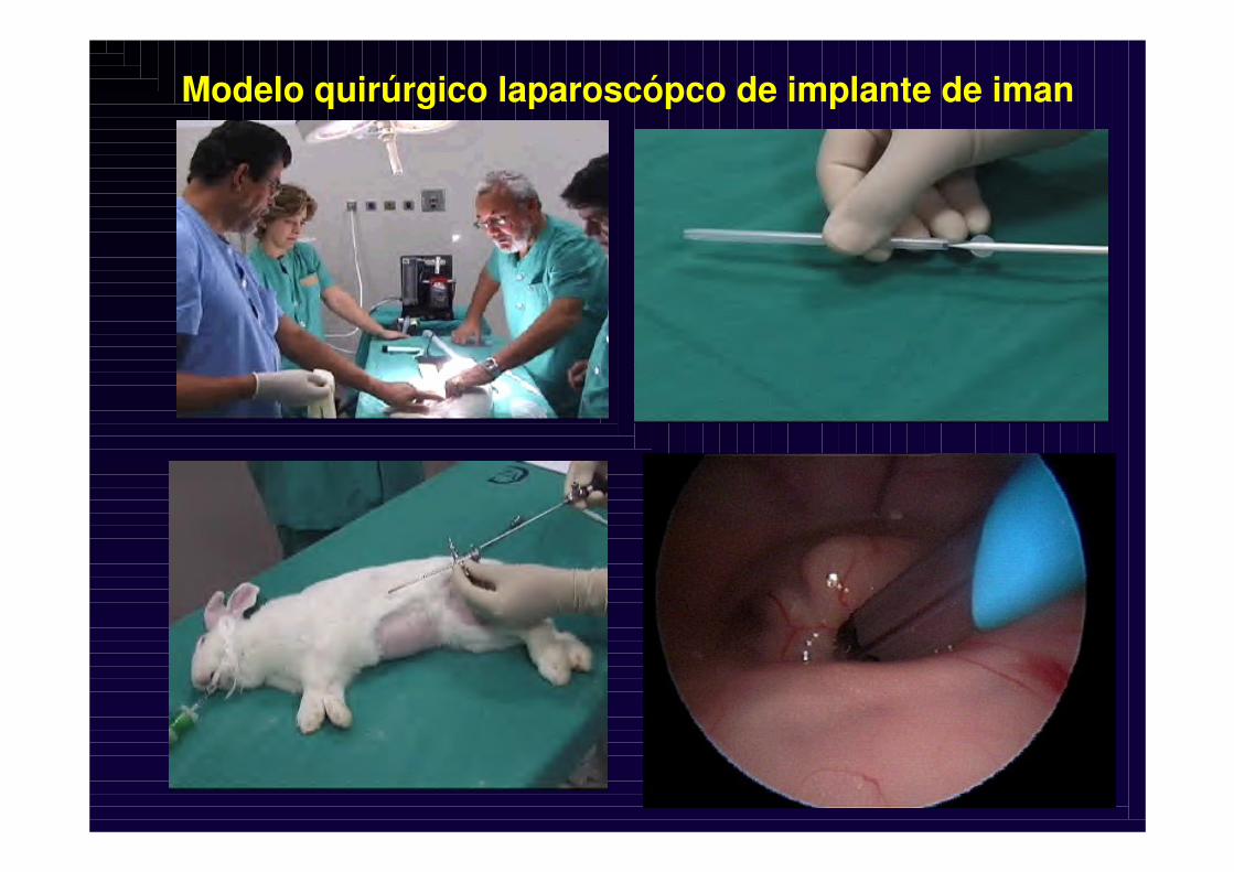
Modelo quirúrgico laparoscópco de implante de iman

Experimentación in vivo de localización de partículas magnéticas mediante administación por via sistémica

Implante imán riñón izquierdo
Riñon derecho con incisión sin implante
Presencia de nanopartículas
Ausencia de nanopartículas

Conejo 22 Conejo 23
RiRiñóñón con implante magnn con implante magnéético: concentracitico: concentracióón de nanopartn de nanopartíículasculas
Conejo 25

CONCLUSIONESCONCLUSIONES
-La aplicación de las nanopartículas magnéticas en medicina abre nuevas expectativas en la clínica humana tanto en el diagnostico precoz como en la terápia
-Se ha probado la biocompatibilidad sanguínea y la capacidad de adsorber y desorber doxorubicina en nanopartículas “core-shell” de Fe&C.
-Se ha comprobado la capacidad de localización con implantes magnéticos
-El reto es hacer la superficie hidrofílica para evadir la detección y captura por el RES sin perder la capacidad de portar la doxorubicina.


CONSOLIDER-Ingenio 2010
Comité de Dirección

SCIENTIFIC CONSOLIDER TEAM
NANOTECHNOLOGIES IN BIOMEDICINE
NANOTHERAPY NANODIAGNOSTIC
Preparation
L. Liz & M.J. Alonso
Funcionalization
A. González
“In-Vitro”
V. Puntes
“In-Vivo”
G. Valdivia
N A N O P A R T I C L E S
Electrochemical
platforms
A. Markoçi
Miniaturized
electrodes
F. Pérez-Murano
Lateral Flow
J.M. De Teresa
B I O S E N S O R S
SPION
X. Batlle
Biological barriers
E. Giralt
Targeting and
Experimental
Models
A. Trés
M R I
Early Cancer Detection
CONSOLIDER-Ingenio 2010

NANOMEDICINA
CIBER
CONSOLIDER CENIT
PROGRAMA INGENIOPROGRAMA INGENIO--20102010

ESCALAS DE LONGITUDESCALAS DE LONGITUDDISTANCIA TIERRA-SOL
HOMBRE
HOJA DE PAPEL DE CANTO
VIRUS
MOLÉCULA ADN
ATOMO
1.5 x 10¹¹ m
1.5 m
10-4 m
5 x 10⁻⁸ m
2 x 10⁻⁹ m
2 x 10⁻¹⁰ m
1 nanómetro = 10-9 m

BARRERAS PARA LA NANOTECNOLOGÍA EN BIOMEDICINA
Físicos-Químicos
Bioquímicos-Farmaceúticos
Médicos
Los tejidos tumorales expulsan las nanopatículas y los sanos las retiran de la circulación
Funcionalización Internalización
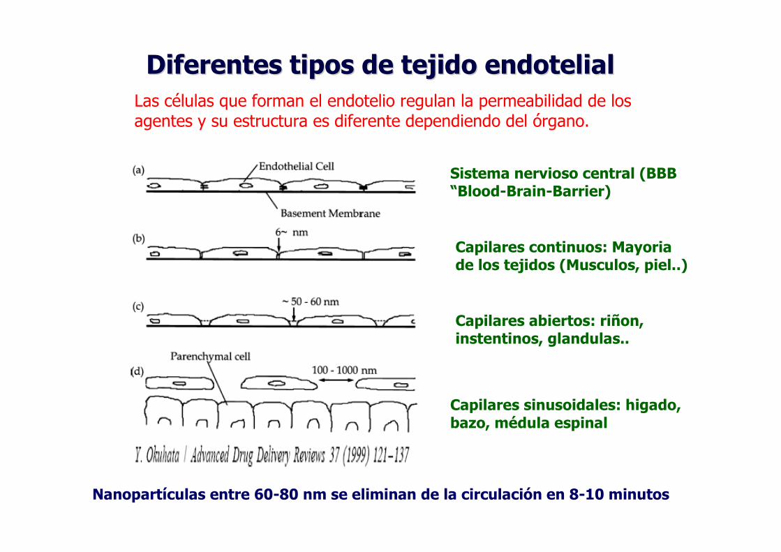
Diferentes tipos de tejido endotelialDiferentes tipos de tejido endotelialLas células que forman el endotelio regulan la permeabilidad de los agentes y su estructura es diferente dependiendo del órgano.
Sistema nervioso central (BBB “Blood-Brain-Barrier)
Capilares continuos: Mayoriade los tejidos (Musculos, piel..)
Capilares abiertos: riñon, instentinos, glandulas..
Capilares sinusoidales: higado, bazo, médula espinal
Nanopartículas entre 60-80 nm se eliminan de la circulación en 8-10 minutos


Magnetic implant in the left kidney
Right kidney witout magnetic implant
Localization of nanoparticles
Lack of nanoparticles

Absorbance spectra of Absorbance spectra of DoxorubicineDoxorubicineafter after desorptiondesorption from carbon coated from carbon coated magnetic nanoparticlesmagnetic nanoparticles
The adsorbent particles were sedimented with a 3 KOe permanent magnet, and the
optical density of the supernatant measured with a UV spectrophotometer (method
proposed by Kuznetsov, A. et al, (1999) J. Mag. Mag. Mat. 194, 22).
144h120h72h60h48h54h

Dynamic of the adsorption and release of Dynamic of the adsorption and release of DoxorubicineDoxorubicine on carbon coated magnetic on carbon coated magnetic
nanoparticlesnanoparticlesSaturation after 20 minutes Complete release after 100 hours
AdsorptionDesorption
-20 0 20 40 60 80 100 120 140 160 180 200
0.0
0.2
0.4
0.6
0.8
1.0
t (min)
Ado
srbe
d do
xoru
bici
n C
(a.
u.)
B
0 20 40 60 80 100 120 140 160 180
0.0
0.2
0.4
0.6
0.8
1.0
Des
robe
d C
(a.
u.)
t (h)
B fit
A 100 µµµµg/ml solution of doxorubicin hydrochloride was mixed with 1 mg/ml of distilled water. The
suspension was incubated on a shaker at room
temperature, and samples were taken after 5, 15, 30,
60, 90, 120 and 180 minutes.
Doxorubicin loaded nanoparticles were mixed with
human blood serum at the concentration of 1 mg/ml.
The suspension was incubated on a shaker at 37 oC,
and samples were taken each 8 hours during four
days and UV absorbance spectra of the supernatant
were obtained.

Optical Microscope Imageof the permanent magnet
coated with gold.
Percutaneus insertion of the permanent magnet
The rabbits were submitted to general anaesthesia with endotracheal intubation.
A laparoscopic optic was introduced by using a 5 mm trocar.
Under endoscopic control, the permanent magnet was implanted in the lower pole of the righ kidney

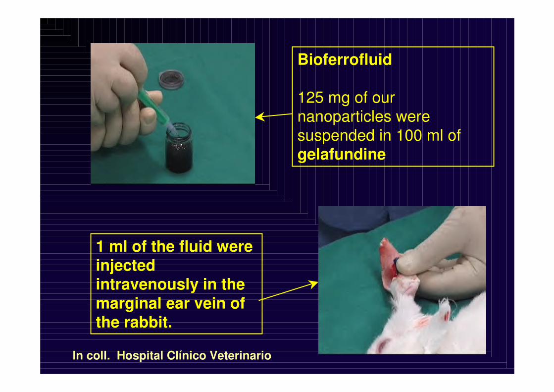
Bioferrofluid
125 mg of ournanoparticles weresuspended in 100 ml of gelafundine
In coll. Hospital Clínico Veterinario
1 ml of the fluid wereinjectedintravenously in the marginal ear vein of the rabbit.

Magnet implant in the left kidney
Right kydney witout magnetic implant
Localization of nanoparticles
Lack of nanoparticles

Rabbit 22 Rabbit 23
KidneyKidney withwith magneticmagnetic implantimplant::
SomeSome concentrationconcentration of nanoparticlesof nanoparticles
In coll. Hospital Clínico Veterinario: Dr. A. Viloria, Dra. MªTeresa García, Dr. Angel Fernandez and Dr. J. A. Jalón

Liver Capture in Kuffer cells Nanoparticles in spleen
Reduced concentration of nanoparticles in lung
Macrophagycapture in
organs


OUTLINE OF THE TALKOUTLINE OF THE TALK
-Introduction
-Encapsulated nanoparticles:preparation andcharaterization
-Morphology, Structure and Magnetism of the nanoparticles
-Bioferrofluids for local drug delivery
-Invivo experiments: magnet implant and magneticlocalization
-Conclusions

Biologicallabeling
Contrastagent
Oftalmology
Hiperthermy
Selective drugdelivery
Bioferrofluid
Medical application of magnetic nanoparticles

Matter manipulationat atomic level
Inteligentnanovectors
Targeting
The The newnew worldworld of nanoscienceof nanoscience

HRTEM images of Fe encapsulated nanoparticles
Fe2O3Nanoparticle
GraphiteEncapsulatio
n
Fe2O3Maghemite
HRTEM
CGraphite
Fernandez Pacheco R., Ibarra M.R.,…J. Proceeding of the NSTI Nanotech 2005 (Anaheim, California) Vol 1 pag 144-147

If K ���� 0
The supermoment followsthe Langevin law
If K>>>
The supermoment followsthe Brillouin J=1/2 law
Classic paramagnet Quantum paramagnet
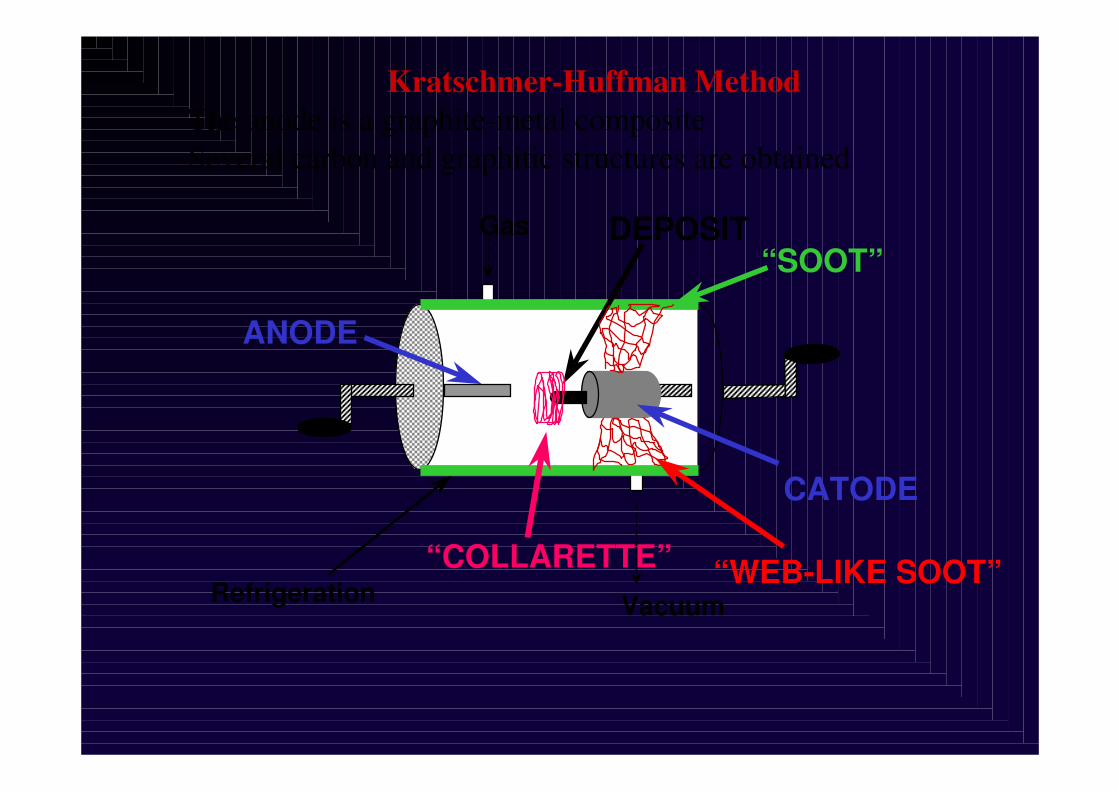
CATODE
Refrigeration
Gas
Vacuum
ANODE
“SOOT”DEPOSIT
“WEB-LIKE SOOT”“COLLARETTE”
Kratschmer-Huffman Method-The anode is a graphite-metal composite
-Several carbon and graphitic structures are obtained
Bioferrofluid
125 mg of ournanoparticles weresuspended in 100 ml of gelafundine
In coll. Hospital Clínico Veterinario
1 ml of the fluid wereinjectedintravenously in the marginal ear vein of the rabbit.


30 nm ���� 5 % atoms at the surface
10 nm ���� 20 % atoms at the surface
3 nm ���� 50 % atoms at the surface
How How smallsmall??

Crítical size for single-domain particle
-Under size reduction the coercive field increases and the the particle becomes single-domain
-When EK=KV as V � 0 then EK � 0 superparamagnetic limit
-At this situation the particle magnetic moment will fluctuate independently of the particle
KV = kBT

Real superparamagnetic system
-No hystheresis
-The isotherm presents a universal H/T behaviour

-40000 -30000 -20000 -10000 0 10000 20000 30000 40000
-200
-150
-100
-50
0
50
100
150
200
M (
emu/
g)
H (Oe)
-1500 -1000 -500 0 500 1000 1500 2000
-60
-50
-40
-30
-20
-10
0
10
20
30
40
50
M (
emu/
g)
H (Oe)
C
EncapsulatedEncapsulated Fe nanoparticlesFe nanoparticles

OUTLINE OF THE TALKOUTLINE OF THE TALK
-Introduction
---EncapsulatedEncapsulatedEncapsulated nanoparticles:preparationnanoparticles:preparationnanoparticles:preparation andandandcharaterizationcharaterizationcharaterization
-Morphology, Structure and Magnetism of the nanoparticles
-Bioferrofluids for local drug delivery
-Invivo experiments: magnet implant and magneticlocalization
-Conclusions

OUTLINE OF THE TALKOUTLINE OF THE TALK
-Introduction
-Encapsulated nanoparticles:preparation andcharaterization
---Morphology, Structure and Magnetism of the Morphology, Structure and Magnetism of the Morphology, Structure and Magnetism of the nanoparticles nanoparticles nanoparticles
-Bioferrofluids for local drug delivery
-Invivo experiments: magnet implant and magneticlocalization
-Conclusions

OUTLINE OF THE TALKOUTLINE OF THE TALK
-Introduction
-Encapsulated nanoparticles:preparation andcharaterization
-Morphology, Structure and Magnetism of the nanoparticles
---BioferrofluidsBioferrofluidsBioferrofluids forforfor local drug local drug local drug deliverydeliverydelivery
-Invivo experiments: magnet implant and magneticlocalization
-Conclusions

OUTLINE OF THE TALKOUTLINE OF THE TALK
-Introduction
-Encapsulated nanoparticles:preparation andcharaterization
-Morphology, Structure and Magnetism of the nanoparticles
-Bioferrofluids for local drug delivery
---InvivoInvivoInvivo experimentsexperimentsexperiments: : : magnetmagnetmagnet implantimplantimplant andandand magneticmagneticmagneticlocalizationlocalizationlocalization
-Conclusions

• Increase of:
– Plasma and blood viscosity
– Red blood cells aggregability
• Activation of Coagulation
– “Factor XII” activation
– Platelets activation
– Vascular endothelium damage
• Withdrawal of particles by phagocytes
Non desire effects of the circulating particles in the blood stream


Si consideramos el sistema circulatorio
como una red de distribución para
nanopartículas:
Asmatulu et al. JMMM, (2004)

Velocidad de flujo en la arteria carótida (Asmatulu
y cols., JMMM 2005)
Fuerza de arrastrede la sangre
Llevando las partículas a donde
se requiere
Campo magnéticoexterno
Es necesario sobrepasar un umbral de fuerza magnética:- Aumentar el campo externo- Aproximar el imán
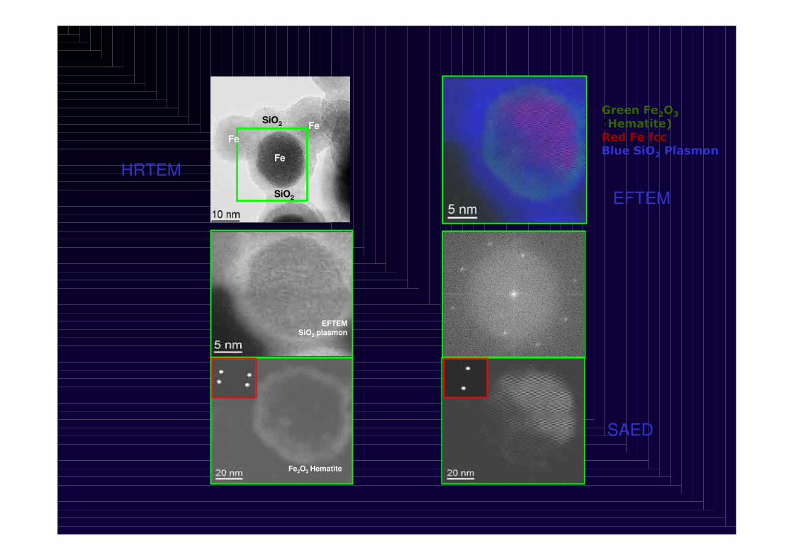
EFTEMSiO2 plasmon
Fe2O3 Hematite
Fe
SiO2 FeFe
SiO2
HRTEM
(100)Fe
(02-3) Fe2O3
(022)Fe2O3
Fe fcc
Green Fe2O3(Hematite)Red Fe fccBlue SiO2 Plasmon
EFTEM
SAED
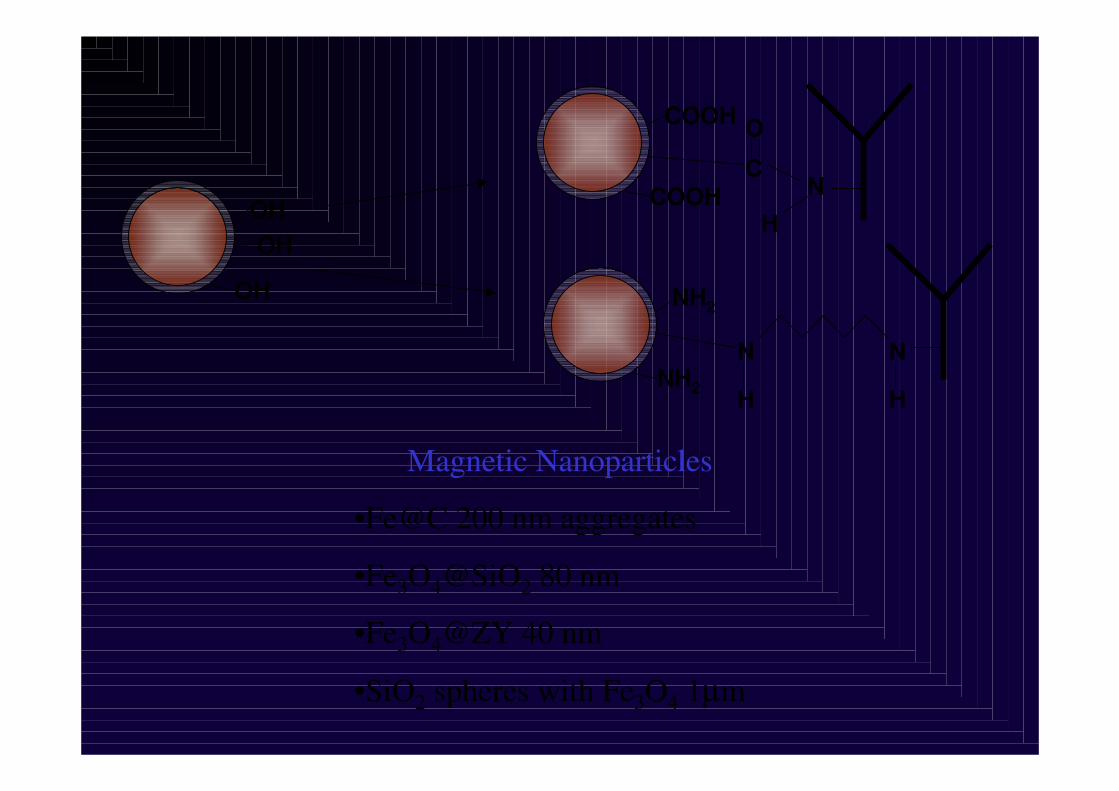
Magnetic Nanoparticles
•Fe@C 200 nm aggregates
•Fe3O4@SiO2 80 nm
•Fe3O4@ZY 40 nm
•SiO2 spheres with Fe3O4 1µm
OH
OH
OH
NC
OCOOH
COOHH
NH2
NH2
NN
H H

GLUTARALDEHYDEGLUTARALDEHYDE
CARBODIIMIDECARBODIIMIDE
FeSiO2 NH2
NH2
NH2NH2
NH2FeSiO2 COOH
COOH
COOHCOOH
COOH

Local drug delivery by using magnetic carriers
Solid tumor
Magnetimplantation
Intravenousadministration of magnetic carriers
New development at the INALapararoscopic implant of a permanent magnet

BIOSENSORS TEST
NANOPARTICLE PREPARATION
-Core Shell Nanoparticles
-Matrix Nanoparticles
-SPIONS
DEVELOP OF MICRO
AND NANODEVICES
FOR BIODETECTION PLATFORMS
-Microcircuitry
-Nanolithography
-Magnetoresistive sensors
-Microcoils
LINEAS DE INVESTIGACIÓN
FUNCTIONALIZATION
“IN-VITRO”“EXVIVO”
“IN-VIVO”
TECHNOLOGICAL TRANSFER

(100)Fe
(02-3) Fe2O3
(022)Fe2O3
EFTEMSiO2
plasmon
Fe2O3
Hematite
Fe fcc
ESTRUCTURA CRISTALINA EN LA NANOESCALAESTRUCTURA CRISTALINA EN LA NANOESCALA
Imagen (espacio real)Imagen (espacio real) Imagen (espacio recImagen (espacio recííproco)proco)
TransformadaFourier
FILTRADO

Nanopartículas-”Nanodevices” del futuro
Los materiales tienen una determinada funcionalidad
La reducción de tamaño la modifica, refuerza o la hace más adecuada para deteminadas aplicaciones
El tamaño es importante pero tambien lo es la superficie
-Aplicaciones electrónicas: espintrónica:
Tamaño-coulomb blockage (Magnetic Spinsingle transistor)
Superficie-Magnetoresistencia elevada (MUND)
-Aplicaciones biomédicas
Tamaño-coexisten con células en la sangre y viajan por todo el cuerpo (Drug delivery)
Superficie-se funcionalizan con grupos peptidicos con acción específica ->Nueva herramienta en ingeniería molecular con implicaciones en farmacia y biomedicina