sugars and polysaccharides. importance of carbohydrates key intermediates of metabolism of food and...
TRANSCRIPT

Sugars and Polysaccharides

Importance of Carbohydrates
• Key intermediates of metabolism of food and energy production (sugars)
• Structural components of plants, animals and bacteria: (cellulose, peptidoglycan, cartilage)
• Central to materials of industrial products: (paper, lumber)
• Key component of food sources: (sugars, flour, fiber)

Outline of Carbohydrates• Part 1: General structure, names, and
stereochemical properties of simple sugars (chpt 11 sec 1)
• Names of simple sugars• Fischer Projections of sugars• Cyclic reactions: Hemiacetal formation
• Part 2: Disaccharides: structure, nomenclature and biology (chpt 11 sec 2)
• Acetal formation• Disaccharide nomenclature
• Part 3: Polysaccharides and Glycoproteins: structures and biology
• Starch, Glycogen structures• Cellulose, Chitin structures• Bacterial cell walls: peptidoglycan• Extracellular matrix: Hyaluronic acid

Classification of Carbohydrates• Carbohydrates- are molecules, consisting only of
carbon (C), hydrogen (H), and oxygen (O), with the empirical formula Cm(H2O)n (where m could be different from n).
– Monosaccharide's (simple sugars) can't be converted into smaller sugars by hydrolysis.
– Disaccharides- comes from two monosaccharides (glucose linked to fructose; sucrose) linked together by an acetal bond.
– Polysaccharides- made of three or more simple sugars connected as acetals (aldehyde and alcohol).

Biological Monosaccharides are classified into two categories
Simple Sugars
Aldoses KetosesMost oxidized
carbon is an aldehyde
Most oxidized carbon is a ketone

D-Glyceraldehyde
3-Carbon
Chiralcenter
4-Carbon
Most Oxidized
carbon
D-Erythrose D-Threose
Three and four carbon Aldoses: Aldotriose, Aldotetriose

Five Carbon Aldoses: Aldopentoses
D-Ribose D-Arabinose D-Xylose D-Lyxose

Six Carbon Aldoses: Aldohexoses
D-Allose D-Altrose D-Glucose D-Mannose
D-Gulose D-Idose D-Galactose D-Talose

Three and four carbon Ketoses: Ketotriose, Ketotetriose
Dihydroxyacetone
3-Carbon 4-Carbon
D-Erythrulose

Five and six Carbon Ketoses
D-Ribulose
D-Xylulose
Ketopentoses Ketohexoses
D-Psicose D-Fructose
D-Sorbose D-Tagatose

Abbreviations for some sugars thatare common components of
polysaccharides
Memorize the shaded abbreviations!!!

Structures you have to memorize!!!
• Glyceraldehyde
• Dihydroxacetone phosphate
• Ribose & Deoxyribose
• Glucose
• Fructose
Write on board

Stereochemisry review
Chiral Centers

How do we deal with multiple chiral centers
• Enantiomers- molecules that are not identical to their mirror images. (This definition includes multiple chiral centers).
• A general rule, a molecule with “N” chiral centers can have 2N stereoisomers.
• N = 6; 26 = 64 possible stereoisomers!!!
• Diastereomers- stereoisomers that are not mirror images.
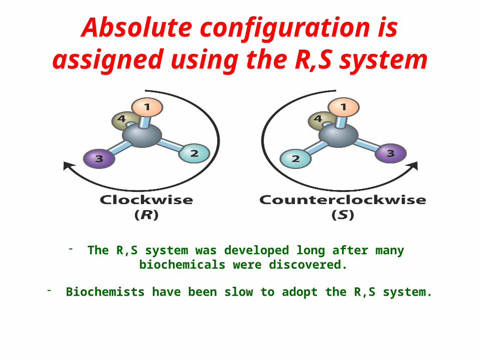
Absolute configuration is assigned using the R,S system
- The R,S system was developed long after many biochemicals were discovered.
- Biochemists have been slow to adopt the R,S system.

Carbohydrate Stereochemistry: Fischer Projections
• A chirality center C is projected into the plane of the paper
• Groups forward from paper are always in horizontal line.
• Vertical bonds represents groups projecting into the plane of paper
Hermann Emil FischerFrom 1852-1919; Nobel Prize in 1902

Fischer convention for carbohydrates (D, L)
• The hydroxyl group at the chiral center farthest from the oxidized end of the sugar determines the stereochemical reference (D, or L).
D-Glyceraldehyde
H
OH
H OH
OC
CH2
Boldwedges
Hashwedges

Stereochemical Reference• A compound is “D” if the hydroxyl group at the
chirality center farthest from the oxidized end of the sugar is on the right or “L” if it is on the left.

“D” is when the hydroxyl group is on the right and “L” is when it is on the left

“D” is when the hydroxyl group is on the right and “L” is when it is on the left

Allowed Movements with Fisher Projections
- Rotation of 90º is not allowed with a fisher projection since this will change the chirality.
- Rotation of 180º is allowed with a fisher projection since this will not change the chirality.

We can change the position of three groups and leave one group the same
without changing the chirality

Specify the sugars as “D” or “L”
L
Most oxidizedcarbon at or
closest to the top
Least oxidizedcarbon at or
closest to the top
Linear chain

Assign “D” or “L” to each Monosaccharide
L D
H
HH
H
O
OHOH
OH
OH
L

Specify the sugar as “D” or “L”
O
OH
OH
H
CH
CH2
LHold
Steady

Epimers are two diastereomers that differ in their configuration around a single carbon

Mechanism of hemiacetal, hemiketal formation

Sugars of five or more carbons readily adopt the cyclic conformations in solution
<1%
62%38%
α-anomerβ-anomer

Haworth Projections
α- D-Glucose α- D-Fructose
β- D-Glucose β- D-Fructose

Haworth Projections
α- D-Glucose α- D-Fructose
β- D-Glucose β- D-Fructose

Sugars can undergo oxidation-reduction reactions at the
anomeric carbon• The exposed C1 (anomeric carbon) is referred to as the
reduced end (carbonyl can be reduced to a carboxyl).
Reducing sugar test is the basis of blood sugar meters.
Reduced end

Carbohydrate Analytical Tests
Oxidizing Agent
Benedict’s solution
Fehling’s Solution
Tollen’s Reagent
Active ingredient
CuSO4 CuSO4 Ag in NH3
Color Deep Blue Deep Blue Mirror
Oxidant Cu+2 ----- Cu+ Cu+2 ----- Cu+ Ag+ ----- Ag(s)
Sugar Product
Oxidized to Carboxylate
Oxidized to Carboxylate
Oxidized to Carboxylate
Test Result
Positive for Aldoses
Positive for Aldoses and
Ketoses
Positive for Aldoses

Carbohydrate Analytical Tests
Positive for all three tests
Positive for Fehling’s
Negative for Tollen’s
Negative for Benedict’s
Negative for all three tests
Fehling’s Tests- Positive for Aldoses and KetosesBenedict’s Test- Positive for Aldoses only
Tollen’s Test- Positive for Aldoses only
All tests are negative if the anomeric carbon is linked to another sugar!!

Mechanism of hemiacetal, acetal formation

Disaccharide nomenclature
• Glycosidic bond- forms when the hydroxyl group of one sugar reacts with the anomeric carbon of the other.
Anomeric carbon
Galactose Glucose(β1 - 4)

ReducingEnd
Non Reducing
End
Naming Rules: nonreducing residue and configuration to reducing residue
Example disaccharide: Maltose
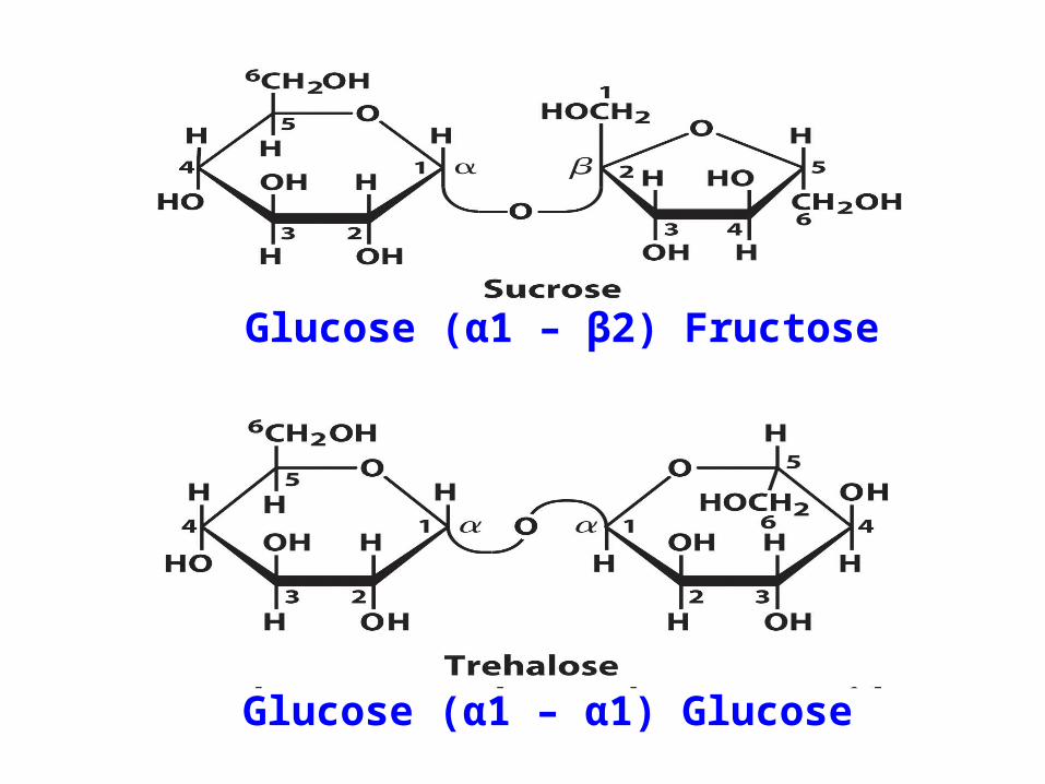
Glucose (α1 – β2) Fructose
Glucose (α1 – α1) Glucose


Types of Homopolysaccharides
Starch- polysaccharides found in plants that contains glucose in two forms:
- Amylose (linear α1-4 linked glucose) (10-30%) - Amylopectin (Linear + branched glucose)
Linear α1-4 linked glucoseBranched α1-6 linked glucose
- Branching occurs every 24-30 residues
Glycogen- polysaccharides found in animals. Linear α1-4 linked glucose
Branched α1-6 linked glucose
- Branching occurs every 8-12 residues
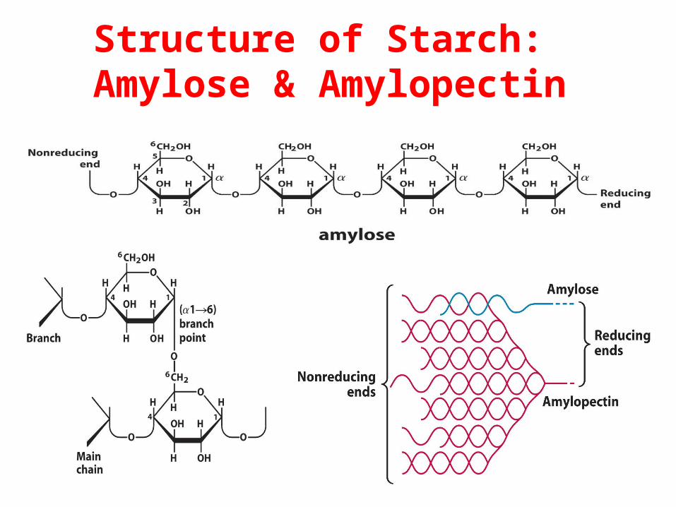
Structure of Starch: Amylose & Amylopectin

3-D Structure of Glycogen and Starch

Structure of Cellulose
Cellulose- is found in cell walls of plants.
- Cellulose uses the β configuration of glucose
- Mammals lack the enzyme required to hydrolyze the β configuration of glucose

Structural Polysaccharides
Composition similar to storage polysaccharides, but small structural differences greatly influence properties
• Cellulose is the most abundant natural polymer on earth
• Cellulose is the principal strength and support of trees and plants
• Cellulose can also be soft and fuzzy - in cotton


Amino Acids
By Doba Jackson, Ph.D.

Outline of Amino Acids, Peptides & Proteins
• Amino Acid Structure (Chpt 4-text)• Backbone• Side Chains
• Acid-Base Properties of A.A’s (Chpt 4-text)
• pKa’s of -COOH, -NH3, side chains
• Levels of Protein Structure (Chpt 7-text; Introduction, p163-164)
– We will skip Chpt 4- section 2 (Optical Activity) and Chpt 4- section 3 (non-standard amino acids)

Amino AcidsBuilding Blocks of Proteins
Amino Group
Carboxyl Group
Side ChainAlphaHydrogen
AlphaCarbon
Chiral Center

Classification of Amino Acids based on the R-group
• Non-polar, Aliphatic (6)
• Non-polar, Aromatic (3)
• Polar, Uncharged (7)
• Polar, Acidic (2)
• Polar, Basic (3)
You should know names, structures, pKa values, 3-letter and 1-letter codes!!!!!


Non-polar, Aliphatic (R-group) Amino Acids

Non-polar, Aromatic (R-group) Amino Acids
*
Tyrosine can also be considered polar, uncharged because of its polar hydroxyl group

Polar, Uncharged (R-group) Amino Acids
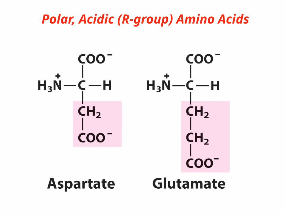
Polar, Acidic (R-group) Amino Acids

Polar, Basic (R-group) Amino Acids
Histidine could be considered aromatic but its absorption is very weak compared to other aromatic amino acids, it is also not aromatic under high pH conditions

At the isoelectric point, the neutral form of the amino acid is the predominant species.


Acid-Base Properties of Amino Acids
-Main species at Low pH (<2)
-Both functional groups contain the maximum #
of protons
-Net charge is +1
-Main species at Neutral pH (7.0)
-Amino group has a proton carboxyl
group loses a proton
-Net charge is zero
Main species at High pH (>12)
-Amino group loses proton
-Net charge is -1

What Is the Fundamental Structural Pattern in Proteins?

“Peptides”• Short polymers of amino acids• 2, 3 residues – dipeptide, tripeptide• 12-20 residues - oligopeptide
What is this peptide sequence? S G Y A L

Levels of Protein Structure
• Primary structure- A description of the covalent bonds linking amino acids in a peptide chain
• Secondary Structure- An arrangement of amino acids giving rise to structural patterns
• Tertiary Structure- Describes all aspects of three dimensional folding of a polypeptide
• Quarternary Structure- The arrangement in space of polypeptide units

Lipids and Biological Membranes

Definition of a Lipid
• A lipids are defined as compounds that have low solubility in water and high solubility in non-polar solvents.
–Hydrophobic (nonpolar only)
–Amphipathic (both polar and nonpolar groups)

Relevant Biology
• Biological membranes
• Energy storage
• Biological recognition on cell membrane
• Cellular signalling: ie. Steroids
• Free radicle scavengers: Vitamin E
• Insulation
• Many unknown functions
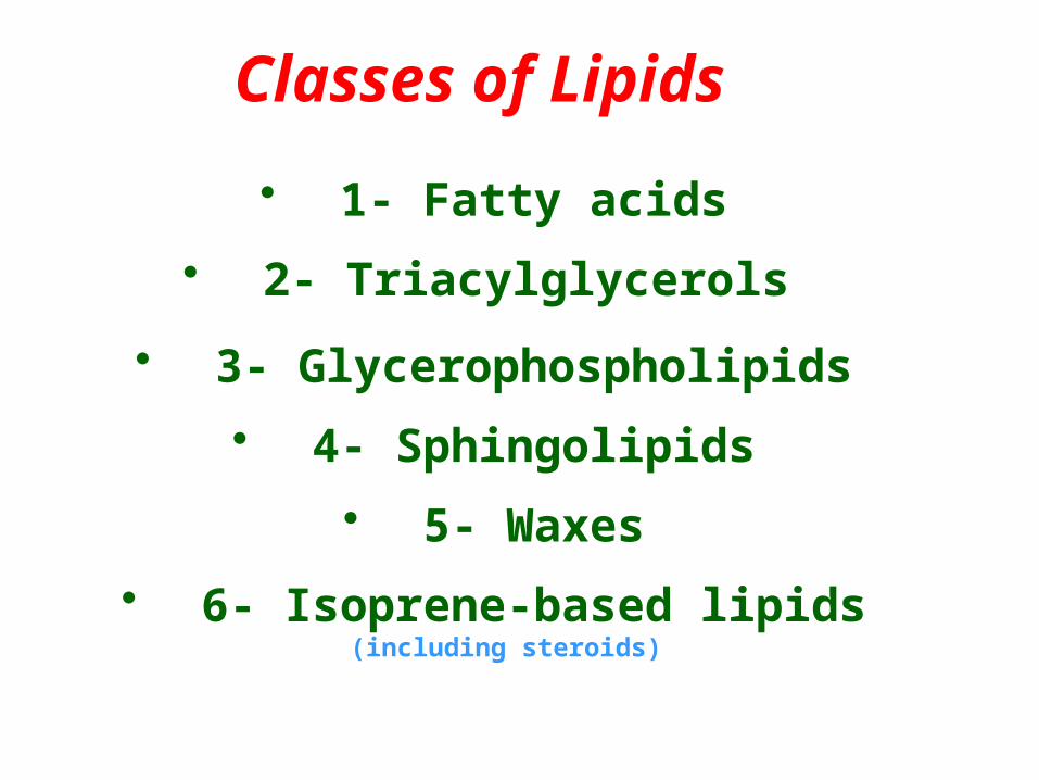
• 1- Fatty acids
• 2- Triacylglycerols
• 3- Glycerophospholipids
• 4- Sphingolipids
• 5- Waxes
• 6- Isoprene-based lipids (including steroids)
Classes of Lipids
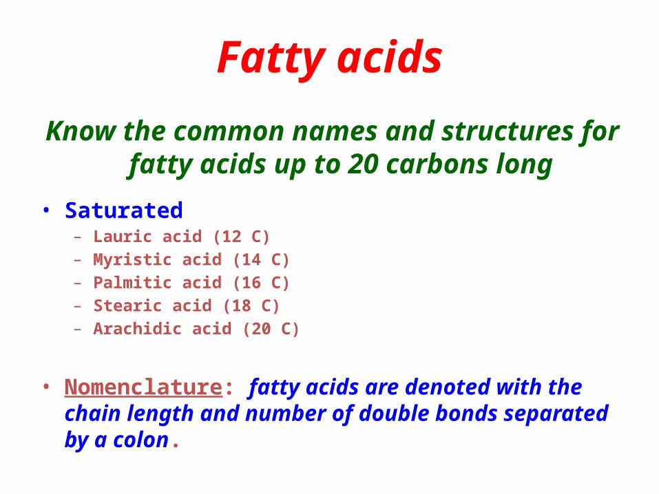
Fatty acids
Know the common names and structures for fatty acids up to 20 carbons long
• Saturated – Lauric acid (12 C) – Myristic acid (14 C) – Palmitic acid (16 C) – Stearic acid (18 C) – Arachidic acid (20 C)
• Nomenclature: fatty acids are denoted with the chain length and number of double bonds separated by a colon.

Note that most natural fatty acids contain an even number of carbon atoms.


Fatty acids• Know the common names and structures for
unsaturated fatty acids up to 20 carbons long
• Unsaturated fatty acids – Palmitoleic acid (16:1 (Δ9))– Oleic acid (18:1 (Δ9)) – Linoleic acid (18:2 (Δ9,12)) – -Linolenic acid (18:3 (Δ9,12,15)) – Arachidonic acid (20:4 (Δ5,8.11,14))
• Nomenclature: position of double bonds are denoted by the Δ symbol next to the first carbon of the double bond.

Structure of unsaturated fatty acids
• Double bonds are never conjugated and always separated by one methylene group.
• Double bonds are always cis in naturally occuring fatty acids.
• Double bonds increase solubility in water because of the decreased ability to pack together.
• Double bonds lower the melting point of the fatty acid.

• The most favorable conformation of a fatty acid is the fully
extended form.
• There is not rotation allowed across a double bond.
• Cis double bonds adds a bend to the fatty acid.
• It takes less energy to disorder poorly ordered arrays of
unsaturatedfatty acids.

Also called triglycerides
• A major energy source for many organisms
• Why? – Most reduced form of carbon in nature– No solvation needed – Efficient packing
Triacylglycerols

Triacylglycerols
• When glycerol has two different fatty acids at C1 and C3 then C2 becomes a chiral
center.
• Simple triacylglycerols with the same fatty acid are
names tripalmitin, tristearin, etc.

Speciallized cells (adipocytes) store large amounts of triacylglycerols that nearly fill the cell.
Adipocytes contain lipases, enzymes that cleave the ester bond and release fatty acids for use as fuel.

Insulation
Metabolic water
Other advantages accrue to users of triacylglycerols


Structure of Lipids in membranes
• Membrane lipids are amphipathic molecules that form bilayers in solution. – Five types of membrane lipids
• Glycerolphospholipids• Glycolipids: Galactolipids & Sulfolipids• Etherlipids (archeabateria)• Spingolipids• Sterols

Glycerolphospholipids
*Glycerolphospholipids have a glycerol backbone esterified to 2 fatty acids a phosphate and
a head group.
*
*
*
*
*
*Charges contribute to the surface charges of the membrane

Sphingolipids are derivatives of Sphingosine
Features of sphingosinesA hydrocarbon backbone
An amide linkage of the fatty acidA free alcohol at C3


Sphingolipids• Sphingomyelins: contain phosphocreatine or phosphocholine. Resembles phosphatidylcholine. Present in significant quantities in
the myelin sheath that surrounds axons.
• Cerebrosides: have a sugar linked to ceramide. Commonly found in plasma membranes.
• Globosides: Neutral lipids with a few linear sugars attached.
• Gangliosides: have sugars attached as heads which terminates with N-acetyl-Neuraminic acid. Commonly found in plasma membranes
and are points of biological recognition.

Some important Gangliosides


Diphytanyl tetraether lipids are found in archeabacteria under extreme
conditions
Able to withstand low pH, high ionic strengths and high temperature
Glycerol dialky glycerol tetraethers

Ether Lipids: found in many tissues (heart) and unicellular
organisms
The ether group is resistant to cleavage by most lipases

Phospholipases breakdown lipids
in the lysosome
When one fatty acid has been removed from the lipid, the second fatty acid is removed by lysophospholipase

Sterols: cholesterols, steroids
The steroid nucleus is a planar rigid ring with no
C-C bond rotation among the nucleus.

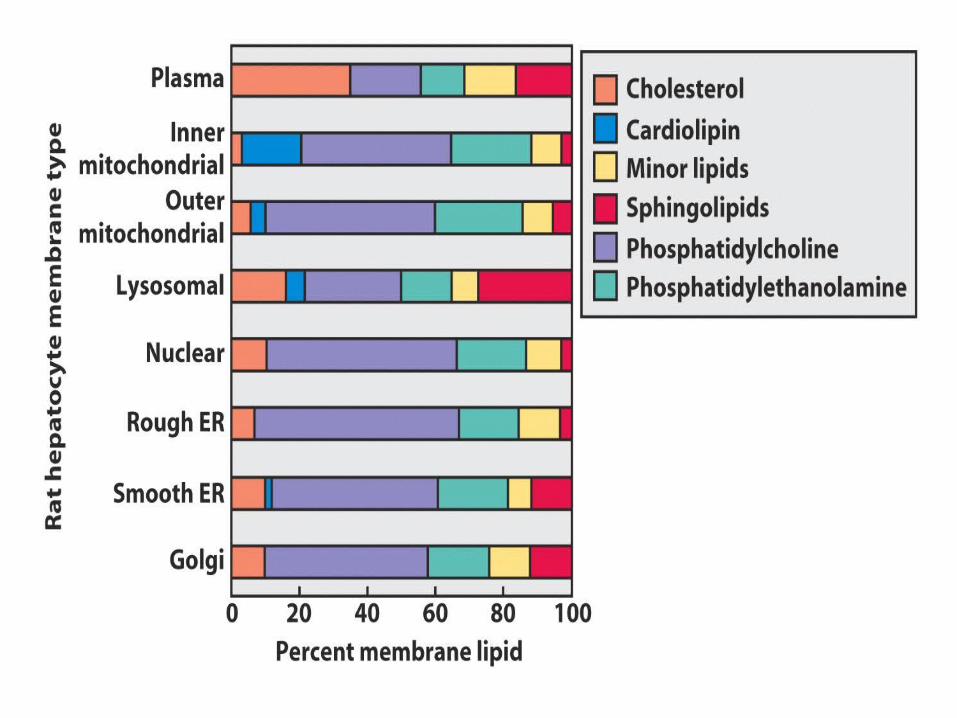
Analysis of lipids in membranes
Relative proportion of components in plasma membranes differ for each species and tissue

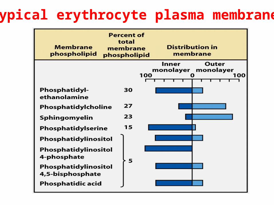
Typical erythrocyte plasma membrane

Low temperature: Thermal motion is constrained
High temperature: Thermal motion is rapid.
Theory: Cells seek to balance these two phases providing enough disorder for lateral movement but less freedom for acyl chains
Tc= when 50% of each phase is
present in solution