sudden oak death and associated diseases caused by
TRANSCRIPT
2003. Plant Management Network. This article is in the public domain. Accepted for publication 6 June 2003. Published 7 July 2003.
Sudden Oak Death and Associated Diseases Caused by Phytophthora ramorum J. M. Davidson, Pacific Southwest Research Station, USDA Forest Service, P.O. Box 245, Berkeley, CA 94701, and Department of Plant Pathology, One Shields Ave., University of California, Davis 95616; S. Werres, Biological Research Centre for Agriculture and Forestry, Institute for Plant Protection in Horticulture, Messeweg 11/12, D-38104 Braunschweig, Germany; M. Garbelotto Department of Environmental Science, Policy and Management, Ecosystem Science Division, 151 Hilgard Hall, University of California, Berkeley 94720; E. M. Hansen Department of Botany and Plant Pathology, Oregon State University, Corvallis 97331; D. M. Rizzo Department of Plant Pathology, One Shields Ave., University of California, Davis 95616 Corresponding author: J. M. Davidson. [email protected]
Davidson, J. M., Werres, S., Garbelotto, M., Hansen, E. M., and Rizzo, D. M. 2003. Sudden oak death and associated diseases caused by Phytophthora ramorum. Online. Plant Health Progress doi:10.1094/PHP-2003-0707-01-DG.
Disease
On tanoak (Lithocarpus densiflora) and some oak species (Quercus spp.): Sudden Oak Death. On other hosts: Ramorum leaf blight and ramorum shoot dieback. Pathogen
Phytophthora ramorum (S. Werres, A. W. A. M. de Cock & W. A. Man in’t Veld) (24). Taxonomy
Phytophthora ramorum was first described as a pathogen of ornamental rhododendron (Rhododendron) and viburnum (Viburnum) in Germany and The Netherlands (24). The morphology and ITS sequence of California and Oregon isolates are identical to European isolates (9,19). Host Range
To date, 23 species in 12 plant families have been found to be naturally infected by Phytophthora ramorum (Table 1) (18,19,24). Species in the Ericaceae and Fagaceae appear to be especially prone to infection. Many more plant species are susceptible to leaf blight in artificial inoculation trials.
Plant Health Progress 7 July 2003
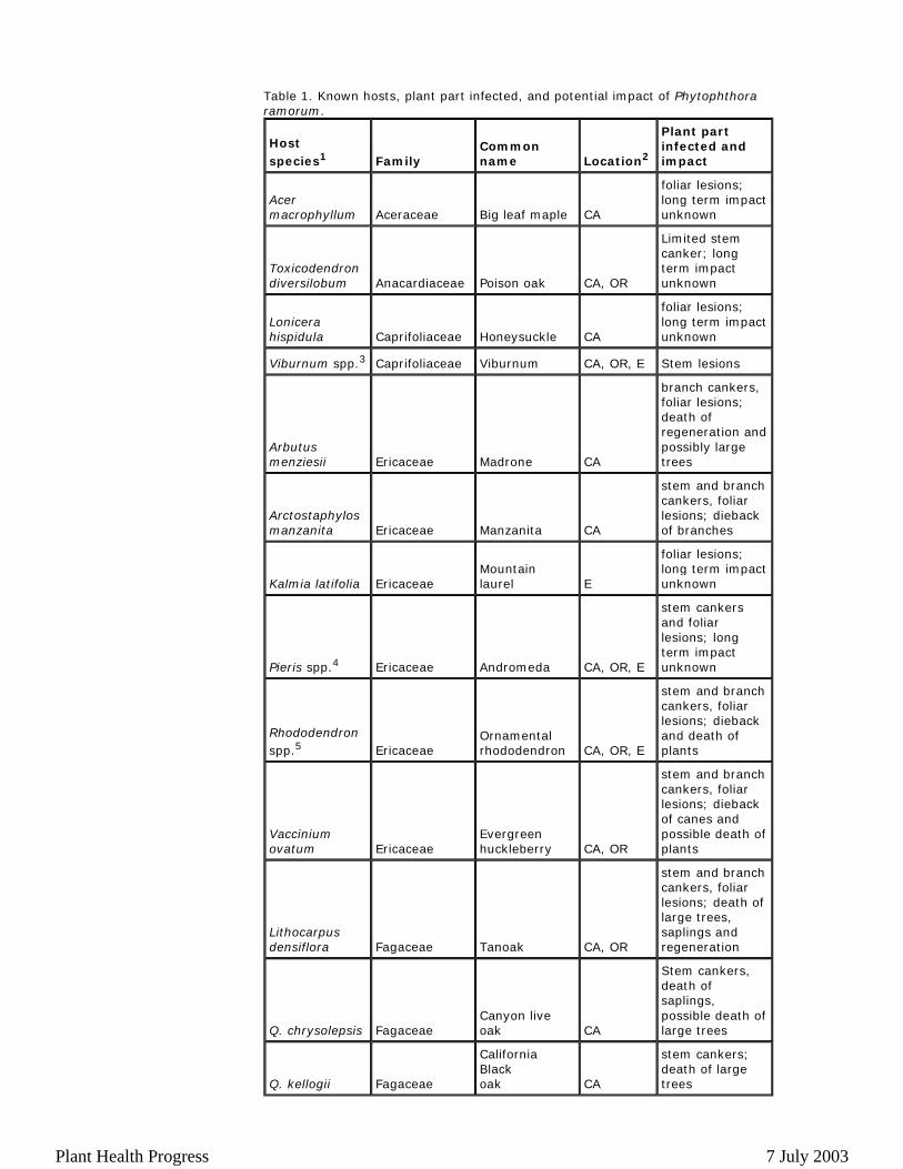
Table 1. Known hosts, plant part infected, and potential impact of Phytophthora ramorum.
Host species1 Family
Common name Location2
Plant part infected and impact
Acer macrophyllum Aceraceae Big leaf maple CA
foliar lesions; long term impact unknown
Toxicodendron diversilobum Anacardiaceae Poison oak CA, OR
Limited stem canker; long term impact unknown
Lonicera hispidula Caprifoliaceae Honeysuckle CA
foliar lesions; long term impact unknown
Viburnum spp.3 Caprifoliaceae Viburnum CA, OR, E Stem lesions
Arbutus menziesii Ericaceae Madrone CA
branch cankers, foliar lesions; death of regeneration and possibly large trees
Arctostaphylos manzanita Ericaceae Manzanita CA
stem and branch cankers, foliar lesions; dieback of branches
Kalmia latifolia EricaceaeMountain laurel E
foliar lesions; long term impact unknown
Pieris spp.4 Ericaceae Andromeda CA, OR, E
stem cankers and foliar lesions; long term impact unknown
Rhododendron spp.5 Ericaceae
Ornamental rhododendron CA, OR, E
stem and branch cankers, foliar lesions; dieback and death of plants
Vaccinium ovatum Ericaceae
Evergreen huckleberry CA, OR
stem and branch cankers, foliar lesions; dieback of canes and possible death of plants
Lithocarpus densiflora Fagaceae Tanoak CA, OR
stem and branch cankers, foliar lesions; death of large trees, saplings and regeneration
Q. chrysolepsis FagaceaeCanyon live oak CA
Stem cankers, death of saplings, possible death of large trees
Q. kellogii Fagaceae
California Black oak CA
stem cankers; death of large trees
Plant Health Progress 7 July 2003
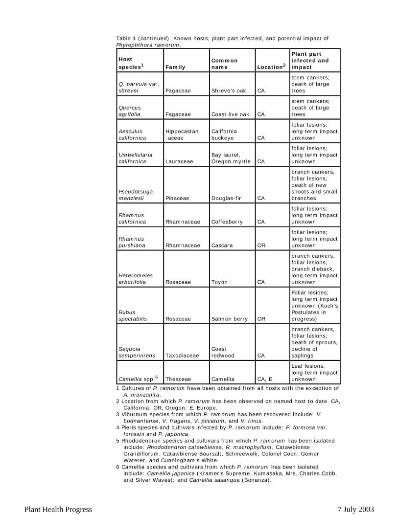
Table 1 (continued). Known hosts, plant part infected, and potential impact of Phytophthora ramorum.
1 Cultures of P. ramorum have been obtained from all hosts with the exception of A. manzanita.
2 Location from which P. ramorum has been observed on named host to date. CA, California; OR, Oregon; E, Europe.
3 Viburnum species from which P. ramorum has been recovered include: V. bodnantense, V. fragans, V. plicatum, and V. tinus.
4 Pieris species and cultivars infected by P. ramorum include: P. formosa var. forrestii and P. japonica.
5 Rhododendron species and cultivars from which P. ramorum has been isolated include: Rhododendron catawbiense, R. macrophyllum, Catawbiense Grandiflorum, Catawbiense Boursalt, Schneewolk, Colonel Coen, Gomer Waterer, and Cunningham’s White.
6 Camellia species and cultivars from which P. ramorum has been isolated include: Camellia japonica (Kramer’s Supreme, Kumasaka, Mrs. Charles Cobb, and Silver Waves); and Camellia sasanqua (Bonanza).
Host species1 Family
Common name Location2
Plant part infected and impact
Q. parvula var. shrevei Fagaceae Shreve’s oak CA
stem cankers; death of large trees
Quercus agrifolia Fagaceae Coast live oak CA
stem cankers; death of large trees
Aesculus californica
Hippocastan -aceae
California buckeye CA
foliar lesions; long term impact unknown
Umbellularia californica Lauraceae
Bay laurel, Oregon myrtle CA
foliar lesions; long term impact unknown
Pseudotsuga menziesii Pinaceae Douglas-fir CA
branch cankers, foliar lesions; death of new shoots and small branches
Rhamnus californica Rhamnaceae Coffeeberry CA
foliar lesions; long term impact unknown
Rhamnus purshiana Rhamnaceae Cascara OR
foliar lesions; long term impact unknown
Heteromeles arbutifolia Rosaceae Toyon CA
branch cankers, foliar lesions; branch dieback, long term impact unknown
Rubus spectabilis Rosaceae Salmon berry OR
Foliar lesions; long term impact unknown (Koch’s Postulates in progress)
Sequoia sempervirens Taxodiaceae
Coast redwood CA
branch cankers, foliar lesions; death of sprouts, decline of saplings
Camellia spp.6 Theaceae Camellia CA, E
Leaf lesions; long term impact unknown
Plant Health Progress 7 July 2003
Geographical Distribution The current known distribution of P. ramorum includes the western USA,
Europe, and Canada. In the USA, the distribution of P. ramorum within natural areas is patchy, with sites extending in a narrow coastal band over 600 km from Monterey County, CA (36°15'N, 121°47'W) to Curry County, OR (42°6'N, 124°15'W) (9,19). The wildland sites currently furthest inland in California are approximately 70 km from the coast in Solano County (38°18'N, 122°12'W). Although these sites include all known hosts, geographic range surveys have thus far used dead oak and tanoak as indicators for disease, and therefore may underestimate the true range by failing to identify areas in which P. ramorum is only present on non-oak hosts. In addition to these wildland sites, P. ramorum has been found on rhododendron, camellia (Camellia), viburnum, and andromeda (Pieris) in nurseries in California, Oregon, Washington, and British Columbia that are within and outside the range of P. ramorum in natural areas (C. Blomquist, unpublished data; E. Hansen, unpublished data; G. Kristjansson, unpublished data; N. Osterbauer, unpublished data; A. Wagner, unpublished data). For the most recent information on the North American geographical distribution of P. ramorum, see www.suddenoakdeath.org.
In Europe, P. ramorum has been isolated from symptomatic rhododendron and viburnum in nurseries, gardens and landscape plantings in Germany, the Netherlands, the United Kingdom, Poland, Spain, France, Belgium, and Sweden (6,7,12,15,17,23,24). P. ramorum has also been found to infect camellia, andromeda (Pieris spp.), and mountain laurel (Kalmia latifolia) in European nurseries (11; P. Beales, unpublished data; T. Brokenshire, unpublished data). Up to now, P. ramorum has not been recovered from plants in forests in Europe. Symptoms and Signs
Oaks (Quercus spp.). To date, only oaks in the red oak group (section Lobatae) have been found to be susceptible to infection by P. ramorum in California (19). These include coast live oak (Quercus agrifolia), California black oak (Quercus kelloggii), and Shreve’s oak (Quercus parvula var. shrevei). The remaining species in the red oak group in California, interior live oak (Q. wislizenii), has been found to be susceptible to infection in greenhouse inoculation trials (D. M. Rizzo, unpublished data). In addition, northern red oak (Quercus rubra) and pinoak (Q. palustris), important species from eastern North America, have been shown to be susceptible in greenhouse inoculation studies (18; Hansen, unpublished data). Canyon live oak (Q. chrysolepsis) in the golden cup group (section Protobalanus) has been found infected by P. ramorum at a single location (16). No species in the white oak group (section Quercus) have been found with the disease in the field in California, Oregon, or Europe.
In oaks, only adult plants are significantly affected. Infection of seedlings is unreported in nature, and infection of saplings appears to be extremely rare. The most useful diagnostic symptom for P. ramorum is the development of cankers on the trunk. Cankers have red-brown to black discoloration and seep dark black to red or amber sap (Figs. 1A and 1B). They usually develop 1 to 2 m off of the ground, although they can be at soil level, or as high as 4 m or greater. Evidence to date suggests that cankers do not extend into the roots below the soil line. Cankers will occasionally extend onto portions of buttress roots if the roots are above the soil line.
Plant Health Progress 7 July 2003

Typically, canker formation begins with isolated bleeding spots on the main
trunk of the tree. At the early stages of canker development, seeping will occur through the intact bark, without any noticeable physical wounding. In later stages, the bark can fracture and exudation occurs both through broken and intact bark. Later, the area will be stained reddish-brown. When liquid, the sap smells like the oak tannin odor of wine barrels. Dark wet cankers on oak trunks with very foul smelling exudates are usually due to bacterial infections (wetwood), not P. ramorum. The sap from a P. ramorum canker may dry and harden into a blackish crust during the summer months. Infection and discoloration is generally more extensive in bark and phloem tissues than in the xylem. In active cankers, the phloem is discolored various shades of brown (Fig. 2A), and cankers are delimited by thin black lines (Fig. 2B). Cankers may be entire, or consist of smaller separate cankers in close proximity. It is unknown at this point whether these represent separate infections or just an uneven spread of the pathogen. Black discoloration can extend to 3 cm or greater into the xylem below the bleeding canker, but more typically extends less than 1 mm. Cankers may range in size from a few cm in diameter to 3 m in length. Phytophthora is typically isolated from the margins of these discolored areas, although it can occasionally be isolated from older portions of the cankers.
Phytophthora ramorum canker disease of oak has been called “Sudden Oak
Death” due to the rapid (2 to 4 weeks) and complete browning of the crown observed on numerous trees at their death (Fig. 3). In these cases, the dead foliage appears without a prolonged period of visible decline. While this sudden browning may occur, death of the tree due to P. ramorum infection usually takes place after an extended period of disease and perhaps more than two years from
A B
Fig. 1. P. ramorum canker on coast live oak. Outer bark shows rust-colored seepage (A, B).
A
B
Fig 2. In coast live oak, removing outer bark reveals diseased phloem tissue and zone lines (A). Thin black zone lines characteristically separate diseased from healthy tissue in oak and tanoak cankers (B).
Plant Health Progress 7 July 2003
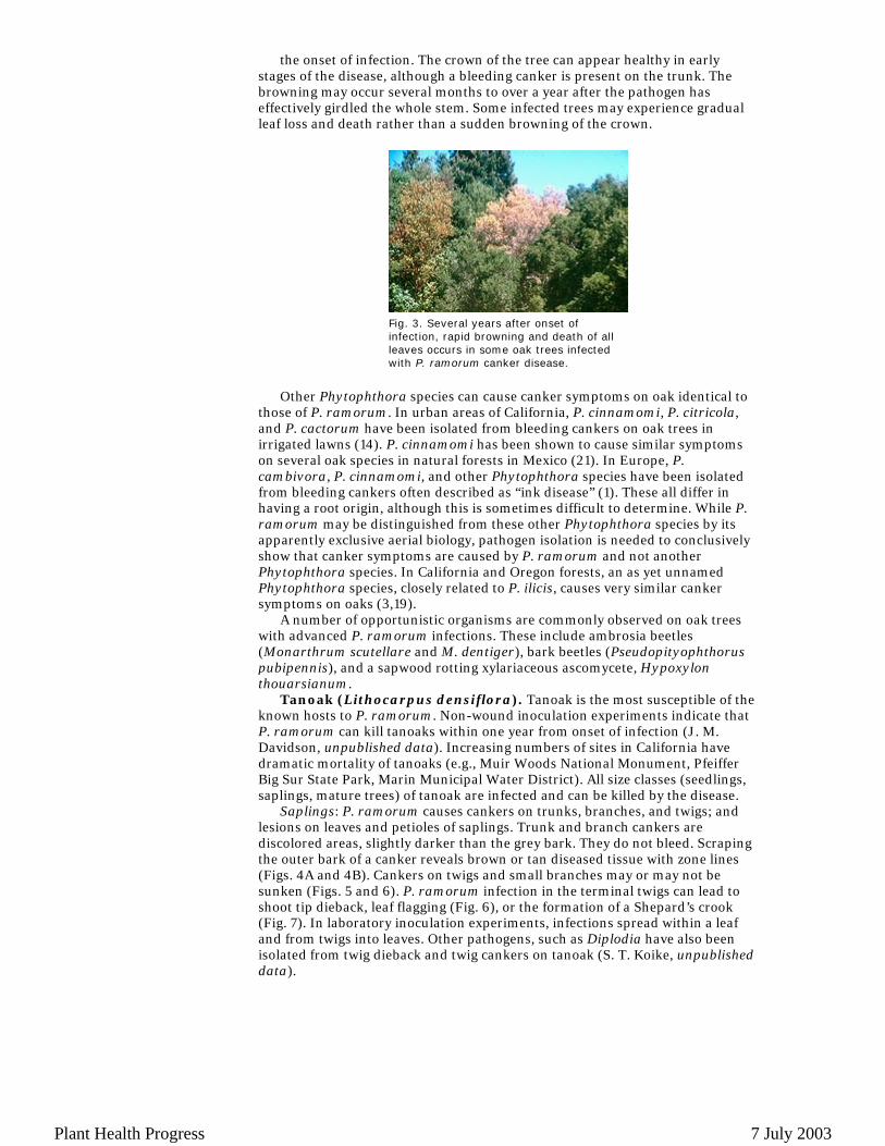
the onset of infection. The crown of the tree can appear healthy in early stages of the disease, although a bleeding canker is present on the trunk. The browning may occur several months to over a year after the pathogen has effectively girdled the whole stem. Some infected trees may experience gradual leaf loss and death rather than a sudden browning of the crown.
Other Phytophthora species can cause canker symptoms on oak identical to
those of P. ramorum. In urban areas of California, P. cinnamomi, P. citricola, and P. cactorum have been isolated from bleeding cankers on oak trees in irrigated lawns (14). P. cinnamomi has been shown to cause similar symptoms on several oak species in natural forests in Mexico (21). In Europe, P. cambivora, P. cinnamomi, and other Phytophthora species have been isolated from bleeding cankers often described as “ink disease” (1). These all differ in having a root origin, although this is sometimes difficult to determine. While P. ramorum may be distinguished from these other Phytophthora species by its apparently exclusive aerial biology, pathogen isolation is needed to conclusively show that canker symptoms are caused by P. ramorum and not another Phytophthora species. In California and Oregon forests, an as yet unnamed Phytophthora species, closely related to P. ilicis, causes very similar canker symptoms on oaks (3,19).
A number of opportunistic organisms are commonly observed on oak trees with advanced P. ramorum infections. These include ambrosia beetles (Monarthrum scutellare and M. dentiger), bark beetles (Pseudopityophthorus pubipennis), and a sapwood rotting xylariaceous ascomycete, Hypoxylon thouarsianum.
Tanoak (Lithocarpus densiflora). Tanoak is the most susceptible of the known hosts to P. ramorum. Non-wound inoculation experiments indicate that P. ramorum can kill tanoaks within one year from onset of infection (J. M. Davidson, unpublished data). Increasing numbers of sites in California have dramatic mortality of tanoaks (e.g., Muir Woods National Monument, Pfeiffer Big Sur State Park, Marin Municipal Water District). All size classes (seedlings, saplings, mature trees) of tanoak are infected and can be killed by the disease.
Saplings: P. ramorum causes cankers on trunks, branches, and twigs; and lesions on leaves and petioles of saplings. Trunk and branch cankers are discolored areas, slightly darker than the grey bark. They do not bleed. Scraping the outer bark of a canker reveals brown or tan diseased tissue with zone lines (Figs. 4A and 4B). Cankers on twigs and small branches may or may not be sunken (Figs. 5 and 6). P. ramorum infection in the terminal twigs can lead to shoot tip dieback, leaf flagging (Fig. 6), or the formation of a Shepard’s crook (Fig. 7). In laboratory inoculation experiments, infections spread within a leaf and from twigs into leaves. Other pathogens, such as Diplodia have also been isolated from twig dieback and twig cankers on tanoak (S. T. Koike, unpublished data).
Fig. 3. Several years after onset of infection, rapid browning and death of all leaves occurs in some oak trees infected with P. ramorum canker disease.
Plant Health Progress 7 July 2003
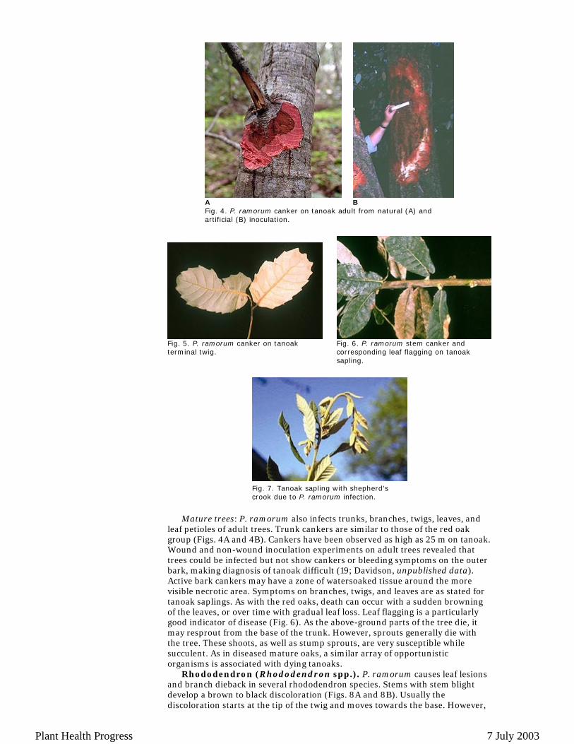
Mature trees: P. ramorum also infects trunks, branches, twigs, leaves, and
leaf petioles of adult trees. Trunk cankers are similar to those of the red oak group (Figs. 4A and 4B). Cankers have been observed as high as 25 m on tanoak. Wound and non-wound inoculation experiments on adult trees revealed that trees could be infected but not show cankers or bleeding symptoms on the outer bark, making diagnosis of tanoak difficult (19; Davidson, unpublished data). Active bark cankers may have a zone of watersoaked tissue around the more visible necrotic area. Symptoms on branches, twigs, and leaves are as stated for tanoak saplings. As with the red oaks, death can occur with a sudden browning of the leaves, or over time with gradual leaf loss. Leaf flagging is a particularly good indicator of disease (Fig. 6). As the above-ground parts of the tree die, it may resprout from the base of the trunk. However, sprouts generally die with the tree. These shoots, as well as stump sprouts, are very susceptible while succulent. As in diseased mature oaks, a similar array of opportunistic organisms is associated with dying tanoaks.
Rhododendron (Rhododendron spp.). P. ramorum causes leaf lesions and branch dieback in several rhododendron species. Stems with stem blight develop a brown to black discoloration (Figs. 8A and 8B). Usually the discoloration starts at the tip of the twig and moves towards the base. However,
A
B
Fig. 4. P. ramorum canker on tanoak adult from natural (A) and artificial (B) inoculation.
Fig. 5. P. ramorum canker on tanoak terminal twig.
Fig. 6. P. ramorum stem canker and corresponding leaf flagging on tanoak sapling.
Fig. 7. Tanoak sapling with shepherd’s crook due to P. ramorum infection.
Plant Health Progress 7 July 2003
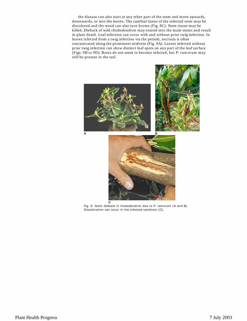
the disease can also start at any other part of the stem and move upwards, downwards, or into the leaves. The cambial tissue of the infected stem may be discolored and the wood can also turn brown (Fig. 8C). Stem tissue may be killed. Dieback of wild rhododendron may extend into the main stems and result in plant death. Leaf infection can occur with and without prior twig infection. In leaves infected from a twig infection via the petiole, necrosis is often concentrated along the prominent midvein (Fig. 9A). Leaves infected without prior twig infection can show distinct leaf spots on any part of the leaf surface (Figs. 9B to 9D). Roots do not seem to become infected, but P. ramorum may still be present in the soil.
A
B
C
Fig. 8. Stem dieback in rhododendron due to P. ramorum (A and B). Discoloration can occur in the infected cambium (C).
Plant Health Progress 7 July 2003

Initially, symptoms of P. ramorum on rhododendron and/or azalea can be
very much like those of other Phytophthora species such as P. cactorum, P. citricola, P. cinnamomi, and P. syringae. There are other damaging agents that may cause twig or stem dieback (e.g., Phomopsis sp., Botryosphaeria sp.) or wilting symptoms (e.g., Cylindrocladium sp., root weevils, drought, or water stress). Leaf lesions caused by P. ramorum can sometimes be confused with early symptoms of rust infection prior to the development of visible spore forms of either pathogen on the undersides of the leaves. In addition, it may be difficult to distinguish between leafspots caused by P. ramorum and those caused by sunburn. However, the margin on Phytophthora lesions tends to be diffuse, while the margin on sunburn is distinct. The extension of necrosis due to P. ramorum along the midvein also is often distinctive.
In Oregon and California, P. ramorum infects native R. macrophyllum (Fig. 9D) growing among infected tanoaks (9). Ornamental rhododendrons in California and Europe also have been found infected with P. ramorum. Susceptible nursery varieties include, but are not limited to: Catawbiense Grandiflorum, Catawbiense Boursalt, Schneewolk, Colonel Coen, Gomer Waterer, and Cunningham’s White. Rhododendron catawbiense has also been found to be susceptible to infection in German nurseries, but P. ramorum does not seem to damage Rhododendron simsii in nurseries. Experiments are underway to investigate the susceptibility of azaleas. Initial inoculation studies under controlled conditions indicate that some azaleas (e.g., Northern Hilights and R. occidentale) are susceptible to P. ramorum. Deciduous azaleas generally seem to be more susceptible than evergreen azaleas (22).
Madrone (Arbutus menziesii). Symptoms and disease progression on madrone are similar to that seen on rhododendron with leaf spots, leaf death, and branch dieback commonly associated with infection by P. ramorum. P. ramorum forms very dark lesions on leaves (Fig. 10), often extending down petioles and into the twig. On stems, the lesions initially are difficult to find and appear as a slight water soaking or darkening of the bark that travels very quickly throughout the stem and kills the tissue, creating a dark lesion (Fig. 11)
A B
C
D
Fig. 9. Infected rhododendron leaves have dark brown lesions. Necrosis is often concentrated along the midvein in leaves infected from a twig infection via the petiole (A). Leaves infected without prior twig infection can have leaf spots on any part of the leaf surface (B, C, and D). 9D shows naturally infected leaves of the native rhododendron, R. macrophyllum, from southwestern Oregon.
Plant Health Progress 7 July 2003
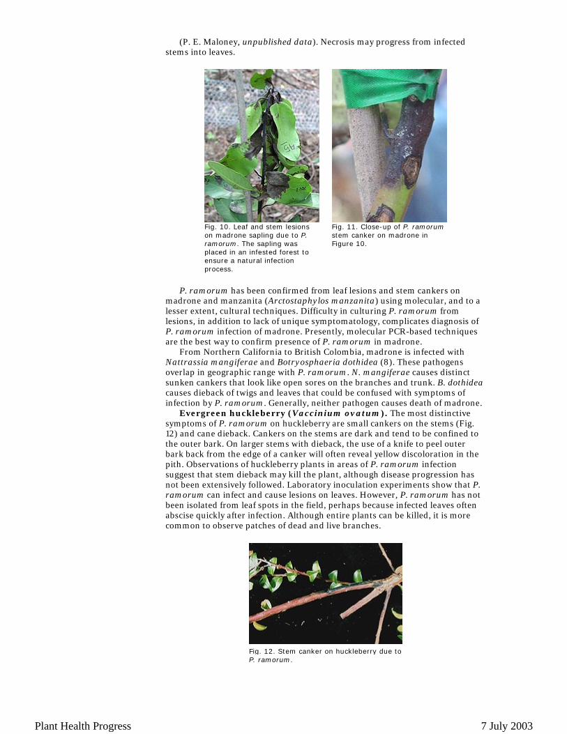
(P. E. Maloney, unpublished data). Necrosis may progress from infected stems into leaves.
P. ramorum has been confirmed from leaf lesions and stem cankers on
madrone and manzanita (Arctostaphylos manzanita) using molecular, and to a lesser extent, cultural techniques. Difficulty in culturing P. ramorum from lesions, in addition to lack of unique symptomatology, complicates diagnosis of P. ramorum infection of madrone. Presently, molecular PCR-based techniques are the best way to confirm presence of P. ramorum in madrone.
From Northern California to British Colombia, madrone is infected with Nattrassia mangiferae and Botryosphaeria dothidea (8). These pathogens overlap in geographic range with P. ramorum. N. mangiferae causes distinct sunken cankers that look like open sores on the branches and trunk. B. dothidea causes dieback of twigs and leaves that could be confused with symptoms of infection by P. ramorum. Generally, neither pathogen causes death of madrone.
Evergreen huckleberry (Vaccinium ovatum). The most distinctive symptoms of P. ramorum on huckleberry are small cankers on the stems (Fig. 12) and cane dieback. Cankers on the stems are dark and tend to be confined to the outer bark. On larger stems with dieback, the use of a knife to peel outer bark back from the edge of a canker will often reveal yellow discoloration in the pith. Observations of huckleberry plants in areas of P. ramorum infection suggest that stem dieback may kill the plant, although disease progression has not been extensively followed. Laboratory inoculation experiments show that P. ramorum can infect and cause lesions on leaves. However, P. ramorum has not been isolated from leaf spots in the field, perhaps because infected leaves often abscise quickly after infection. Although entire plants can be killed, it is more common to observe patches of dead and live branches.
Fig. 10. Leaf and stem lesions on madrone sapling due to P. ramorum. The sapling was placed in an infested forest to ensure a natural infection process.
Fig. 11. Close-up of P. ramorum stem canker on madrone in Figure 10.
Fig. 12. Stem canker on huckleberry due to P. ramorum.
Plant Health Progress 7 July 2003
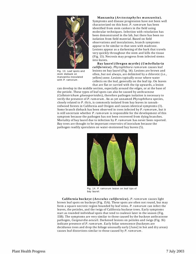
Manzanita (Arctostaphylos manzanita).
Symptoms and disease progression have not been well characterized on this host. P. ramorum has been identified from stem cankers in the field using molecular techniques. Infection with reisolation has been demonstrated in the lab, but there has been no isolation from field material. Based on field observations and inoculations, branch symptoms appear to be similar to that seen with madrone. Lesions appear as a darkening of the bark that travels very quickly throughout the stem and kills the tissue (Fig. 13). Necrosis may progress from infected stems into leaves.
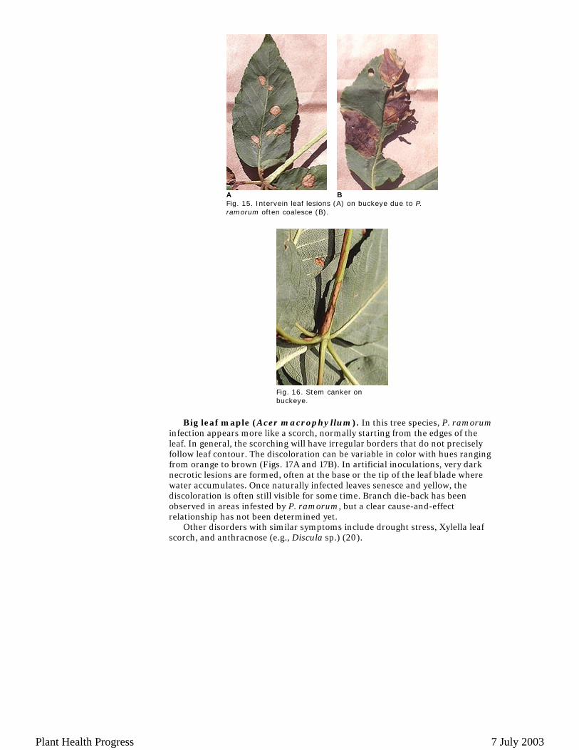
Bay laurel (Oregon myrtle) (Umbellularia californica). Phytophthora ramorum causes leaf lesions on bay laurel (Fig. 14). Lesions are brown and often, but not always, are delimited by a chlorotic (i.e., yellow) zone. Lesions typically occur where water collects on the leaf, generally on the leaf tip. On leaves that are flat or carried with the tip upwards, a lesion
can develop in the middle section, especially around the edges, or at the base of the petiole. These types of leaf spots can also be caused by anthracnose (Colletotrichum gloeosporioides), therefore pathogen isolation is necessary to verify the presence of P. ramorum. An as yet unnamed Phytophthora species, closely related to P. ilicis, is commonly isolated from bay leaves in tanoak-redwood forests in California and Oregon and causes identical symptoms (3). Some branch dieback has been observed in trees infected by P. ramorum, but it is still uncertain whether P. ramorum is responsible for the development of this symptom because the pathogen has not been recovered from dying branches. Mortality of bay laurel due to infection by P. ramorum has never been reported. Bay trees are thought to be important reservoirs of inoculum because the pathogen readily sporulates on water-moistened bay leaves (5).
California buckeye (Aesculus californica). P. ramorum causes light
brown leaf spots on buckeye (Fig. 15A). These spots are often not round, but may form a square necrotic region bounded by leaf veins. P. ramorum can infect the leaves, the petioles, and the twigs of California buckeye trees. Early symptoms start as rounded individual spots that tend to coalesce later in the season (Fig. 15B). The symptoms are very similar to those caused by the buckeye anthracnose pathogen, Guignardia aesculi. Darkened lesions on petioles and twigs (Fig. 16) indicate presence of P. ramorum. Early foliar senescence (buckeyes are deciduous trees and drop the foliage unusually early [June] in hot and dry areas) causes leaf distortions similar to those caused by P. ramorum.
Fig. 13. Leaf spots and stem dieback on manzanita inoculated with P. ramorum.
Fig. 14. P. ramorum lesion on leaf tips of bay laurel.
Plant Health Progress 7 July 2003
Big leaf maple (Acer macrophyllum). In this tree species, P. ramorum
infection appears more like a scorch, normally starting from the edges of the leaf. In general, the scorching will have irregular borders that do not precisely follow leaf contour. The discoloration can be variable in color with hues ranging from orange to brown (Figs. 17A and 17B). In artificial inoculations, very dark necrotic lesions are formed, often at the base or the tip of the leaf blade where water accumulates. Once naturally infected leaves senesce and yellow, the discoloration is often still visible for some time. Branch die-back has been observed in areas infested by P. ramorum, but a clear cause-and-effect relationship has not been determined yet.
Other disorders with similar symptoms include drought stress, Xylella leaf scorch, and anthracnose (e.g., Discula sp.) (20).
A
B
Fig. 15. Intervein leaf lesions (A) on buckeye due to P. ramorum often coalesce (B).
Fig. 16. Stem canker on buckeye.
Plant Health Progress 7 July 2003
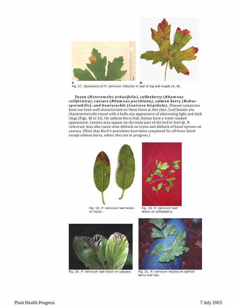
Toyon (Heteromeles arbutifolia), coffeeberry (Rhamnus
californica), cascara (Rhamnus purshiana), salmon berry (Rubus spectabilis), and honeysuckle (Lonicera hispidula). Disease symptoms have not been well characterized on these hosts at this time. Leaf lesions are characteristically round with a bulls-eye appearance of alternating light and dark rings (Figs. 18 to 21). On salmon berry leaf, lesions have a water-soaked appearance. Lesions may appear on the main part of the leaf or leaf tip. P. ramorum may also cause stem dieback on toyon and dieback of basal sprouts on cascara. (Note that Koch’s postulates have been completed for all hosts listed except salmon berry, where they are in progress.)
A B
Fig. 17. Symptoms of P. ramorum infection in leaf of big leaf maple (A, B).
Fig. 18. P. ramorum leaf lesion on toyon.
Fig. 19. P. ramorum leaf lesion on coffeeberry.
Fig. 20. P. ramorum leaf lesion on cascara. Fig. 21. P. ramorum lesions on salmon berry leaf tips.
Plant Health Progress 7 July 2003
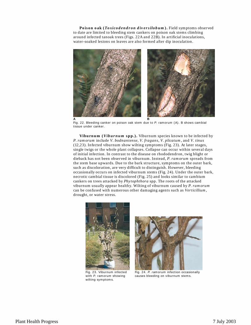
Poison oak (Toxicodendron diversilobum). Field symptoms observed to date are limited to bleeding stem cankers on poison oak stems climbing around infected tanoak trees (Figs. 22A and 22B). In artificial inoculations, water-soaked lesions on leaves are also formed after dip inoculation.
Viburnum (Viburnum spp.). Viburnum species known to be infected by
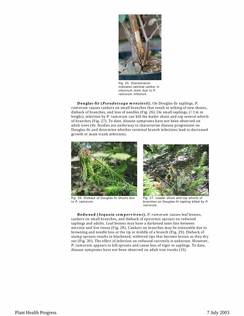
P. ramorum include V. bodnantense, V. fragans, V. plicatum, and V. tinus (12,23). Infected viburnum show wilting symptoms (Fig. 23). At later stages, single twigs or the whole plant collapses. Collapse can occur within several days of initial infection. In contrast to the disease on rhododendron, twig blight or dieback has not been observed in viburnum. Instead, P. ramorum spreads from the stem base upwards. Due to the bark structure, symptoms on the outer bark, such as discoloration, are very difficult to distinguish. However, bleeding occasionally occurs on infected viburnum stems (Fig. 24). Under the outer bark, necrotic cambial tissue is discolored (Fig. 25) and looks similar to cambium cankers on trees attacked by Phytophthora spp. The roots of the attacked viburnum usually appear healthy. Wilting of viburnum caused by P. ramorum can be confused with numerous other damaging agents such as Verticillium, drought, or water stress.
A
BFig. 22. Bleeding canker on poison oak stem due to P. ramorum (A). B shows cambial tissue under canker.
Fig. 23. Viburnum infected with P. ramorum showing wilting symptoms.
Fig. 24. P. ramorum infection occasionally causes bleeding on viburnum stems.
Plant Health Progress 7 July 2003
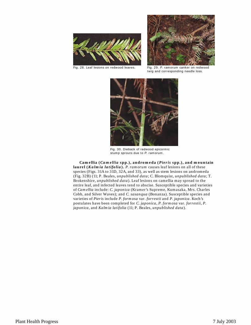
Douglas-fir (Pseudotsuga menziesii). On Douglas-fir saplings, P.
ramorum causes cankers on small branches that result in wilting of new shoots, dieback of branches, and loss of needles (Fig. 26). On small saplings, (<1 m in height), infection by P. ramorum can kill the leader shoot and top several whorls of branches (Fig. 27). To date, disease symptoms have not been observed on adult trees (4). Studies are underway to characterize disease progression on Douglas-fir and determine whether terminal branch infections lead to decreased growth or main trunk infections.
Redwood (Sequoia sempervirens). P. ramorum causes leaf lesions,
cankers on small branches, and dieback of epicormic sprouts on redwood saplings and adults. Leaf lesions may have a darkened zone line between necrotic and live tissue (Fig. 28). Cankers on branches may be noticeable due to browning and needle loss at the tip or middle of a branch (Fig. 29). Dieback of stump sprouts results in blackened, withered tips that become brown as they dry out (Fig. 30). The effect of infection on redwood currently is unknown. However, P. ramorum appears to kill sprouts and cause loss of vigor in saplings. To date, disease symptoms have not been observed on adult tree trunks (13).
Fig. 25. Discoloration indicates cambial canker in viburnum stem due to P. ramorum infection.
Fig. 26. Dieback of Douglas-fir shoots due to P. ramorum.
Fig. 27. Leader shoot and top whorls of branches on Douglas-fir sapling killed by P. ramorum.
Plant Health Progress 7 July 2003
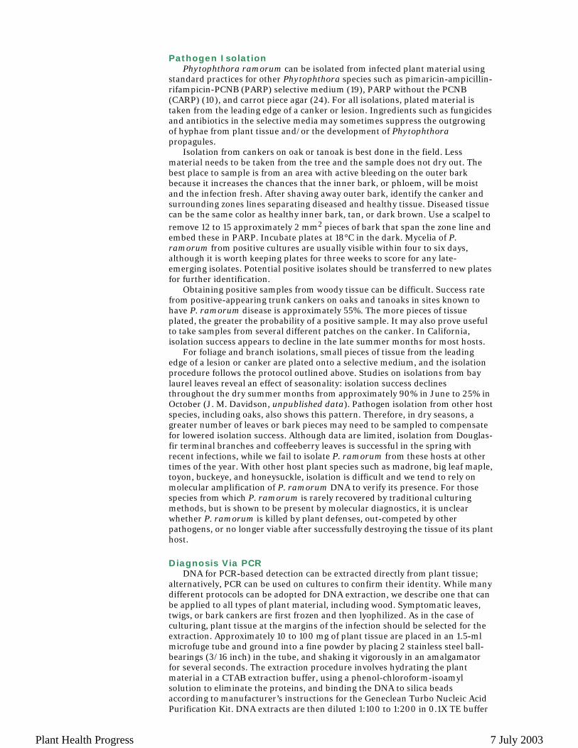
Camellia (Camellia spp.), andromeda (Pieris spp.), and mountain
laurel (Kalmia latifolia). P. ramorum causes leaf lesions on all of these species (Figs. 31A to 31D, 32A, and 33), as well as stem lesions on andromeda (Fig. 32B) (11; P. Beales, unpublished data; C. Blomquist, unpublished data; T. Brokenshire, unpublished data). Leaf lesions on camellia may spread to the entire leaf, and infected leaves tend to abscise. Susceptible species and varieties of Camellia include: C. japonica (Kramer’s Supreme, Kumasaka, Mrs. Charles Cobb, and Silver Waves); and C. sasanqua (Bonanza). Susceptible species and varieties of Pieris include P. formosa var. forrestii and P. japonica. Koch’s postulates have been completed for C. japonica, P. formosa var. forrestii, P. japonica, and Kalmia latifolia (11; P. Beales, unpublished data).
Fig. 28. Leaf lesions on redwood leaves. Fig. 29. P. ramorum canker on redwood twig and corresponding needle loss.
Fig. 30. Dieback of redwood epicormic stump sprouts due to P. ramorum.
Plant Health Progress 7 July 2003
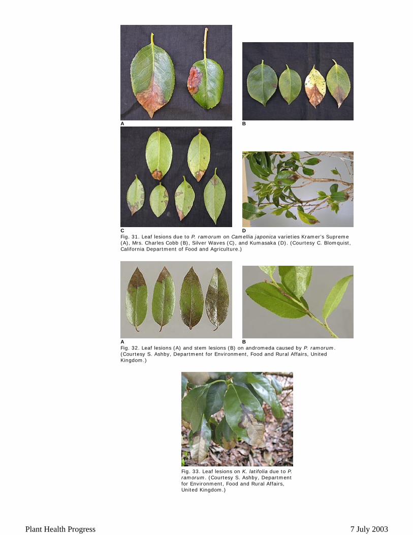
Fig. 31. Leaf lesions due to P. ramorum on Camellia japonica varieties Kramer’s Supreme (A), Mrs. Charles Cobb (B), Silver Waves (C), and Kumasaka (D). (Courtesy C. Blomquist, California Department of Food and Agriculture.)
Fig. 32. Leaf lesions (A) and stem lesions (B) on andromeda caused by P. ramorum. (Courtesy S. Ashby, Department for Environment, Food and Rural Affairs, United Kingdom.)
A B
C D
A B
Fig. 33. Leaf lesions on K. latifolia due to P. ramorum. (Courtesy S. Ashby, Department for Environment, Food and Rural Affairs, United Kingdom.)
Plant Health Progress 7 July 2003
Pathogen Isolation
Phytophthora ramorum can be isolated from infected plant material using standard practices for other Phytophthora species such as pimaricin-ampicillin-rifampicin-PCNB (PARP) selective medium (19), PARP without the PCNB (CARP) (10), and carrot piece agar (24). For all isolations, plated material is taken from the leading edge of a canker or lesion. Ingredients such as fungicides and antibiotics in the selective media may sometimes suppress the outgrowing of hyphae from plant tissue and/or the development of Phytophthora propagules.
Isolation from cankers on oak or tanoak is best done in the field. Less material needs to be taken from the tree and the sample does not dry out. The best place to sample is from an area with active bleeding on the outer bark because it increases the chances that the inner bark, or phloem, will be moist and the infection fresh. After shaving away outer bark, identify the canker and surrounding zones lines separating diseased and healthy tissue. Diseased tissue can be the same color as healthy inner bark, tan, or dark brown. Use a scalpel to remove 12 to 15 approximately 2 mm2 pieces of bark that span the zone line and embed these in PARP. Incubate plates at 18°C in the dark. Mycelia of P. ramorum from positive cultures are usually visible within four to six days, although it is worth keeping plates for three weeks to score for any late-emerging isolates. Potential positive isolates should be transferred to new plates for further identification.
Obtaining positive samples from woody tissue can be difficult. Success rate from positive-appearing trunk cankers on oaks and tanoaks in sites known to have P. ramorum disease is approximately 55%. The more pieces of tissue plated, the greater the probability of a positive sample. It may also prove useful to take samples from several different patches on the canker. In California, isolation success appears to decline in the late summer months for most hosts.
For foliage and branch isolations, small pieces of tissue from the leading edge of a lesion or canker are plated onto a selective medium, and the isolation procedure follows the protocol outlined above. Studies on isolations from bay laurel leaves reveal an effect of seasonality: isolation success declines throughout the dry summer months from approximately 90% in June to 25% in October (J. M. Davidson, unpublished data). Pathogen isolation from other host species, including oaks, also shows this pattern. Therefore, in dry seasons, a greater number of leaves or bark pieces may need to be sampled to compensate for lowered isolation success. Although data are limited, isolation from Douglas-fir terminal branches and coffeeberry leaves is successful in the spring with recent infections, while we fail to isolate P. ramorum from these hosts at other times of the year. With other host plant species such as madrone, big leaf maple, toyon, buckeye, and honeysuckle, isolation is difficult and we tend to rely on molecular amplification of P. ramorum DNA to verify its presence. For those species from which P. ramorum is rarely recovered by traditional culturing methods, but is shown to be present by molecular diagnostics, it is unclear whether P. ramorum is killed by plant defenses, out-competed by other pathogens, or no longer viable after successfully destroying the tissue of its plant host. Diagnosis Via PCR
DNA for PCR-based detection can be extracted directly from plant tissue; alternatively, PCR can be used on cultures to confirm their identity. While many different protocols can be adopted for DNA extraction, we describe one that can be applied to all types of plant material, including wood. Symptomatic leaves, twigs, or bark cankers are first frozen and then lyophilized. As in the case of culturing, plant tissue at the margins of the infection should be selected for the extraction. Approximately 10 to 100 mg of plant tissue are placed in an 1.5-ml microfuge tube and ground into a fine powder by placing 2 stainless steel ball-bearings (3/16 inch) in the tube, and shaking it vigorously in an amalgamator for several seconds. The extraction procedure involves hydrating the plant material in a CTAB extraction buffer, using a phenol-chloroform-isoamyl solution to eliminate the proteins, and binding the DNA to silica beads according to manufacturer’s instructions for the Geneclean Turbo Nucleic Acid Purification Kit. DNA extracts are then diluted 1:100 to 1:200 in 0.1X TE buffer
Plant Health Progress 7 July 2003
before setting up the PCR reaction. In the case of cultures, it is sufficient to place a minute amount of hyphal tissue in a microfuge tube containing 100 to 200 µl of 0.1X TE, vortex it, pellet the cells down by a 2-minute spin in a microfuge, and use the supernatant as template DNA. A single round of 35 cycles is normally sufficient to detect P. ramorum DNA in Rhododendron spp., and bay laurel leaves. Diagnosis from wood and from other hosts requires more sensitive approaches such as increasing the number of cycles to 40 or a second round of nested PCR using the PCR products from the first round as template. PCR Reaction
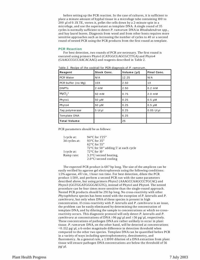
For best detection, two rounds of PCR are necessary. The first round is executed using primers Phyto1 (CATGGCGAGCGCTTGA) and Phyto4 (GAAGCCGCCAACACAAG) and reagents described in Table 2. Table 2. Recipe of the cocktail for PCR-diagnosis of P. ramorum.
PCR parameters should be as follows:
The expected PCR product is 687 bp long. The size of the amplicon can be
easily verified by agarose gel electrophoresis using the following conditions: 1.5% agarose, 4V/cm, 1 hour run time. For best detection, dilute the PCR product 1:500, and perform a second PCR run with the same parameters described above, but using primers Phyto2 (AAAGCCAAGCCCTGCAC) and Phyto3 (GGTGGATGGGGACGTG), instead of Phyto1 and Phyto4. The nested procedure can be four times more sensitive than the single-round approach. Nested PCR products should be 291 bp long. No cross-reactivity with other Phytophthora species has been noted with the exception of P. lateralis and P. cambivora, but only when DNA of these species is present in high concentration. If cross-reactivity with P. lateralis and P. cambivora is an issue, the problem can be easily eliminated by determining the concentration of template DNA, and by diluting the sample to concentrations at which no cross-reactivity occurs. This diagnostic protocol will only detect P. lateralis and P. cambivora at concentrations of DNA >96 pg/µl and >56 pg/µl, respectively. These concentrations of pathogen DNA are rather unlikely to occur in plant tissue. P. ramorum DNA, on the other hand, will be detected at concentrations <0.152 pg/µl, a 6-order magnitude difference in detection threshold when compared to the other two species. Template DNA can be quantified before PCR in a variety of ways including spectrophotometry, densitometry, and fluorometry. As a general rule, a 1:1000 dilution of a DNA extraction from plant tissue will ensure pathogen DNA concentrations are below the threshold of 56 pg/µl.
Reagent Stock Conc. Volume (µl) Final Conc.
PCR Water N/A 12.25 N/A
PCR buffer (no Mg) 10X 2.50 1X
DNPTs 2 mM 2.50 0.2 mM
MgCl2* 50 mM 0.75 2.0 mM
Phyto1 50 µM 0.25 0.5 µM
Phyto4 50 µM 0.25 0.5 µM
Taq polymerase 5 U/µl 0.25 0.05 U/µl
Template DNA 6.25
Total Volume 25
1 cycle at: 94°C for 1’25”34 cycles at: 93°C for 35” 62°C for 55” 72°C for 50” adding 5’ at each cycle1 cycle at: 72°C for 10’Ramp rate: 3.3°C/second heating,
2.0°C/second cooling
Plant Health Progress 7 July 2003
Precautions and controls. At all stages (material preparation, lyophilization, DNA extraction, first PCR round, nested PCR round), negative control samples should be included. Extreme care should be exercised when handling and diluting the first round of PCR as DNA contamination is likely to occur at this stage. A universal primer pair can be added to the traditional PCR reaction as a control on the efficacy of the DNA extraction or PCR reaction (26). Control primers will amplify an amplicon approximately 175 bp in size from all plant and fungal material, and will not interfere with P. ramorum primers. Control primers UnivForw (ggaacgtgagctgggtttag) and UnivRev (ttctgacttagaggcgttcag) can be added to reach a final 0.5 µM concentration in the traditional PCR approach. Currently, DNA sequencing is extremely inexpensive and both the 687 bp and the 291 bp amplicons are informative enough to differentiate P. ramorum from all other known Phytophthora species. We recommend sequencing confirmation for critical samples. Although the diagnostic procedures here described are highly specific for P. ramorum, unwanted deviations from the described protocols due to varying quality of reagents and DNA extracts, or to pipette and Thermocycler malfunction, may result in non-specific amplification.
Several other PCR-based procedures based on a range of nuclear and mitochondrial sequences are now available, including Real Time PCR, single strand conformation polymorphism (SSCP) detection, as well as other traditional PCR protocols. The procedure described above in detail can be employed by all laboratories, without the need for sophisticated equipment, and is the only technique based on primers fully matching P. ramorum DNA that has been extensively tested with environmental samples. Pathogen Identification
Phytophthora ramorum has a nearly unique set of morphological characteristics and a unique molecular sequence at the ITS region (24). The most distinguishing feature of P. ramorum is the presence of large (22 to 72 µm) chlamydospores that are mainly terminal (Figs. 34A and 34B). Chlamydospores change from hyaline to cinnamon brown as they mature. This change is especially pronounced in media with host material. Sporangia are semi-papillate, caducous, and will form in clusters on agar with flooding (Fig. 34C). They are highly deciduous in soil water at 20°C. P. ramorum has highly branching hyphae on PARP that become septate in older cultures (Fig. 34B). This pathogen appears to be heterothallic. Mating in vitro has been achieved through crosses with other Phytophthora species (Fig. 34D) (24). Pairings of European and American isolates in vitro on carrot agar (C. M. Brasier and S. Kirk, unpublished data) and in vivo within rhododendron twigs (25) have lead to production of oospores.
Plant Health Progress 7 July 2003
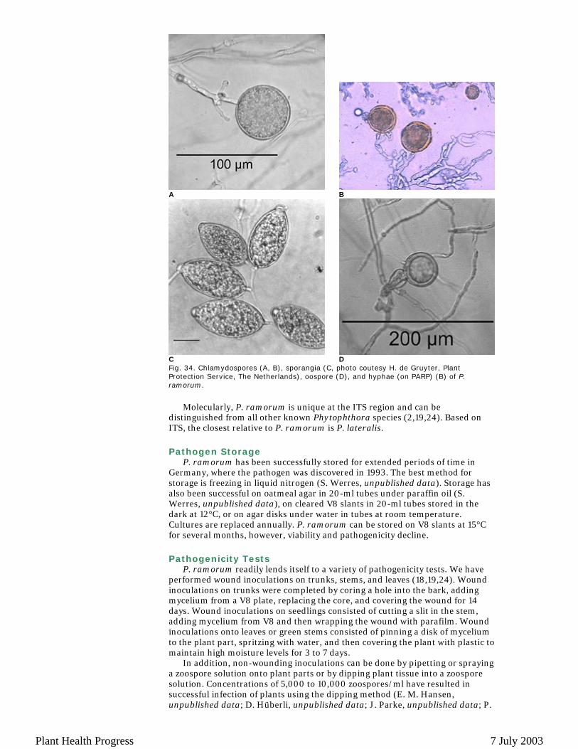
Fig. 34. Chlamydospores (A, B), sporangia (C, photo coutesy H. de Gruyter, Plant Protection Service, The Netherlands), oospore (D), and hyphae (on PARP) (B) of P. ramorum.
Molecularly, P. ramorum is unique at the ITS region and can be
distinguished from all other known Phytophthora species (2,19,24). Based on ITS, the closest relative to P. ramorum is P. lateralis. Pathogen Storage
P. ramorum has been successfully stored for extended periods of time in Germany, where the pathogen was discovered in 1993. The best method for storage is freezing in liquid nitrogen (S. Werres, unpublished data). Storage has also been successful on oatmeal agar in 20-ml tubes under paraffin oil (S. Werres, unpublished data), on cleared V8 slants in 20-ml tubes stored in the dark at 12°C, or on agar disks under water in tubes at room temperature. Cultures are replaced annually. P. ramorum can be stored on V8 slants at 15°C for several months, however, viability and pathogenicity decline. Pathogenicity Tests
P. ramorum readily lends itself to a variety of pathogenicity tests. We have performed wound inoculations on trunks, stems, and leaves (18,19,24). Wound inoculations on trunks were completed by coring a hole into the bark, adding mycelium from a V8 plate, replacing the core, and covering the wound for 14 days. Wound inoculations on seedlings consisted of cutting a slit in the stem, adding mycelium from V8 and then wrapping the wound with parafilm. Wound inoculations onto leaves or green stems consisted of pinning a disk of mycelium to the plant part, spritzing with water, and then covering the plant with plastic to maintain high moisture levels for 3 to 7 days.
In addition, non-wounding inoculations can be done by pipetting or spraying a zoospore solution onto plant parts or by dipping plant tissue into a zoospore solution. Concentrations of 5,000 to 10,000 zoospores/ml have resulted in successful infection of plants using the dipping method (E. M. Hansen, unpublished data; D. Hüberli, unpublished data; J. Parke, unpublished data; P.
A B
C D
Plant Health Progress 7 July 2003
Tooley, unpublished data). Tissue should be kept at high humidity for 5 to 7 days after inoculation. P. ramorum mycelial disks on V8 agar readily produce sporangia in soil water (or to a lesser extent in distilled water) in 24 hours at 15 to 20°C. Soil water level should be even with, but not over the top of mycelial disks. Zoospore release is induced by placing a sporangial culture at 5°C for 30 minutes, and then returning to room temperature for 30 minutes. Acknowledgements
We are grateful to the Gordon and Betty Moore Foundation, the USDA Forest Service Pacific Southwest Research Station, USDA Forest Service Forest Health Management, and the California Department of Forestry for providing funding to support the research underlying this Diagnostic Guide. Literature Cited 1. Brasier, C. M. 1999. The role of Phytophthora pathogens in forests and semi-natural
communities in Europe and Africa. Pages 6-13 in: Phytophthora Diseases of Forest Trees. E. M. Hansen and W. Sutton, eds. Forest Research Laboratory, Oregon State University, Corvallis, OR.
2. Cooke, D. E. L., Drenth, A., Duncan, J. M., Wagels, G., and Brasier, C. M. 2000. A molecular phylogeny of Phytophthora and related oomycetes. Fungal Genet Biol 30:17-32.
3. Davidson, J. M., Garbelotto, M., Hansen, E. M., Reeser, P., and Rizzo, D. M. 2002. Another canker-causing Phytophthora from California and Oregon forest trees. Phytopathology 92:S17-S8.
4. Davidson, J. M., Garbelotto, M., Koike, S. T., and Rizzo, D. M. 2002. First report of Phytophthora ramorum on Douglas-fir in California. Plant Dis. 86:1274.
5. Davidson, J. M., Rizzo, D. M., Garbelotto, M., Tjosvold, S., and Slaughter, G. W. 2002. Phytophthora ramorum and Sudden Oak Death in California: II. Transmission and survival. Pages 741-9 in: Proceedings of the Fifth Symposium on Oak Woodlands: Oak Woodlands in California's Changing Landscape. 2001 October 22-25; San Diego, CA. General Technical Report PSW-GTR-184. R. B. Standiford, D. McCreary, and K. L. Purcell, eds. Pacific Southwest Research Station, Albany, CA, Forest Service, U.S. Department of Agriculture.
6. De Merlier, D., Chandelier, A., and Cavelier, M. 2003. First Report of Phytophthora ramorum on Viburnum bodnantense in Belgium. Plant Dis. 87:203.
7. Delatour, C., Saurat, C., Husson, C., Loos, R., and Schenck, N. 2002. Discovery of Phytophthora ramorum on Rhododendron sp. in France and experimental symptoms on Quercus robur. Presented at Sudden Oak Death, A Science Symposium: The State of Our Knowledge, Monterey, CA.
8. Elliott, M. 2001. The decline of Pacific madrone (Arbutus menziesii Pursh) in urban and natural environments: Its causes and management. M.S. thesis. University of Washington, Seattle, WA.
9. Goheen, E. M., Hansen, E. M., Kanaskie, A., McWilliams, M. G., Osterbauer, N., and Sutton, W. 2002. Sudden oak death caused by Phytophthora ramorum in Oregon. Plant Dis. 86:441.
10. Hansen, E. M., and Hamm, P. B. 1996. Survival of Phytophthora lateralis in infected roots of Port Orford cedar. Plant Dis. 80:1075-8.
11. Inman, A. J., Townend, V. C., Barnes, A. V., Lane, C. R., Hughes, K. J. D., Griffin, R. L., and Eales, S. J. 2003. First report of Ramorum dieback (Phytophthora ramorum) on Pieris in England. Online. New Dis. Rep. 7. British Society for Plant Pathology.
12. Lane, C. R., Beales, P. A., Hughes, K. J. D., Griffin, R. L., Munro, D., Brasier, C. M., and Webber, J. F. 2003. First outbreak of Phytophthora ramorum in England, on Viburnum tinus. Plant Pathol. 52:414.
13. Maloney, P. E., Rizzo, D. M., Koike, S. T., Harnik, T. Y., and Garbelotto, M. 2002. First report of Phytophthora ramorum on coast redwood in California. Plant Dis. 86:1274.
14. Mircetich, S. M., Campbell, R. N., and Matheron, M. E. 1977. Phytophthora trunk canker of coast live oak and cork oak trees in California. Plant Dis. Rep. 61:66-70.
15. Moralejo, E., and Werres, S. 2002. First report of Phytophthora ramorum on Rhododendron sp. in Spain. Plant Disease 86:1052.
16. Murphy, S. K., and Rizzo, D. M. 2003. First report of Phytophthora ramorum on Canyon Live Oak in California. Plant Dis. 87:315.
17. Orlikowski, L. B., and Szkuta, G. 2002. First record of Phytophthora ramorum in Poland. Phytopathol. Polonica 25:69-79.
18. Rizzo, D. M., Garbelotto, M., Davidson, J. M., Slaughter, G. W., and Koike, S. T. 2002. Phytophthora ramorum and Sudden Oak Death in California: I. Host relationships. Presented at 5th Symposium on California Oak Woodlands, San
Plant Health Progress 7 July 2003
Diego, CA. 19. Rizzo, D. M., Garbelotto, M., Davidson, J. M., Slaughter, G. W., and Koike, S. T.
2002. Phytophthora ramorum as the cause of extensive mortality of Quercus spp. and Lithocarpus densiflorus in California. Plant Dis. 86:205-14.
20. Sinclair, W. A., Lyon, H. H., and Johnson, W. T. 1987. Diseases of Trees and Shrubs. Cornell University Press, Ithaca, NY.
21. Tainter, F. H., O'Brien, J. G., Hernandez, A., Orozco, F., and Rebolledo, O. 2000. Phytophthora cinnamomi as a cause of oak mortality in the state of Colima, Mexico. Plant Dis. 84:394-8.
22. Tjosvold, S. A., Koike, S. T., Davidson, J. M., and Rizzo, D. M. 2002. Susceptibility of azalea (Rhododendron) to Phytophthora ramorum. Presented at Sudden Oak Death, A Science Symposium: The State of Our Knowledge, Monterey, CA.
23. Werres S. 2002. Phytophthora ramorum - erste Ergebnisse zum Wirtspflanzenspektrum in Deutschland. Dtsch. Baumsch. 7:46.
24. Werres, S., Marwitz, R., Man in 't Veld, W. A., and De Cock, A. W. A. M., Bonants P. J. M., De Weerdt, M., Themann, K., Ilieva, E., and Baayen, R. P. 2001. Phytophthora ramorum sp. nov., a new pathogen on Rhododendron and Viburnum. Mycol. Res. 105:1155-65.
25. Werres, S., and Zielke, B. 2003. First studies on the pairing of Phytophthora ramorum. J. Plant Dis. Prot. 110:129-30.
26. Winton, L. M., and Hansen, E. M. 2001. Molecular diagnosis of Phytophthora lateralis in trees, water, and foliage baits using multiplex polymerase chain reaction. For. Pathol. 31:275-83.
Plant Health Progress 7 July 2003