successful treatment for bilateral femoral neck
TRANSCRIPT
CASE REPORT Open Access
Successful treatment for bilateral femoralneck insufficiency fractures: a rare lesioncase report and an updated review of theliteratureXu-yi Tan1,2†, Ting Lei3†, Guan-bao Wu2, Hai-en Luo2, Gang Huang2, Can-yu He2, Min Lu1* and Peng-fei Lei3*
Abstract
Background: The incidence of insufficiency fracture (IF) at femoral neck is low, accounting for about 5% of allinsufficiency fractures, and IF at bilateral femoral neck is less common with more occurrence in athlete orserviceman. With the aging of populations, more cases of bilateral femoral neck IF have occurred recently, while thestandard clinical treatment still remains lacking due to the complexity of these patients.
Case presentation: A 55-year-old male patient complained pain in his bilateral hip, with no history of trauma,glucocorticoid hormone consumption or radiotherapy, and imaging examination revealed fracture nonunion andshortening in his left femoral neck, and double fracture line on the right femoral neck. The patient received acementless THA for the left femoral neck fracture and conservative treatment for the right side, followed byElcatonin injection and oral administration of Carbonate D3 Granules. After 4 months of fellow-up, the patientpresented improved functional scorings in bilateral hip joints, with no signs of prothesis infection or loosening.
Conclusion: We present a rare case of bilateral femoral neck IF in a middle-aged male and the treatment issuccessful. The timely CT and MRI examinations of bilateral hip joints for patients was necessary for orthopedists toselect proper therapeutic regimen. In addition, the choice for therapeutic regimen of bilateral femoral IF should notonly be based on the professional judgement of orthopedists, but also on the wishes of patients.
Keywords: Insufficiency fractures, Femoral neck fracture, Bilateral, Case report
BackgroundInsufficiency fractures (IF), which was first proposed bythe professor Pentecost in 1964, was caused by normalor physiological stress applied to bone with decreasedbone mineral content and deficient elastic resistancewhich would result in a weakened zone of the bone [1].IF often occurs in the sacrum and ilium, while the inci-dence of IF at femoral neck is rare, accounting for about5% of all stress fractures, not to mention bilateral femoral
neck fracture [2–4]. Several femur neck IF cases have beenreported [5], which generally occur in athlete and service-man. Cases about femur neck IF occurring in other adultpopulations have been rarely reported, especially for bilat-eral femur neck IF. With the aging of populations, morecases of bilateral femoral neck IF have occurred recently,while the standard clinical treatment still remains lackingdue to the complexity of these patients.Herein, we presented a bilateral femur neck IF case of a
middle-aged male, who suffered from bilateral femoral neckIF. Meanwhile, we also reported relevant treatment for thepatient which could serve as a reference for other doctors.
Case presentationA 55-year-old man, with body mass index (BMI) of18.36 kg/m2, was admitted into our hospital for left hip
© The Author(s). 2020 Open Access This article is distributed under the terms of the Creative Commons Attribution 4.0International License (http://creativecommons.org/licenses/by/4.0/), which permits unrestricted use, distribution, andreproduction in any medium, provided you give appropriate credit to the original author(s) and the source, provide a link tothe Creative Commons license, and indicate if changes were made. The Creative Commons Public Domain Dedication waiver(http://creativecommons.org/publicdomain/zero/1.0/) applies to the data made available in this article, unless otherwise stated.
* Correspondence: [email protected]; [email protected]†Xu-yi Tan and Ting Lei contributed equally to this study and share the firstauthorship.1Affiliated First Hospital of Hunan University of TCM, Changsha 410007,Hunan, China3Xiangya Hospital of Centre-south University, Changsha 410008, Hunan,ChinaFull list of author information is available at the end of the article
Tan et al. BMC Musculoskeletal Disorders (2020) 21:102 https://doi.org/10.1186/s12891-020-3107-x
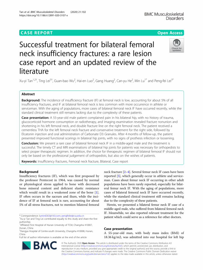
pain for 6 months. The symptom started with moderatepain after walking. Over the last few days, the pain gotworsen with no remission after rest. The patient deniedany history of trauma, glucocorticoid hormone con-sumption or radiotherapy. Examinations before admis-sion revealed old fracture on the left femoral neck(Fig. 1) and an increase in blood uric acid. After admis-sion, the function evaluations of his bilateral hip jointwere summarized as follows: Visual Analogue Scale(VAS) score was 4, Harris score was 23 in the left hipjoint, while VAS score was 0, Harris score was 90 in theright hip joint. The imaging examinations of the pelvis,including radiographs (Fig. 2a), CT (Fig. 2b) and MRIscan (Fig. 2c, d), confirmed the existence of bilateralfemoral neck fractures, with fracture nonunion and fem-oral neck shortening in his left femoral neck and doublefracture lines in his right femoral neck. The Dual energyX-ray absorptiometry (DXA) examination of the lumbarspine showed reduced bone mineral density (BMD) andsevere osteoporosis (L1–L4: BMD0.514 g/cm2, T score =− 4.6). In addition, tumor markers, tuberculosis anti-bodies, alkaline phosphatase and parathyroid hormonelevels were found to be normal.We recommended a left cementless THA, and a can-
nulated screws fixation on the right side. And the uricacid lowering treatment was performed on the patient.However, the patient only agreed to the left THA, whileon the right side, conservative treatment was selected forpatient because of financial reasons. The left THAthrough the lateral approach was performed for the patientunder general anesthesia (Fig. 3a). During the surgery, wefound sclerosis, fatty degeneration and necrosis of the frac-ture end of the left femoral neck (Fig. 3b). We usedimpactive bone grafting to strengthen the acetabularon the fracture region (Fig. 3c).
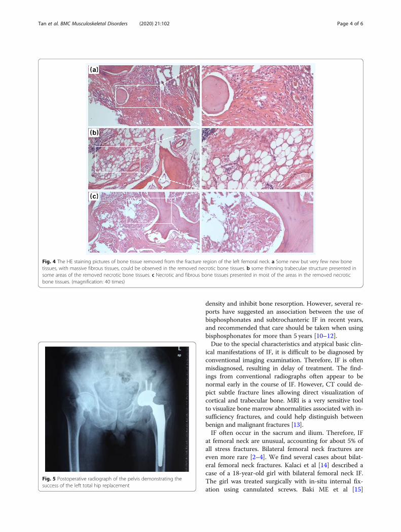
After the operation, the patient was treated with 20 UElcatonin injection (Sidinuo®, Shangdong Luye Pharma-ceutical Co.LTD, Yantai, China) once a week, and 1 tab-let of Calcium Carbonate D3 Granules (Langdi®, BeijingKangyuan Pharmaceutical CO.LTD, Beijing, China) con-taining calcium carbonate (500 mg) and vitamin D (200IU), twice a day. Meanwhile, He was required to exercisehis quadriceps and calf muscle on bed, move with thehelp of wheelchairs, and avoid walking on crutches. Thepostoperative histological examination, as showed inFig. 4, revealed that the left fracture gap was filled withfibrous tissues, with few new bone tissues and a largeamount of necrotic bone tissue observed. Meanwhile,the plain radiograph of the pelvis one day after surgeryrevealed that the total hip replacement arthroplasty wasvery successful (Fig. 5). Generally, the patient recoveredwell during the hospitalization and was successively dis-charged home.Four months after discharge, the radiograph image
showed no signs of implant loosening or infection in hisleft hip (Fig. 6a). It was worth noting that CT imagesrevealed fracture union of the right femoral neck, asshown in Fig. 6b and Fig. 6c. In addition, function evalu-ations of his bilateral hips at four months after surgerywas satisfactory and summarized as follows: VAS scorewas 0, Harris score was 98 in the left hip joint and VASscore was 0, Harris score was 95 in the right hip joint.
Discussion and conclusionPatients with IF often have multiple chronic diseases,such as osteoporosis, diabetes, chronic gastritis, chronicnephritis, organ transplantation, or rheumatoid arthritisneeding immunosuppressive treatment, or tumor requir-ing radiation and chemotherapy. IF is more common inthe elderly, and less common in men than women. Re-cently, Bakker et al [6] published a study about the diag-nose of Sacral IF for 130 patients, who aged between 46and 98 years (mean, 79.8 years), with 117 females and 13males. Melton et al [7] analyzed the contribution of gen-der on the incidence of pelvic insufficiency fractures in aretrospective review of a Mayo Medical database, ofwhich total of 198 patients with 204 fractures, showedthe incidence of pelvic insufficiency fractures was nearlytwice as common in women (47.5/100000) as it is inmen (24.4/100000).IF was produced by normal or physiological stress ap-
plied to bone with decreased bone mineral content anddeficient elastic resistance, resulting in a weakened zoneof the bone. As such, this kind of fracture are oftencaused by daily activities or even light exercise, and usu-ally are not associated with trauma [8]. Osteoporosis isthe most common predisposing factor for IF [9]. Bispho-sphonates are considered the first-line therapy of post-menopausal osteoporosis, as they could improve bone
Fig. 1 Plain radiograph of the pelvis indicating old fracture of theleft femoral neck (red arrow) at a hospital in Foshan,Guangdong Province
Tan et al. BMC Musculoskeletal Disorders (2020) 21:102 Page 2 of 6

Fig. 2 Imaging examinations of the patient in the hip region after admission into our hospital. a Plain radiograph of the pelvis indicating fractureof left femoral neck, with obvious displacement, nonunion and femoral neck shortening, and old fracture of the right femoral neck. b CT imageof the coronal plane revealing bilateral femoral neck insufficiency fractures, with obvious displacement, nonunion and femoral neck shortening inthe left femoral neck and double fracture line (arrows) in his right femoral neck. c Coronal T1-weighted image showing low signal intensity in thefracture region of bilateral femoral neck, with double fracture lines (arrows) on the right side. d Coronal T2-weighted image showing swelling onthe right femoral neck and interruption of cortex on bilateral femoral neck
Fig. 3 The operation procedure performed for the patient. a The lesion region revealed through the lateral approach of the left hip joint. b Thenecrosis bone tissue with fatty degeneration removed from the left femoral neck. c Impactive bone grafting was carry out due to acetabularbone mass was poor
Tan et al. BMC Musculoskeletal Disorders (2020) 21:102 Page 3 of 6
density and inhibit bone resorption. However, several re-ports have suggested an association between the use ofbisphosphonates and subtrochanteric IF in recent years,and recommended that care should be taken when usingbisphosphonates for more than 5 years [10–12].Due to the special characteristics and atypical basic clin-
ical manifestations of IF, it is difficult to be diagnosed byconventional imaging examination. Therefore, IF is oftenmisdiagnosed, resulting in delay of treatment. The find-ings from conventional radiographs often appear to benormal early in the course of IF. However, CT could de-pict subtle fracture lines allowing direct visualization ofcortical and trabecular bone. MRI is a very sensitive toolto visualize bone marrow abnormalities associated with in-sufficiency fractures, and could help distinguish betweenbenign and malignant fractures [13].IF often occur in the sacrum and ilium. Therefore, IF
at femoral neck are unusual, accounting for about 5% ofall stress fractures. Bilateral femoral neck fractures areeven more rare [2–4]. We find several cases about bilat-eral femoral neck fractures. Kalaci et al [14] described acase of a 18-year-old girl with bilateral femoral neck IF.The girl was treated surgically with in-situ internal fix-ation using cannulated screws. Baki ME et al [15]
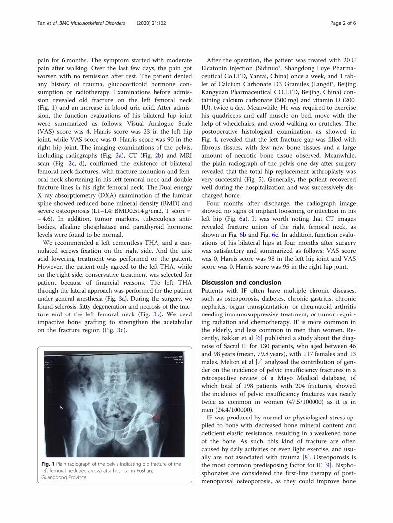
Fig. 4 The HE staining pictures of bone tissue removed from the fracture region of the left femoral neck. a Some new but very few new bonetissues, with massive fibrous tissues, could be observed in the removed necrotic bone tissues. b some thinning trabeculae structure presented insome areas of the removed necrotic bone tissues. c Necrotic and fibrous bone tissues presented in most of the areas in the removed necroticbone tissues. (magnification: 40 times)
Fig. 5 Postoperative radiograph of the pelvis demonstrating thesuccess of the left total hip replacement
Tan et al. BMC Musculoskeletal Disorders (2020) 21:102 Page 4 of 6

reported a 22-year-old female case with bilateral femoralneck fractures, of whom the diagnosis was delayed be-cause the patient was pregnant and could not receiveimaging examination. Finally, the female patient wastreated surgically with internal fixation using cannulatedscrews and received medical treatment for vitamin D de-ficiency. Ahn DK et al [16] reported a bilateral femoralneck IF case of a 78-year-old woman who had a longhistory of using anti-resorptive drug, and bilateral in-ternal fixations using cannulated screws were performedfor the patient. Vaishya R et al [2] reported a 50-year oldmale patient who simultaneously suffered from chronickidney disease and bilateral femoral neck IF with minortrochanter IF. Finally, the patient was managed with can-nulated screws at unusual sites.In our case, the patient had a 7-year history of drink-
ing, about 60 g per day, and had a poor appetite withfew breakfasts in daily life. These bad life styles possiblycaused malnutrition and finally contributed to the devel-opment of bilateral femoral neck IF [17]. Although noexaminations related to nutrition evaluations were car-ried out, such as Vitamin D, the reduced BMD of hislumbar spine reflected a poor nutrition condition of thepatient, which was a potential risk for the occurrence offemoral neck IF. However, the poor nutrition conditioncannot fully explain the occurrence of bilateral femoralfracture. We suspected that the patient had fallen ordone physically demanding work, while the patient de-nied this which he may forget.In conclusion, from our experience with the patient
with bilateral femoral neck IF, he complained of pain inleft hip when walking, which led to suspicion of a fem-oral neck fracture. Although radiological examinationwas performed and indicated old fracture of the left
femoral neck. The immediate CT and MRI examinationwas not followed for further examination, which is con-sidered great value to evaluate fracture healing. Becauseof the pain on the left side, the upper body weight over-load on the right side may have caused the subsequentIF on the right side. Due to the increase of the averageage of the population, as the incidence of IF is increas-ing, it is of great importance to perform CT and MRIscan additionally.
AbbreviationsBMD: Bone mineral density; CT: Computed tomography; IF: InsufficiencyFracture; MRI: Magnetic resonance imaging; THA: Total hip arthroplasty
AcknowledgementsNone
Authors’ contributionsPL, XT and ML conceived the original ideas of this manuscript. GW, GH andCH participated in the surgical and medical treatment. XT, HL and MLexecuted the fellow-up examination and materials collection. XT and MLanalyzed the examination results. TL, XT and PL analyzed the data andprepared the Figs. TL, PL and XT prepared the manuscript. All authors haveread and approved the manuscript.
FundingThis study was supported by the Natural Science Foundation of HunanProvince, China (Grant No. 2018JJ3844), the Scientific Research Project ofHealth and Family Planning Commission of Hunan Province, China (GrantNo. B2019188), the Youth Science Foundation of Xiangya Hospital CentralSouth University (Grant No. 2017Q07), the Postdoctoral Research Program ofXiangya Hospital Central South University (Grant No. 223551), thePostdoctoral Research Program of Hunan university of TCM (Grant No.194780). Fundings include the cost of collecting the fellow-up data andpublication fee.
Availability of data and materialsAll relevant data was presented within the manuscript and the datasets usedand/or analyzed during the current study are available from thecorresponding author on reasonable request.
Fig. 6 Imaging examination of the hip region for the patient four months after operation. a Radiograph of the pelvis showing well postoperativeeffect. b, c CT images of the right femoral neck showing fracture healing
Tan et al. BMC Musculoskeletal Disorders (2020) 21:102 Page 5 of 6

Ethics approval and consent to participateNot applicable.
Consent for publicationThe authors have obtained the patient’s written informed consent of hispersonal clinical data and all images in this study for print and electronicpublication.
Competing interestsThe authors have no competing interests.
Author details1Affiliated First Hospital of Hunan University of TCM, Changsha 410007,Hunan, China. 2Affiliated Hospital of Hunan Academy of Chinese MedicalScience, Changsha 410006, Hunan, China. 3Xiangya Hospital of Centre-southUniversity, Changsha 410008, Hunan, China.
Received: 4 May 2019 Accepted: 3 February 2020
References1. Pentecost RL, Murray RA, Brindley HH. Fatigue, insufficiency, and pathologic
fractures. Jama. 1964;187(13):1001–4.2. Vaishya R, Agarwal AK, Banka PK, Vijay V, Vaish A. Insufficiency fractures at
unusual sites: a case series. J Orthop Case Rep. 2017;7(4):76.3. Matcuk GR, Mahanty SR, Skalski MR, Patel DB, White EA, Gottsegen CJ. Stress
fractures: pathophysiology, clinical presentation, imaging features, andtreatment options. Emerg Radiol. 2016;23(4):365–75.
4. Wild A, Jaeger M, Haak H, Mehdian S. Sacral insufficiency fracture, anunsuspected cause of low-back pain in elderly women. Arch OrthopTrauma Surg. 2002;122(1):58–60.
5. Bailie DS, Lamprecht DE. Bilateral femoral neck stress fractures in anadolescent male runner: a case report. Am J Sports Med. 2001;29(6):811–3.
6. Bakker G, Hattingen J, Stuetzer H, Isenberg J. Sacral insufficiency fractures :how to classify? J Korean Neurosurg Soc. 2018;61(2):258–66.
7. Melton LJ 3rd, Sampson JM, Morrey BF, Ilstrup DM. Epidemiologic featuresof pelvic fractures. Clin Orthop Relat Res. 1981;155:43–7.
8. O'Connor TJ, Cole PA. Pelvic insufficiency fractures. Geriatr Orthop SurgRehabil. 2014;5(4):178–90.
9. Iba K, Wada T, Takada J, Yamashita T. Multiple insufficiency fractures withsevere osteoporosis. J Orthop Sci. 2003;8(5):717–20.
10. Seraphim A, Al-Hadithy N, Mordecai SC, Al-Nammari S. Do bisphosphonatescause femoral insufficiency fractures? J Orthop traumatol. 2012;13(4):171–7.
11. 강수용, 오정희, 최경원, 백지훈, 하용찬. Sequential Bilateral InsufficiencyFractures of the Femur Neck in Patients Treated with ProlongedBisphosphonate Therapy - A Case Report. J Bone Metab 2011; 18(1):65–68.
12. Capeci CM, Tejwani NC. Bilateral low-energy simultaneous or sequentialfemoral fractures in patients on long-term alendronate therapy. J Bone JointSurg Am Vol. 2009;91A(11):2556–61.
13. Krestan CR, Nemec U, Nemec S. Imaging of insufficiency fractures. SeminMusculoskelet Radiol. 2011;15(3):198–207.
14. Kalaci A, Yanat AN, Sevinc TT, Dogramaci Y. Insufficiency fractures of bothfemoral necks in a young adult caused by osteoporosis: a case report. ArchOrthop Trauma Surg. 2008;128(8):865–8.
15. Baki ME, Uygun H, Ari B, Aydin H. Bilateral femoral neck insufficiencyfractures in pregnancy. Eklem Hastalik Cerrahisi. 2014;25(1):60–2.
16. Ahn D-K, Kim J-H, Lee J-I, Kim J-W. Bilateral femoral neck insufficiencyfractures after use of a long-term anti-resorptive drug therapy forosteoporosis: a case report. Hip Pelvis. 2015;27(2):115–9.
17. Carpintero P, Lopez-Soroche E, Carpintero R, Morales R. Bilateral insufficiencyfracture of the femoral neck in a male patient with anorexia nervosa. ActaOrthop Belg. 2013;79(1):111–3.
Publisher’s NoteSpringer Nature remains neutral with regard to jurisdictional claims inpublished maps and institutional affiliations.
Tan et al. BMC Musculoskeletal Disorders (2020) 21:102 Page 6 of 6