substitution of malachite green with nigrosin - eosin ... · prepared slide was examined...
TRANSCRIPT
African Health Sciences Vol 7 No 1 March 2007 33
Substitution of Malachite Green with Nigrosin - Eosin YellowStain in the Kato-Katz method: microscopical appearance of
the helminth eggs
Emmanuel I.Odongo-Aginya1; Narcis Kabatereine2; Siefert Ludwig3; Henry Wabinga4; Alan Fenwick5; Antonio Montresor6.
1) Uganda Virus Research Institute P.O.Box 42 Entebbe Uganda (East Africa).2) Vector Control Department Ministry of Health P.O.Box 1666 Kampala Uganda.3) Faculty of Veterinary Medicine Makerere University, Wild Life Conservation Department P.O.Box 7062 Kampala4) Faculty of Medicine Department of Pathology Makerere Medical School P.O Box 7062 Kampala Uganda.5) St.Mary,s Campus, Norfolk Place, Paddington, London W2 1PG, UK6) WHO Vietnam PO Box 52 10000 Hanoi Vietnam.
AbstractBackground: The Kato-Katz thick smear technique is the standard technique recommended by the World Health Organisation for thequantitative diagnosis of Schistosoma mansoni and other intestinal helminth infections. The major problem of the technique is that a fewhours after the preparation of slides hookworm eggs over clear and disappear due glycerin.Objective: To illustrate clear visibility of different helminth eggs microscopically in Odongo-Aginya method, substitution of malachitegreen with 7.5% nigrosin in 10% formalin and 5% eosin in 10% formalin.Method: Measured, strained stool specimen was stained with mixture of nigrosin/ eosin and covered with cellophane cover slips. Theprepared slide was examined immediately microscopically.Result: Slides prepared with Odongo-Aginya method can be examined immediately or later without compromising the visibility ofparasite eggs and larvae. Hookworm eggs remain visible for a long time.Conclusion: The present publication shows microscopic appearance of the helminth eggs using the Odongo-Aginya modification.Keywords: Kato - Katz method, pictorial illustration, Odongo-aginya modification, helminth eggs, Uganda.African Health Sciences 2007; 7(1) 33-36
Corresponding authorAntonio MontresorWorld Health Organization63 Tran Hung Dao StreetMail P.O. Box 52Ha Noi - VietnamTel +(84 4) 943 3734 /5 /6 ext 29Fax +(84 4) 943 3740e-mail [email protected]
Introduction
The Kato-Katz thick smear technique is the standard tech-nique recommended by World Health Organisation(WHO) for the quantitative diagnosis of Schistosomamansoni and other intestinal helminth infections1.
The glycerin, in the malachite green in KatoKatz’s technique, functions as a clearing agent while themalachite green besides being a dye, is bactericidal 2,3 .The Kato-Katz methods require between 1 to 2 hoursbefore the glycerin clears the background of the stoolsmear on the slide for accurate visualization of mosthelminth eggs 1. The major problem of the technique isthat few hours after the preparation of the slidehookworm eggs are difficult to recognize due to over-clarification by glycerin 1.
The aim of this paper is to show the appearanceof the helminth eggs when malachite green is replacedwith a stain comprised of nigrosin and eosin yellow informalin4. Several field studies confirm the simplicity,quality, and cost effectiveness of the proposed modifica-tion 5-9, a visual reference of the results of the methodcan be useful to facilitate the recognition of parasite eggsby microscopists willing to adopt this methodology.
Material and Method
The malachite green in the Kato-Katz technique issubstituted by the following compound stain: 5% eosinyellow in 10% formalin, mixed 1:1 with 7.5% nigrosinin 10% formalin4. Cellophane cover slips cut in 25x40mm pieces are soaked in 50% glycerin. Stool specimensare strained through a stainless steel sieve as used in Kato-Katz’s method. Through a template, providing 41.7 mgof feces, the strained stool is delivered on a microscopeslide and a drop of the compound stain (about 10-50µl)is added to the stool on the slide and stirred using thecorner of another slide. A cellophane cover slip

African Health Sciences Vol 7 No 1 March 2007 34
presoaked in 50% glycerin is picked with a pair of for-ceps and excess glycerin on it is blotted out on an absor-bent paper. The cellophane cover slip is then placed onthe stained stool on the slide. The slide is inverted upsidedown and pressed down gently on tissue paper or anyabsorbent paper to spread out the stool smeared on theslide and to remove excess of the stain from the slide. Awell-prepared slide has a pinkish background and is thinenough to read the face of a wristwatch through it.Photographs of the helminth eggs are presented in fi-gure 1, 2 and 3. The photographs were taken using anOlympus Photomicroscope B 20 from slides preparedin the Department of Pathology Makerere MedicalSchool according to Kato-Katz and Odongo-Aginya’smethods. The stool specimens were collected from
Kigungu residents as a part of Odongo-Aginya, Doctorof Philosophy (Ph.D) study. Kigungu is a fishing villageon Lake Victoria shoreline, Entebbe Uganda.Photographs were taken immediately after preparationsof slides, after one hour and one week later.Ethic: Faculty of Medicine, Higher Degree and EthicCommittee; Uganda National Council of Science andTechnology, approved the study. Consent was requestedbefore the residents were recruited in the study. Allailments were treated. For S.mansoni, praziquantel wasused. Privacy was observed. The doctor in the treatmentroom released results to individuals and risk was mini-mal.
Figure 1 Microscopical appearance hookworm egg with eosin-nigrosin stain (x 40 objective)
a- Immediately after preparation b- 24 hours after preparation c- One week after preparation.
Figure 2 Microscopic appearance Hymenolepis nana egg with eosin-nigrosin stain (x 40 objective) 1 hour afterpreparation
African Health Sciences Vol 7 No 1 March 2007 35
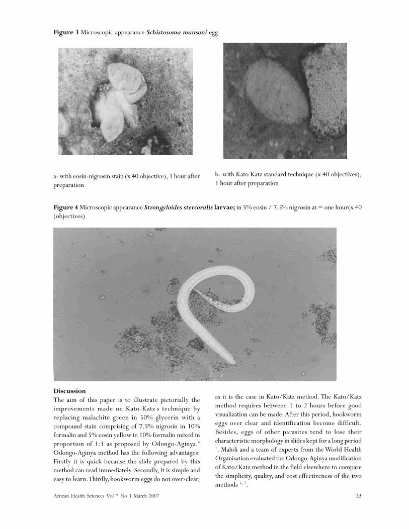
Figure 3 Microscopic appearance Schistosoma mansoni egg
a- with eosin-nigrosin stain (x 40 objective), 1 hour afterpreparation
b- with Kato Katz standard technique (x 40 objectives),1 hour after preparation
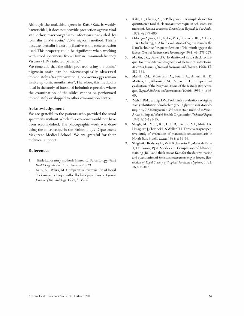
Figure 4 Microscopic appearance Strongyloides stercoralis larvae; in 5% eosin / 7.5% nigrosin at = one hour(x 40(objectives)
DiscussionThe aim of this paper is to illustrate pictorially theimprovements made on Kato-Katz’s technique byreplacing malachite green in 50% glycerin with acompound stain comprising of 7.5% nigrosin in 10%formalin and 5% eosin yellow in 10% formalin mixed inproportion of 1:1 as proposed by Odongo-Aginya.4
Odongo-Aginya method has the following advantages:Firstly it is quick because the slide prepared by thismethod can read immediately. Secondly, it is simple andeasy to learn. Thirdly, hookworm eggs do not over-clear,
as it is the case in Kato/Katz method. The Kato/Katzmethod requires between 1 to 2 hours before goodvisualization can be made. After this period, hookwormeggs over clear and identification become difficult.Besides, eggs of other parasites tend to lose theircharacteristic morphology in slides kept for a long period1. Mahdi and a team of experts from the World HealthOrganisation evaluated the Odongo-Aginya modificationof Kato/Katz method in the field elsewhere to comparethe simplicity, quality, and cost effectiveness of the twomethods 6, 7.
African Health Sciences Vol 7 No 1 March 2007 36
Although the malachite green in Kato/Katz is weaklybactericidal, it does not provide protection against viraland other microorganism infections provided byformalin in 5% eosin/ 7.5% nigrosin method. This isbecause formalin is a strong fixative at the concentrationused. This property could be significant when workingwith stool specimens from Human ImmunodeficiencyViruses (HIV) infected patients.4
We conclude that the slides prepared using the eosin/nigrosin stain can be microscopically observedimmediately after preparation. Hookworm eggs remainvisible up to six months later4. Therefore, this method isideal in the study of intestinal helminth especially wherethe examination of the slides cannot be performedimmediately or shipped to other examination centre.
AcknowledgementWe are grateful to the patients who provided the stoolspecimens without which this exercise would not havebeen accomplished. The photographic work was doneusing the microscope in the Pathothology DepartmentMakerere Medical School. We are grateful for theirtechnical support.
References
1. Basic Laboratory methods in medical Parasitology. WorldHealth Organisation. 1991 Geneva 25- 29
2. Kato, K., Miura, M. Comparative examination of faecalthick smear technique with cellophane paper covers .JapaneseJournal of Parasitolology. 1954, 3: 35-37.
3. Katz, K., Chaves, A., & Pellegrino, J. A simple device forquantitative tool thick smears technique in schistomiasismansoni. Revista do institut De medicine Tropical de Sao Paulo.1972; 4: 397-400
4. Odongo-Aginya, EI., Taylor, MG., Sturrock, RF., Ackers,JP & Doehring, E. A field evaluation of Aginya stain in theKato Technique for quantification of Helminth eggs in thefaeces. Tropical Medicine and Parasitology 1995; 46: 275-277.
5. Martin, LK., Beaver, PC. Evaluation of Kato’s thick techni-que for quantitative diagnosis of helminth infections.American Journal of tropical Medicine and Hygiene. 1968; 17:382-391.
6. Mahdi, RM., Montresor, A., Foum, A., Ameri, H., DiMatteo, L., Albonico, M., & Savioli L. Independentevaluation of the Nigrosin-Eosin of the Kato-Katz techni-que. Tropical Medicine and International Health, 1999; 4 1: 46-49.
7. Mahdi, RM., & Luigi DM. Preliminary evaluations of Aginyastain (substitution of malachite green/glycerin in Kato tech-nique by 7.5% nigrosin / 5% eosin stain method in WonjiArea (Ethiopia). World Health Organisation Technical Report1996; A16-181-15.
8. Sleigh, AC, Mott, KE, Hoff R, Barreto ML, Mota EA,Hmaguire J, Sherlock I, & Weller TH. Three years prospec-tive study of evaluation of mansoni’s schistosomiasis inNorth-East Brazil. Lancet 1985; II 63-66.
9. Sleigh AC, Rodyney H, Mott K, Barreto M, Maisk de PaivaT, De Souza, PJ & Sherlock I. Comparison of filtrationstaining (Bell) and thick smear Kato for the determinationand quantitation of Schistosoma mansoni egg in faeces. Tran-saction of Royal Society of Tropical Medicine Hygiene. 1982;76:403-407.