subcutaneous nerve stimulation for rate control in
TRANSCRIPT

MANUSCRIP
T
ACCEPTED
1
2
3
4
5
6
7
8
9
10
11
12
13
14
15
16
17
18
19
20
21
22
ACCEPTED MANUSCRIPT
Subcutaneous nerve stimulation for rate control in ambulatory dogs with persistent atrial
fibrillation
Short title: Subcutaneous stimulation for AF rate control
Yuan Yuan, MD,1, 2 Xiao Liu, MD,1 Juyi Wan, MD,1, 3 Johnson Wong, BS,1 Amanda A. Bedwell,
MS,4 Scott A. Persohn, BA,4 Changyu Shen, PhD,5 Michael C. Fishbein, MD,6 Lan S. Chen,
MD,7 Zhenhui Chen, PhD,1 Thomas H. Everett IV, PhD, FHRS,1 Paul R. Territo, PhD,4 and
Peng-Sheng Chen, MD, FHRS1
1. The Krannert Institute of Cardiology and Division of Cardiology, Department of
Medicine, Indiana University School of Medicine, Indianapolis, IN
2. Department of Cardiothoracic Surgery, Xinhua Hospital, Shanghai Jiaotong University
School of Medicine, Shanghai, China
3. Department of Cardiothoracic Surgery, the Affiliated Hospital of Southwest Medical
University, Luzhou, Sichuan Province , China
4. Department of Radiology and Imaging Sciences, Indiana University School of Medicine,
Indianapolis, IN
5. Richard and Susan Smith Center for Outcomes Research in Cardiology, Beth Israel
Deaconess Medical Center, Harvard Medical School, Boston, MA
6. The Department of Pathology and Laboratory Medicine, David Geffen School of
Medicine at UCLA, Los Angeles, CA
7. Department of Neurology, Indiana University School of Medicine, Indianapolis, IN
Corresponding Author:
Peng-Sheng Chen, MD, 1800 N. Capitol Ave, E475, Indianapolis, IN 46202
23
___________________________________________________________________
This is the author's manuscript of the article published in final edited form as:Yuan, Y., Liu, X., Wan, J., Wong, J., Bedwell, A. A., Persohn, S. A., … Chen, P.-S. (2019). Subcutaneous nerve stimulation for rate control in ambulatory dogs with persistent atrial fibrillation. Heart Rhythm. https://doi.org/10.1016/j.hrthm.2019.05.029

MANUSCRIP
T
ACCEPTED
2
Source of Funding 1
This study was supported by NIH Grants R42DA043391, TR002208-01, R01 HL139829, 2
American Heart Association Grant #18TPA34170284 /ZC/2018a, a Medtronic-Zipes Endowment 3
of the Indiana University and the Indiana University Health-Indiana University School of 4
Medicine Strategic Research Initiative. 5
6
Disclosures 7
Johnson Wong and Thomas Everett have equity interest in Arrhythmotech, LLC. Other authors 8
do not have conflicts. 9
10
Word Count: 4980 11
12
13

MANUSCRIP
T
ACCEPTED
3
Abstract 1
Background: Subcutaneous nerve stimulation (ScNS) damages the stellate ganglion and 2
improves rhythm control of atrial fibrillation (AF) in ambulatory dogs. 3
Objective: To test the hypothesis that thoracic ScNS can improve rate control in persistent AF. 4
Methods: We created persistent AF in 13 dogs and randomly assigned them to ScNS (N=6) and 5
sham control groups (N=7). 18F-2-Fluoro-2-deoxyglucose (18F-FDG) positron emission 6
tomography / magnetic resonance imaging of the brain stem was performed at baseline and at the 7
end of the study. 8
Results: The average stellate ganglion nerve activity (aSGNA) reduced from 4.00±1.68 µV after 9
the induction of persistent AF to 1.72±0.42 µV (p=0.032) after ScNS. In contrast, the aSGNA 10
increased from 3.01±1.26 µV during AF to 5.52±2.69 µV after sham stimulation (p=0.023). The 11
mean ventricular rate during persistent AF reduced from 149±36 bpm to 84±16 bpm (p=0.011) in 12
ScNS group but no changes were observed in control. Left ventricular ejection fraction (LVEF) 13
remained unchanged in ScNS group but reduced significantly in sham control group. 14
Immunostaining showed damaged ganglion cells in bilateral stellate ganglia and increased brain 15
stem glial cell reaction in the ScNS group but not in the controls. The 18F-FDG uptake in pons 16
and medulla was significantly (p=0.011) higher in the ScNS group than the sham control group 17
at the end of the study. 18
Conclusions: Thoracic ScNS causes neural remodeling in the brain stem and stellate ganglia, 19
controls the ventricular rate and preserves the LVEF in ambulatory dogs with persistent AF. 20
21
Keywords: Subcutaneous nerve stimulation; Autonomic nervous system; Neuromodulation; 22
Persistent atrial fibrillation; Positron emission tomography; Magnetic resonance imaging 23
MANUSCRIP
T
ACCEPTED
4
Introduction: 1
Both rhythm and rate controls are acceptable strategies in managing patients with atrial 2
fibrillation (AF).1, 2 Rapid ventricular rate (VR) may result in left ventricular dysfunction. Beta 3
blockers are recommended by the current guidelines for both rate and rhythm control of AF.2 4
While pharmacological agents are mostly effective in rate control of AF, atrioventricular nodal 5
ablation may be needed in patients refractory to drug therapy.2, 3 Alternative strategies of VR 6
control include vagal nerve stimulation, which reduces the VR during AF in dogs.4 Stimulating 7
the auricular branch of the vagal nerve for one hour can suppresses AF and decreases 8
inflammatory cytokines in patients with paroxysmal AF.5 The acute effects of vagal nerve 9
stimulation in those studies are attributed in part to parasympathetic activation. However, vagal 10
nerve stimulation can also rapidly activate the stellate ganglion in ambulatory dogs probably 11
through the activation of sympathetic nerve fibers within the vagal nerve.6, 7 Rapid neuronal 12
activation can cause neurotoxicity through intracellular calcium accumulation.8, 9 Consistent with 13
the latter observation, chronic intermittent vagal nerve stimulation can control the VR, reduce the 14
stellate ganglion (SG) nerve activity (SGNA) and cause neurotoxicity in the SG.10 If the effects 15
of chronic vagal nerve stimulation are mediated through rapid excitation of the sympathetic 16
nerve fibers within the vagal nerves, then it follows that stimulating any sympathetic nerve fibers 17
connected to the SG may result in the same effects. The thoracic subcutaneous nerves in dogs 18
originated primarily from the SG.11 We recently showed that thoracic subcutaneous nerve 19
stimulation (ScNS) from three different sites can rapidly activate the SG, suppress SGNA and 20
result in rhythm control of atrial tachyarrhythmias in chronic canine models.12, 13 The purpose of 21
the present study was to test the hypothesis that ScNS can damage SG, reduce SGNA and control 22
VR in ambulatory dogs with persistent AF. 23
24

MANUSCRIP
T
ACCEPTED
5
Methods 1
The animal protocol was approved by the Institutional Animal Care and Use Committee of the 2
Indiana University School of Medicine and the Methodist Research Institute, Indianapolis, IN, 3
and conformed to the Guide for Care and Use of Laboratory animals. 4
5
Surgical procedures 6
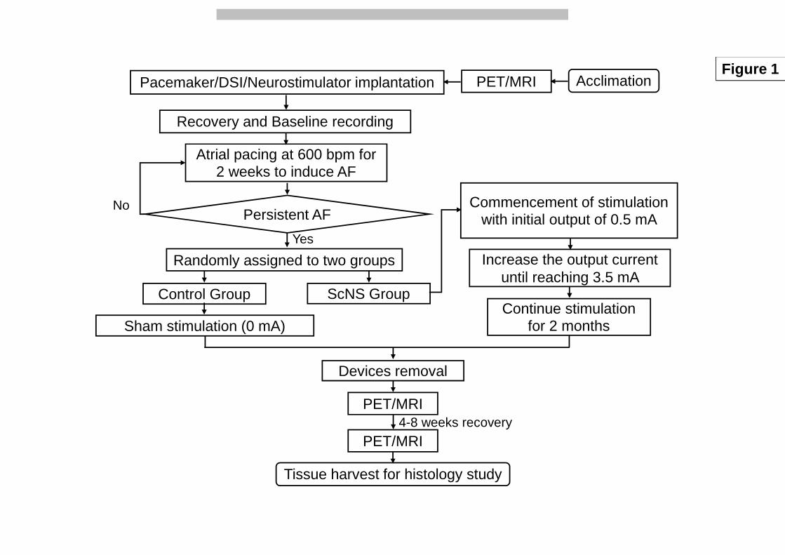
Figure 1 shows the research protocol. Thirteen mongrel dogs (23-30 kg) underwent isoflurane 7
inhalation general anesthesia and sterile left lateral thoracotomy through the forth intercostal 8
space. A radiotransmitter (D70EEE, Data Sciences International, St. Paul, MN) was implanted to 9
record SGNA and vagal nerve activity (VNA). A modified Medtronic Secura pacemaker 10
(Medtronic Inc, Minneapolis, MN) was implanted during the same surgery for intermittent rapid 11
atrial pacing through a pacing lead sutured onto the left atrial (LA) appendage. The skin incision 12
was then extended to the back to reach left Xinshu acupoint (BL15, approximately 5 cm lateral 13
to the spine at T5 level) as in a previous study.12 The subcutaneous space was explored to locate 14
visible subcutaneous nerves and blood vessels in that area. A Cyberonics Model 304 bipolar 15
vagal stimulating lead was implanted around these small subcutaneous nerves and connected to a 16
subcutaneously positioned Cyberonics Demipulse neurostimulator (Cyberonics Inc, Houston, 17
TX) (Online supplement Figure 1). A third pair of the bipolar recording electrodes from the 18
D70EEE radiotransmitter was placed in the subcutaneous tissue to record from the nerves being 19
stimulated, with the two electrodes bracketing the point of stimulation. The latter bipolar leads 20
have an interelectrode distance of > 4 cm. The chest was then closed. 21
22
Pacing protocol 23
The DSI radiotransmitter was turned on two weeks after surgery to record baseline rhythm and 24

MANUSCRIP
T
ACCEPTED
6
subcutaneous nerve activity (ScNA). After baseline recording, high- rate (600 beats/min, twice 1
the diastolic threshold) atrial pacing was then initiated and continued for 2 weeks. The 2
pacemaker was then turned off to determinate if there was persistent (>48 hours) of AF. If not, 3
the atrial pacing was reinitiated until persistent AF was documented. 4
5
Subcutaneous nerve stimulation 6
After persistent AF was induced, the dogs were randomly assigned to ScNS group (N=6) and 7
sham control group (N=7). The neurostimulator was turned on and programmed to 14-s ON and 8
1.1-min OFF (10 Hz, 500 µs pulse duration) based on the parameters used in a clinical trial.14 9
The output current (mA) was progressively increased to 3.5 mA over 10 weeks. The dogs 10
tolerated 3.5 mA stimulation without showing signs of discomfort or reduced appetite. After 10 11
weeks of ScNS, the stimulator was turned off for an additional 24 hours recording without ScNS. 12
The sham control group underwent the same surgery, but the output current was set at 0 mA. 13
14
Functional imaging of brain stem 15
All dogs underwent brain stem 18F-2-Fluoro-2-deoxyglucose (18F-FDG) positron emission 16
tomography (PET)–magnetic resonance imaging (MRI) for the functional survey before surgery 17
(baseline). After 10 weeks of ScNS or sham stimulation, all implanted devices were removed 18
during a third sterile surgery. The dogs then underwent repeat PET/MRI imaging. A final 19
PET/MRI imaging was performed after an additional 4-8 weeks of recovery. All PET/MRI 20
images were imported and registered to their anatomical reference and baseline time point using 21
a normalized entropy algorithm15 implemented in Analyze 11 (AnalyzeDirect, Stilwell KS). 22
Fusion images were created between the registered PET and their respective MRI images. Image 23
MANUSCRIP
T
ACCEPTED
7
volumes were then manually segmented in 3D (Analyze 11; AnalyzeDirect, Stilwell KS) to 1
obtain object maps for the pons and the medulla. Images were then quantified for amount of 2
uptake in percent of injected dose per gram (%ID/g) within the region of interest in pons and 3
medulla at baseline and follow up. A representative coronal slice was then exported and 4
subjected to voxel-wise analysis to compute percentage change in %ID/g (∆%ID/g) from 5
baseline, and then mapped using custom developed software on anatomical MRI images yielding 6
parametric maps which illustrate the changes in %ID/g. 7
8
Immunohistochemical Studies 9
After the dogs were euthanized, both SG were harvested and fixed in 4% formalin for 45-60 min, 10
followed by storage in 70% alcohol for at least 48 hours. The tissues were processed routinely, 11
paraffin embedded and cut into 5-µm thick sections. Immunohistochemical staining was 12
performed with antibodies against tyrosine hydroxylase (TH) using mouse monoclonal anti-TH 13
(Accurate Chemical, Westbury, NY). All slides were examined under a microscope to determine 14
if there were regions of damage, characterized by decreased ganglionic cell density, pyknotic cell 15
bodies, decreased TH staining, increased fibrosis and hypereosinophilia of neurons on Masson’s 16
trichrome staining. Digital photographs were taken from five roughly even spaced fields per slide 17
with a 20X objective. The mean percentage of TH-negative ganglion cells was calculated 18
manually in both SG using the same methods reported elsewhere. Terminal deoxynucleotidyl 19
transferase dUTP nick end labeling (TUNEL) staining was performed to probe for cell death. A 20
confocal microscope was used to detect TUNEL-positive cells. For quantitative analyses, we 21
randomly selected 5 high power (40X objective) fields for image acquisition. We then manually 22
counted the TUNEL-positive ganglion cells in each high power fields for analyses. 23
MANUSCRIP
T
ACCEPTED
8
Data Analyses 1
The signals were manually analyzed using custom-written software to determine the temporal 2
relationship between nerve activities and VR changes. Data from 3 recording electrodes were 3
high-pass filtered at 150 Hz to obtain nerve activities, which were quantified by integrating the 4
absolute value of the filtered signal over 20-s windows. The integrated nerve activities were then 5
divided by the total number of samples (i.e., the product of sampling rate and 20) in each 6
window to calculate the average SGNA (aSGNA), average vagal nerve activity (aVNA) and 7
average subcutaneous nerve activity ScNA (aScNA). To quantify the hourly nerve activities over 8
a 24-hr period, we selected for analyses 2-min of data at the beginning, 20 min, and 40 min past 9
each hour when the stimulation was off. Artifacts or noises during that 2-min period were 10
manually excluded from analyses. Nerve activities and VR were compared between different 11
time points. 12
13
Statistical Analyses 14
The data were reported as mean ± Standard deviation (SD). Paired t tests were performed to 15
compare the differences between HR, integrated nerve activities and the number of PAT episodes 16
at different stages of experiments. Because paired t and signed rank reach similar p-values, only 17
the paired t result is reported. The statistics were computed using the PASW Statistic (version 22; 18
SPSS Inc, Chicago, IL). A two-sided p value of ≤0.05 was considered statistically significant. 19
20
Results 21
Effects of ScNS on nerve activities and ventricular rate during persistent AF 22
Figures 2A and 2B show the serial changes in the ratio of aSGNA and VR at different stages of 23

MANUSCRIP
T
ACCEPTED
9
the experiments in ScNS and sham control groups. Because the absolute values of aSGNA and 1
VR vary among dogs, we used the ratio to baseline to display the relative changes during the 2
course of the study. There was progressive reduction of aSGNA and VR in ScNS group but not 3
in the sham control group. Compared to AF before ScNS, aSGNA was significantly reduced after 4
6 weeks of ScNS (p=0.045) and persisted afterwards. VR was significantly suppressed after 7 5
weeks of ScNS (p=0.011) and persisted afterwards. Asterisks indicate significant differences of 6
aSGNA and VR between ScNS group and control group. Figure 2C and 2D show typical 7
recordings of SGNA, VNA and ScNA at baseline and after the induction of AF, respectively. 8
VNA and ScNA could either activate simultaneously (blue arrows) or at different times 9
(asterisks) with SGNA. AF causes significant increase of SGNA. Figure 2E shows the acute 10
effects of ScNS on SGNA. In that episode, SGNA was abruptly suppressed (red dot) followed by 11
the progressive reduction of VR (black arrow). 12
Figures 3 shows a summary of all dogs studies. In ScNS group (Figure 3A), aSGNA was 13
4.00±1.68 µV immediately after induction of AF. The aSGNA then significantly reduced to 14
2.81±1.19 µV (p=0.030) at week 5 of stimulation, and persisted afterwards. In the final week of 15
study, the aSGNA was 1.72±0.42 µV (p=0.011). Figure 3B shows the data in sham control group. 16
aSGNA was 4.23±1.48 µV after the induction of AF. Weekly analyses showed non-significant 17
increase of aSGNA to 4.72±0.95 µV at week 5 (p=0.229) and to 5.90±2.20 µV at the final week 18
of study (p=0.072). The mean VR was 149±36 bpm after the induction of AF. The VR 19
significantly decreased to 84±16 bpm (p=0.011) at the final week of ScNS. Figure 3B shows in 20
sham control group, the mean VR was 170±33 bpm after the induction of AF. At the final week 21
of monitoring, the mean VR of sham control group reduced insignificantly to 153±25 bpm 22
(p=0.262), which was significantly higher than the final VR of the ScNS group (p<0.001). The 23
MANUSCRIP
T
ACCEPTED
10
aVNA and aScNA did not change significantly during the study in either group. Figure 4 shows 1
examples of 24-hr SGNA at baseline sinus rhythm, after AF induced and after either 3.5 mA or 2
sham (0 mA) ScNA in the ScNS group (A) and in the sham control group (B). SGNA was 3
significantly increased after persistent AF in both groups. Compared to AF, SGNA was 4
suppressed after ScNS (Figure 4A), but did not change in the sham control group (Figure 4B). 5
6
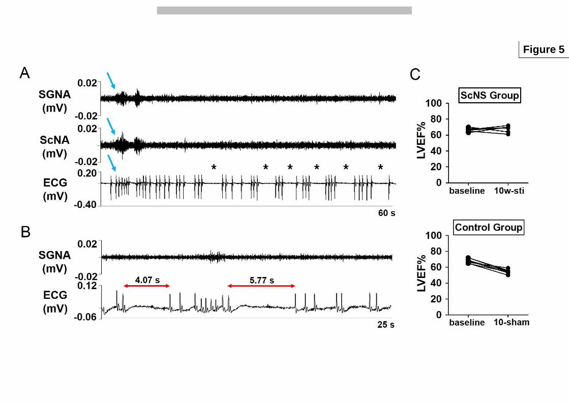
Prolonged pauses during persistent AF 7
Prolonged (>3s) pauses were observed with increased frequency in the ScNS group but not in the 8
sham control group. Figure 5A shows SGNA is associated with VR acceleration during AF 9
(arrow). Prolonged pauses (asterisks) happened frequently after ScNS in the absence of SGNA 10
bursts. The average number of prolonged pauses after the induction of persistent AF (but before 11
ScNS) was similar in the ScNS group (5±4 episodes/d) and in the sham control group (5±7/d, 12
p=0.93). After completing the ScNS protocol, the number of prolonged paused was 38±19/d in 13
the ScNS group and 10±8 times in the sham control group (p=0.028). 14
The left ventricular ejection fraction (LVEF) was 68±2% in the ScNS group and 69±3% 15
in the sham control group during baseline sinus rhythm (p=0.823). After the induction of 16
persistent AF and completing the ScNS protocols, LVEF was 68±4% in the ScNS group, 17
significantly higher than that in the sham control group (55±3%, p<0.001) (Figure 5C). 18
19
SG remodeling after ScNS in persistent AF dogs 20
Bilateral SG were successfully harvested for histology analyses. The SG from the ScNS group 21
showed large damaged regions (Figure 6A and 6C). Multiple ganglion cells in damaged regions 22
showed pyknotic nuclei, contracted and hypereosinophilic cytoplasm in the Masson trichrome 23

MANUSCRIP
T
ACCEPTED
11
stained sections. The damaged regions could be either confluent or multifocal. These changes 1
were not observed in SG from the sham control group. TH staining showed abundant of TH-2
negative ganglion cells in the damaged regions of the ScNS group. Within the damaged region, 3
the percentage of TH-negative ganglion cells was 28.89±15.22% in the left SG and 4
26.56±22.13% in the right SG in the ScNS group, which were significantly higher than that of 5
the sham control group (6.40±10.04%, p=0.013 and 4.02±5.41%, p=0.036, respectively). 6
7
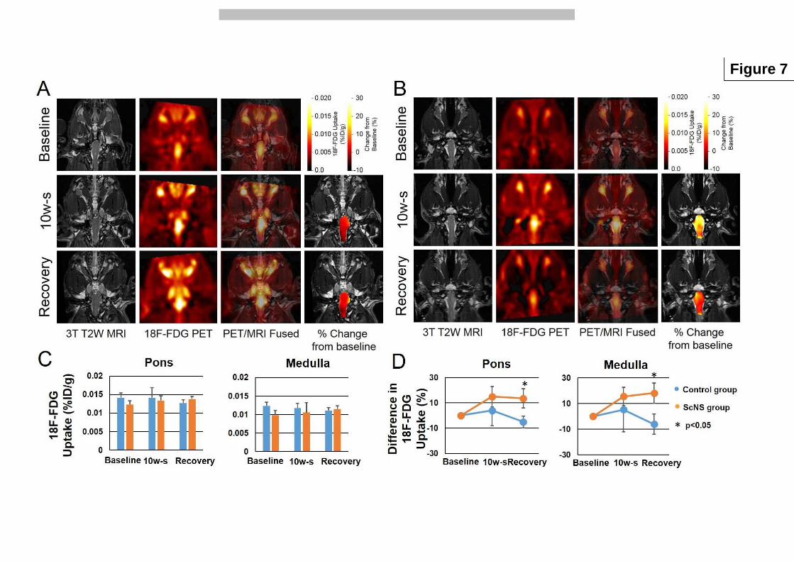
Brain stem remodeling 8
Figure 7A (Sham control group) and 7B (ScNS group) from left to right show 3T T2W 9
SPACE3D MRI, 18F-FDG PET, PET/MRI Fused, and percent changes from baseline. The rows 10
from top to bottom show the images obtained at baseline, after ScNS and after recovery, 11
respectively. Blinded volumetric analyses showed 18F-FDG uptake in pons and medulla of the 12
sham control group had no statistically significant time-dependent changes. In contrast, 18F-FDG 13
uptake in pons and medulla of the ScNS group of dogs continued to increase (Figure 7C). The 14
mean ∆%ID/g in the pons of the ScNS group went from 14.84% to 13.54% and in the medulla 15
15.64% to 18.22%. In the sham control group the mean ∆%ID/g went from 3.92% to -5.07% in 16
pons and 5.27% to -6.07%. The final differences in 18F-FDG uptake between these two groups 17
were statistically significant (p=0.011, Figure 7D). 18
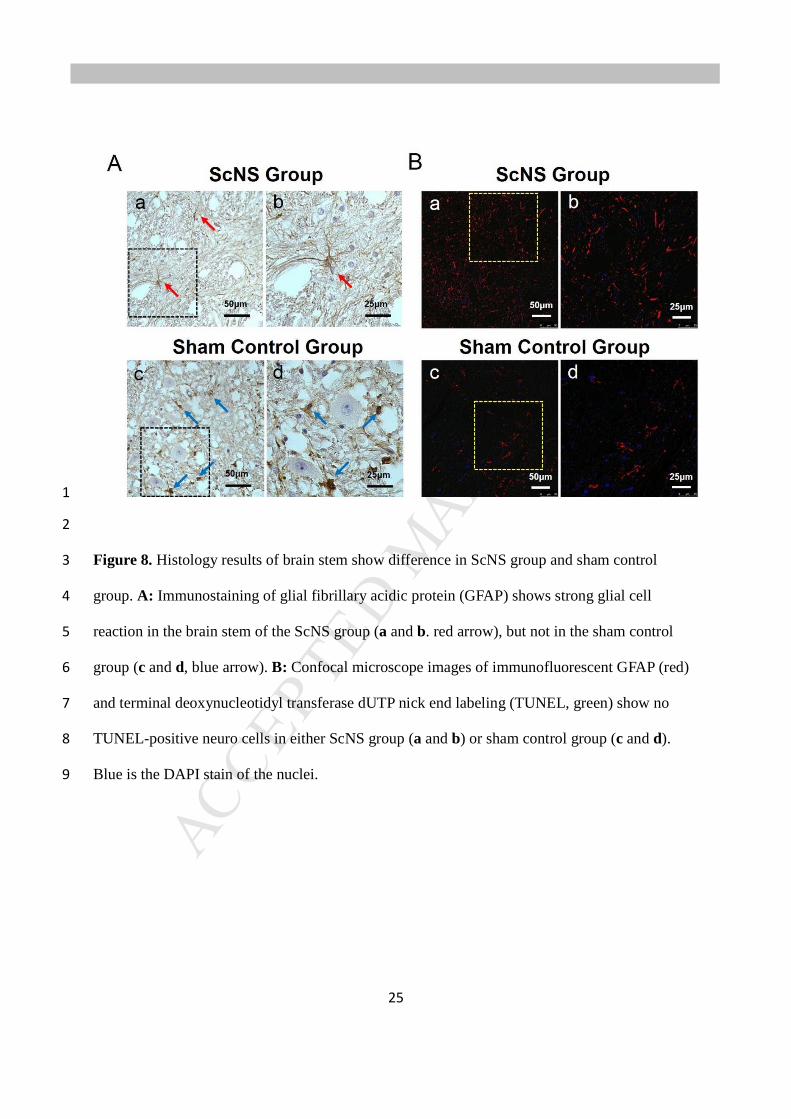
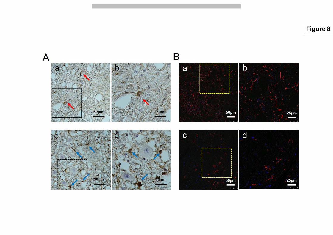
All brain stems were successfully harvested for histology analyses. Figure 8A shows the 19
GFAP staining of the brain stem from the ScNS group (a and b) and from the sham control group 20
(c and d). Brown color indicates the GFAP-positive glial cells. The densities of GFAP 21
immunoreactivity were significantly higher in the ScNS group (32700±12900 µm2/mm2) than 22
that in the sham control group (16900±7300 µm2/mm2, p=0.026). Figure 8B shows terminal 23
MANUSCRIP
T
ACCEPTED
12
deoxynucleotidyl transferase dUTP nick end labeling (TUNEL) staining with GFAP double 1
immunofluorescence staining. The red colors shows positive GFAP staining while the green 2
color is expected for TUNEL positive cells. There were more GFAP activity in the ScNS group 3
(a and b) than in the sham control group (c and d). No TUNEL-positive neuron cells were 4
observed in either group. 5
6
Discussion 7
We found that ScNS reduces VR and aSGNA but increases the frequencies of long pauses during 8
persistent AF. These changes are associated with significant brain stem and SG remodeling. 9
10
Neural Remodeling 11
Electrical stimulation is a commonly used method for managing human diseases, including 12
cardiac arrhythmias.15 Because the autonomic nervous system is highly plastic, chronic electrical 13
stimulation is likely to result in significant neural remodeling. Chronic stimulation of the vagal 14
nerves and the subcutaneous tissues can be associated with significant remodeling of the SG.10, 12, 15
16 Electrical stimulation applied to the left cymba conchae could produce significant activation of 16
the classical central vagal projections in functional magnetic resonance imaging.17 Like that 17
found in the present study, the SG changes include increased percentage of TH-negative cells and 18
TUNEL-positive neurons. Radiofrequency catheter ablation of the renal sympathetic nerves is 19
associated with similar changes in the SG.18 However, contrary to that found in dogs underwent 20
renal denervation, we found that the 18F-FDG uptake in the brain stem is increased. While there 21
are significant glial cell reactions, there are no TUNEL-positive cells in the brain stem. The 22
combined use of PET and MRI for functional studies of the brain has undergone significant 23

MANUSCRIP
T
ACCEPTED
13
evolution in recent years.19 The glucose analog 18F-FDG is a surrogate marker for glucose 1
metabolism that is generally increased in malignant tumors and inflammation. Because neuronal 2
activity is dependent upon glucose metabolism,20 the findings in this study suggest that there is 3
increased brain stem activity in dogs with ScNS as compared with sham controls. Viral 4
transneuronal labeling studies have found that brain stem is connected with the sympathetic 5
preganglionic neurons at the lateral horn, which project to the SG.21 It is possible that the SG 6
remodeling could lead to brain stem remodeling through these direct connections. 7
8
ScNS versus drug therapy for ventricular rate control 9
Dosdall et al22 reported that a combination of digoxin and metoprolol can reduce the average 10
ventricular rate in canine pacing-induced chronic AF from 172 bpm to 130 bpm after 6 months, 11
along with a reduction of LVEF from 54% to 33%. In comparison, we showed that ScNS 12
reduced the VR from 149 bpm to 84 bpm in 6 weeks with no change of LVEF. These findings 13
suggest that ScNS may be more effective than drug therapy in controlling ventricular rate. 14
However, due to shorter duration of follow up, whether or not ScNS can preserve LVEF after 6 15
months of AF remains unclear. 16
17
Clinical implications 18
Ventricular rate control is an important strategy in managing patients with AF.1 When drug 19
therapy fails to achieve rate control, nonpharmacological therapy such as catheter ablation of the 20
atrioventricular node may be needed. ScNS may provide an alternative to atrioventricular node 21
ablation. Instead of using subcutaneously implanted electrodes, it is also possible that 22
transcutaneous electrical nerve stimulation can achieve similar therapeutic effects. However, we 23

MANUSCRIP
T
ACCEPTED
14
do not have data to test that hypothesis. 1
Similar to chronic vagal nerve stimulation,10 chronic ScNS can increase the frequencies 2
of long pauses during AF. These iatrogenic long (> 3 s) pauses may lead to a need for pacemaker 3
insertions.23 Our recent studies24 showed that the strength of electrical stimulation is important in 4
determining the electrophysiological responses. Very low dose (0.25 mA) increases serum 5
norepinephrine, causes nerve sprouting and is proarrhythmic. Intermediate dose (2.5 mA) do not 6
increase norepinephrine levels and appears to be similarly antiarrhythmic as 3.5 mA. In that 7
study, the dogs were in sinus rhythm. Whether or not the data are applicable to rate control of AF 8
remains unclear. If there is a future clinical trial on ScNS or TENS in AF rate control, the 9
relationship between stimulation strength and the bradycardic complications will need to be 10
carefully considered. 11
12
Limitations 13
The study duration is insufficient to determine the persistent efficacy or the reversibility of the 14
neural remodeling induced by ScNS. 15
Acknowledgement 16
We thank Nicole Courtney, Christopher Corr and David Adams for their assistance. 17
18
References 19
1. Wyse DG, Waldo AL, DiMarco JP, et al. A comparison of rate control and rhythm control 20
in patients with atrial fibrillation. N.Engl.J.Med. 2002;347:1825-1833. 21
2. January CT, Wann LS, Alpert JS, et al. 2014 AHA/ACC/HRS guideline for the 22
management of patients with atrial fibrillation: a report of the American College of 23
MANUSCRIP
T
ACCEPTED
15
Cardiology/American Heart Association Task Force on Practice Guidelines and the Heart 1
Rhythm Society. J Am Coll Cardiol. 2014;64:e1-76. 2
3. Ozcan C, Jahangir A, Friedman PA, et al. Long-term survival after ablation of the 3
atrioventricular node and implantation of a permanent pacemaker in patients with atrial 4
fibrillation. N Engl J Med. 2001;344:1043-1051. 5
4. Zhuang S, Zhang Y, Mowrey KA, et al. Ventricular rate control by selective vagal 6
stimulation is superior to rhythm regularization by atrioventricular nodal ablation and 7
pacing during atrial fibrillation. Circulation. 2002;106:1853-1858. 8
5. Stavrakis S, Humphrey MB, Scherlag BJ, et al. Low-level transcutaneous electrical vagus 9
nerve stimulation suppresses atrial fibrillation. J Am Coll Cardiol. 2015;65:867-875. 10
6. Rhee KS, Hsueh CH, Hellyer JA, et al. Cervical vagal nerve stimulation activates the 11
stellate ganglion in ambulatory dogs. Korean Circ J. 2015;45:149-157. 12
7. Onkka P, Maskoun W, Rhee KS, et al. Sympathetic nerve fibers and ganglia in canine 13
cervical vagus nerves: Localization and quantitation. Heart Rhythm. 2013;10:585–591. 14
8. Olney JW, deGubareff T, Sloviter RS. "Epileptic" brain damage in rats induced by 15
sustained electrical stimulation of the perforant path. II. Ultrastructural analysis of acute 16
hippocampal pathology. Brain Res Bull. 1983;10:699-712. 17
9. Olney JW. Excitotoxicity, apoptosis and neuropsychiatric disorders. Curr Opin 18
Pharmacol. 2003;3:101-109. 19
10. Chinda K, Tsai WC, Chan YH, et al. Intermittent Left Cervical Vagal Nerve Stimulation 20
Damages the Stellate Ganglia and Reduces Ventricular Rate During Sustained Atrial 21
Fibrillation in Ambulatory Dogs. Heart Rhythm. 2016;13:771-780. 22
11. Taniguchi T, Morimoto M, Taniguchi Y, Takasaki M, Totoki T. Cutaneous distribution of 23

MANUSCRIP
T
ACCEPTED
16
sympathetic postganglionic fibers from stellate ganglion: A retrograde axonal tracing 1
study using wheat germ agglutinin conjugated with horseradish peroxidase. J Anesth. 2
1994;8:441-449. 3
12. Yuan Y, Jiang Z, Zhao Y, et al. Long-term intermittent high-amplitude subcutaneous 4
nerve stimulation reduces sympathetic tone in ambulatory dogs. Heart Rhythm. 5
2018;15:451-459. 6
13. Zhao Y, Yuan Y, Tsai WC, et al. Antiarrhythmic effects of stimulating the left dorsal 7
branch of the thoracic nerve in a canine model of paroxysmal atrial tachyarrhythmias. 8
Heart Rhythm. 2018;15:1242-1251. 9
14. Dicarlo L, Libbus I, Amurthur B, Kenknight BH, Anand IS. Autonomic regulation 10
therapy for the improvement of left ventricular function and heart failure symptoms: the 11
ANTHEM-HF study. J Card Fail. 2013;19:655-660. 12
15. Chen PS, Chen LS, Fishbein MC, Lin SF, Nattel S. Role of the autonomic nervous 13
system in atrial fibrillation: pathophysiology and therapy. Circ Res. 2014;114:1500-1515. 14
16. Shen MJ, Hao-Che Chang X, Park HW, et al. Low-level vagus nerve stimulation 15
upregulates small conductance calcium-activated potassium channels in the stellate 16
ganglion. Heart Rhythm. 2013;10:910-915. 17
17. Frangos E, Ellrich J, Komisaruk BR. Non-invasive Access to the Vagus Nerve Central 18
Projections via Electrical Stimulation of the External Ear: fMRI Evidence in Humans. 19
Brain Stimul. 2015;8:624-636. 20
18. Tsai W-C, Chan YH, Chinda K, et al. Effects of renal sympathetic denervation on the 21
stellate ganglion and the brain stem in dogs. Heart Rhythm. 2017;14:255-262. 22
19. Broski SM, Goenka AH, Kemp BJ, Johnson GB. Clinical PET/MRI: 2018 Update. AJR 23

MANUSCRIP
T
ACCEPTED
17
Am J Roentgenol. 2018;211:295-313. 1
20. Barros LF, Deitmer JW. Glucose and lactate supply to the synapse. Brain Res Rev. 2
2010;63:149-159. 3
21. Jansen ASP, Wessendorf MW, Loewy AD. Transneuronal labeling of CNS neuropeptide 4
and monoamine neurons after pseudorabies virus injections into the stellate ganglion. 5
Brain Res. 1995;683:1-24. 6
22. Dosdall DJ, Ranjan R, Higuchi K, et al. Chronic atrial fibrillation causes left ventricular 7
dysfunction in dogs but not goats: experience with dogs, goats, and pigs. Am J Physiol 8
Heart Circ Physiol. 2013;305:H725-731. 9
23. Epstein AE, Dimarco JP, Ellenbogen KA, et al. ACC/AHA/HRS 2008 guidelines for 10
Device-Based Therapy of Cardiac Rhythm Abnormalities: executive summary. Heart 11
Rhythm. 2008;5:934-955. 12
24. Wan J, Chen M, Yuan Y, et al. Antiarrhythmic and proarrhythmic effects of subcutaneous 13
nerve stimulation in ambulatory dogs. Heart Rhythm. 2019:pii: S1547-5271(1519)30149-14
30143. doi: 30110.31016/j.hrthm.32019.30102.30027. [Epub ahead of print]. 15
16

MANUSCRIP
T
ACCEPTED
18
1
2
Figure 1. Schematic of the study protocol. The first 18F-FDG PET/MRI was performed before 3
surgery. The baseline nerve activity and ECG were recorded two weeks after radiotransmitter 4
implantation. After persistent AF was induced, the dogs were randomly assigned to thoracic 5
subcutaneous nerve stimulation (ScNS) and sham control group. 6

MANUSCRIP
T
ACCEPTED
19
1
Figure 2. Effects of ScNS on SGNA, VR, VNA and ScNA. The serial changes in the ratio of 2
average SGNA (A) and VR (B) at different times after persistent AF in the ScNS group (filled 3
columns) and in the sham control group (unfilled columns). In comparison with baseline, there 4
are significant decreases in aSGNA after 6 weeks and in VR after 7 weeks of ScNS. C: 5
Examples of baseline nerve discharges (asterisks). D: SGNA, VNA and ScNA co-firing (blue 6
arrow) discharges during persistent AF. E: Abrupt (red dot) reduction of SGNA during ScNS 7
ON-time (box) after 10 weeks of ScNS, which is associated with a reduction of VR (black 8
arrow). Asterisks indicate significant differences of aSGNA and VR between ScNS group and 9
control group. 10
11
12

MANUSCRIP
T
ACCEPTED
20
1
2
Figure 3. Changes of nerve activities and VR in ScNS group and in sham control group. A: 3
aSGNA increased after persistent AF and reduced gradually after ScNS (red arrow) in ScNS 4
group. The reduction was statistically significant compared to baseline after 4 or more weeks of 5
ScNS. Ventricular rate significantly increased after persistent AF and gradually reduced after 6
ScNS onset (red arrow). Average VNA (aVNA) and average ScNA (aScNA) did not change 7
significantly by ScNS. B: aSGNA increased after persistent AF and kept increasing in the 8
following weeks in sham control group. Ventricular rate increased significantly after persistent 9
AF and then fluctuated but did not show a stable trend of increase or decrease. 10
11
12
13
14
15
16

MANUSCRIP
T
ACCEPTED
21
1
Figure 4. Nerve activity over a 24-hr period. A: SGNA at baseline (upper panel), during 2
persistent AF (middle panel) and after 11 weeks of ScNS (lower panel) in the ScNS group. B: 3
SGNA at baseline (upper panel), during persistent AF (middle panel) and after 11 weeks of 4
follow up (lower panel) in sham control group. 5
6
MANUSCRIP
T
ACCEPTED
22
1
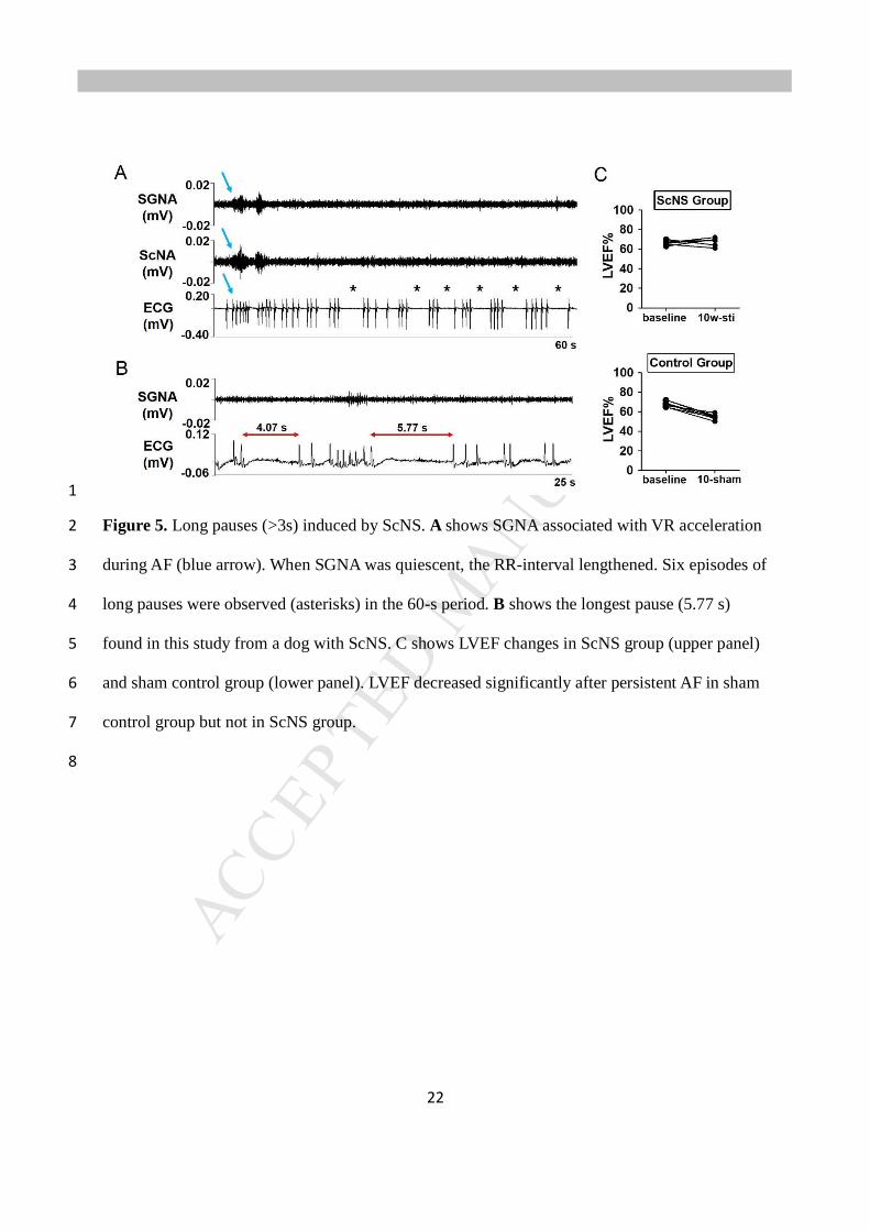
Figure 5. Long pauses (>3s) induced by ScNS. A shows SGNA associated with VR acceleration 2
during AF (blue arrow). When SGNA was quiescent, the RR-interval lengthened. Six episodes of 3
long pauses were observed (asterisks) in the 60-s period. B shows the longest pause (5.77 s) 4
found in this study from a dog with ScNS. C shows LVEF changes in ScNS group (upper panel) 5
and sham control group (lower panel). LVEF decreased significantly after persistent AF in sham 6
control group but not in ScNS group. 7
8
MANUSCRIP
T
ACCEPTED
23
1
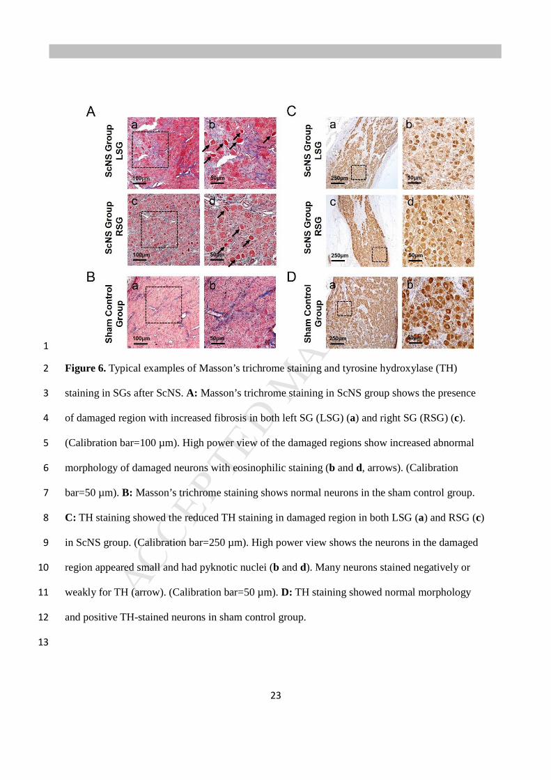
Figure 6. Typical examples of Masson’s trichrome staining and tyrosine hydroxylase (TH) 2
staining in SGs after ScNS. A: Masson’s trichrome staining in ScNS group shows the presence 3
of damaged region with increased fibrosis in both left SG (LSG) (a) and right SG (RSG) (c). 4
(Calibration bar=100 µm). High power view of the damaged regions show increased abnormal 5
morphology of damaged neurons with eosinophilic staining (b and d, arrows). (Calibration 6
bar=50 µm). B: Masson’s trichrome staining shows normal neurons in the sham control group. 7
C: TH staining showed the reduced TH staining in damaged region in both LSG (a) and RSG (c) 8
in ScNS group. (Calibration bar=250 µm). High power view shows the neurons in the damaged 9
region appeared small and had pyknotic nuclei (b and d). Many neurons stained negatively or 10
weakly for TH (arrow). (Calibration bar=50 µm). D: TH staining showed normal morphology 11
and positive TH-stained neurons in sham control group. 12
13

MANUSCRIP
T
ACCEPTED
24
1
Figure 7. Functional imaging of brain stem in ScNS group and sham control group. A (sham 2
control group) and B (ScNS group) from left to right show 3T T2W SPACE3D MRI, 18F-2-3
Fluoro-2-deoxyglucose (18F-FDG), PET/MRI Fused, and percent changes from baseline. The 4
rows from top to bottom show the images obtained at baseline, after ScNS and after recovery, 5
respectively. Blinded volumetric analyses showed 18F-FDG uptake in pons and medulla of the 6
sham control group had no statistically significant time-dependent changes. In contract, 18F-FDG 7
uptake in pons and medulla of ScNS group dogs continued to increase (C). After 4-8 weeks of 8
recovery, the final differences in 18F-FDG uptake between these two groups were statistically 9
significant (D). 10
11
MANUSCRIP
T
ACCEPTED
25
1
2
Figure 8. Histology results of brain stem show difference in ScNS group and sham control 3
group. A: Immunostaining of glial fibrillary acidic protein (GFAP) shows strong glial cell 4
reaction in the brain stem of the ScNS group (a and b. red arrow), but not in the sham control 5
group (c and d, blue arrow). B: Confocal microscope images of immunofluorescent GFAP (red) 6
and terminal deoxynucleotidyl transferase dUTP nick end labeling (TUNEL, green) show no 7
TUNEL-positive neuro cells in either ScNS group (a and b) or sham control group (c and d). 8
Blue is the DAPI stain of the nuclei. 9
MANUSCRIP
T
ACCEPTED
AcclimationPET/MRIPacemaker/DSI/Neurostimulator implantation
Recovery and Baseline recording
Atrial pacing at 600 bpm for 2 weeks to induce AF
Persistent AFNo
Yes
Randomly assigned to two groups
Control Group ScNS Group
Sham stimulation (0 mA)
Commencement of stimulation with initial output of 0.5 mA
Increase the output current until reaching 3.5 mA
Continue stimulation for 2 months
Tissue harvest for histology study
PET/MRI
Devices removal
4-8 weeks recovery
PET/MRI
Figure 1

MANUSCRIP
T
ACCEPTED
Figure 2
1 2 3 4 5 6 7 8 9 10 11 12 130
1
2
3
ScNSSham-control
1 2 3 4 5 6 7 8 9 10 11 12 130
1
2
3
ScNSSham-control
40 sec
A
ScNS ScNSweek week
BaSGNA VR
EC D0.04
0.04SGNA(mV)
0.04
0.04VNA(mV)
0.04
0.04ScNA(mV)
0.10
-0.20ECG(mV)
400
0VR(bpm)
0.04
0.04SGNA(mV)
0.04
0.04VNA(mV)
0.04
0.04ScNA(mV)
0.20
-0.50ECG(mV)
400
0VR(bpm)
40 sec
0.04
0.04SGNA(mV)
0.04
0.04VNA(mV)
0.04
0.04ScNA(mV)
0.20
-0.10ECG(mV)
400
0VR(bpm)
40 sec
ScNS
* *
*
***
**
* * ** *
*
3
2
1
0
3
2
1
0BSAF 1 2 3 4 5 6 7 8 9 10 11 BSAF 1 2 3 4 5 6 7 8 9 10 11

MANUSCRIP
T
ACCEPTED
Figure 3
VR
0
2
4
6
8
10
0
50
100
150
200
250
0
2
4
6
8
10
0
2
4
6
8
10
0
2
4
6
8
10
0
2
4
6
8
10
0
2
4
6
8
10
0
50
100
150
200
250
A
B
aSGNA
aSGNA
aVNA aScNA
aVNA aScNA VR
µV µV µV bpm
µV µV µV bpm
*
*
*
*
Dog1 Dog3 Dog7 Dog8 Dog11 Dog12
Dog2 Dog4 Dog5 Dog6 Dog9 Dog10 Dog13
week2B
SA
F 1 3 4 5 6 7 8 91011week
2BS
AF 1 3 4 5 6 7 8 91011
week2B
SA
F 1 3 4 5 6 7 8 91011week
2BS
AF 1 3 4 5 6 7 8 91011
week2B
SA
F 1 3 4 5 6 7 8 91011week
2BS
AF 1 3 4 5 6 7 8 91011
week2B
SA
F 1 3 4 5 6 7 8 91011week
2BS
AF 1 3 4 5 6 7 8 91011

MANUSCRIP
T
ACCEPTED
Figure 4
MANUSCRIP
T
ACCEPTED
Figure 5

MANUSCRIP
T
ACCEPTED
Figure 6
MANUSCRIP
T
ACCEPTED
Figure 7
MANUSCRIP
T
ACCEPTED
Figure 8