subcutaneous fat necrosis: clinical presentation and pathophysiological ... · subcutaneous fat...
TRANSCRIPT

April 2017
Subcutaneous fat necrosis:
clinical presentation and
pathophysiological aspects
of altered calcium
homeostasis
SWISS SOCIETY OF NEONATOLOGY

Juvet C, Aijjou A, Diebold P, Service de Pédiatrie (JC,
AA, DP), Hôpital d’Aigle, Hôpital Riviera-Chablais, Aigle,
Switzerland
Title figure:
Cholesterol crystals (clefts) and multinucleated giant cells
(Source: www.humpath.com)
© Swiss Society of Neonatology, Thomas M Berger, Webmaster

This female infant was born after an uncompli-
cated pregnancy at 41 5/7 weeks of gestation to a
31-year-old mother. Screening examinations had been
un remarkable except for positive GBS cultures for
which the mother was treated with 3 doses of anti-
biotics prior to delivery. Membranes had ruptured
9 hours before birth and amniotic fluid was noted to
be meconium-stained. Vaginal delivery was vacuum-
assisted because of failure to progress and shoulder
dystocia. Her birth weight was 4220 g (P 90 – 95).
Initially, she had no spontaneous breathing, was hypo-
tonic and non-reactive. She was stimulated, suctioned
and bag-mask ventilated, first with an FiO2 of 21%,
then up to 100% for 5 minutes. Apgar scores were 2,
4 and 6 at 1, 5 and 10 minutes, respectively. Arterial
and venous umbilical cord pH values were 7.16 and
7.21, respectively. At 5 minutes of life, there was spon-
taneous breathing, and CPAP treatment was initiated
with a PEEP of 5 cmH2O. At 10 minutes of life, she
still presented respiratory distress, with suprasternal,
intercostal and subcostal retractions, as well as nos-
tril flaring; SpO2 was 89 – 92% in room air. She was
hypotonic with a diminished reactivity, but her heart
rate always remained above 100 beats per minute and
blood pressure was normal.
At 30 min of life, there was marked mixed acidosis
with a pH of 6.93, a pCO2 of 10.7 kPa (80 mmHg),
a bicarbonate of 9.5 mmol/l, a BE of –14.8 mmol/l,
CASE REPORT
3

4
and a lactate of 10.7 mmol/l. Because of hypoglycemia
(blood glucose 1.9 mmol/l) dextrose 10% was started.
Arterial hypotension (MAP 29 mmHg) responded to a
single fluid bolus.
On physical examination, right-sided upper brachial
plexus palsy was noted. In addition, a discrete indura-
ted lesion on the distal part of the left arm appeared
48 hours later. A chest X-ray showed no signs of
pneumo thorax or meconium aspiration. She rapidly
recovered and was discharged home at 7 days of life
with a prescription for Vitamin D3 400 UI/day.
One week later, she was referred to the hospital by her
pediatrician because of numerous indurated skin lesi-
ons, similar to the one noted initially on her left arm.
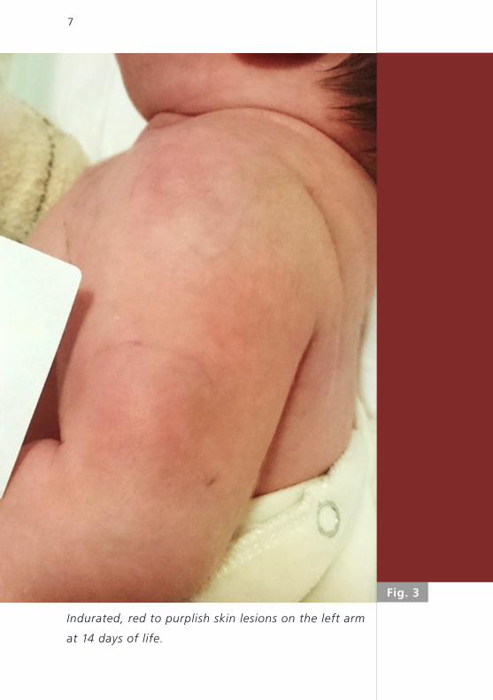
On admission, several indurated, red-purplish subcu-
taneous lesions, located predominantly on the trunk
and the arms were noted (Fig. 1 – 3). A diagnosis of a
subcutaneous fat necrosis was made.
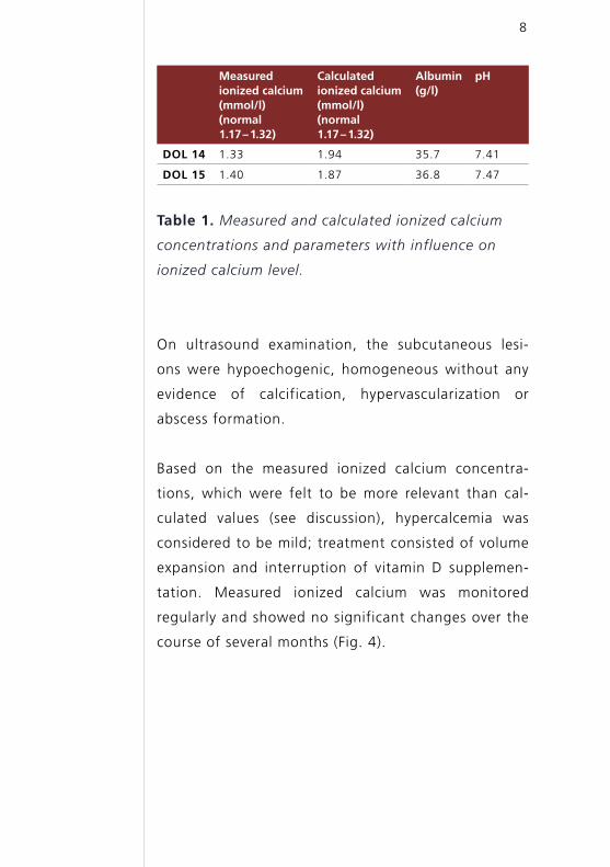
Laboratory examinations were normal except for
thrombocytosis (736 G/l). Serum calcium levels were
elevated, but with a significant discrepancy between
measured and calculated ionized calcium concentra-
tions (Table 1). 25-OH-vitamin D2/D3 was normal (63.9
mmol/l), whereas parathyroid hormone was markedly
decreased (0.7 pmol/l, normal 1.3-6.8 pmol/l). Alkaline
phosphatase was within the normal range.

5
Indurated, red to purplish skin lesions on the trunk at
14 days of life.
Fig. 1

Fig. 2
6
Indurated, red to purplish skin lesions on the left arm
and axilla at 14 days of life.
7
Indurated, red to purplish skin lesions on the left arm
at 14 days of life.
Fig. 3
8
Measured ionized calcium (mmol/l) (normal 1.17 – 1.32)
Calculated ionized calcium (mmol/l) (normal 1.17 – – 1.32)
Albumin (g/l)
pH
DOL 14 1.33 1.94 35.7 7.41
DOL 15 1.40 1.87 36.8 7.47
Table 1. Measured and calculated ionized calcium
concentrations and parameters with influence on
ionized calcium level.
On ultrasound examination, the subcutaneous lesi-
ons were hypoechogenic, homogeneous without any
evidence of calcification, hypervascularization or
abscess formation.
Based on the measured ionized calcium concentra-
tions, which were felt to be more relevant than cal-
culated values (see discussion), hypercalcemia was
considered to be mild; treatment consisted of volume
expansion and interruption of vitamin D supplemen-
tation. Measured ionized calcium was monitored
regularly and showed no significant changes over the
course of several months (Fig. 4).

Fig. 4
1 2 3 4
measured Ca2+
calculated Ca2+
ion
ized
cal
ciu
m c
on
cen
trat
ion
(mm
ol/
l)
months of life
9
Evolution of blood calcium concentrations over the
first months of life.
2
1.5
1
0.5
0

10
DISCUSSION Subcutaneous fat necrosis (SFN) is a rare form of
panniculitis presenting with purplish-erythematous
indurated nodules and plaques in the first weeks of
life; it is typically located on the face, bottom, arms,
thighs and trunk. It is usually seen in term or near term
infants (1). Several risk factors have been reported
in the literature, including macrosomia, perinatal
trauma, asphyxia, (therapeutic) hypothermia, gestati-
onal diabetes, preeclampsia and hypoglycemia (1 – 9).
The incidence of SFN in children undergoing therapeu-
tic hypothermia is about 1%, and thus much higher
than in the general neonatal population (5). It is very
likely that hypothermia favors SFN as it facilitates fat
crystallization (see below). Notably, many of these risk
factors can occur simultaneously, and, given the low
incidence of SFN, it is difficult to assess whether they
are independent of each other.
The diagnosis of SFN mostly relies on its clinical presen-
tation. However, histological examination is sometimes
performed when lesions are not unequivocal. It typi-
cally shows fat cell necrosis, along with needle-shaped
crystals or clefts and inflammatory cell infiltration,
including multinucleated giant cells. These crystals are
mostly found in fat cells (1), but occasionally described
in multinucleated giant cells (6). Calcium deposition is
inconsistently being reported (3, 6). Interestingly, Tran
et al. reported on calcifications found incidentally at 1
month of life in the context of marked hypercal cemia,
while SFN lesions had resolved and biopsy in the ini-

11
tial phase had failed to show calcium depositions. This
indicates that calcification could be a later event, pos-
sibly linked to hypercalcemia (3).
The differential diagnosis of SFN includes sclerema
neonatorum (SN), lipogranulomatosis and cold panni-
culitis (4). Like SFN, SN appears in the first few days of
life, but it is always associated with severe disorders
and bad outcome (1). Histological findings also differ:
while crystals are usually present, necrosis, inflamma-
tion and giant cells are usually absent (2, 3).
Hypercalcemia is the most feared complication of SFN
and can occur up to 6 months after the resolution
of the skin lesions. Clinically, patients may present
with lethargy, hypotonia and poor feeding. Persistent
hypercalcemia can lead to nephrocalcinosis with sub-
sequent renal failure. This complication has predomi-
nantly been described in the most severely affected
children; it usually resolves over a period of several
months (9).
Other complications that have been described in
pa tients with SFN include thrombocytopenia, hyper-
triglyceridemia, hypoglycemia, and late subcutane-
ous lipoatrophy (3 – 6, 9). Thrombocytopenia usually
appears before or concomitantly with the skin lesions
(3), and, given the fact that bone marrow examina-
tions are usually unremarkable, is thought to be of
peripheral origin, such as sequestration (10). Dysli-

12
pidemia is inconsistently observed. While changes in
fat composition in newborns are an element in the
current understanding of SFN, as will be discussed
below, it is unclear whether dyslipidemia is a cause or
a consequence of SFN, or even an unrelated finding. In
a case series of 16 patients with SFN, 9 patients (56%)
developed hypercalcemia, 1 patient (6%) renal failure,
and 1 patient (6%) dyslipidemia. Notably, all of the 6
children available for long-term follow-up had develo-
ped subcutaneous atrophy (9).
The pathophysiology SFN is only incompletely under-
stood (Fig. 5). However, the fatty acid composition
of neonatal adipocytes may play an important role:
higher saturated fatty acid concentrations result in a
higher melting point, and therefore facilitate crystalli-
zation in conditions of hypothermia (either in the con-
text of asphyxia and hypoperfusion or induced (i.e.,
therapeutic) hypothermia) (3, 5, 7, 8, 10).
Necrosis and crystal formation lead to the formation
of granulomatous tissue with multinucleated giant
cells. These cells are also present in other diseases,
such as sarcoidosis, tuberculosis or silicone-induced
granulomas. In these conditions, they are known to
1-hydroxylate and therefore activate 25-OH vitamin
D (3, 6), potentially leading to hypercalcemia. Publi-
shed data on 1,25-OH vitamin D are not unequivocal
as both high and normal levels have been described.
However, considering the PTH suppression observed in

Fig. 5
adipocyte
multinucleated giant cell
lymphocyte
histiocyte
hypoxia hypoperfusion
hypercalcemiadiphosphonatesfluid
furosemide calcitonin
low calcium diet
stop vitamin D supplementation
corticosteroids
PGE 1,25-OH vitamin D
increased bone resorption increased calcium absorption
1 newborns have higher saturated fatty acid concentrations leading to a higher melting point
pressure trauma
inflammatory reaction with granuloma formation (see B)
hypothermia
adipocyte lesion/necrosis
fat1 crystallization
± calcium deposition
A) primary insult
B) inflammatory reaction
13
Principal hypotheses of SFN pathophysiology and
potential treatment options (red boxes):
A) primary insult with adipocyte lesions and necrosis;
B) inflammatory reaction with multinucleated giant
cells, lymphocytes and histiocytes.

14
this context, even normal levels can be considered as
elevated, (3, 4, 6, 7, 11).
Another hypothesis to explain hypercalcemia relates to
enhanced prostaglandin E (PGE) production, causing
increased bone resorption (3, 4, 6).
Vitamin D levels were normal in our patient, but only
25-OH-vitamin D2-D3 was measured, which does not
represent the most active form, which is 1,25-OH vita-
min D. Decreased PTH levels measured in our patient
suggest that the increase in calcium concentration was
mediated by another molecule than PTH (1,25-OH vita-
min D and/or PGE). An increased tubular phosphate
reabsorption rate of 99.85% (normal 78 – 97%) with
normal serum phosphate levels might indicate increa-
sed 1,25-OH vitamin D activity.
We observed marked differences between measured
and calculated ionized calcium (Table 1). The calcula-
ted ionized calcium concentration is derived from the
measured total calcium concentration and albumin
concentration. Notably, automated calculations use
the higher adult albumin norms (therefore conside-
ring normal neonatal albumin levels as hypoalbumine-
mia), and consequently overestimate ionized calcium
concentrations. Furthermore, this method does not
correct for pH deviations. Alkalosis and acidosis,
however, have a significant impact on calcium-protein
binding. For these reasons, measured ionized calcium
15
concentrations are likely to be more reliable than cal-
culated ionized calcium concentrations.

16
CONCLUSION SFN is a rare form of neonatal panniculitis, which
should be kept in mind when evaluating a child with
indurated skin lesion and history of perinatal asphyxia
and/or trauma. SFN can frequently be diagnosed based
on clinical presentation alone. In cases of atypical
presentation, a skin biopsy can confirm the diagnosis.
The most important complication of SFN is hypercalce-
mia, which requires monitoring over several months,
as it can occur up to 6 month of age. Prognosis of
these skin lesions is very good as they usually resolve
spontaneously without sequelae, except for possible
lipoatrophy.
See also COTM 02 2003: subcutaneous fat necrosis of
the newborn.

1. Burden AD, Krafchik BR. Subcutaneous fat necrosis of the new-
born: a review of 11 cases. Pediatr Dermatol 1999;16:384 – 387
(Abstract)
2. Parvathidevi GK, Vijayashankar MR, Belagavi CS, et al.
Cytological diagnosis of subcutaneous fat necrosis of newborn:
a case report. Dermatol Online J 2005;11:20 (Abstract)
3. Tran JT, Sheth AP. Complications of subcutaneous fat necrosis
of the newborn: a case report and review of the literature.
Pediatr Dermatol 2003;20:257 – 261 (Abstract)
4. Barbier C, Cneude F, Deliège R, El Kohen R, Kremy O, Leclerc
F. Subcutaneous fat necrosis in the newborn: a risk for severe
hypercalcemia. Arch Pediatr 2003;10:713 – 715 (Abstract)
5. Martins J, Maxaud A, Bah AG, Prophette B, Maillard H,
Bénéton N. Subcutaneous fat necrosis after moderate thera-
peutic hypothermia in two newborns of African origin.
Arch Pediatr 2015;22:191 – 194 (Abstract)
6. Cook JS, Stone MS, Hansen JR. Hypercalcemia in association
with subcutaneous fat necrosis of the newborn: studies of
calcium-regulating hormones. Pediatrics 1992;90:93 – 96
(Abstract)
7. Karochristou K, Siahanidou T, Kakourou-Tsivitanidou T,
Stefanaki K, Mandyla H. Subcutaneous fat necrosis asso-
ciated with severe hypocalcaemia in a neonate. J Perinatol
2006;26:64 – 66 (Abstract)
8. Vonk J, Janssens PM, Demacker PN, Folkers E. Subcutaneous
fat necrosis in a neonate, in association with aberrant plasma
lipid and lipoprotein values. J Pediatr 1993;123:462 – 464
(Abstract)
REFERENCES
17

9. Mahé E, Girszyn N, Hadj-Rabia S, Bodemer C, Hamel-Teillac D,
De Prost Y. Subcutaneous fat necrosis of the newborn:
a systematic evaluation of risk factors, clinical manifestations,
complications and outcome of 16 children. Br J Dermatol
2007;156:709 – 715 (Abstract)
10. Wolach B, Raas-Rothschild A, Vogel R, Choc L, Metzker A. Sub-
cutaneous fat necrosis with thrombocytopenia in a newborn
infant. Dermatologica 1990;181:54 – 55 (Abstract)
11. Kruse K, Irle U, Uhlig R. Elevated 1,25-dihydroxy vitamin D
serum concentrations in infants with subcutaneous fat necro-
sis. J Pediatr 1993;122:460 – 463 (Abstract)
18

SUPPORTED BY
CONTACT
Swiss Society of Neonatology
www.neonet.ch
con
cep
t &
des
ign
by
mes
ch.c
h