study of the cellexercise: observe methylene blue and potassium permanganate) • diffusion across...
TRANSCRIPT

Study of the Cell

Cell
• Cell is structural and functional unit• Activity of organism is dependant upon cell
structure and function• Activity of cell us dependant upon its
internal metabolism• Life and health is dependant upon function
of cells

General Features of the Cell• Cell (plasma) membrane
– Boundary of the cell (phospholipid framework)• Cytoplasm
– Substance within the cell (other than the nucleus)• Organelles
– Any of the various “living” structures of the cell• Inclusions
– Any substance found within the cell such as a chemical or deposit (or as a foreign body)

Cells and Environment• Cells of the body are
– Somatic cells (2n or 23 pairs of homologous chromosomes)
– Gametes (n or 23 chromosomes, one-half of the homologous set)
• Extracellular environment– Extracellular fluid (ECF)
• Interstitial fluid (IF)• Plasma and lymph (in vessels)• CSF, synovial fluids, etc
• Intracellular environment– Intracellular fluid
• Cytoplasm

Microscopic Observations of Cells
• Improve your observations– Get a good slide preparation– Know what you are looking for on the preparation– Clean the lenses and the slide preparation– Adjust the light
• At what magnifications are you making the observations?– How much area can you observe?– How much of the details can you observe?– Is a higher magnification necessarily the best
magnification?

Observations• Stratified squamous epithelium• Exfoliated epithelium• Blood• Sperm• Smooth muscle• Adipose tissue• Ciliated columnar epithelium• Protozoa (live and as a wet mount)

Microscope Slides
• Label• Cover glass• Specimen

Stratified Squamous Epithelium

Exfoliated Squamous Cells

Blood

Sperm

Ciliated Columnar Epithelium

Smooth Muscle

Adipose Tissue

Generalized Cells?• Are the following features shared by all the
observed cells? What are your observations and explanations of each?– Cell (plasma) membrane– Cytoplasm– Organelles– Inclusions

Generalized Cell• Cell membrane
– Gap junctions– Desmosomes– Tight junctions– Microvilli
• Cytoplasm– cytosol
• Organelles– Mitochondria– Lysosomes– Ribosomes
– Endoplasmic reticulum
• Smooth ER• Rough ER
– Cilia– Flagella– Golgi
apparatus– Peroxisomes– Centrosome
• Centrioles– Nucleus
• Nuclear membrane
• Nucleolus• Chromatin
• Inclusions• Lipids• Melanin

Functions of Cell Membrane• Physical boundary • Regulation of exchange (transport)
– Diffusion, facilitated diffusion– Osmosis– Filtration– Vesicular transport (exocytosis and endocytosis)– Receptor mediated endocytosis
• Structural support• Cell-to-cell connections• Microvilli and enzymes• Maintains membrane potential (excitable cells)

Structure of Cell Membrane• Framework consists of bilayer of phospholipids
– Amphiphatic (hydrophobic vs. hydrophilic)– Other lipids include cholesterol and glycolipids

Cytoplasm
• Between cell membrane and nucleus• Fluid called cytosol contains
– Water and solutes such as salts, proteins, amino acids, sugars, ions, etc.
• Organelles are functional (living) structures• Inclusions are chemicals (or foreign bodies)
within the cell
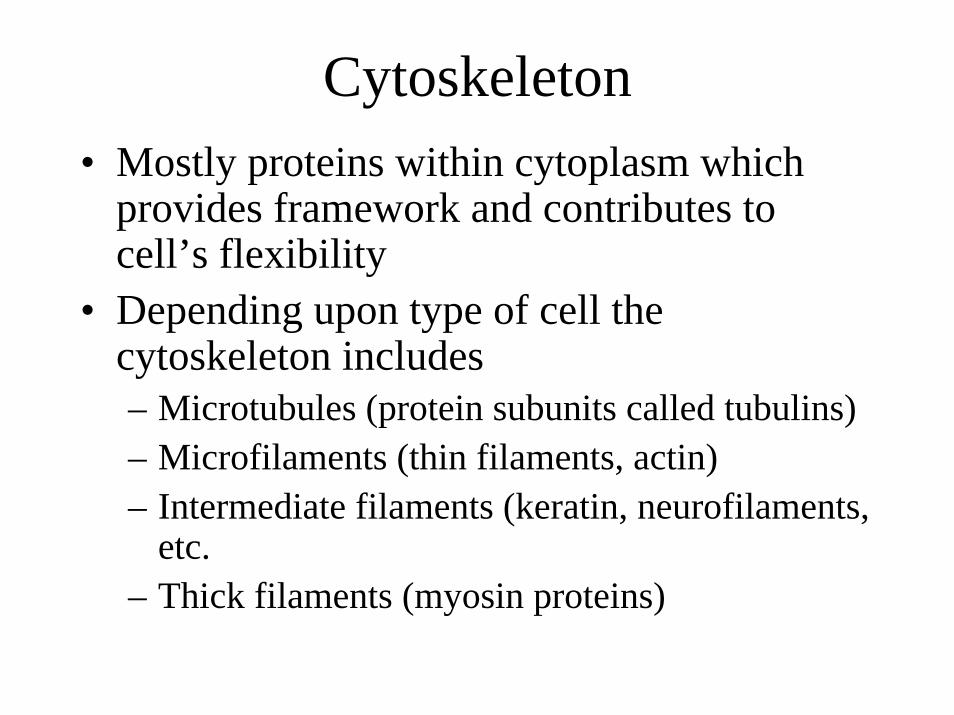
Cytoskeleton• Mostly proteins within cytoplasm which
provides framework and contributes to cell’s flexibility
• Depending upon type of cell the cytoskeleton includes– Microtubules (protein subunits called tubulins)– Microfilaments (thin filaments, actin)– Intermediate filaments (keratin, neurofilaments,
etc.– Thick filaments (myosin proteins)

Microtubules
• Microtubules– Primary component of cytoskeleton– Assembly and disassembly provides for
movements– Attach to and move materials along their axis– Form spindle during cell division– Form the components of centrioles, cilia, and
flagella

Thick Filaments
• Major structural and contractile protein of muscle cells consisting of protein, myosin– Interact with microfilament called actin (part of
the thin filament of muscle contraction)

Centrioles• Two structures located near nucleus which
are composed mostly of microtubules• Form mitotic spindle during mitosis
– Are not found in all cells. Skeletal muscle, cardiac muscle, RBCs, and the typical neurons do not have centrioles and do not divide
• Area surrounding centrioles is called centrosome

Cilia• Extension of the cell which contain
microtubules.• Coordinated movements move materials
across surface of cell.• Found in upper respiratory airways,
fallopian tubes, etc.

Flagella
• Same structure as cilia except longer and fewer in number
• Only human flagellated cell is sperm

Ribosomes• Sites of protein synthesis• Originate from the nucleolus (region of nucleus)
as two subunits• Consist of rRNA and proteins• Either found
– free in the cytoplasm or – Fixed (attached) to endoplasmic reticulum
• Proteins produced by free ribosomes become part of the cytoplasm
• Proteins produced by fixed ribosomes moved into the RER (May be exocytosed by secretoryvesicles, formed into lysosomes, or transported to the cell membrane by transport vesicles.)

Endoplasmic Reticulum• Membranes partitions and membrane
channels throughout cytoplasm • Functions in
– Synthesis of proteins, carbohydrates, lipids– Storage of substances (skeletal muscle it stores
calcium ions)– Transport of materials (in channels)– Detoxification of materials by enzymatic action

RER and SER
• Rough endoplasmic reticulum (RER) has ribosomes attached
• Smooth endoplasmic reticulum (SER) does not have ribosomesattached

SER• FUNCTIONS INCLUDE:• Lipid metabolism
– Phospholipids and cholesterol– Steroid hormones (androgens and
estrogens)– Glycerides (adipose and liver)
• Carbohydrate metabolism– Synthesis of glycogen (liver and
skeletal muscle)– Storage and release of Ca++
(sarcoplasmic reticulum of skeletal muscle)
• Detoxification (medications, etc.)

RER• Functions in initial packaging of proteins
– Proteins are moved into ER from its fixed (attached) ribosomes– Portions of ER are pinched off as transport vesicles– Transport vesicles are moved to Golgi apparatus for processing– Leave Golgi apparatus as
• Lysosomes• Secretory vesicles• Membrane bound transport vesicles

Golgi Apparatus• Series of membranous sacs
stacked one upon the other.• Functions in
– Modifications of proteins (example, adding carbohydrate chains)
– Packaging of proteins into vesicles for exocytosis
– Parking of proteins into vesicles for maintenance of cell membrane

Lysosomes
• Contain powerful digestive enzymes• Produced by the Golgi apparatus• Fuse with
– vesicles from endocytosis– damaged organelles– enzymes may be exocytosed (osteoblasts)
• Products from digestion can be used for– Energy– Building blocks of other molecules
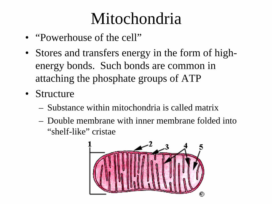
Mitochondria• “Powerhouse of the cell”• Stores and transfers energy in the form of high-
energy bonds. Such bonds are common in attaching the phosphate groups of ATP
• Structure– Substance within mitochondria is called matrix– Double membrane with inner membrane folded into
“shelf-like” cristae

Energy Production
• Energy rich molecule such as glucose or fatty acid is converted to acetyl group.
• Acetyl group enters Krebs cycle (TCA, or tricarboxylic acid cycle) and energy is transferred in form of H+ (attached to carrier molecules) into electron transport system (ETS). CO2 is produced during initial stages.
• ETS transfers energy to ADP + Pi ATP. Oxygen is used to capture hydrogen ions and water is produced.

Nucleus• Houses chromosomes which
normally appear in dispersed form called chromatin
• Membrane is double with numerous pores– Allows passage of ions, small
molecules, mRNA, rRNA, etc. but not DNA (chromosomes)
• Region called nucleolus manufactures rRNA and associated proteins (histones) for ribosomes– Ribosomal subunits move out into
cytoplasm to form the ribosomes

Chromosomes• Non-dividing cell
– DNA strands become associated with proteins called histones. Further organization produces chromatin
• Dividing cell– DNA is supercoiled
into chromosomes– Each chromosome is
replicated

Transcription– One DNA strand (a gene) is
used as template in assembly of mRNA by complementary base pairing (RNA lacks thymine, substitution is uracil). RNA polymerase promotes bonding
• Gene– Nitrogen base sequence (or
triplet sequence) used in manufacture of a protein (or polypeptide)

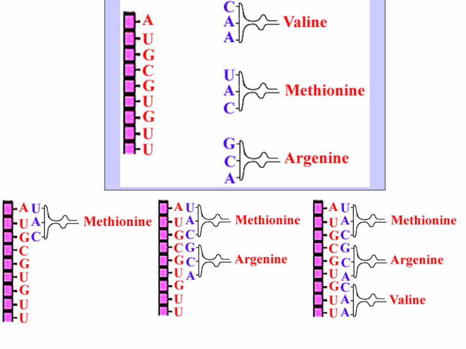
Complementary Pairing• Triplet is sequence of three nitrogen
bases of DNA• Codon is sequence of three nitrogen
bases of mRNA• Genetic code is “triplet code.”
However, actual assembly of protein is from mRNA so codons are used in specifying the amino acids (by using codon table).– What is the 2nd triplet?– What is the 3rd codon?– How do we determine which amino acid
is “called for” by triplet #3?

Translation• The transfer of information
encoded in mRNA into the construction of a protein (or polypeptide).
• Transfer RNA (tRNA) moves appropriate amino acids to codon
• Ribosome serves as site for incoming tRNAs and provides enzymes for bonding of amino acids (peptide bonds).

Translation • Initiation• mRNA binds to ribosome• tRNA carrying methionine
(anticodon UAC) binds
• Elongation• 2nd tRNA arrives• Amino acids joined with
peptide bond • Enzymes split off first amino
acid from mRNA and ribosome moves one codondown, process continues….
• Termination• Ribosome reaches “stop”
signal, the stop codon, UGA. Release.

DNA Control• Direct control by the manufacture of proteins (or
polypeptides) dictated by triplets of genes.– Some proteins exist directly from synthesis as structural
proteins of the cell• In-direct control by producing proteins which
mediate reactions, the enzymes.– Carbohydrates, lipids, etc. of the cell are under
enzymatic (indirect) control.• DNA responsible for production of ribosomes • DNA responsible for its own replication - mitosis

Movements Across/Throughthe Cell Membrane
• Passive Processes– Diffusion (simple)– Facilitated diffusion– Osmosis– Filtration
• Active processes– Solute pumps (membrane potential)– Vesicular transport
• Exocytosis• Endocytosis
– Phagocytosis– Pinocytosis
• Receptor mediated endocytosis

Molecular Movement
• Kinetic energy is the energy of motion.• Molecules are in motion
– Lab exercise: observe with microscope; examples are milk and India ink, and paint.
• Speed of motion depends upon– Temperature (kinetic energy)– Size– Nature of environment
• Particle movement is random

Diffusion• Random movement from an area of high
concentration to an area of low concentration– Follows concentration gradient– Rate depends upon molecular movement
• Size, temperature, nature of environment, etc. (Lab exercise: observe methylene blue and potassium permanganate)
• Diffusion across cell membrane depends upon solubility, size, concentration gradient, etc.

Diffusion Across Cell Membrane
• Cell membrane is semi-permeable (or selectively permeable). Substances such as oxygen, carbon dioxide, most steroid hormones easily cross the membrane.
• Diffusion of molecules– Nonpolar, lipid soluble, and small can easily
diffuse across membrane– Polar, lipid insoluble, and large molecules do
not easily diffuse across membrane

Osmosis
• Diffusion of water across a selectively permeable membrane– Water diffuses from area of high concentration to
area of low concentration– Concentration of water differs because of the
concentration of solute/s (the substance dissolved in the water, the solvent).

Osmometer • Solutions are named due to their ability to change shape of cell according to their concentration of SOLUTE
• Hypertonic solution– More solute (less water), thus,
water moves from “cell” into the solution.
• Hypotonic solution– Less solute (more water); thus,
water moves from this solution into the “cell.”
• Isotonic solution– Equal solute and solution; thus, no
net movement of water.

Hypertonic Solutions
• Solution with more SOLUTE; thus, less water than the “cell.” Water movement is OUT of cell (moves into the solution). Cell will shrink, shrivel, or RBC becomes crenated
• Lab: place slice of potato into 10% sugar or 10% NaClsolution. Firmness of potato will decrease.
RBCs in hypertonic solution

Hypotonic Solutions
• Solution with less SOLUTE; thus, more water than the cell. Water movement is INTO the cell and (leaves the solution). Cell will swell or plump and might break (undergo lysis, cytolysis)
• Lab: place a slice of potato into distilled water. Firmness of potato will increase
RBCs in hypotonic solution

Isotonic Solutions
• Solution has the same solute concentration as within the cell. There is no net movement of water; thus, no change in cell shape. An example is “normal saline.”
RBCs in isotonic solution


Dialysis membrane
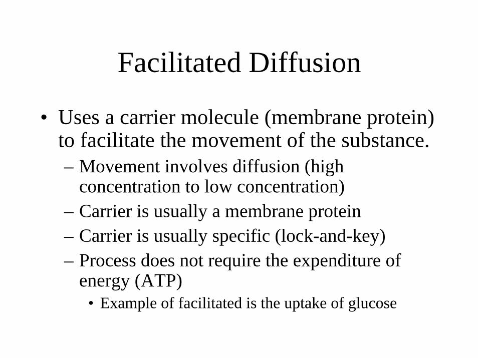
Facilitated Diffusion
• Uses a carrier molecule (membrane protein) to facilitate the movement of the substance.– Movement involves diffusion (high
concentration to low concentration)– Carrier is usually a membrane protein– Carrier is usually specific (lock-and-key)– Process does not require the expenditure of
energy (ATP)• Example of facilitated is the uptake of glucose

Filtration• Process requires a “filtration membrane” which
regulates passage by the size of the membrane’s “pores”
• Process requires a driving pressure such as hydrostatic pressure (blood pressure)– Filtration occurs from high pressure to low pressure
Capillary

Active Transport Mechanisms
• Requires the expenditure of ATP• May move a substance across the membrane against its
concentration gradient (from low to high)• May involve “bulk transport/movement” of materials• Active transport includes
– Solute pumps– Endocytosis
• Pinocytosis• Phagocytosis• Receptor mediated
– Exocytosis

Solute Pumps• Maintain concentration gradients of ions (in
excitable tissues)– Sodium (Na+) is the most abundant extracellular cation– Potassium (K+) is the most abundant intracellular
cation• Movement is against concentration gradient; thus,
sodium is moved out of the cell into the higher extracellular environment, etc.
• Sodium/potassium pump maintains balance between extracellular sodium and intracellular potassium concentrations. Pump is common in excitable tissues, such as neural and muscular tissues

Phagocytosis
• Engulfment of large solid particles by the cell. Often described as “cell eating.”
• Membrane pinches inward and forms phagosomeor “food vacuole.” Lysosomes fuse with phagosome; thus, releasing their digestive enzymes for digestion of the engulfed material.
• White blood cells called macrophages (monocytes) are examples

Pinocytosis(bulk-phase endocytosis)
• Engulfment of extracellular fluidsf• Forms a pinocytotic vesicle. Lysosome
fusion releases digestive enzymes to complete the digestion of materials (solutes) in the fluid.
• Examples are the cells which line the intestines

Receptor-Mediated Endocytosis
• Substance binds to surface receptors• Surface receptors are specific
– Engulfed material and receptors are called a “coated pit”
– Include substances such as insulin and LDLs– Lysosome fusion results in incorporation of
usage of the substance and the receptors are recycled.

Life Cycle of the Cell• Interphase
– Stage in the life of the cell when is not dividing• Mitosis
– Stage in the life of the cell when it is actively dividing. For convenience, divided into four phases
• Prophase• Metaphase• Anaphase• Telophase

Interphase• Cell is undergoing normal life
processes• DNA is duplicated
– Duplication of the chromosome involves separation of the original DNA molecule into two strands. Complementary base pairing reforms original DNA structure
– A duplicated chromosome consists of two chromatids held together by a centromere. (Each chromatid is part “original” and part “new.”) Separation of the chromatidsproduces two chromosomes

Interphase
• Chromosomes replicated• Centrioles replicated• Nucleoli are observed

Cell Division• Mitosis
– Nuclear division which produces two cells, each with identical DNA
• Meiosis– Nuclear division which produces cells which
have a homologous set (n) of the parents chromosomes (2n).
• Cytokinesis– Division of the cytoplasm which produces
daughter cells, each with nuclear material.

Prophase
• Nuclear membrane disappears• Nucleoli disappear• Centrioles form poles• Spindle fibers form• Aster fibers form

Metaphase
• Chromosome alignment a equator

Anaphase
• Chromosome separate• Move toward poles

Telophase
• Begins when chromosome movement stops• Chromosomes take on chromatin appearance• Nucleus reforms• Nucleoli reform• Spindle disappears

Cytokinesis
• Division of cytoplasm produces two daughter cells.
• Cells begin growth and replication of organelles. Mitochondria are self replicating.