study of the effect of ginger and turmeric on osteoporosis
TRANSCRIPT

African J. Biol. Sci., 17 (1): 83-110 (2021)
ISSN 1687-4870 e- ISSN 2314-5501 (online)
www.ajbs.journals.ekb.eg E.mail: [email protected]
Study of the effect of ginger and turmeric on osteoporosis in female rats
Eman G. Mohamed1*
, Zenab M. Mosa1, Samah M. Esmail
1 Adel Bakeer Khloussy
2 , Sahar
O. Ahmed3 and Naglaa A. Abdelkader
4
1-Nutrition and Food Sciences, Home Economics Dept., Fac. of Education. Ain-Shams
2- Pathology Dept., Fac. of Veterinary, Cairo University.
3- Food Technology Research Institute.
4 - Surgery, Anaethesiologh and Radiology Dept., Fac. of Veterinary, Cairo University
*Email; [email protected]
ABSTRACT
Osteoarthritis is the most common form of arthritis, involving inflammation and major
structural changes of the joint, causing pain and functional disability. Pain and stiffness,
particularly after exercise, are the major symptoms, resulting in considerable impact on ability to
perform activities of daily living. There is discordance between symptoms and radiographic
changes, with some sufferers not experiencing symptoms, but showing osteoarthritic changes on
X-ray. The present study was performed to examine the effect of ginger and turmeric
consumption on liver function (ALT, AST), phosphorus, total calcium , ionized calcium, x-ray
and histopathologyon osteoporosis rats induced by prednisone acetate at a dose of 4 mg / kg bw
three time a week for three weeks. On the other hand, the chemical constituent’s moisture,
protein, fat, crude fibre, total digestible nutrients, ash, carbohydrate, phosphorous, calcium was
determined for the tested ginger and turmeric. In addition to, volatile compounds and analysis of
phytochemicals was determined for the tested ginger and turmeric.
This work was carried out on 48 non-pregnant female albino rats (age 6 to 8 weeks and
about 160 to 210g body weight) classified into two main groups. The first main group (6) fed on
basal diet and the second main group (42 rats)injected with prednisone acetate at a dose of 4 mg /
kg bw three time a week for three weeks to cause osteoporosis and divided into seven subgroups
such as each group consists of (6rats). Then fed on basal diet containing 10% -15% ginger, 10%
-15% turmericand10% -15% ginger and turmeric. Results revealed that all osteoporosis groups
administrated with different levels of ginger and turmeric (10-15%) had significant decrease liver
function (ALT, AST), phosphorus, total calcium, ionized calcium comparing with the positive
control group. On the other hand, x-ray and histopathologyof the positive control group after two
months revealed bone loss of different part such as fibula, tibia and femur in addition to bone
demineralization and femoral fracture and fibula bone trabexculae showed dystrophy and
resorption and osteoporosis. These findings revealed that ginger andturmeric treatment
attenuated and treated degrees to osteoporosis in compare to positive control group.
Keywords: Medicinal Herbs, Osteoporosis, Ginger, Turmeric Rhizome, Phytochemicals
Analysis, Biochemical Analysis.
INTRODUCTION
Osteoarthritis (OA) is the most
common form of arthritis, involving
inflammation and major structural changes
of the joint, causing pain and functional
disability. Pain and stiffness, particularly
Received: Dec. 12, 2020; Accepted: Feb. 28, 2021; Available online: March 10, 2021

84
Eman G. Mohamed et al.
after exercise, are the major symptoms,
resulting in considerable impact on ability to
perform activities of daily living. There is
discordance between symptoms and
radiographic changes, with some sufferers
not experiencing symptoms, but showing
osteoarthritic changes on X-ray. It is known
that OA is more common in women than in
men, and the prevalence of OA increases
steeply with age (Busija et al., 2010).
Osteoporosis is worldwide defined as a
systemic skeletal disease characterized by
low bone density and micro architectural
deterioration of bone tissue, which leads to
increased bone fragility and risk of fracture
(Genant et al., 1999; Anbinder et al., 2006).
In healthy rats, both simvastatin and
fenofibrate treatment showed a negative
effect on the trabecular bone located at the
level of femoral diaphysis. These results are
consistent with other studies which
concluded that to a certain extent, statins
inhibit bone resorption and promote bone
formation (Chang et al., 2011; Gradosova et
al., 2011). Glucocorticoids act directly on
bone cells and one of their principal actions
is to reduce osteoblasts function and number
by apoptosis (Chang et al., 2009).The Bax
expression by ostoblasts increase in the
glucocorticoid-induced osteoporosis (GIO)
as showed by (Lucinda et al., 2013). It`s
well known that apoptosis is regulated by an
intrinsic process involving activation of
genes that can promote cell death (Bras et
al., 2005).
Ginger (Zingiber officinale Roscoe,
Zingiberacae) is widely used around the
world in foods as a spice. For cen-turies, it
has been an important ingredient in Chinese
.Ayurvedic and Tibb-Unani herbal
medicines for the treat-ment of catarrh,
rheumatism, nervous diseases, gingivitis
toothache, asthma, stroke, constipation and
diabetes (Awang, 1992; Wang and Wang,
2005; Tapsell et al., 2006). The constituents
of ginger are numerous and vary depending
on the place of origin and whether the
rhizomes are fresh or dry. The odor of
ginger depends mainly on its volatile oil, the
yield of which varies from 1% to 3%. Over
50 components of the oil have been
characterized and these are mainly
monoterpenoids. Some of the oil
components are converted into less odor-
defining compounds on drying (Langner et
al., 1998; Evans, 2004). The pungency of
fresh ginger is due primarily to the
gingeroils, which are a homologous series of
phenols. Ginger and compounds isolated
there from include immuno-modulatory,
anti-tumorigenic, anti-inflammatory, anti-
apoptotic, anti-hyperglycemic, anti-
lipidemic and anti-emetic actions. Ginger is
a strong anti-oxidant substance and may
either mitigate or prevent generation of free
radicals. It is considered a safe herbal
medicine with only few and insignificant
adverse side effects (Badreldin et al., 2008)
Turmeric has anti-inflammatory
(Jurenka, 2009) and anti-cancer (Ravindran
et al.,2009) properties, which have been
mainly attributed to curcumin, a
diarylheptanoid compound. However,
turmeric oil containing ar-turmerone,
turmerone and curlone showed antioxidant
effects and may provide an explanation for
their antimutagenic action (Jayaprakasha et
al., 2002).This turmeric oil also has anti-
bacterial activity (Negi et al., 1999).
Turmeric is a rich source of various volatile
oils, including turmerone, atlantone,
zingiberone, and other constituents such as
sugars, proteins, resins, lignin, salts, resins.
The root contains 10% resin which is a
glucoside (Dulbecco et al., 2014).
Phytochemical studies of turmeric have
shown the presence of curcumin, demethoxy
curcuminbisde methoxy curcumin,
zingiberene, curcumenol, curcumol,
eugenol, tetrahydrocurcumin-
triethylcurcumin, turmerin, turmerones, and
turmeronols. Turmeric is made up three

85
Study of the effect of ginger and turmeric on osteoporosis in female rats
curcuminoids: 75% diferuloyl methane
(also called curcumin), 16% demethoxy
curcumin, and 8% bisdemothy curcumin.
The present data revealed that most of the
therapeutic effects of Turmeric are due to
presence of curcumin. Curcumin is also the
component that gives turmeric its yellow
colour. Curcumin , a polyphenol compound
with a molecular formula C21H20 O6, can
exist in two tautomeric forms: a keto form
(an aldehyde) and a stable enol form (an
alcohol) (Balaji and Chempakam, 2010).
Based on ginger and turmeric data
from white ginger (rhizome root and leaf),
yellow ginger (rhizome, root and leaf) and
turmeric (rhizome and leaf released to the
NCBI database), we selected putative mono-
and sesquiterpene synthases and cloned and
expressed them with GPP and FPP as
substrates in E. coli or yeast. Although many
of these enzymes were found to be insoluble
when expressed in these systems, we were
able to identify the functions for some of
them. We also analyzed why some that are
paralogs produce different products even
though their sequences are very similar
according to protein structural modeling.
Both ginger and turmeric produce α-
zingiberene and β-sesquiphellandrene
However, only turmeric synthesizes α-
turmerone, β-turmerone, which is also
described. astumerone and curlone,
respectively, in some papers (Hiserodt et al.,
1996). Curcuma-containing products
consistently demonstrated statistically
significant improvement in osteoarthritis-
related endpoints compared with placebo,
with one exception. When compared with
active control, curcuma-containing products
were similar to nonsteroidal anti-
inflammatory drugs, and potentially to
glucosamine (Kimberly et al., 2017). The
research aimed at study of effect of ginger
and curcumin on osteoporosis in rats.
MATERIALS AND METHODS
Materials:
The fresh ginger and turmeric
rhizomes were purchased from herbal
market, the ginger and turmeric were dried
and powdered in the air temperature. The
betamethasone (4mg/1kg bw) three times a
week for three weeks was purchased from
Pharmaceutical industries El Obour City- in
Egypt, Dexaglobe Ampoules.
Biological experiment
Animal, housing and diets:
48 non-pregnant female albino rats
(age 6 to 8 weeks and about 160 to 210g
body weight) were obtained from the animal
house in Agriculture Research Center,
Cairo, Egypt were housed (6 rats per cage)
in the animal room under controlled lighting
(12-hour light:12-hour darkness) and
temperature (20°C ± 2°C) conditions and
had free access to laboratory food and tap
water They were kept under normal healthy
conditions and fed on the commercial diet
(Table 1) without any treatment for one
week for acclimatization. Experimental diet
(Table 2) and water were offered ad libitum
all over the experimental period. The first
group of rats, the control (-) fed on
commercial diet for 8 weeks (total period of
experimental). The remained 42 rats were
injected with beta methasone at a dose of 4
mg / kg bw three time a week for three
weeks to cause osteoporosis then divided to7
groups of six rats each (Liao et al., 2003).
The second group after injected fed on
commercial diet (control +). The third and
fourth groups after injected fed on 10 and 15
% of ginger, respectively. Fifth and sixth
groups after injected fed on 10 and 15 % of
turmeric, respectively. Seventh and eighth
groups after injected fed on mixture of 10
and 15% ginger and turmeric, respectively.

86
Eman G. Mohamed et al.
Table (1): Composition of commercial diet.
Ingredients Percentage %
Protein: [soy flour meal+ sun flower meal + gluten] 21.00
Fat 03.26
Crude fibre 03.29
Dl. Methionine 00.40
Vitamins mixed 01.00
Minerals mixed 04.00
Carbohydrates 67.05
Table (2). Composition of Experimental diet as follows: Groups Experimental diets
Frist Commercial diet (control (-) group)
Second Beta methasone + Commercial diet (control (+) group)
Third Beta methasone + commercial diet contain 10% of the ginger.
Fourth Beta methasone + commercial diet contain 15% of the ginger.
Fifth Beta methasone + commercial diet contain 10% of the turmeric.
Sixth Beta methasone + commercial diet contain 15% of the turmeric
Seventh Beta methasone + commercial diet contain10% of ginger and turmeric
Eighth
Nineth:
Beta methasone + commercial diet contain15% of ginger and turmeric
were injected with Beta methasone at a dose of 4 mg / kg bw three time a week to three a week
then slaughtering
At the end of the experiment after 8
weeks the rats mended before slaughtering
and all the blood done from each rat
separately after anesthesia and conduct a
blood centrifuge to get the serum. Blood
samples were withdrawn from orbital
plexus venous by using fine capillary glass
tubes. Blood samples were collected into
plain tubes without anticoagulant and
allowed to clot, Blood samples were
centrifuged at 3000 rpm for 10 min at 4°C,
to obtain clear serum. Blood samples were
centrifuged at 3000 rpm for 10 min at 4°C,
to obtain clear serum. Serum was frozen at
-18oC until analyzed. The animals were
anesthetized with ether and sacrificed.
Liver, kidney, and femoral bone will be
separated from each rat and will be
weighed to calculate the percentage of
increase in organ weight. These organs
were weighed and then kept until
histological investigations.
Methods:
Chemical analysis
Moisture, protein, fat, crude fibre,
total digestible nutrients and ash were
determined according to the method of
AOAC (2007). All determinations were
done in triplicate. Phosphorous, calcium,
atomic absorption spectrophotometer
according to the method of AOAC (1998).
All determinations were done in triplicate.
The carbohydrate contents were
tested quantitatively by the phenol–sulphuric
acid method (Chaplin and Kennedy, 1986).
The absorbance was measured at a
wavelength of 490 nm using UV-Vis
Shimadzu Spectrophotometer (UV-1601
PC).
Volatile compounds Hydro distillation (Extraction of essential oil)
About 100 g of cleaned and dried
plant material was powdered using metal
mortar and placed in a round bottom flask
fitted with condenser hydro distilled for
about 3hrs at atmospheric pressure and
constant temperature. The strongly aromatic
oil was separated from the water layer using

87
Study of the effect of ginger and turmeric on osteoporosis in female rats
diethyl ether and the solvent was removed
by sodium sulfate anhydrous and
concentrated by rotary evaporator.
Phytochemicals Analysis:
Phytochemical analysis for
qualitative detection of alkaloids, tannins,
saponins, flavonoids and phenol was
performed on the powder of ginger
andturmeric rhizome as follows:
Total tannins content in the
lyophilized plant extract was determined by
a modified method of Polshettiwar et al.,
(2007).The total phenolic content was
determined using Folin-Ciocalteau regent
(Mc-Donald et al., 2001). The total
flavonoids in the beverage were determined
using aluminium chloride colorimetric
method (El-Olemy et al., 1994). Alkaloids
and Saponins were determined using method
of Oloyed (2005).
Histopathology Technique
Autopsy samples were taken from
the liver, kidney and femur bone of rats in
different groups and fixed in 10% formol
saline for twenty four hours. The bone was
decalcified by formic acid. Washing was
done in tap water then serial dilutions of
alcohol (methyl, ethyl and absolute ethyl)
were used for dehydration. Specimens were
cleared in xylene and embedded in paraffin
at 56 degree in hot air oven for twenty four
hours. Paraffin bees wax tissue blocks were
prepared for sectioning at 4 microns
thickness by slidge microtome. The obtained
tissue sections were collected on glass
slides, deparaffinized, stained by
hematoxylin and eosin stain for routine
examination through the light electric
microscope (Banchroft et al., 1996).
Scanning X-ray
Determination the right femurs
and LV5 of rats were wrapped with saline-
saturated gauze to maintain their moisture
and stored at 20C. After thawed at room
temperature, the bones were moisturized by
soaking them in saline solution with the
residual muscle removed. The whole
femoral BMD was scanned with Prodigy
Dual-Energy X-ray Absorptiometry scanner
(GE Healthcare, Little Chalfont, UK) to
measure the bone mineral content (BMC,
g/cm2) and bone area (BA, cm
2). The BMD
was calculated as BMC/BA (Bagi et al.,
2011). .
Biological Determination
Biological evaluation of the different
tested diets was carried by determination of
body weight gain% (BWG %) and organs
weight/body weight% according to Chapman
et al. (1959). BWG% = [(Final weight - Initial
weight) / (Initial weight)] X 100
Organ weight/ body weight % = (Organ
weight / Final weight) X 100
Biochemical analysis
Blood samples were withdrawn from
orbital plexus venous by using fine capillary
glass tubes, placed in centrifuge tubes
without anticoagulant and allowed to clot.
After the serum prepared by centrifugation
(3000 rpm for 15 min), serum samples were
analyzed by biodiagnostic kits:
- Alanine aminotransferase (ALT)
activities were determined colori-
metrically using spectrophotometer
(model DU 4700) at 505 nm according
to the method of Reitman and Frankel
(1975).
- Aspartate Aminotransferase (AST)
activities were determined colori-
metrically using spectrophotometer
(model DU 4700) at 540 nm according
to the method of Reitman and Frankel
(1975).
- Phosphorus, inorganic and calcium O-
cpcactivities were determined calori-
metrically using spectrophotometer at

88
Eman G. Mohamed et al.
340 nm according to the method of
Young (1990).
- Calcium O-cpcactivities were
determined calorimetrically using
spectrophotometer (model DU 4700) at
540 nm according to the method of
Young (1990).
RESULTS AND DISCUSSION
According to the WHO Osteoporosis
is a disease characterized by low bone mass
and microarchitectural deterioration of bone
tissues, leading to enhanced fragility and
consequent increase in fracture risk that
results in fractures with minimal trauma.
There is imbalance between bone formation
(osteoblastic activity) and bone resorption
process (osteoclastic activity) due to various
causes such as deficiency of estrogen
hormone as in post-menopausal
osteoporosis, aging and oxidative stress
(Yan Zhang et al., 2007). Ginger (Zingiber
officinale Roscoe) is one of the most
commonly consumed dietary condiments in
the world. The main active phytochemicals
present in ginger are gingerols, shogaols and
paradols, and they have strong antioxidant
and chemopreventive properties (Halvorsen
et al., 2002). The medicinal, chemical and
pharmacological properties of ginger has
been extensively reviewed (Ali et al .,
2008). Ginger extracts have been
extensively studied for a broad range of
biological activities including antibacterial,
anticonvulsant, analgesic, antiulcer, gastric
antisecretory, antitumor, antifungal,
antispasmodic, antithrombotic,
hypocholesterolemic, antiallergic,
antiserotonergic , anticholinergic and other
beneficial activities (Tchombé et al ., 2012).
Turmeric (Curcuma longa) is a
dietary spice belonging to the family
zingiberaceae. It is a coloring and flavouring
agent in foods, and has been reported to
possess antioxidant properties both in vitro
and animal studies. Aqueous extracts of
turmeric showed antioxidant and
antimicrobial activity due to the presence of
curcumin (5%), a polyphenolic compound.
It is known that the phenolic character of
curcumin is responsible for its anti-oxidant
properties (Varunraj et al., 2011). Fresh root
contains good levels of vitamin-C. Other
phytochemicals in turmeric include
tumerone, zingiberene, cineole, d-
phellandrene, d-sabinene, borneol and other
curcuminoids. The majority of the
phytochemicals found in turmeric occur in
the volatile oil that makes up 7 percent of its
weight. Dry rhizomes yield 5.8% essential
oil including sesquiterpene (e.g.
Zingeberene), sesquiterpene alcohols and
ketones, and monoterpenes. Fresh turmeric
contains 0.24% oil containing zingiberene.
The most Curcumin is a known
bacteriostatic agent whereas the essential oil
of turmeric is bactericidal and fungistatic.
The active principle, curcumin is known for
its inhibitory action on micro-organisms
(Niamsa and Sittiwet 2009).
Chemical composition
Results in Table (3) indicated that
the chemical composition of ginger was
9.25%, 4.64%, 70.71%, 9.1%, 6.3%, 5.89%
and 67.60% for protein, fat, Carbohydrates,
moistures, ash, crud fiber and TDN
respectively. On the other hand, the values
of protein, fat, Carbohydrates, moistures,
ash, crud fiber and TDN in turmeric were
7.56%, 3.73%, 72.81%, 12%, 3.9%, 2.95%
and 69.32% respectively. The mean values
of calcium and phosphorous were in ginger
(0.30% and 0.21) while calcium and
phosphorous were in turmeric (0.29% and
0.24%). Many studies have proved that
ginger is endowed with strong antioxidant
(Nirmalaet al., 2008; Nirmala et al., 2012).
Antigenotoxic, antimutagenic and
anticarcinogenic properties both in vitro and
in vivo studies Powdered ginger rhizome
contains 3.6% fatty oil, 9% protein, 60-70%

89
Study of the effect of ginger and turmeric on osteoporosis in female rats
carbohydrates, 3.8% crude fiber, 8% ash,
9-12% water and other terpenes and
terpenoids. Fresh ginger contains 80.9%
moisture, 23% protein, 0.9% fat, 1.2%
minerals, 2.4% fiber, and 12.3%
carbohydrates. Ginger has been shown to be
effective against the growth of both gram-
positive and gram-negative bacteria
including Escherichia coli, Proteus vulgaris,
Salmonella typhi, Staphylococcus aureus
and Streptococcus viridians (Mascolo et al.,
1989).
Table (3): The Chemical composition of Ginger and Turmeric
Chemical composition % Samples
Ginger Turmeric
Protein 9.25 7.56
Fat 4.64 3.73
Carbohydrates 70.71 72.81
Moistures 9.1 12
Ash 6.3 3.9
Crude Fiber 5.89 2.95
TDN 67.60 69.32
Calcium 0.30 0.29
Phosphorous 0.21 0.24
Phytochemicals Phytochemicals are component of
plant foods play an important role in the
treatment of diseases and as a major. The
type and amount of various phytochemical
in ginger and Turmeric presented in Table
(4). The obtained data showed that ginger is
a rich source of alkaloids 9.76% and
Tannins 2.59% as well as Saponins 0.38%
While Flavonoids 4.12 % and total phenolic
0.13% addition of ginger showed high
significant in Alkaloids in all
phytochemicals then Flavonoids. The date
also showed that Turmeric is a rich source of
alkaloids 4.26% and Tannins 2.03% as well
as Saponins6.17% While Flavonoids 2.31%
and total phenolic 3.05% addition of
Turmeric showed high significant in
Saponins in all phytochemicals then
Alkaloids.
Phytochemicals are non-nutritive
plant chemicals that have protective or
disease preventive properties, they are found
generally in plants. The Phytochemical
screening in (Tijjani et al.,2009) study were
agree with our result, who showed that all
leaves contain phenolics, Tannins,
Alkaloids, Saponins, Flavonoids, Steroid
and does not contain phylobatanin, and
tripertenes (Deokar et al., 2016). The
findings revealed that the knowledge of the
antimicrobial activity of the extracts
obtained from ginger can be very useful and
can be applied in different areas of research
such as the pharmaceutical and food
industries Phytochemical constituents such
as steroids, alkaloids, flavonoids, tannins,
phenol and several other aromatic
compounds are secondary metabolites of
plants that serve a defense mechanism
against prediction by many microorganisms,
insects and herbivores. These secondary
metabolites exert antimicrobial activity
through different mechanisms. The alkaloids
contain in plants are used in medicine as
anesthetic agents. Ginger rhizome extract
and their components can be used as
alternative and effective novel therapeutic
strategy.
Turmeric (Curcuma longa) is a
dietary spice coloring and flavouring agent
in foods and has been reported to possess

90
Eman G. Mohamed et al.
antioxidant properties both in vitro and
animal studies. Aqueous extracts of turmeric
showed antioxidant and antimicrobial
activity due to the presence of curcumin
(5%), a polyphenolic compound. It is known
that the phenolic character of curcumin is
responsible for its anti-oxidant properties
(Varunraj et al., 2011). Fresh root contains
good phytochemicals in turmeric include
tumerone, zingiberene, cineole d-
phellandrene, d-sabinene, borneol and other
curcuminoids. The majority of the
phytochemicals found in turmeric occur in
the volatile oil that makes up 7 percent of its
weight. Dry rhizomes yield 5.8% essential
oil including. Fresh turmeric contains 0.24%
oil containing zingiberene. The most
significant curcuminoid is curcumin. It has
been reported that turmeric has an
antimutagenic effect on bacteria in vitro.
Curcumin is a known bacteriostatic agent
whereas the essential oil of turmeric is
bactericidal and fungistatic. The active
principle curcumin is known for its
inhibitory action on micro-organisms
(Virendra et al., 2013).
Table (4): Phytochemical analyses of Ginger and Turmeric. Phytochemicals (%) Ginger Turmeric
Alkaloids 9.76±0.32 4.26±0.28
Tannins 2.59±0.08 2.03±0.19
Saponins 0.38±0.04 6.17±0.25
Flavonoids 4.12±0.17 2.31±0.16
Phenols 0.13±0.05 3.05±0.29
Volatile compounds
The present data given in Tables (5
& 6) indicated the volatile compounds of
ginger and turmeric essential oil. Volatile
compounds of ginger essential oil
(Concentration) as well as values are
expressed as relative area percentage while
(K1) Kovat index on DB5 were analyzed by
compounds identified by GC-MS (MS) and
(KI) of standard compounds run under
similar GCMS conditions. Recorded the 29
volatiles were identified in Concentrationthe
most abundant identified volatile
compounds were Zingiberene which
represent 41.05% then α-Cubebene, which
represent 21.31%respectively. But (KI)
recorded the best result in β-Curcumene
then β-Bisabolene which represent 1519,
1514respectively. Also, Heptanol (0.03 %)
was found in very low amounts.Volatile
compounds of turmeric essential oil
(Concentration) as well as values are
expressed as relative area percentage while
(K1) Kovat index on DB5 were analyzed by
compounds identified by GC-MS because it
was recorded the best result in sensory
evaluation. A total of 17 volatiles were
identified in Concentration the most
abundant identified volatile compounds
were Tumerone which represent 43.87%
then Zingiberenol, which represent
14.69%respectively. While (KI) recorded
the Xanthorhizol then Curcumenol which
represent 1748, 1731respectively.Also
Eudesmol (0.43%) was found in very low
amounts. Shaguftanaz et al. (2010) indicated
that these oils contain volatile substances
which are terpenes and their oxygenated
derivatives usually known as camphor. The
chemical constituents of turmeric rhizomes
include volatiles (ar-tumerone, zingiberene,
turmerone and curlone) and non-volatiles
which are colorings agents and rich source
of phenolics. The aroma of the turmeric is
curcumin and its analogues account for its
bright yellow color. El-Baroty et al. (2010)
indicated that ginger is a characterized oil
(GEO) with high content of sesquiterpene
hydrocarbons, including β-

91
Study of the effect of ginger and turmeric on osteoporosis in female rats
sesquiphellandrene (27.16%), caryophyllene
(15.29) % zingiberene (13.97%), α-
farnesene (10.52%) and ar-curcumin
(6.62%). Fijelu Frank et al. (2013) found
that the major components of essential oil
from Curcuma longa analysed by GC/MS
were ar-turmerone (33.2%), α-turmerone
(23.5%) and β-turmerone (22.7%). The
antifungal activities of the oil were studied
with regard to Aspergillus flavus growth
inhibition.
Table (5). Volatile compounds of ginger essential oil
Volatile compounds KIa
Concentration b
Identification method c
Heptanol 897 0.03 MS, KI, SD
α-Pinene 936 0.88 MS, KI
Camphene 941 3.28 MS, KI
2-Methyl nonane 967 0.63 MS, KI, SD
Myrcene 971 1.82 MS, KI
Pinene 974 0.05 MS, KI
α-Phellandrene 982 0.05 MS, KI
Limonene 1013 0.04 MS, KI, SD
β-Phellandrene 1017 4.43 MS, KI
1,8-ceneole 1031 2.16 MS, KI
α-Terpinolene 1062 0.08 MS, K
n-Nonenal 1129 0.58 MS, KI, SD
2-Methyl undecane 1165 0.16 MS, KI
3-Methyl butanol 1198 18.94 MS, KI
α-Cubebene 1345 21.31 MS, KI, SD
p-Menth-1-en-8-ol acetate 1349 1.25 MS, KI
Geranyl acetate 1381 0.07 MS, KI
Methyl eugenol 1402 0.06 MS, KI
Geranyl propionate 1428 0.27 MS, KI
α-Farnesene 1432 0.04 MS, KI, SD
γ-Elemene 1439 0.04 MS, KI
Neryl acetone 1442 0.27 MS, KI
Germacrene D 1457 1.90 MS, KI
Zingiberene 1492 41.05 MS, KI
Valencene 1498 0.05 MS, KI
Citronellyl n-butyrate 1503 0.04 MS, KI
α-Bisabolene 1509 0.09 MS, KI
β-Bisabolene 1514 0.05 MS, K
β-Curcumene 1519 0.17 MS, KI a: Kovat indices;
b:Values are expressed as relative area percentage;
c:compounds identified by
GC-MS (MS) and / or Kovat index on DB5 (KI) and / or by comparison of MS and KI of
standard compounds run under similar GCMS conditions.

92
Eman G. Mohamed et al.
Table (6). Volatile compounds of turmeric essential oil Volatile compounds KI
a Concentration
b Identification method
c
Camphene 958 1.26 MS, KI, SD
-Fenchene 961 1.24 MS, KI
Pinene 976 1.69 MS, KI
α –phellanderene 983 1.82 MS, KI, SD
3-Thujene 992 3.03 MS, KI
Zingiberenol 1612 14.69 MS, KI
Turmerone 1629 8.00 MS, KI
Tumerone 1651 43.87 MS, KI, SD
Eudesmol 1658 0.43 MS, KI
Atlantone 1667 1.12 MS, KI
Eudesmol 1669 0.79 MS, KI
Curcumenone 1672 3.55 MS, KI, SD
Germacrone 1688 5.04 MS, KI
ZZ-Farnesol 1691 0.60 MS, KI
ZE-Farnesol 1702 2.90 MS, KI, SD
Curcumenol 1731 9.19 MS, KI
Xanthorhizol 1748 0.77 MS, KI a: Kovat indices;
b:Values are expressed as relative area percentage;
c:compounds identified by GC-MS (MS) and /
or Kovat index on DB5 (KI) and / or by comparison of MS and KI of standard compounds run under similar GCMS
conditions.
Biological Determination
Data in Table (7) indicated that the two
groups of rats fed on 10% and 15% turmeric and
ginger, respectively showed increase in their
body weight gain comparing with the positive
control group. The body weight gain was higher
in group (4) with level 15% ginger
(57.261±8.733), while it was lower in group (5)
that fed on basal diet of 10% turmeric
(30.077±10.59) comparing with the positive
control group. This result was in agreement with
that obtained by Saber Sakr et al. (2011).
Many studies were carried out on ginger
and its pungent constituents, fresh and dried
rhizome. Among the pharmacological effects
demonstrated is anti-platelet, antioxidant,
anti-tumour, anti- rhinoviral, anti-
hepatotoxicity and anti arthritic effect
(Fisher-Rasmussen et al., 1991; Sharma et
al., 1994; Kamtchouing et al., 2002). Ginger
was found to have hypocholesterolaemic
effects and cause decrease in body weight,
blood glucose, serum total cholesterol and
serum alkaline phosphatase in adult male
rats.
Table (7): Effect of ginger and turmeric on weight gains of rats suffering from osteoporosis. Parameters
Groups body weight gain % Initial weight (g) Final weight (g)
24.866c±7.391 177.677
a±13.571 221.833
c±110.241 Group (1):negative control
39.011b±7.192 177.333
a±12.711 246.53
b±17.833 Group (2): positive control
48.211ab
±9.211 177.333a±12.471 262.833
a±18.311 Group (3): 10% ginger
57.261a±8.733 176.672
b±11.691 277.833
a±22.162 Group(4): 15% ginger
30.077c±10.59 176.833
ab±9.831 230
c±114.291 Group (5): 10% turmeric
39.112b±11.377 176.831
ab±10.311 246
b±122.652 Group (6): 15% turmeric
34.461bc
±11.611 177a±8.292 238
bc±117.281 Group (7): 10% ginger and turmeric
53.911a±12.477 176.833
ab±8.492 272.172
a±14.591 Group (8): 15% ginger and turmeric
0.297 0.119 0.305 ANOVA ( F)
* * * Sig.
- Values are expressed as mean ± SD. - Significant at p<0.05 using one way ANOVA test.
- Values which have different letters in each column differ significantly, while those with have similar or partially
are not significant.

93
Study of the effect of ginger and turmeric on osteoporosis in female rats
The results in Table (8) indicated the
effect of ginger and turmeric on relative
organs weight of rats suffering from
osteoporosis. The mean values of relative
weights of liver and kidney in positive
control group was lower than the all
experimental groups. But the mean values of
liver and kidney for rats in group (6) were
higher than those of the positive control
group (8.491±1.552g, 5.342±0.591g and
1.728±0.165g, 0.957±0.746g, respectively).
On the other hand, the mean values of
relative weights of femoral bones in positive
control group was similar to group (6)
(3.917±0.493g and 3.962±1.162g) while the
positive control group in relative weights of
femoral bones was lower than group (8)
(3.917±0.493g and 6.72±1.101g) the results
agreed with that obtained by (Stanley
Iheanacho et al., 2017).
Table (8): Effect of ginger and turmeric on relative organs weight of rats suffering from
osteoporosis. Parameters
Groups OW/ BWG Relative weights of
femoral bones (g)
OW/ BWG Relative
weights of
kidney(g)
OW/BWG Relative
weights of
liver(g)
1.609b±4.641 3.57c±0.828 0.565b±5.115 1.255bc±0.16 2.648b±4.837 5.881c±0.551 Group (1):negative control
1.589b±4.737 3.917bc±0.493 0.388c±4.558 0.957c±0.746 2.164c±4.668 5.342c±0.591 Group (2):positive control
1.698b±5.819 4.462b±0.981 0.529b±6.347 1.392b±0.235 2.192c±5.798 5.766c±1.011 Group (3):10% ginger
1.755b±5.704 4.875b±0.666 0.503b±6.047 1.397b±0.181 2.449b±5.848 6.811ab±0.462 Group(4):15% ginger
2.921a±7.077 6.718a±0.582 0.723a±7.324 1.664a±0.233 2.814ab±6.81 6.471b±0.952 Group (5):10% turmeric
1.611b±7.223 3.962bc±1.162 0.702a±7.928 1.728a±0.165 3.450a±6.947 8.491a±1.552 Group (6):15% turmeric
2.785a±8.049
6.628a±0.227 0.587ab±8.057
1.396b±0.216 2.808ab±7.89
6.681b±0.441 Group (7):10% ginger and
turmeric
2.469ab±8.044
6.72a±1.101 0.601a±8.700 1.635a±0.173 2.424b±8.546
6.592b±0.391 Group (8):15% ginger and turmeric
0.053
0.017
0.354
0.169
0.436
0.049
ANOVA ( F)
** ** * * * ** Sig.
- Values are expressed as mean ± SD. - Significant at p<0.05 using one way ANOVA test.
- Values which have different letters in each column differ significantly, while those with have similar or partially
are not significant.
From the data shown in Table (9), it
could be observed that the level of ALT in
group (7) fed on 10% ginger and turmeric
was lower than the positive control group
(7.4±5.639 U/L, 13.667±4.589U/L) The
results agreed with that obtained by Uma
Bhandari et al. (2003). While the mean level
of AST in group (8) fed on 15% ginger and
turmeric was lower than the positive control
group (14.2±3.421 U/L, 18.5±1.225 U/L).
The results agreed with that obtained by
(Stanley et al., 2017). On the contrary, the
mean value of P was similar in the
experimental group except group (7) fed on
10% ginger and turmeric was higher than
the positive control group (5.367±0.907,
5.2±0.1). Treating animals with water
extract of ginger and adriamycin led to
an improvement in the histological
changes induced by adriamycin together
with significant decrease in ALT and AST
activity. Moreover, ginger reduced the level
of malondialdehyde and increased the
activity of superoxide dismutase. The
results of the present work indicated that
ginger had protective effect against liver
damage induced by adriamycin and this is
due to its antioxidant activities.(Saber et al .,
2011).

94
Eman G. Mohamed et al.
Table (9): Effect of ginger and turmeric on liver function and phosphorusof rats suffering from
osteoporosis. Parameters
Groups P
AST
U/L ALT U/L
4.8a±0.3 21.4a±2.191 9.2b±7.120 Group (1):negative control
5.2a±0.1 18.5a±1.225 13.667a±4.589 Group (2): positive control
4.733b±0.153 18a±1.549 10.833b±3.601 Group (3):10% ginger
4.767ab±0.115 17ab±1.549 13.833ab±3.764 Group(4):15% ginger
4.867a±0.289 14.5b±5.282 8.833c±4.997 Group (5):10% turmeric
4.633b±0.153 16b±3.0 14.2a±4.087 Group (6):15% turmeric
5.367a±0.907 14.4b±4.979 7.4c±5.639 Group (7):10% ginger and turmeric
4.667b±0.115 14.2b±3.421 9.8b±4.919 Group (8): 15% ginger and turmeric
0.566 0.163 0.834 ANOVA ( F)
* * Sig.
- Values are expressed as mean ± SD. - Significant at p<0.05 using one way ANOVA test.
- Values which have different letters in each column differ significantly, while those with have similar or partially
are not significant.
The results in Table (10) indicated
that the mean values of the total Ca in
positive control group was higher than that
in group (4) fed on15% ginger (8±1.082 and
6.967±0.462, respectively), and Ca++ in
positive control group was higher than that
in group (3) fed on10% ginger (1.288a±
0.081 and 1.236±0.021, respectively). On
the other hand, total Ca and Ca++ levels in
all experimental groups fed on ginger and
turmeric were significantly less than that in
positive control group. On the contrary, the
mean value of Ca/Ca++
in the positive
control group was significantly less than the
all experimental groups fed on ginger and
turmeric. In healthy rats, both simvastatin
and fenofibrate treatment showed a negative
effect on the trabecular bone located at the
level of femoral diaphysis. These results are
consistent with other studies which
concluded that to a certain extent, statins
inhibit bone resorption and promote bone
formation, but have no significant effect on
bone mineral density in healthy rats (Chang
et al., 2011; Gradosova et al., 2011).
Effects of extra-skeletal estrogen
deficiency are mainly based upon increased
renal calcium excretion and decreased
intestinal calcium absorption (Khosla et al.,
1997). Estrogen plays an important role in
calcium absorption in the gut (Gennari et al.,
1990) and its reabsorption in the kidney
(McKane et al., 1995). The presence of
estrogen receptors in the intestine has been
reported and has been shown to increase
intestinal calcium absorption in both rats and
humans. Curcumin (Diferuloylmethane, 1,
7- bis (4-Hydroxy-3-methoxyphenyl)-1, 6-
heptadiene-3, 5 - dione) is the active
polyphenolic compound extracted from the
rhizomes of turmeric (Curcuma longa L.,
Zingiberaceae), grown in tropical Southeast
Asia (Jagetia and Aggarwal 2007; Padhye et
al., 2010). Some studies demonstrated the
efficacy of turmeric extracts in the
prevention of bone loss in animal models of
rheumatoid arthritis and postmenopausal
osteoporosis (Wright et al., 2010). It was
found in vitro investigations that the anti-
inflammatory effects of curcumin prevent
osteoclast differentiation (Bharti et al.,
2004; von Metzler et al., 2009). Thus,
curcumin produces beneficial changes in
bone turnover and increase in bone strength
using the ovary ectomized mature rat model
of postmenopausal osteoporosis (Houet et
al., 2016).

95
Study of the effect of ginger and turmeric on osteoporosis in female rats
Table (10): Effect of ginger and turmeric on total and ionized calcium of rats suffering from
osteoporosis. Parameters
Groups Ca/Ca++
Ca++
Total Ca
6.436a±1.519 1.323
a±0.049 8.567
a±2.369 Group (1):negative control
2.999c±3.295 1.288
a±0.081 8
a± 1.082 Group (2):positive control
6.313a±0.709 1.236
b±0.021 7.8
a±0.781 Group (3):10% ginger
5.488b±0.407 1.27
a±0.017 6.967
b±0.462 Group(4):15% ginger
6.187ab
±0.691 1.263ab
±0.046 7.8a±0.693 Group (5):10% turmeric
5.716b±0.902 1.25
b±0.026 7.133
b±1.012 Group (6):15% turmeric
6.173ab
±0.957 1.253b±0.0321 7.733
ab±1.168 Group (7):10% ginger and turmeric
6.205a±1.044 1.24
b±0.0458 7.667
b±1.026 Group (8):15% ginger and turmeric
0.588 0.176 0.168 ANOVA ( F)
* * * Sig.
- Values are expressed as mean ± SD. - Significant at p<0.05 using one way ANOVA test.
- Values which have different letters in each column differ significantly, while those with have similar or partially
are not significant.
X-ray and Histopathology
In the present study; radiographic
imaging of the negative control group
radiographic view showed normal
radiographic finding of tibia and distal
extermity of femur.and showing normal
histological structure of the periosteium,
compact shaft of long bone and bone
trabeculae with bone marrow in between
(Figs. 1,2, 3).
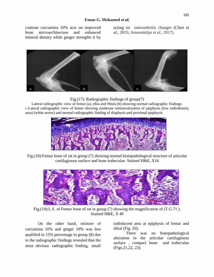
Fig.(1): Radiographic findings of group (1). a- Lateral radiographic view showed Normalradiographic finding of femur.
b-Lateral radiographic view showed Normalradiographic finding of tibia and disital extermity of femur .
Fig.(2): L.S. of Femur bone of rat in group (1) showing normal histological structure of the
periosteium, compact shaft of long bone and bone trabeculae with bone marrow in between.
Stained Hx.E, X40.
a b

96
Eman G. Mohamed et al.
b a
Fig.(3): L.S. of Femur bone of rat in group(1) showing normal histological structure of the
cartilaginous articular surface of the condyle. Stained Hx.E, X40.
In the present study; radiographic
imaging of the positive control group after
glucocorticoids (GC) administration for
three weeks revealed bone loss of different
part such as fibula, tibia and femur in
addition to bone demineralization and
thinning of femoral cortex in rats, disappear
of cortex of tibia in others and fibula and
these agree with the observations of
Hallberg et al. (2009), Weinstein (2001) and
Sipos et al. (2015). Osteoporosis is
characterized as a reduction in bone mass
and an impairment of bone architecture
resulting a bone thinning with direct effects
on increased cortical porosity, bone fragility
and fracture risk. GC therapy is the most
common cause of osteoporosis, leading to
osteonecrosis of the femoral head and
fractures, which may also be associated with
fracture-related morbidity and a decreased
quality of life.
Radiographic findings of group (2)
show demineralization of tibia and bone loss
of proximal part of fibula (Fig 4).
Histopathologically, there were bone
trabeculae osteoporosis and resorption
associated with dystrophy of the articular
cartilaginous surface and congestion of the
blood vessels, these findings were confirmed
the radiological findings (Figs. 5, 6).
Fig.(4): Radiographic findings of group (2).
a. Lateral radiographic view of tibia and fibula showing demineralization of tibia (red
arrow) and bone loss of proximal part of fibula (white arrow).
b.Lateral radiographic view of femur and pelvic bone showing demineralization and partial
bone loss of ilium and femoral head fracture (white arrow).

97
Study of the effect of ginger and turmeric on osteoporosis in female rats
a b c
Fig.(5):L.S. of Femur bone of rat in group (2) showing osteoporosis and resorption or the bone
trabeculae. Stained H&E, X16.
Fig.(6):L.S. of Femur bone of rat in group (2) showing cartilaginous dystrophy of the articular
surface with congestion of the blood vessels. H&E, X16.
Radiologically in treated group by
ginger 10%in group (3) after two months
revealed mineralization improvement of
bony loss and demineralized area as
following remineralization at proximal
extremity of femur and normal architecture
without disruption at distal extremity of
femur, rebuilding of fibula with
mineralization in compare to untreated
group and Remineralization Bridge at tibia
with different degree of callus formation but
still there was thinning in fibula (Fig. 7).
Histopathologically, there was no
histopathological alteration (Figs.,8, 9).
Fig. (7):Radiographic findings of group (3) a-Lateral radiographic view of tibia, fibula and disital extremity of femur showing normal radiographic
findings and architecture (thinning in fibula) Remineralization Bridge at tibia
b-Lateral radiographic view of femur showing remineralization at proximal extremity (black arrow) and
normal architecture without disruption at distal extremity
c-Lateral radiographic view of tibia showing remineralization and small callus formation at lost bony part
(white arrow).

98
Eman G. Mohamed et al.
Fig.(8):L.S. of Femur bone of rat in group (3) showing normal histopathological structure of
articular cartilaginous surface and under lying coupaet bone. Stained H&E, X 16.
Fig.(9):Femur bone of rat in group (3) showing in fact normal histopathological structure of bone
trabeculae. Stained H&E, X 16.
In parallel to it the treated group by
ginger 15% in group (4) after two months
revealed continuation of rebuilding of
different demineralized parts such as
difficult notification of remineralization and
callus formation over epiphyseal fracture,
traces of callus formation at tibial lost bony
part, normal radiographic findings of fibula
and femoral discontinuation of proximal
cortical surface, and disital part but there
was small callus formation at femoral lost
bony part at distal epiphysis which
confirmed histopathologically resorption of
the bone trabeculae was detected. These
findings revealed that ginger has important
role in remineralization and improvement of
osteoprotic changes of tibia, fibula and
femur especially at extremities of femur and
tibia (articular surface) due to ginger has
anti inflammatory effect in osteoarthritis
cases in agree with Baer et al. (2005) and
Amorndoljai et al. (2017) who indicated that
ginger has been used for time immemorial
for treatment of rheumatic disorder due to its
anti-inflammatory properties and ability to
inhibit arachidonic acid metabolism. It
relieve joint pain and improve problematic
symptoms and the quality of life in knee
patients, it is effective as 1% Diclofenac gel
and it can be considered as complementary
therapy in patients with osteoarthritis of
knee, in addition to it reduce the risk of
systemic toxic (Figs. 10, 11).

99
Study of the effect of ginger and turmeric on osteoporosis in female rats
Fig.(10):Radiographic findings of group (4) a-Lateral radiographic view of tibia showing remineralization and bridge formation over epiphyseal fracture (black
arrow) and normal radiographic findings of fibula
b-Lateral radiographic view of femur showing discontinuation with small callus formation at lost bony part at distal
epiphysis (white arrow)and discontinuation of proximal cortical surface (white arrow).
Fig.(11):L.S. of Femur bone of rat in group (4) showing bone resorption of the trabeculae.
Stained H&E, X 16.
Radiologically in treated group by
curcumina 10% in group(5) after two
months revealed good mineralization
improvement of bony loss and rebuilding
bone process as following no radiographic
findings of tibia, fibula, proximal femur and
still there were moderate remineralization of
tibial epiphysis and bridged callus formation
at oblique epiphyseal fracture of tibia (Fig.
12), Histopathologically the bone trabeculae
showed dystrophy and resorption.
these findings revealed that curcumin
treatment attenuated and treated degrees to
osteoporosis induced by GC in compare to
untreated group in agree with (Yang et al.,
2011; Chen et al., 2015) who demonstrated
that curcumin improved bone
microarchitecture and enhanced mineral
density in APP/PS1 transgenic mice, and it
attenuated GIOP by inhibiting
osteocyticapoptosis (Fig.13).
a b

100
Eman G. Mohamed et al.
Fig.(12):Radiographic findings of group (5)
a. Lateral radiographic view of tibia and fibula showing moderate remineralization of epiphysis
and normal radiographic finding of fibula
b.Lateral radiographic view of femur showing discontinuation of femoral cortex
c. Lateral radiographic view of femur, tibia and fibula showing normal radiographic findings of
femur, fibula and bridged callus formation at oblique epiphyseal fracture of tibia (white arrow) .
Fig.(13):Femur bone of rat in group (5) showing dystrophy and resorption of the bone
trabeculae. Stained H&E, X 16.
While in the treated group by
curcumina 15%in group (6) after two
months revealed some repairs to damaged
bony parts as normal radiographic findings
of femur (Fig. 14). There was no
histopathological alteration in the articular
cartilaginous surface and bone treabeculae
and thick radiopaque tibial cortex but there
were also bony loss of proximal part of
fibula, in complete remineralization of fibula
and discontinuation of femoral cortex, these
findings in compare to untreated group
revealed that the curcumina 15% stimulate
rebuilding process (Yang et al., 2011; Chen
et al., 2015) but not as curcumina 10%, the
curcumina 10% was more effective (Figs.
15,16).
a b c

101
Study of the effect of ginger and turmeric on osteoporosis in female rats
Fig.(14):Radiographic findings of group (6)
a. Lateral radiographic view of femur, tibia and fibula showing normal radiographic
findings of femur and incomplete remineralization of fibula (white arrow).
b. Lateral radiographic view of tibia and fibula showing bony loss of proximal part of
fibula (still incomplete remineralization).
Fig.(15):L.S. of Femur bone of rat in group (6) showing normal histopathological structure of
articular cartilaginous surface and treabeculae. Stained H&E, X 16.
Fig.(16): L.S. of Femur bone of rat in group(6) showing the magnification of (T.G.65).
Stained H&E, X40.
In the present study, the radiological
findings of treated group by curcumina 10%
and ginger 10% in group (7) indicated
normal radiographic findings of femur, tibia
and fibula (Fig. 17). Also, there was no
histopathological alteration (Figs. 18, 19).
Along with moderate
remineralization of tibial and femoral
epiphysis. These findings revealed that the
mixture of both curcumina 10% and ginger
10% has more compelling effect against
osteoperotic changes than each one alone, in
a b
102
Eman G. Mohamed et al.
contrast curcumina 10% acts on improved
bone microarchitecture and enhanced
mineral density while ginger strengths it by
acting on osteoarthritis changes (Chen et
al., 2015; Amorndoljai et al., 2017).
Fig.(17): Radiographic findings of group(7) Lateral radiographic view of femur (a), tibia and fibula (b) showing normal radiographic findings.
c-Lateral radiographic view of femur showing moderate remineralization of epiphysis (low radiodensity
area) (white arrow) and normal radiographic finding of diaphysis and proximal epiphysis
Fig.(18):Femur bone of rat in group (7) showing normal histopathological structure of articular
cartilaginous surface and bone trabeculae. Stained H&E, X16
Fig.(19):L.S. of Femur bone of rat in group (7) showing the magnification of (T.G.71 ).
Stained H&E, X 40
On the other hand, mixture of
curcumina 10% and ginger 10% was less
qualified in 15% percentage in group (8) due
to the radiographic findings revealed that the
most obvious radiographic finding, small
radiolucent area at epiphysis of femur and
tibial (Fig. 20).
There was no histopathological
alteration in the articular cartilaginous
surface , compact bone and trabeculae
(Figs.21,22, 23).
a b c

103
Study of the effect of ginger and turmeric on osteoporosis in female rats
a b
Fig.(20): Radiographic findings of group (8). a and b: Lateral radiographic view of femur, tibia
and fibula showing normal radiographic findings.
Fig.(21): L.S. Femur bone of rat in group (8) showing the magnification of (T.G.74 ) to identify
the bone trabeculae. Stained H&E, X40
Fig.(22): L.S of Femur bone of rat in group (8) showing the magnification of (T.G.74 ) to
identify the in fact compact bone. Stained H&E, X80
Fig.(23): L.S. Femur bone of rat in group (8) showing the magnification of (T.G.74 ) to identify
the in fact bone cartilaginous structure. Stained H&E, X40.

104
Eman G. Mohamed et al.
On the other hand, the radiological
and histological findings of the group (9)
bone loss of different part such as fibula,
tibia and femur in addition to bone
demineralization and femoral fracture and
fibula bone trabexculae showed dystrophy
and resorption and osteoporosis (Figs. 24,
25, 26). This agrees with the findings of
Henneicke et al. (2011) and Sipos, et
al.(2015) who found that using of oral
corticosteroids is associated with serious
side effects, including osteoporosis and
consequently an increase in fractures.
Fig.(24): Radiographic findings of group (9). a. Lateral radiographic view of femur and tibia showing fractured distal extremities of femur
(white arrow) and demineralization and loss of proximal part of fibula (white arrow).
b. Lateral radiographic view of tibia and fibula showing bony loss of fibula (white arrow).
Fig.(25): L.S. of Femur bone of rat in group (9) showing zesorption,dystrophy and osteoporosis
of the bone trabeuclae. Stained H&E, X16
Fig.(26): L.S. of Femur bone of rat in group (9) showing resorption , dystrophy and osteoporosis
of the bone trabeuclae. Stained H&E,X16
b a

105
Study of the effect of ginger and turmeric on osteoporosis in female rats
Conclusion
In the present study; Osteoporosis is
characterized as a reduction in bone mass
and an impairment of bone architecture
resulting a bone thinning with direct effects
on increased cortical porosity, bone fragility
and fracture risk. GC therapy is the most
common cause of osteoporosis, leading to
osteonecrosis of the femoral head and
fractures, which may also be associated with
fracture-related morbidity and a decreased
quality of life. ginger has important role in
remineralization and improvement of
osteoprotic changes of tibia, fibula and
femur especially at extremities of femur and
tibia (articular surface) due to ginger has
anti-inflammatory effect in osteoarthritis
cases.And also these findings revealed that
curcumin treatment attenuated and treated
degrees to osteoporosis induced by GC in
compare to untreated group. Also findings
revealed that the mixture of both curcumina
and ginger has more compelling effect
against osteoperotic changes than each one
alone.
REFERENCES
Ali, B.H.; Blunden, G.; Tanira, M.O. and
Nemmar, A. (2008). Some
phytochemical pharmacological and
toxicological properties of ginger
(Zingiberofficinale Roscoe): a
review of recent research. Food
Chem. Toxicol., 20-409: (2)46.
Anbinder, A.L.; Prado, M.A.; Spalding, M.;
Balducci, I; Carvalho, Y.R. and
Rocha, R.F. (2006). Estrogen
Deficiency and Periodontal
Conditionin Rats- A Radiographic
and Macroscopic Study. Braz. Dent.
J., 17(3): 201-207.
Amorndoljai, P.; Taneepanichskul, S.;
Niempoog, S. and Nimmannit, U.
(2017). A Comparative of Ginger
Extract in Nanostructure Lipid
Carrier (NLC) and 1% Diclofenac
Gel for Treatment of Knee
Osteoarthritis (OA). J. Med. Assoc.
Thai., 100(4): 447-456.
AOAC (1998). Official methods of Analysis
of the Association of Official
Analytical Chemists, 20th
ed.
AOAC. (2007). Official Methods of
Analysis, 18th
Ed. Association of
Official Analytical chemists,
Gaithersburg, MD, USA.
Awang, D.V.C. (1992). Ginger. Can. Pharm.
J., 125:309–311.
Badreldin, H. Ali.; G. B.; Musbah, O.T. and
Abderrahim N. (2008). Some
phytochemical, pharmacological and
toxicological properties of ginger
(Zingiber officinale Roscoe): A
review of recent research. Food
Chem. Toxicol., 46:409–420.
Baer, P.A.; Thomas, L.M. and Shainhouse,
Z. (2005). Treatment of osteoarthritis
of the knee with a topical diclofenac
solution: a randomised controlled,
6week trial [ISRCTN53366886].
BMC Musculos kelet Disord, 6: 44.
Balaji, S. and Chempakam, B.
(2010).Toxicity prediction of
compounds from turmeric (Curcuma
longa L). Food Chem. Toxicol.,
48(10):2951-2959.
Banchroft , J.D.; Stevens, A. and Turner,
D.R. (1996).Theory and Practice of
Histological Techniques. Fourth Ed.
Churchil Livingstone ,New York ,
London , San Francisco , Tokyo.
Bharti, A.C. and Takada, Y. B.B. (2004).
Aggarwal Curcumin
(diferuloylmethane) inhibits receptor
activator of NF-kappa B ligand-
induced NF-kappa B activation in
osteoclast precursors and suppresses

106
Eman G. Mohamed et al.
osteoclastogenesis. J. Immunol.,
172(10): 5940-7.
Bras, M.; Queenan, B. and Susin, S.
(2005).Programmed cell death via
mitochondria: different modes of
dying. Biochemistry, 70: 231-239.
Bagi, C.M.; Berryman, E.and Moalli, M.R.
(2011). Comparative bone anatomy
of commonly used laboratory
animals: implications for drug
discovery. Comp Med 61:76–85
Busija, L.; Bridgett, L.; Williams, S.R., et
al. (2010). Osteoarthritis. Best. Pract.
Res. Clin. Rheumatol., 24:757–68.
Chang, B.; Yang, J.; Li, H.; Lu, S.; Chen, L.;
Fang, P.(2011). Effects
ofatorvastatin on bone metabolism
and bone mineral density in Wistar
rats, Pharmazie, 2011, 66(7), 535-7.
Chaplin, M.F and Kennedy,J.F .(1986).
Monosaccharides. In: Chaplin, M.F.,
Kennedy, J.F( Eds.), Carbohydrate
Analysis A Practical Approach. The
Practical Approach Series. Oxford
University Press, Oxford, pp. 1–42.
Chen, Z.; Xue, J.; Shen, T.; Ba, G.; Yu,
D.;Fu, Q.(2015).Curcumin alleviates
glucocorticoid-induced osteoporosis
by protecting osteoblasts from
apoptosis in vivo and in vitro. Clin.
Exp. Pharmacol. Physiol., 30: pages
268-276.
Chapman, D.G.; Castilla, R. ; Campbell,
J.A. (1959). Evalution of protein in
food. I- A method for the
determination of protein efficiency
ratio. Can. J Biochem. Physiol., 37:
679 – 686.
Deokar, S.B.; Pawar R.M.; Tambe, A.R.
(2016). Evaluation of a ntimicrobial
a ctivity and phytochemical a nalysis
of Zingiber officinale (Ginger)
rhizome extract. IJAPRR, 3(4):1-9.
Dulbecco, P., and Savarino, V. (2014)
.Therapeutic potential of curcumin in
digestive diseases. World journal of
gastroenterology: WJG, 19(48):
9256.
El-Baroty, G. S.; Abd El-Baky, H.H.;
Farag, R.S. and M.A. Saleh (2010).
Characterization of antioxidant and
antimicrobial compounds of
cinnamon and ginger essential oils.
Afr. J. Biochem. Res., 4(6):167-174.
at
http://www.academicjournals.org/AJ
BR.
El-Olemy, M.M.; Farid, J.A. and Abdel-
Fattah, A.A. (1994). Ethanol Extract
of P. stratiotes. NISEB J. 1(1): 51-
59.
Evans, M. L.; Dick, M. J.; Lewallen, L. P.;
& Jeffrey, C. (2004). Modified
breastfeed-ing attrition prediction
tool: Prenatal and postpartum tests.
Journal of Perinatal Education, 13, 1-
8. doi:10.1624/105812404X109348.
Fisher-Rasmussen, W.; Kjaer, S.K.; Dahl,
C.; Asping, U. (1991). Ginger
treatment of Hyperemesis
gravidarm. Eur. J. Obstet.
Gynecol. Rep. Biol., 38(1):19-24.
Fijelu, F.; Yanshun, X.; Qixing, J. and
Wenshui, X. (2013). Protective
effects of garlic (Allium sativum) and
ginger (Zingiber officinale) on
physicochemical and microbial
attributes of liquid smoked silver
carp (Hypophthalmichthys molitrix)
wrapped in aluminium foil during
chilled storage. Afr. J. Food Sci.,
52:302-310.DOI:
10.5897/AJFS2013.1030.
Genant, H.K.; Cooper, C.; Poor, R.; Reid, I.;
Ehilich ,G.; Kanis, J., et al. (1999).
Interim report and recommendations
of the World Health Organization
task-force for osteoporosis.
Ostoporos Int.,10:259-264.
Gennari, C.; Agnusde, D.; Nardm P. and
Civitell, R. (1990). Estrogen
preserves a normal intestinal

107
Study of the effect of ginger and turmeric on osteoporosis in female rats
responsiveness to 1,25-dihydroxy
vitamin D3 in oophorectomized
women. J. Clin. Endocrinol. Metab.,
71(5): 1288-1293.
Gradosova, I.; Zivna, H.; Svejkovska, K.;
Palicka,V.; Pichy, A. and Zivny, P.
(2011).The role of atorvastatin in
bone metabolism in male albino
Wistar rats. Pharmazie, 66(8): 606-
10.
Halvorsen, B.L.; Holte, K.; Myhrstad, M.C.;
Barikino, I.; Hvattum, E.; Remberg,
S.F.; Wold, A.B.; Haffner, A.
(2002).A systemic screening of total
antioxidants in dietary plants. J.
Nutr., 2002; 132:461-471.
Hiserodt, R..; Thomas, G.H.; Chi-Tang, H.
and Robert, T.R.(1996).
Characterization of powdered
turmeric by liquid chromatography
mass spectrometry and gas
chromatography mass spectrometry.
J. Chromatogr., 740(1):51-63.
Hou, M.; Song, Y.; Li, Z.; Luo, C.; Ou J.-S.;
Yu H.; Yan, J.; Lu, L.(2016).
Curcumin attenuates osteogenic
differentiation and calcification of rat
vascular smooth muscle cells. Mol.
Cell Biochem., 420 (1-2):151-60 .
Hallberg, I.; Bachrach-Lindström, M.;
Hammerby, S.; Toss, G .; Ek,
A.C.(2009). Health-related quality of
life after vertebral or hip fracture: a
seven-year follow-up study. BMC
Musculoskelet. Disord., 10: 135,
2009.
HenneickeHolger, M.; Robert, K.; Tara,
C.B.; Uta, H.; Nicky, B.; Robert,
E.D.; Dörte, H.; Frank, B.; Colin,
R.D.; and Hong, Z.( 2011).
Corticosterone selectively targets
endocortical surfaces by an
osteoblast-dependent mechanism.
Bone, 49: 733-742.
Jagetia, G.C. and B.B.(2007).Aggarwal
“Spicing up” of the immune system
by curcumin. J. Clin. Immunol.,
27(1):19-35.
Jayaprakasha, G.K.; Bhabani, S.J.; Pradeep,
S.N. and Kunnumpurath, K.S. (
2002). Evaluation of antioxidant
activities and antimutagenicity of
turmeric oil: a byproduct from
curcumin production. Z. Naturforsch
[C], 57(9-10): 828-35.
Jurenka, J.S. (2009). Anti-inflammatory
properties of curcumin, a major
constituent of Curcuma longa: a
review of preclinical and clinical
research. Altern. Med.:141-153.(
Kamtchouing, P.; Fandio,G.Y.M.; Dimo, T.
and Jatsa, H.B. (2002). Evaluation of
androgenic activity of Zingiber
officinale and Pentadiplandra
brazzeana in male rats. Asian J.
Androl., 4: 299-301.
Khosla, S.; Atkinson, E.J.; Melton, L.J. and
Riggs, B.L.(1997). Effects of age
and estrogen status on serum
parathyroid hormone levels and
biochemical markers of bone
turnover in women: a population-
based study. J. Clin. Endocrinol.
Metab., 82 (5):1522-1527.
Kimberly, P.; William, S.; Robert, D. and
Beckett. (2017). Efficacy of
Curcuma for treatment of
osteoarthritis. J. Evidence-Based
Complementary & Alternative Med.,
22(1) pages 156-165 .
Langner, E.; Greifenberg, S. and Gruenwald,
J. (1998). Ginger: history and use.
Advances in Therapy, 15: 25–44.
Lucinda, L.M.; Vieira, B.J.; Peters, V.M.;
Reis, J.E.P.; Oliveira, R. and Guerra,
M.O. (2013). The effect of the
Ginkgo biloba extract in the
expression of Bax, Bcl-2 and bone
mineral content of Wistar rats with
glucocorticoid-induced osteoporosis.
Phytother. Res., 27: 515-520.

108
Eman G. Mohamed et al.
Liao, E.Y.; Deng, X.G. and Zhou, Z.G.(
2003). Study on the treatment of
postmenopausal osteoporosis with
the combination of nylestriol and
progestogen. Chin. J. Endocrin.
Metabolism, 18(2):95–98.
Mascolo, N.; Jain, S.C. and Capasso, P.
(1989). Ethnopharmacologic
investigation of ginger (Zingiber
officinale). J. Ethnopharmacol.,
27:129-140.
McDonald, S.; Prenzler, P.D.; Autolovich,
M. and Robards, K. (2001). Phenolic
content and
antioxidant activity of olive extracts:
Food Chem., 73: 73‐84.
McKane, W.R.; Khosla, S.; Burritt, M.F .;
Kao, P.C.; Wilson, D.M.; Ory, S.J.
and Riggs B.L. (1995). Mechanism
of renal calcium conservation with
estrogen replacement therapy in
women in early postmenopause - a
clinical research center early
postmenopause - a clinical research
center study. J. Clin.
Endocrinol.Metab, 80 (12), 3458.
Negi, P.S., Jayaprakasha G. K.; Jagan M.;
Rao, L. and Sakariah K. K. (1999).
Antibacterial activity of turmericoil:
a byproduct from curcumin
manufacture. J. Agric. Food Chem.,
47(10): 4297-300.
Niamsa, N. and Sittiwet, C. (2009).
Antibacterial activity of Curcuma
longa aqueous extract. J. Pharmacol.
Toxicol., 4(4):173-177.
Nirmala, K.; Virendra ,V.P.; Rajakumar,
A.K.; Bhaskar,V. and Polasa, K.
(2012). Dose-dependent effect in the
inhibition of oxidative stress and
anticlastogenic potential of ginger in
STZ induced diabetic rats, Food
Chem., 135:2954-2959.
Nirmala, K.; Prasanna, K.T. and Polasa, K.
(2008). Alterations in antioxidant
status following intake of ginger
through diet. Food Chem., 106:991-
996.
Oloyed, O.I. (2005). Chemical profile of
unripe pulp of Carica pagaya. Pak.
J. Nutr., 4: 379-381.
Padhye, S.; Chavan, D.; Pandey, S.;
Deshpande J.; Swamy, K.V.; Sarkar,
F.H. (2010). Perspectives on
chemopreventive and therapeutic
potential of curcumin analogs in
medicinal chemistry. Mini Rev.
Med. Chem., 10 (5): 372-387.
Polshettiwar, S.A.; Ganjiwale, R.O.;
Wadher, S.J.; Yeol, P.G. (2007).
Spectrophoto metrice stimation of
total tannins in some
ayurvedic eye drops: Ind. J. Pharm. S
ci., 69(4): 574‐76.
Reitman, S. and Frankel, S. (1975). A
colorimetric method for the
determination of serum glutamic
oxalacetic and glutamic pyruvic
transaminases.Am.J.Clin.Path, 28:65. Ravindran, J.; Prasad, S. and B.B. (2009).
Aggarwal, Curcumin and Cancer
Cells: How Many Ways Can Curry
Kill Tumor Cells Selectively?Aaps.
J., 10:10.
Saber, A.; Sakr, Sobhy, E.; Hassab El Nabi.;
Yosry, A.; Okdah.; Islam, M.; El-
Garawani, I,M; Ahmed, M.; El-
Shabka, H.A.(2016). Cytoprotective
effects of aqueous ginger (Zingiber
officinale extract agianst
carbimazole-indused toxcisity in
albino rats. Dept. Zool. Eur. J.
Pharm. Med. Res., ejpmr, 3(7): 489-
497.
Sharma , J.N.; Srivastava, K.C. and Gan ,
E.K. (1994). Suppressive effects of
eugenol and ginger oil on arthritic
rats. Pharmacol., 49(5): 314-318.
Shagufta, N.A.Z.; Saiqa,l.; Zahida, p, and
Sumera, j. (2010). Chemical
Analysis of essential oils from
turmeric Curcuma longa Rhizome

109
Study of the effect of ginger and turmeric on osteoporosis in female rats
through GC-MS. Asian J. Chem.,
22(4):3153-3158.
Stanley, C.I.; Johnny, O.O.; Emmanuel,
O.O.; Lucy, A.N.; Ifebundu, O
Nnatuanya.; Stephen, N. O.;
Christian. E.M.; Ibrahim, B.U. and
Musa, H. (2017). Comparative
assessment of ampicillin antibiotic
and ginger (Zingiber officinale)
effects on growth, haematology and
biochemical enzymes of Clarias
gariepinus juvenile. J.
Pharmacognosy Phytochem., 6(3):
761-767.
Sipos, R.S.; Fechete, R.; Moldovan, D.; Sus,
I.; Szasz, S. and Pávai, Z. (2015).
Assessment of femoral bone
osteoporosis in rats treated with
simvastatin or fenofibrate. Open Life
Sci., 10: 379–387.
Tchombé, N.L.; Louajri, A. and Benajiba,
M.H. (2012).Therapeutic effects of
Ginger. ISESCO J. Sci. Technol.,
8(14):64-69.
Tapsell, L.C.; Hemphill, I.; Cobiac, L.;
Patch, C.S.; Sullivan, D.R.; Fenech,
M.; Roodenrys, S.; Keogh, J.B.;
Clifton, P.M.; Williams, P.G.; Fazio,
V.A. and Inge, K.E. (2006). Health
benefits of herbs and spices: the past,
the present, the future. Med. J. Aust.,
185(4): S4-24.
Tijjani, M. B.; Bello, I. A.; Aliyu, A. B.;
Olurishe, T.; Maidawa, S. M.;Habila,
J. D.; Balogun, E. O. (2009).
Phytochemical and antibacterial
studies of root extract of
Cochlospermum tinctorium A. Rich
(Cochlospermaceae). Res. J. Med.
Plant, 3(1): 16-22.
Uma. B. A.; Anwer, S. K.; Pillai, K. and
M.S.Y. Khan.(2003).
Antihepatotoxic Activity of ginger
ethanol extract in rats. Pharm. Biol.,
41(1): 68–71.
Varunraj, S.; One, K.C.; Youngwoo, S.;
Don-Shik, K. (2011). Antimicrobial
and Antioxidant activities of
polyphenols against Streptococcus
mutans free radicals and
antioxidants, 11; 1(3):48-55.
Virendra, V.; Panpatil, …...; Shalini, T.;
Nirmala, K.; Chetan, N. and
Kalpagam, …..(2013). In vitro
evaluation on antioxidant and
antimicrobial activity of spice
extracts of ginger, turmeric and
garlic. J. Pharmacognosy
Phytochem., 2 (3): 143-148.
Von Metzler, I.; Krebbel, H.; Ulrike, K.;
Heider, U.; Jakob ,C.; Kaiser, M.;
Fleissner, C.; Terpos, E. and Seze, O.
(2009). Curcumin diminishes human
osteoclastogenesis by inhibition of
the signalosome-associated I kappaB
kinase. J. Cancer Res. Clin. Oncol.,
135 (2): 173-9 .
Wang, W.H. and Wang, Z.M. (2005).
Studies of commonly used traditional
medicine-ginger. Zhongguo Zhong
Yao Za Zhi, 30: 1569–1573.
Weinstein, R.S. (2001). Glucocorticoid-
induced osteoporosis. Rev Endocr.
Metab. Disord., 2: 65-73.
Wright, L.E.; Beischel, Frye, J.;
Timmermann, B.N. and Funk, J.L.
(2015). Medicinal Zingiberaceae in
the prevention of menopausal bone
loss. Planta Med., 74 - PH10DOI:
10.1055/s-0028-1084855.
Wright, L.E.; Fry, J.B.; Timmermann, B. N.
and Funk, J.L.(2010). Protection of
trabecular bone in ovariectomized
rats by turmeric (Curcuma longa L.)
is dependent on extract composition.
J. Agric Food Chem., 58 (17): 9498-
504.
Young, D.S.(1990).Effects of drugs on
clinical laboratory tests. Third
edition 1990:3:6-12.
110
Eman G. Mohamed et al.
Yang, M.W.;Wang, T.H.; Yan, P.P.; Chu,
L.W.; Yu, J.; Gao, Z.D.; Li, Y.Z. and
Guo, B.L. (2011). Curcumin
improves bone microarchitecture and
enhances mineral density in
APP/PS1 transgenic mice.
Phytomedicine, 18: 205-213.
Yan, Z.; Xiao-Li, L.; Wan-Ping, L.; Bin, C.;
Hung-Kay, C.; Chun-Fu, W.; Nai-Li,
W.; Xin-Sheng, Y. and Man-Sau, W.
(2007). Anti-osteoporotic effect of
Erythrina variegata L. in ovary
ectomized rats. J. Ethnopharmacol.,
109(1):165-169.
فئرا انتجبرةأنبث عهي يرض هشبشة انعظبو نذى دراسة تأثير انسنججيم وانكركى
ايب جبل يحذ1
،
زينت يصطفي يوسي1
سبح يحذ اسبعيم، 1
عبدل ثكير خهوصي، 2
سحر عثب احذ، 3
،
نجلاء عجذ انقبدر4
عبؼخ ػ شس- ميخ اىزشثخ اىىػخ - قس الاقزصبد اىضى- رغزخ وػيى أغؼخ- 1
عبؼخ اىقبهشح. ، ميخ اىطت اىجطشاىجبصىىىعقس - 2
ؼهذ ثحىس رنىىىعب اىغزاء- 34
اىطت اىجطش عبؼخ اىقبهشح. قس اىغشاحخ و اىزخذش و الأشؼخ-
انستخهص
هى اىشنو الأمضش شىػب لاىزهبة اىفبصو ، واىز شو الاىزهبة واىزغشاد اىهنيخ اىشئسخ هشبشخ اىؼظب
ؼزجش الأى واىزصيت ، خبصخ ثؼذ اىزش ، الأػشاض اىشئسخ ، ب ؤد . ىيفصو ، ب سجت الأى واىؼغض اىىظف
هبك اخزلاف ث الأػشاض واىزغشاد الإشؼبػخ ، حش لا ؼب . إى رأصش مجش ػي اىقذسح ػي أداء أشطخ اىحبح اىىخ
.ثؼط اىصبث أػشاظب ، وىن ظهشو رغشاد هشبشخ اىؼظب ػي الأشؼخ اىسخ
، اىفىسفىس ، (ALT ،AST)اىذساسخ ىفحص رأصش اسزهلاك اىضغجو واىنشم ػي وظبئف اىنجذ هز أعشذ
اىضغجو واىنشم ػي سمت اىسغ ىؼشغخ رأصش اسزخذاالأشؼخ اىسخ واىذور اسزخذا اىني ، واىنبىسى اىزأ
مغ وص اىغس صلاس شاد / يغ 4 خلاه اىحق ثشذضو أسزذ ثغشػخ اىحذس ثهشبشخ اىؼظب اىفئشا اىصبثخ
بحخ أخشي ، ر رحذذ اىزشمت اىنبئ اىشغىثخ واىجشور واىذهى والأىبف واىغزبد . ف الأسجىع ىذح صلاصخ أسبثغ
ثبلإظبفخ إى رىل ، ر رحذذ . اىنيخ اىقبثيخ ىيهع واىشبد واىنشثىهذساد واىفىسفىس واىنبىسى ىيضغجو واىنشم اىخزجش
.اىشمجبد اىزطبشح ورحيو اىىاد اىنبئخ اىجبرخ ىيضغجو واىنشم اىخزجش
إى 160 أسبثغ وحىاى 8 إى 6اىؼش ) ػي صبخ وأسثؼ فأسغش اىحبو الإبس اىجعبء اعشاء اىذساسخر
رزغزي ػي اىظب اىغزائ اىقبػذ (6)اىغىػخ اىشئسخ الأوى . صفخ إى غىػز سئسز ( ع وص اىغس210
مغ وص اىغس صلاس شاد ف / يغ 4ر حقهب ثشذضو أسزذ ثغشػخ ( فأس42)واىغىػخ اىشئسخ اىضبخ
ص . ( فئشا6)الأسجىع ىذح صلاصخ أسبثغ ىزسجت هشبشخ اىؼظب ورقس إى سجغ غىػبد فشػخ رزنى مو غىػخ
.٪ صغجو ومشم15-10٪ مشم و 15 -10٪ صغجو ، 15 -10رزغزي ػي ظب غزائ أسبس حزى ػي
-10)أظهشد اىزبئظ أ عغ غىػبد هشبشخ اىؼظب اىز ر ربوىهب ثسزىبد خزيفخ اىضغجو واىنشم
، واىفىسفىس ، واىنبىسى اىني ، واىنبىسى اىزأ (ALT ،AST)أدد إى اخفبض ؼى ف وظبئف اىنجذ (15٪
بحخ أخشي ، مشفذ الأشؼخ اىسخ واىزششح اىشظ ىيغىػخ اىعبثطخ الإغبثخ . قبسخ ثغىػخ اىسطشح الإغبثخ
ثؼذ شهش ػ فقذا اىؼظب لأعضاء خزيفخ ضو اىشظخ واىقصجخ وػظ اىفخز ثبلإظبفخ إى إصاىخ اىؼبد اىؼظب ومسش
أظهشد اىزبئظ أ ػلاط اىضغجو واىنشم خفف . اىفخز وأظهشد رشاثنسنىلا ػظ اىشظخ ظىس واسرشبف وهشبشخ اىؼظب
.وؼبىظ شض هشبشخ اىؼظب قبسخ ثغىػخ اىسطشح الإغبثخ
لأػشبة اىطجخ ، هشبشخ اىؼظب ، اىضغجو ، اىنشم ، اىزحيو اىنبئ ، اىزقطش اىبئ ، رحيو اىىاد ا: انكهبت انذانة
.اىنبئخ اىجبرخ ، اىزحيو اىنبئ اىحى