study of precursor-dependent cus nanostructures
TRANSCRIPT
Study of precursor-dependent CuS nanostructures: crystallographic,morphological, optical and photocatalytic activity
AMANJOT KAUR1,*, BALWINDER KAUR2, KARAMJIT SINGH1, RAJESH KUMAR3
and SUBHASH CHAND4
1Physics Department, Punjabi University, Patiala 147002, India2Chemistry Department, Punjabi University, Patiala 147002, India3Department of Chemistry, GDC Sugh-Bhatoli, Kangra 176022, India4Department of Chemistry, L.R.D.A.V. College, Jagraon 142026, India
*Author for correspondence ([email protected])
MS received 27 March 2021; accepted 23 July 2021
Abstract. Diethylenetriamine (DETA)-assisted solvothermal method is explored for the synthesis of CuS nanostructures
(NSs). Cu(ethylxanthate)2, Cu(morpholine-4-dithiocarbamate)2 and Cu(piperazine-1-dithiocarbamate)2 are used as effi-
cient single source precursors for the synthesis of CuS NSs. DETA acts as stabilizing as well as reducing agent.
Diffraction and electron microscopy techniques are used for the analyses of crystallographic and morphological features
of the synthesized NSs. Synthesized NSs crystalize in hexagonal crystal structure, having average crystallite size *28.95,
32.85 and 33.30 nm for CuS prepared from Cu(ethylxanthate)2, Cu(morpholine-4-dithiocarbamate)2 and Cu(piperazine-1-
dithiocarbamate)2 precursors, respectively. Transmission electron microscope analysis shows the formation of multi-
faceted NSs. Topographical studies carried out with the help of field-emission scanning electron microscope reveal the
agglomeration of layered NSs. Photocatalytic activity of synthesized CuS NSs is assessed by evaluating the degradation of
methylene-blue dye aqueous solution under visible light irradiation. Photocatalytic activity dependence on morphology is
thoroughly studied.
Keywords. Copper sulphide; nanostructures; crystallography; morphology; solvothermal synthesis.
1. Introduction
Owing to the size-tunable properties of nanomaterials,
they have attracted extensive attention by the various
research groups in recent years. Size-tunable properties
of nanomaterials are because of two chief reasons:
increase in surface to volume ratio with decrease in
particle size and quantum confinement effect. This
makes them attractive for widespread applications, for
example, in industries i.e., textiles, cosmetics, construc-
tion, military, nano-medicine, green technology, etc. A
lot of research has been done on semiconductor metal
sulphide nanomaterials like CdS [1], PbS [2], HgS [3],
SnS [4], etc., but the toxicity of these nanomaterials
limits their extensive applications. Among the plethora
of chalcogenide-based nanomaterials, copper sulphide
exhibits unique electronic, optical and chemical proper-
ties. Copper sulphide has captured attention of various
research groups in recent years because of its broad
stoichiometric composition from copper-deficient (CuS2)
region to copper- rich (Cu2S) region with various mor-
phology evolutions [5]. CuxS (x = 1.0–2.0) is a non-
toxic p-type semiconductor having a wide range of
bandgap (1.2–2.5 eV) [6], thus making it suitable in
various modern applications like imaging and in cancer
therapy [7], lithium ion batteries [8], UV-protection,
electromagnetic interference shielding [9], energy storage
[10], photo-fento reaction [11], solar cells [12], photo-
catalysts, etc.
Among all applications of nanomaterials, photocatalysis
is a simple and economical way for degrading hazardous
industrial effluents, and organic and inorganic pollutants
as they cause a major threat to the environment. How-
ever, various sophisticated methods are opted to treat
pollutants such as chemical treatment, wastewater treat-
ment, microbial degradation and photocatalytic degrada-
tion. Out of all the reported methods, the photocatalysis
is gaining much interest because of cost effectiveness. In
recent years, there has been extensive research in the
direction for the development of visible light-active
photocatalysts, as the UV light comprises only 4% of
solar energy, whereas visible light constitutes about 50%.
Till date, there are various visible light dynamic inor-
ganic semiconductor photocatalysts like WO3 [13], CdS
[14], BiVO4 [15], AgI [16], Ag3PO4 [17], CdSe [18],
CuS [19], etc.
Bull. Mater. Sci. (2021) 44:268 � Indian Academy of Scienceshttps://doi.org/10.1007/s12034-021-02558-4Sadhana(0123456789().,-volV)FT3](0123456789().,-volV)
Further, various morphologies of CuS such as nanorods
[20], nanoribbons [21], spheres [22], nanoparticles [23],
nanoplates [24], quantum dots [25], nanoflower-like struc-
tures [26] have been reported. These are prepared via
numerous synthesis routes like co-precipitation method
[27], sol–gel method, solvothermal technique [9], sono-
chemical method [28], dealloying [11], chemical vapour
deposition [29], polyol method [30], template-assisted
growth, microemulsion [31], wet chemical method [32],
microwave-assisted method [33], hydrothermal method
[34], etc. Out of all these techniques, hydrothermal method
is gaining interest because of high efficiency and could be
scaled up [35]. Studies have also shown that morphology of
the NSs can be tailored by various factors, such as precur-
sors concentration [36], reaction time [37], temperature
[38], capping agent [39], etc.
In the present investigation, a hydrothermal method has
been opted for the synthesis of various morphologies of CuS
NSs using Cu(ET)2, Cu(M4DTC)2 and Cu(Pi1DTC)2(where ET, M4DTC and Pi1DTC are ethylxanthate, mor-
pholine-4-dithiocarbamate and piperazine-1-dithiocarba-
mate, respectively) as single source precursors. The
synthesized nanoparticles were characterized by X-ray
diffraction (XRD), transmission electron microscope
(TEM), field emission scanning electron microscope (FE-
SEM), energy X-ray dispersive spectroscope (EDS) and
UV–visible (UV–vis) spectroscope. Furthermore, photo-
catalytic potential properties of synthesized NSs were
checked by degrading organic pollutant under visible light
irradiation.
2. Experimental
2.1 Materials
All the analytical reagent grade chemicals; potassium
hydroxide (KOH), diethyl ether (C2H6O), morpholine
(C4H8ON), carbon disulphide (CS2), sodium hydroxide
(NaOH), piperazine (C4H8N2), diethylenetriamine (DETA;
N3C4H12), ethanol (C2H5OH) and methylene blue dye
(C16H18N3SCl) were procured from S.D. Fine Chemical
Limited. All the reagents were used without further
purification.
2.2 Preparation of potassium ethylxanthate, morpholine-4-dithiocarbamate and piperazine-1-dithiocarbamate
Potassium ethylxanthate, piperazine-1-dithiocarbamate and
morpholine-4-dithiocarbamate were synthesized by the
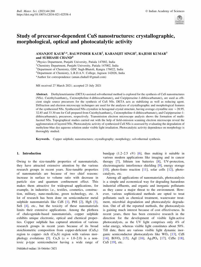
procedure as already described by Kaur et al [40]. Sche-matic synthesis route of potassium ethylxanthate is repre-
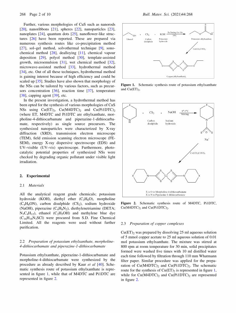
sented in figure 1, while that of M4DTC and Pi1DTC are
represented in figure 2.
2.3 Preparation of copper complexes
Cu(ET)2 was prepared by dissolving 25 ml aqueous solution
of 5 mmol copper acetate to 25 ml aqueous solution of 0.01
mol potassium ethylxanthate. The mixture was stirred at
800 rpm at room temperature for 30 min, solid precipitates
formed were washed five times with 10 ml distilled water
each time followed by filtration through 110 mmWhatmann
filter paper. Similar procedure was applied for the prepa-
ration of Cu(M4DTC)2 and Cu(Pi1DTC)2. The schematic
route for the synthesis of Cu(ET)2 is represented in figure 1,
while for Cu(M4DTC)2 and Cu(Pi1DTC)2 are represented
in figure 2.
Figure 1. Schematic synthesis route of potassium ethylxanthate
and Cu(ET)2.
Figure 2. Schematic synthesis route of M4DTC, Pi1DTC,
Cu(M4DTC)2 and Cu(Pi1DTC)2.
268 Page 2 of 10 Bull. Mater. Sci. (2021) 44:268

2.4 Preparation of CuS NSs
Copper sulphide NSs were prepared from single source
copper precursors: [Cu(ET)2], [Cu(M4DTC)2] and
[Cu(Pi1DTC)2] via hydrothermal method. CuS NSs were
prepared by transferring 0.5 g of copper precursors and 15
ml of DETA in Teflon-lined autoclave maintained at 170�Cfor 5 h. DETA acts as stabilizing and reducing agent. The
black product was separated by centrifugation, washed five
times by 10 ml deionized water and 10 ml ethanol each
time, then dried in hot air oven for 3 h. The schematic
synthesis route of CuS NSs by using Cu(ET)2 as precursor
is represented in figure 3, and using Cu(M4DTC)2 and
Cu(Pi1DTC)2 as precursors are represented in figure 4.
2.5 Instrument
For the detailed crystallographic characterization of
synthesized NSs and powder XRD patterns were recorded
using Rigaku Miniflex-600 Powder X-ray diffractometer, in
2h range of 20�–80� at scan speed of 4 min–1 keeping
step size 0.02�. For morphological analyses, electron
micrographs were recorded on Hitachi (H-7650) electron
microscope at accelerating voltage of 80 kV. For TEM
studies, the samples were prepared by dispersing 1 mg syn-
thesized NSs in 5 ml ethanol followed by 15 min of ultra-
sonication at room temperature. A drop of well-dispersed
ethanolic solution was placed on carbon-coated copper grid.
Then the dried sample was placed on the electron microscope
for further studies. The UV–vis absorption spectra
(k = 300–700 nm) of synthesized CuS NSs were recorded
using Hitachi (2J1-0004) UV–visible absorption spec-
trophotometer by dissolving 2 mg of nanopowder in 20 ml
ethanol at room temperature. The topographical and chemical
composition analyses were done using FE-SEM with EDS
attachment (NOVA NANOSEM-450).
2.6 Measurement of photocatalytic activity
The photocatalytic activity of the synthesized nano-photo-
catalysts was investigated using methylene blue (MB) dye
as a test contaminant in aqueous media under visible light
irradiation. First, dye stock solution was prepared in aque-
ous media by dissolving 6 mg dye in 1000 ml of water.
Then 14 mg of CuS NSs was suspended in 100 ml of
aqueous dye solution followed by vigorous stirring in dark
for 60 min for equilibration of solution to ensure maximum
adsorption of dye on the surface of nano-photocatalyst.
Then the solution illuminated in visible light for total 90
min along with collection of 5 ml sample after every 10 min
of irradiation, the collected aliquots were analysed using
UV–vis absorption spectrophotometer to check the photo-
degradation of MB dye. The characteristic absorption peak
of MB dye at 663 nm was used to assess the extent of
degradation. The degradation efficiency of photocatalyst is
defined by equation 1
g% ¼ Ao � At
Ao
� 100; ð1Þ
where g = degradation efficiency, Ao and At are the absor-
bance of MB at 663 nm in dark and under light irradiation
(at t min), respectively.Figure 3. Schematic synthesis route of CuS NSs prepared from
Cu(ET)2.
Figure 4. Schematic synthesis route of CuS NSs prepared from
Cu(M4DTC)2 and Cu(Pi1DTC)2.
Bull. Mater. Sci. (2021) 44:268 Page 3 of 10 268
3. Results and discussions
The growth of CuS NSs by using different ligands was done
to know the effect of the same on the morphology of the
products. In this regard, ET, M4DTC and Pi1DTC were
used as ligands. A detailed morphological difference and its
importance to photocatalysis are discussed below.
3.1 Crystallographic analyses
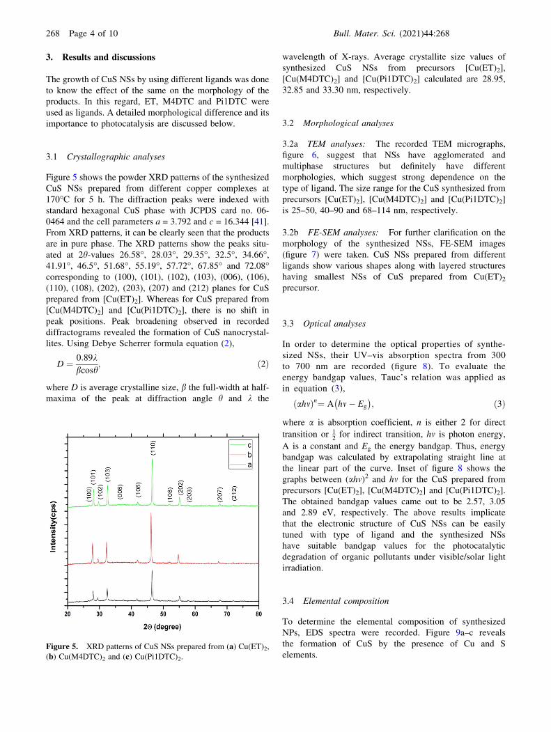
Figure 5 shows the powder XRD patterns of the synthesized
CuS NSs prepared from different copper complexes at
170�C for 5 h. The diffraction peaks were indexed with
standard hexagonal CuS phase with JCPDS card no. 06-
0464 and the cell parameters a = 3.792 and c = 16.344 [41].
From XRD patterns, it can be clearly seen that the products
are in pure phase. The XRD patterns show the peaks situ-
ated at 2h-values 26.58�, 28.03�, 29.35�, 32.5�, 34.66�,41.91�, 46.5�, 51.68�, 55.19�, 57.72�, 67.85� and 72.08�corresponding to (100), (101), (102), (103), (006), (106),
(110), (108), (202), (203), (207) and (212) planes for CuS
prepared from [Cu(ET)2]. Whereas for CuS prepared from
[Cu(M4DTC)2] and [Cu(Pi1DTC)2], there is no shift in
peak positions. Peak broadening observed in recorded
diffractograms revealed the formation of CuS nanocrystal-
lites. Using Debye Scherrer formula equation (2),
D ¼ 0:89kbcosh
; ð2Þ
where D is average crystalline size, b the full-width at half-
maxima of the peak at diffraction angle h and k the
wavelength of X-rays. Average crystallite size values of
synthesized CuS NSs from precursors [Cu(ET)2],
[Cu(M4DTC)2] and [Cu(Pi1DTC)2] calculated are 28.95,
32.85 and 33.30 nm, respectively.
3.2 Morphological analyses
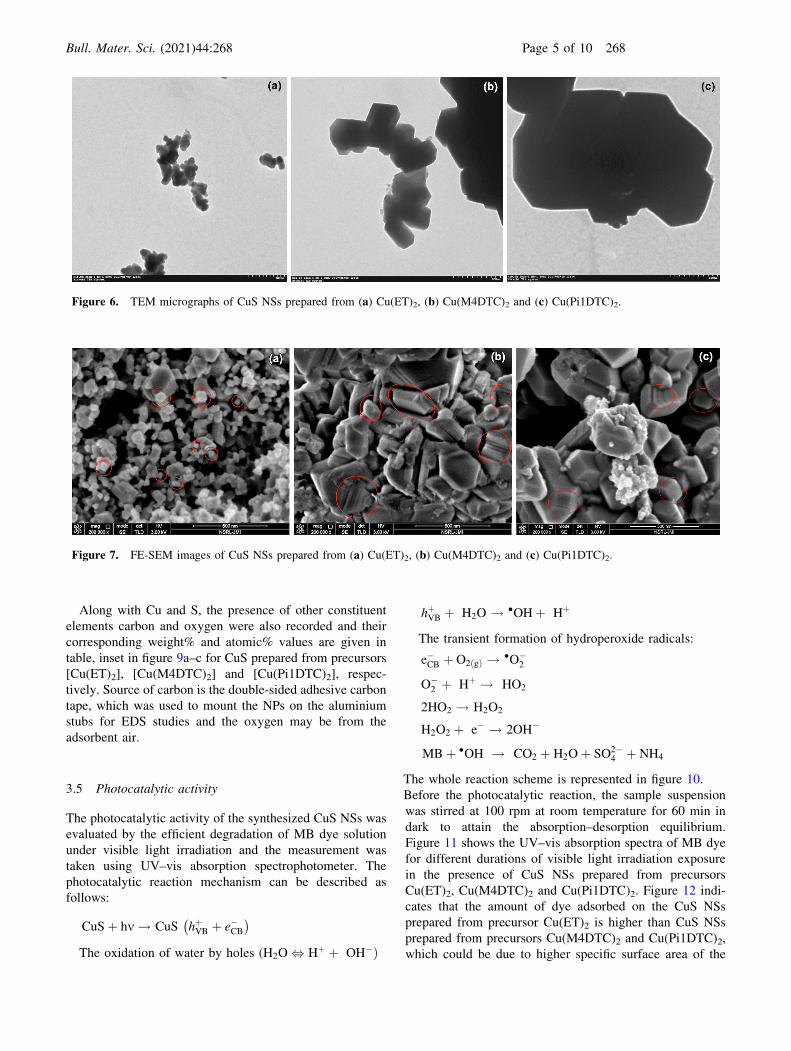
3.2a TEM analyses: The recorded TEM micrographs,
figure 6, suggest that NSs have agglomerated and
multiphase structures but definitely have different
morphologies, which suggest strong dependence on the
type of ligand. The size range for the CuS synthesized from
precursors [Cu(ET)2], [Cu(M4DTC)2] and [Cu(Pi1DTC)2]
is 25–50, 40–90 and 68–114 nm, respectively.
3.2b FE-SEM analyses: For further clarification on the
morphology of the synthesized NSs, FE-SEM images
(figure 7) were taken. CuS NSs prepared from different
ligands show various shapes along with layered structures
having smallest NSs of CuS prepared from Cu(ET)2precursor.
3.3 Optical analyses
In order to determine the optical properties of synthe-
sized NSs, their UV–vis absorption spectra from 300
to 700 nm are recorded (figure 8). To evaluate the
energy bandgap values, Tauc’s relation was applied as
in equation (3),
ahmð Þn¼ A hm� Eg
� �; ð3Þ
where a is absorption coefficient, n is either 2 for direct
transition or 12for indirect transition, hm is photon energy,
A is a constant and Eg the energy bandgap. Thus, energy
bandgap was calculated by extrapolating straight line at
the linear part of the curve. Inset of figure 8 shows the
graphs between (ahm)2 and hm for the CuS prepared from
precursors [Cu(ET)2], [Cu(M4DTC)2] and [Cu(Pi1DTC)2].
The obtained bandgap values came out to be 2.57, 3.05
and 2.89 eV, respectively. The above results implicate
that the electronic structure of CuS NSs can be easily
tuned with type of ligand and the synthesized NSs
have suitable bandgap values for the photocatalytic
degradation of organic pollutants under visible/solar light
irradiation.
3.4 Elemental composition
To determine the elemental composition of synthesized
NPs, EDS spectra were recorded. Figure 9a–c reveals
the formation of CuS by the presence of Cu and S
elements.Figure 5. XRD patterns of CuS NSs prepared from (a) Cu(ET)2,(b) Cu(M4DTC)2 and (c) Cu(Pi1DTC)2.
268 Page 4 of 10 Bull. Mater. Sci. (2021) 44:268
Along with Cu and S, the presence of other constituent
elements carbon and oxygen were also recorded and their
corresponding weight% and atomic% values are given in
table, inset in figure 9a–c for CuS prepared from precursors
[Cu(ET)2], [Cu(M4DTC)2] and [Cu(Pi1DTC)2], respec-
tively. Source of carbon is the double-sided adhesive carbon
tape, which was used to mount the NPs on the aluminium
stubs for EDS studies and the oxygen may be from the
adsorbent air.
3.5 Photocatalytic activity
The photocatalytic activity of the synthesized CuS NSs was
evaluated by the efficient degradation of MB dye solution
under visible light irradiation and the measurement was
taken using UV–vis absorption spectrophotometer. The
photocatalytic reaction mechanism can be described as
follows:
CuSþ hm ! CuS hþVB þ e�CB� �
The oxidation of water by holes (H2O , Hþ þ OH�Þ
hþVB þ H2O ! �OHþ Hþ
The transient formation of hydroperoxide radicals:
e�CB þ O2 gð Þ ! �O�2
O�2 þ Hþ ! HO2
2HO2 ! H2O2
H2O2 þ e� ! 2OH�
MBþ �OH ! CO2 þ H2Oþ SO2�4 þ NH4
The whole reaction scheme is represented in figure 10.
Before the photocatalytic reaction, the sample suspension
was stirred at 100 rpm at room temperature for 60 min in
dark to attain the absorption–desorption equilibrium.
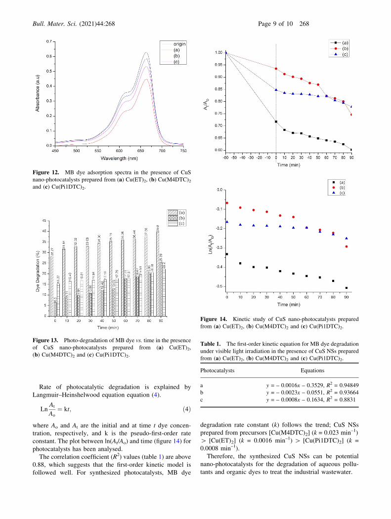
Figure 11 shows the UV–vis absorption spectra of MB dye
for different durations of visible light irradiation exposure
in the presence of CuS NSs prepared from precursors
Cu(ET)2, Cu(M4DTC)2 and Cu(Pi1DTC)2. Figure 12 indi-
cates that the amount of dye adsorbed on the CuS NSs
prepared from precursor Cu(ET)2 is higher than CuS NSs
prepared from precursors Cu(M4DTC)2 and Cu(Pi1DTC)2,
which could be due to higher specific surface area of the
Figure 6. TEM micrographs of CuS NSs prepared from (a) Cu(ET)2, (b) Cu(M4DTC)2 and (c) Cu(Pi1DTC)2.
Figure 7. FE-SEM images of CuS NSs prepared from (a) Cu(ET)2, (b) Cu(M4DTC)2 and (c) Cu(Pi1DTC)2.
Bull. Mater. Sci. (2021) 44:268 Page 5 of 10 268

Figure 8. UV–vis absorption spectra of CuS NSs prepared from (a) Cu(ET)2, (b) Cu(M4DTC)2 and (c) Cu(Pi1DTC)2; inset: Tauc’srelation curves.
268 Page 6 of 10 Bull. Mater. Sci. (2021) 44:268

Figure 9. EDS images of CuS NSs prepared from (a) Cu(ET)2, (b) Cu(M4DTC)2 and (c) Cu(Pi1DTC)2;inset: composition table.
Bull. Mater. Sci. (2021) 44:268 Page 7 of 10 268

nano-photocatalyst. It can be clearly seen from figure 13
that CuS NSs prepared from precursor Cu(ET)2 show
maximum photocatalytic activity. After 90 min of visible
light irradiation, the degradation of dye is 39.8%, whereas
for same duration of time dye degradation values observed
in case of CuS NSs prepared from precursors Cu(M4DTC)2and Cu(Pi1DTC)2 are 25.39 and 22.2%, respectively. The
higher photocatalytic activity of CuS NSs prepared from
precursor Cu(ET)2 could be attributed to small size, which
leads to high surface to volume ratio, hence lesser proba-
bility for volume recombination due to which chances of
surface charge transfer increases. UV–vis absorption spectra
of CuS NSs prepared from precursor Cu(ET)2 show broad
absorption profile, which is well matched with source
emission profile and has a high absorption yield. Hence,
implying more number of photo-excited carriers are gen-
erated, leading to higher photocatalytic activity. In the case
of CuS NSs prepared from precursor Cu(M4DTC)2, the
absorption yield is lower than CuS NSs precursor
Cu(Pi1DTC)2. This has abruptly increased towards the
lower wavelength and the absorption profile is not broad,
leading to the generation of lower number of photo-excited
carriers, which is the reason for lower photocatalytic
activity of CuS NSs prepared from precursor Cu(Pi1DTC)2as compared to others.
Figure 10. Schematic illustration of degradation of MB dye.
Figure 11. Graphical plot of MB dye absorption spectra in the
presence of CuS NSs prepared from (a) Cu(ET)2, (b) Cu(M4DTC)2and (c) Cu(Pi1DTC)2.
268 Page 8 of 10 Bull. Mater. Sci. (2021) 44:268
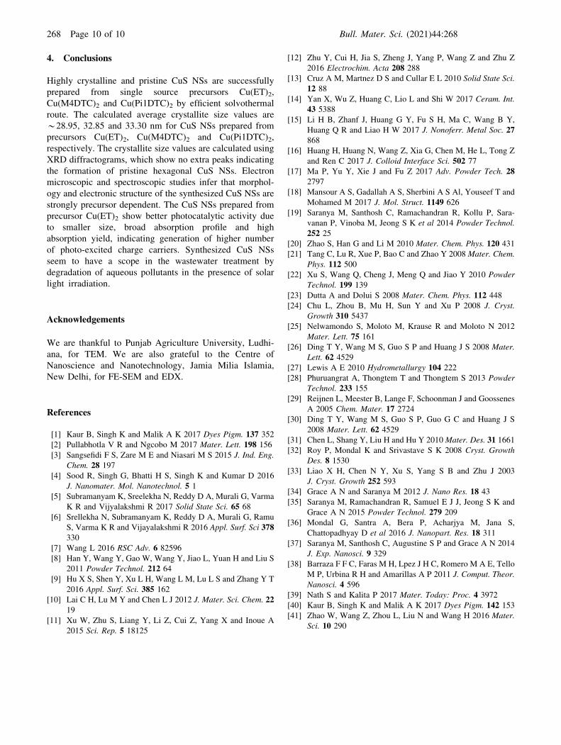
Rate of photocatalytic degradation is explained by
Langmuir–Heinshelwood equation equation (4).
LnAt
Ao
¼ kt; ð4Þ
where Ao and At are the initial and at time t dye concen-
tration, respectively, and k is the pseudo-first-order rate
constant. The plot between ln(At/Ao) and time (figure 14) for
photocatalysts has been analysed.
The correlation coefficient (R2) values (table 1) are above
0.88, which suggests that the first-order kinetic model is
followed well. For synthesized photocatalysts, MB dye
degradation rate constant (k) follows the trend; CuS NSs
prepared from precursors [Cu(M4DTC)2] (k = 0.023 min–1)
[ [Cu(ET)2] (k = 0.0016 min–1) [ [Cu(Pi1DTC)2] (k =
0.0008 min–1).
Therefore, the synthesized CuS NSs can be potential
nano-photocatalysts for the degradation of aqueous pollu-
tants and organic dyes to treat the industrial wastewater.
Figure 12. MB dye adsorption spectra in the presence of CuS
nano-photocatalysts prepared from (a) Cu(ET)2, (b) Cu(M4DTC)2and (c) Cu(Pi1DTC)2.
Figure 13. Photo-degradation of MB dye vs. time in the presence
of CuS nano-photocatalysts prepared from (a) Cu(ET)2,
(b) Cu(M4DTC)2 and (c) Cu(Pi1DTC)2.
Figure 14. Kinetic study of CuS nano-photocatalysts prepared
from (a) Cu(ET)2, (b) Cu(M4DTC)2 and (c) Cu(Pi1DTC)2.
Table 1. The first-order kinetic equation for MB dye degradation
under visible light irradiation in the presence of CuS NSs prepared
from (a) Cu(ET)2, (b) Cu(M4DTC)2 and (c) Cu(Pi1DTC)2.
Photocatalysts Equations
a y = – 0.0016x – 0.3529, R2 = 0.94849
b y = – 0.0023x – 0.0551, R2 = 0.93664
c y = – 0.0008x – 0.1634, R2 = 0.8831
Bull. Mater. Sci. (2021) 44:268 Page 9 of 10 268
4. Conclusions
Highly crystalline and pristine CuS NSs are successfully
prepared from single source precursors Cu(ET)2,
Cu(M4DTC)2 and Cu(Pi1DTC)2 by efficient solvothermal
route. The calculated average crystallite size values are
*28.95, 32.85 and 33.30 nm for CuS NSs prepared from
precursors Cu(ET)2, Cu(M4DTC)2 and Cu(Pi1DTC)2,
respectively. The crystallite size values are calculated using
XRD diffractograms, which show no extra peaks indicating
the formation of pristine hexagonal CuS NSs. Electron
microscopic and spectroscopic studies infer that morphol-
ogy and electronic structure of the synthesized CuS NSs are
strongly precursor dependent. The CuS NSs prepared from
precursor Cu(ET)2 show better photocatalytic activity due
to smaller size, broad absorption profile and high
absorption yield, indicating generation of higher number
of photo-excited charge carriers. Synthesized CuS NSs
seem to have a scope in the wastewater treatment by
degradation of aqueous pollutants in the presence of solar
light irradiation.
Acknowledgements
We are thankful to Punjab Agriculture University, Ludhi-
ana, for TEM. We are also grateful to the Centre of
Nanoscience and Nanotechnology, Jamia Milia Islamia,
New Delhi, for FE-SEM and EDX.
References
[1] Kaur B, Singh K and Malik A K 2017 Dyes Pigm. 137 352
[2] Pullabhotla V R and Ngcobo M 2017 Mater. Lett. 198 156
[3] Sangsefidi F S, Zare M E and Niasari M S 2015 J. Ind. Eng.Chem. 28 197
[4] Sood R, Singh G, Bhatti H S, Singh K and Kumar D 2016
J. Nanomater. Mol. Nanotechnol. 5 1
[5] Subramanyam K, Sreelekha N, Reddy D A, Murali G, Varma
K R and Vijyalakshmi R 2017 Solid State Sci. 65 68
[6] Srellekha N, Subramanyam K, Reddy D A, Murali G, Ramu
S, Varma K R and Vijayalakshmi R 2016 Appl. Surf. Sci 378330
[7] Wang L 2016 RSC Adv. 6 82596
[8] Han Y, Wang Y, Gao W, Wang Y, Jiao L, Yuan H and Liu S
2011 Powder Technol. 212 64
[9] Hu X S, Shen Y, Xu L H, Wang L M, Lu L S and Zhang Y T
2016 Appl. Surf. Sci. 385 162
[10] Lai C H, Lu M Y and Chen L J 2012 J. Mater. Sci. Chem. 2219
[11] Xu W, Zhu S, Liang Y, Li Z, Cui Z, Yang X and Inoue A
2015 Sci. Rep. 5 18125
[12] Zhu Y, Cui H, Jia S, Zheng J, Yang P, Wang Z and Zhu Z
2016 Electrochim. Acta 208 288
[13] Cruz A M, Martnez D S and Cullar E L 2010 Solid State Sci.12 88
[14] Yan X, Wu Z, Huang C, Lio L and Shi W 2017 Ceram. Int.43 5388
[15] Li H B, Zhanf J, Huang G Y, Fu S H, Ma C, Wang B Y,
Huang Q R and Liao H W 2017 J. Nonoferr. Metal Soc. 27868
[16] Huang H, Huang N, Wang Z, Xia G, Chen M, He L, Tong Z
and Ren C 2017 J. Colloid Interface Sci. 502 77
[17] Ma P, Yu Y, Xie J and Fu Z 2017 Adv. Powder Tech. 282797
[18] Mansour A S, Gadallah A S, Sherbini A S Al, Youseef T and
Mohamed M 2017 J. Mol. Struct. 1149 626
[19] Saranya M, Santhosh C, Ramachandran R, Kollu P, Sara-
vanan P, Vinoba M, Jeong S K et al 2014 Powder Technol.252 25
[20] Zhao S, Han G and Li M 2010 Mater. Chem. Phys. 120 431
[21] Tang C, Lu R, Xue P, Bao C and Zhao Y 2008 Mater. Chem.Phys. 112 500
[22] Xu S, Wang Q, Cheng J, Meng Q and Jiao Y 2010 PowderTechnol. 199 139
[23] Dutta A and Dolui S 2008 Mater. Chem. Phys. 112 448
[24] Chu L, Zhou B, Mu H, Sun Y and Xu P 2008 J. Cryst.Growth 310 5437
[25] Nelwamondo S, Moloto M, Krause R and Moloto N 2012
Mater. Lett. 75 161
[26] Ding T Y, Wang M S, Guo S P and Huang J S 2008 Mater.Lett. 62 4529
[27] Lewis A E 2010 Hydrometallurgy 104 222
[28] Phuruangrat A, Thongtem T and Thongtem S 2013 PowderTechnol. 233 155
[29] Reijnen L, Meester B, Lange F, Schoonman J and Goossenes
A 2005 Chem. Mater. 17 2724
[30] Ding T Y, Wang M S, Guo S P, Guo G C and Huang J S
2008 Mater. Lett. 62 4529
[31] Chen L, Shang Y, Liu H and Hu Y 2010Mater. Des. 31 1661[32] Roy P, Mondal K and Srivastave S K 2008 Cryst. Growth
Des. 8 1530
[33] Liao X H, Chen N Y, Xu S, Yang S B and Zhu J 2003
J. Cryst. Growth 252 593
[34] Grace A N and Saranya M 2012 J. Nano Res. 18 43
[35] Saranya M, Ramachandran R, Samuel E J J, Jeong S K and
Grace A N 2015 Powder Technol. 279 209
[36] Mondal G, Santra A, Bera P, Acharjya M, Jana S,
Chattopadhyay D et al 2016 J. Nanopart. Res. 18 311
[37] Saranya M, Santhosh C, Augustine S P and Grace A N 2014
J. Exp. Nanosci. 9 329
[38] Barraza F F C, Faras M H, Lpez J H C, Romero M A E, Tello
M P, Urbina R H and Amarillas A P 2011 J. Comput. Theor.Nanosci. 4 596
[39] Nath S and Kalita P 2017 Mater. Today: Proc. 4 3972
[40] Kaur B, Singh K and Malik A K 2017 Dyes Pigm. 142 153
[41] Zhao W, Wang Z, Zhou L, Liu N and Wang H 2016 Mater.Sci. 10 290
268 Page 10 of 10 Bull. Mater. Sci. (2021) 44:268