study of antimicrobial, biochemical and nanotechnological ... · study of antimicrobial,...
TRANSCRIPT
Study of antimicrobial, biochemical and
nanotechnological aspects of novel sulfur, selenium
and tellurium compounds
Dissertation
zur Erlangung des Grades
des Doktors der Naturwissenschaften
der Naturwissenschaftlich-Technischen Fakultät III
Chemie, Pharmazie, Bio- und Werkstoffwissenschaften
der Universität des Saarlandes
von
Uma Maheswari Viswanathan
Saarbrücken
2014

Tag des Kolloquiums: 27 August 2014
Dekan: Prof. Dr. Volkhard Helms
Berichterstatter: Prof. Dr. Claus Jacob
Prof. Dr. Ingolf Bernhardt
Vorsitz: Prof. Dr. Heiko Zimmermann
Akad. Mitarbeiter: Dr. Frank Hannemann

Diese Dissertation entstand unter der Anleitung von Prof. Dr. Claus Jacob in der
Arbeitsgruppe für Bioorganische Chemie, Fachrichtung 8.2 Pharmazie der
Naturwissenschaftlich-Technischen Fakultät III der Universität des Saarlandes im Zeitraum
von Juli 2011 bis Juni 2014.

Dedicated to my loving family

Contents
iii
Table of Contents
Acknowledgements .................................................................................................................... 7
Abstract ...................................................................................................................................... 9
Kurzfassung .............................................................................................................................. 10
List of Abbreviations ................................................................................................................ 11
PART I ..................................................................................................................................... 14
1. Introduction .................................................................................................................. 15
1.1 Sulfur and its biological significance ............................................................... 15
1.2 A brief history of Allium species ...................................................................... 15
1.3 Garlic based therapeutics and therapies ........................................................... 16
1.3.1 Antimicrobial activity ........................................................................... 17
1.3.2 Anticancer effect ................................................................................... 19
1.4 Chemistry/Reactivity of Polysulfanes .............................................................. 20
1.5 From simple polysulfanes to pharmaceutically interesting molecules ............. 22
2 Objectives ..................................................................................................................... 24
3. Results and Discussion ................................................................................................. 25
3.1 Synthesis of unsymmetrical tetrasulfanes ........................................................ 25
3.2 Physico-chemical properties ........................................................................... 29
3.2.1 Electrochemistry ................................................................................... 30
3.2.2 Sita tensiometry measurements ........................................................... 31
3.2.3 Densitometric measurements ............................................................... 32
3.3 Molecular Non-Covalent Interaction Studies ................................................... 34
3.3.1 Haemolytic assay .................................................................................. 34
3.3.2 FRET measurements ............................................................................. 36
3.3.3 Circular dichroism measurements ........................................................ 39
3.4 Biological activities of the compounds ............................................................ 40
3.4.1 Activity against microbes and small organisms ................................... 41
3.4.1.1 Nematode assay ................................................................................. 41
3.4.1.2 Toxicity against different yeast strains .............................................. 44
3.4.1.3 Activity against different filamentous fungi ...................................... 50
3.4.1.4 Botrytis assay ..................................................................................... 52
Contents
iv
3.4.1.5 Antibacterial assay ............................................................................. 53
3.4.1.6 Anti-bacteriophage assay ................................................................... 58
3.5 Activity in Primary Cell lines ........................................................................... 61
3.5.1 Activity in human synovial fibroblast cell lines .................................. 61
3.5.2 Activity in neuronal cell lines of rats .................................................... 62
4. Summary and Outlook ................................................................................................. 63
PART II .................................................................................................................................... 66
1. Introduction .................................................................................................................. 67
2 Objectives ..................................................................................................................... 68
3. Results and Discussion of Se- and Te-containing compounds .................................... 70
3.1 Physico- Chemical Properties .......................................................................... 70
3.1.1 Ring and Sita Tensiometry ................................................................... 70
3.2 Non-Covalent Membrane Interaction Studies .................................................. 73
3.2.1 FRET Measurements ............................................................................ 73
3.3 Biological activities of the selenium and tellurium compounds ...................... 75
3.3.1 Toxicity against yeast strains ................................................................ 75
3.3.2 Antibacterial Assay ............................................................................... 80
4. Summary and Outlook ................................................................................................. 87
5. Experimental Part ......................................................................................................... 88
5.1 Materials and Methods ..................................................................................... 88
5.2 NMR Spectroscopy .......................................................................................... 88
5.3 Mass spectroscopy ............................................................................................ 88
5.4 Calculation of clogP ......................................................................................... 89
5.5 Synthesis of unsymmetrical tetrasulfanes ........................................................ 89
5.5.1 General procedure for the synthesis of tetrasulfanes (I) ....................... 89
5.5.2 General procedure for the synthesis of UT (II) .................................... 89
5.5.2.1 1, 4-diphenyltetrasulfane (DPhTTS) ................................................. 90
5.5.2.2 3-(propyltetrasulfanyl)-propanoic acid (PrTTSPs) ............................ 90
5.5.2.3 3-(benzyltetrasulfanyl)-propanoic acid (BnTTSPs) .......................... 91
5.5.2.4 3-(phenyltetrasulfanyl)-propanoic acid (PhTTSPs) ........................... 91
5.5.2.5 1-benzyl-4-phenyltetrasulfane (BnTTSPh) ....................................... 91
5.5.2.6 1-propyl-4-benzyltetrasulfane (PrTTSBn) ........................................ 92

Contents
v
5.5.2.7 1-propyl-4-phenyltetrasulfane (PrTTSPh) ......................................... 92
5.6 Physico-Chemical Measurements .................................................................... 93
5.6.1 Electrochemistry ................................................................................... 93
5.6.2 Ring tensiometry ................................................................................... 93
5.6.3 Sita tensiometry .................................................................................... 94
5.6.4 Densitometry ........................................................................................ 94
5.7 Molecular Non-Covalent Interaction Studies ................................................... 95
5.7.1 Haemolysis assay .................................................................................. 95
5.7.1.1 Preparation of buffer .......................................................................... 95
5.7.1.2 Preparation of human blood .............................................................. 95
5.7.1.3 Preparation of red blood cells ............................................................ 95
5.7.1.4 Procedure ........................................................................................... 95
5.7.2 FRET measurements ............................................................................. 96
5.8 Biological Assays ............................................................................................. 97
5.8.1 Nematode assay .................................................................................... 97
5.8.2 Anti-yeast experiment ........................................................................... 98
5.8.2.1 Preparation of Saburo broth and media ............................................. 98
5.8.2.2 Re-cultivation of yeast ....................................................................... 98
5.8.2.3 Procedure ........................................................................................... 98
5.8.3 Antifungal experiment ...................................................................................... 99
5.8.3.1 Preparation of Wort broth and media ................................................ 99
5.8.3.2 Spore collection ................................................................................. 99
5.8.3.3 Procedure ........................................................................................... 99
5.8.4 Antibacterial experiment ................................................................................ 100
5.8.4.1 Preparation of LB broth and media ................................................. 100
4.8.4.2 Re-cultivation of bacteria ................................................................ 100
4.8.4.3 Procedure ......................................................................................... 100
5.8.5 Anti-bacteriophage experiment ...................................................................... 101
5.8.5.1 Preparation of LB broth and media ................................................. 101
5.8.5.2 Re-cultivation of E. coli C-T4 ......................................................... 101
5.8.5.3 Isolation of T4-bacteriophage .......................................................... 101
5.8.5.4 Procedure ......................................................................................... 101
Contents
vi
6. References .................................................................................................................. 103
List of own publications ............................................................................................. 111
Appendix .................................................................................................................... 113
Curriculum Vitae ........................................................................................................ 127
Acknowledgements
7
Acknowledgements
My heartfelt gratitude goes to Prof. Dr. Claus Jacob for accepting me as his PhD student. I am
thankful to him for his supervision, constant support and suggestions throughout my work. He
was very friendly and kindly helped me at each step and motivated me to accomplish my
studies successfully. I am extremely grateful to Prof. Dr. Ingolf Bernhardt, my second
supervisor. I would like to express my sincere thanks to Dr. Torsten Burkholz for his valuable
academic advice and suggestions during my research. He also helped me to solve many
bureaucratic problems on an official level, without his support it would not have been a nice
stay for a foreigner like me in Germany.
My sincere thanks go to all my colleagues in the group of Prof. Jacob, with whom I worked
throughout my PhD. They are Dr. Aisha Lalla Ba, Dr. Brigitte Czepukojc, Dr. Mandy
Doering, Dr. Ada Nucci, Dr. Ethiene Estevam, Dr. Enrique Domínguez, Dr. Khairan Khairan,
Dr. Zhanjie Xu, Alexandra Hoffmann, Lucienne Ngimatio, Lisa Faulstich, Marina Hakenesch,
Sandra Hübgen, Peng Du, Sayed Ali, Ahsan Raza, Omar Elakad, Jawad Nasim, and Adel Al-
Marby.
I would like to express my deepest thanks to Prof. Dr. Shiraz Markarian from the Yerevan
State University (YSU), who accepted me as an exchange student and allowed me to perform
experiments in his laboratory. All of my colleagues at YSU, Dr. Liana Gabrielyan, Dr.
Ashkhen Zatikyan, Naira Tonoyan, Zakar Papanyan, Dr. Karine Grigoryan, Karen
Amirbekyan, Gohar Shahinyan, Levon Sargsyan, Hasmik Shilajyan and Dr. Alla Terzyan in
the physical chemistry department were wonderful and helped me throughout, which turned
my stay fruitful.
I am also thankful to Prof. Dr. Armen Trchounian, who allowed me to perform detailed
microbiological experiments in his laboratory, which formed an important part of my thesis. I
am extremely thankful to Dr. Inga Bazukyan, who supervised me in these experiments and
her valuable ideas helped me to succeed. My thanks and respect go to Dr. Hovik Panosyan,
Armine Margaryan and Dr. Lilyth Gabrielyan, Anushik Arakelyan and Margarit Petrosyan
who were my good friends and colleagues in this group.

Acknowledgements
8
I would like to specially thank Prof. Dr. Mark Schneider, who allowed me to work in his lab
to carry out Ring tensiometry experiments. I would also like to gratefully acknowledge the
University of Saarland for funding my PhD studies. My special thanks go to Academiacs
International.
I am extremely grateful to Prof. Dr. Mangalraj Devanesan, Dr. Ponpandian Nagamony, and
Dr. Viswanathan Chinnuswamy, who helped during my Master studies and were the genuine
motivation to perform my PhD studies. I am thankful to Dr. Dickson Joseph, Dr. Sooraj
Ravindran, Padmapriya Sooraj, Dr. Biju Kuyyadi, Rajasekhar Duggineni and Sajid Hussain
who helped me during the initial days of my research. I would also like to thank Dr. Anja
Philippi, Dr. Kristina Riehemann, Caroline Blumenthal and Birgit Wiegand for all of their
help.
My special thanks also go out to my family; my father, Viswanathan Arumugam, mother,
Subadhra Viswanathan, and brother, Vineeth Arumugam, Davathamizhan Godandaramen, as
well as my friends Anjana Kenath, Aswathy Jayadev, Vishnu Ramani, Ganesh Ram, Nisha
Sajad, and Lakshmy Balu.

Abstract
9
Abstract
Chalcogen chemistry is widely interdisciplinary, especially in the field of biology, material
sciences and supra-molecular chemistry and biology. Whilst oxygen and sulfur are fairly
omnipresent in biology selenium has its nutritional role in animals and humans; however, for
tellurium, this is yet to be discovered. The common behaviour of all three elements is related
to their antioxidant properties and their therapeutic effects. Most drugs for present day
diseases are either natural or derivatives of compounds found in natural products. Allium plant
species have a wide range of effective therapeutics benefits. Garlic and its components,
namely allicin, polysulfanes, thiosulfinates, thiosulfonates and dithienes, form an important
class of natural compounds and represent a field where tremendous research has been ongoing
for the past decades.
As part of this thesis, a series of unsymmetrical tailor-made tetrasulfanes have been
synthesised. They were tested along with some other symmetrical tetrasulfanes, selenium and
tellurium compounds for their physico-chemical properties and therapeutic benefits, in both
microorganisms and primary cell lines. Biological activities were investigated by testing the
tetrasulfanes in nematodes, bacteria, yeast, fungi and bacteriophages. The effects on red blood
cells and neuronal cell lines of rats were also studied. A cytotoxicity assay and ELISA were
performed on human synovial fibroblast cell lines.
Kurzfassung
10
Kurzfassung
Die Chemie der Chalkogene ist sehr interdisziplinär, besonders, wenn Biologie, Medizin,
Materialwissenschaften und supramolekulare Wissenschaften miteinbezogen werden.
Schwefel und Selen tragen bei Mensch und Tier zur Bedarfsdeckung bei; ob dies auch auf
Tellur zutrifft muss hingegen noch bewiesen werden. Diesen drei Elementen sind ihre
antioxidativen Eigenschaften und therapeutischen Effekte gemeinsam. Die meisten
Medikamente für aktuelle Krankheiten sind entweder auf Naturstoffen basierend oder
Derivate dieser Stoffe. Alle Allium-Arten weisen eine große Anzahl an potenten Wirkstoffen
mit therapeutischen Wirkungen auf. Knoblauch und seine Bestandteile, nämlich Allicin,
Polysulfane, Thiosulfinate, Thiosulfate und Dithiene, formen wichtige Gruppen natürlicher
Stoffe, zu denen in den letzten Jahrzenten enorme Forschungsarbeiten geführt worden sind.
Ein Teil dieser Arbeit beschäftigt sich mit der Synthese einer Reihe unsymmetrischer und
maßgeschneiderter Tetrasulfane. Sie wurden gemeinsam mit anderen symmetrischen Tetra-
und Polysulfanen, Selen- und Tellur-Gemischen auf Grund ihrer physikalisch-chemischen
Eigenschaften und ihres möglichen therapeutischen Nutzens in Mikroorganismen und
Primärzelllinien getestet. Die biologische Aktivitäten der Tetrasulfane wurden in Nematoden,
Bakterien, Pilzen (v.a. Hefe) und Bakteriophagen untersucht. Die Auswirkungen auf rote
Blutzellen wurden mit Hilfe von Hämoglobinassays erforscht. Studien zur Zytotoxizität,
sowie weiterführende Experimente und Analysemethoden (z.B. ELISA) wurden an
menschlichen synovialen Fibroblasten durchgeführt.
.

Abbreviations
11
List of Abbreviations
AChE acetylcholine esterase
Ag/AgCl silver/silver chloride electrode
BnTTSPh 1-benzyl-4-phenyltetrasulfane
BnTTSPs 3-(benzyltetrasulfanyl)-propanoic acid
BSA bovine serum albumin
CDCl3 deuterochloroform
CH2Cl2 dicholoromethane
CHCl3 chloroform
CMC critical micelle concentration
DADS diallyldisulfide
DAS diallylsulfide
DATS diallyltrisulfane1
DATTS diallyltetrasulfane
DBTTS 1, 4-dibenzyltetrasulfane
DEETTS 1, 4-bis(2-ethoxyethyl)tetrasulfane
DMSO dimethylsulfoxide
DNA deoxyribonucleic acid
DPhTTS 1, 4-diphenyltetrasulfane
DPSEETTS diethyl 3, 3’-tetrasulfanediyldipropanoate
DPSTTS 3, 3’-tetrasulfanediyldipropanoic acid
DPTTS dipropyltetrasulfane
DU145 human prostate cancer cell line
1 The compounds with chemical formula RSxR, where R ≠ H and x ≥ 3 are termed as polysulfanes. These compounds are
sometimes incorrectly referred to as ‘polysulfides’ in bio-chemical publications.
Abbreviations
12
E1/2 half wave potential
Epa anodic oxidation potential
Epc cathodic reduction potential
ESI electrospray ionisation
GO garlic oil
GPx glutathione peroxidase
GSH glutathione
GSSG glutathione disulfide
h hours
H2O2 dihydrogen peroxide
HRMS high-resolution mass spectroscopy
IC50 half-maximal inhibitory concentration
Ksv Stern Volmer constant
LD50 lethal dose (50 %)
m/z mass by charge ratio
MDA-MB-231 human mammary gland carcinoma cell lines
MgSO4 magnesium sulfate
MIC minimum inhibitory concentration
NMR nuclear magnetic resonance
OSCs organosulfur compounds
PC3 prostate cancer cell line 3
PEG polyethylene glycol
PhTTSPs 3-(phenyltetrasulfanyl)-propanoic acid
pKa logarithmic acid dissociation constant
PrTTSBn 1-benzyl-4-propyltetrasulfane
PrTTSPh 1-phenyl-4- propyltetrasulfane

Abbreviations
13
PrTTSPs 3-(propyltetrasulfanyl)-propanoic acid
RBCs red blood cells
Rf retention factor
S2Cl2 sulfur monochloride
SDS sodium dodecyl sulfate
S-S sulfur-sulfur bond
ST symmetrical tetrasulfanes
TLC thin layer chromatography
UT unsymmetrical tetrasulfanes

Part I
14
PART I
Study of tetrasulfanes

Introduction
15
1. Introduction
The elements in group 16 (formerly group 6) of the Periodic Table are called chalcogens. This
group contains Oxygen (O), Sulfur (S), Selenium (Se), Tellurium (Te) and the radioactive
element Polonium (Po).
1.1 Sulfur and its biological significance
Sulfur is an important constituent of various amino acids, the most important being cysteine
and methionine. Sulfur is also part of cellular enzymes and proteins, which take part in
substantial biochemical and cellular signalling pathways; hence, small changes in their
amount and activity may cure many diseases [1]. It is therefore important to keep an eye on
the optimum intake of sulfur. Sulfur is found in a large number of natural products, with
Allium species being the most significant, as well as in other micro- and marine organisms in
the form of various organo sulfur compounds (OSCs) [2] [3] [4]. The Allium species with
enormous amounts of OSCs play an importunate role as dietary supplements for individuals
[5].
1.2 A brief history of Allium species
One of the oldest cultivated vegetables, Allium cepa, commonly known as onion, has been
reported for over 5000 years by now. In India, reports of onions are found in writings from the
6th century onwards [6]. Onion was cultivated and became widespread as a crop in Europe,
only during the Middle ages. The second most important vegetable of Allium species is Allium
sativum, commonly known as garlic, has been cultivated for over 5000 years [7]. One of the
most accepted history is that garlic originated from Central Asia and then became widespread
from there to different parts of the world. Garlic has been grown by village people for both
culinary and medicinal purposes [6] [8] .

Introduction
16
1.3 Garlic based therapeutics and therapies
Before the invention of antibiotics, preparations of garlic were used to treat diseases [9] [10]
[11] such as cholera, tuberculosis, dysentery, diphtheria etc [12] [13]. To some extent,
Dengue fever was also treated with the same techniques [13].
Figure 1 : Collection of organo sulfur compounds and their intermediates present in Garlic. Allicin (1), 1
propene sulfenic acid (2), 3-Vinyl-3,4-dihydro-1,3-dithiin (3), 2-Vinyl-2,4-dihydro-1,3-dithiin (4),
((allyldisulfanyl)methyl)tetrahydro-2H-thiopyran (5), 2-((allyldisulfanyl)methyl)-3,4-dihydro-2H-
thiopyran (6), E-ajoene (7), thioacrolein (8), 1,6diallylhexasulfane (9), diallyldisulfide (10), Z-ajoene (11),
2,2’-(propane-1,2-diyl)bis(1-allyldisulfane) (12), 2 propenesulfenic acid (13). Figure modified in reference
to [4].
The OSCs found in garlic (Figure 1) have properties such as cancer prevention, antimicrobial
activity, insect and animal attractive/repulsive activity, olfactory-gustatory-lachrymatory
properties, effects on lipid metabolism, and platelet aggregation [4] [14] [15] [16] [17]. All of
these different properties of sulfur compounds are due to their ability to undergo diverse
chemical reactions such as reduction, oxidation, reactions involving sulfur radicals, pericyclic
and re-arrangement reactions [18] [19].

Introduction
17
1.3.1 Antimicrobial activity
Anti-microbial effects probably represent the most prominent activity of garlic and its
components [20] [21] [22]. Garlic shows a growth inhibitory effect against both Gram-
positive and Gram-negative bacteria. Allicin, even at 1:100,000 dilutions, has been proven to
exhibit inhibitory effects against microbes [21] [23]. Methyl methanethiosulfinate [24], propyl
propane-thiosulfinate [25] and thiosulfinates of onion also exhibit activity against the bacteria,
but this is definitely weaker than that of allicin [26] [27] [28] [29]. Other than the anti-
bacterial activity, garlic has also shown activities against fungi and yeast. The pathogenic
strains of Candida albicans and Cryptococcus neoformans for instance were also affected by
garlic extracts [26] [30] [31] [32].
Table 1: Different constituents of garlic oil and their respective concentrations in mg/g of dry weight of
garlic oil. Table adapted from [33].
Analysis of undiluted GO
GO component Concentration (mg/g)* of dry
weight
Diallyl monosulfide 106 ± 7 (10.6)
Diallyl disulfide 530 ± 7 (53.0)
Diallyl trisulfide 115 ± 4 (11.5)
Diallyl tetrasulfide 43 ± 2 (4.3)
Diallyl pentasulfide 10.5 ± 0.4 (1.1)
Diallyl hexasulfide 0.14 ± 0.01 (0.01)
Methyl allyl disulfide 44.1 ± 2 (4.4)
Methyl allyl trisulfide 69.9 ± 2.2 (7)
Methyl allyl tetrasulfide 24.6 ± 2.0 (2.5)
Methyl allyl pentasulfide 6.3 ± 0.6 (0.6)
Methyl allyl hexasulfide 1.5 ± 0.1 (0.2)
Dimethyl trisulfide 12.0 ± 1.3 (1.2)
Dimethyl tetrasulfide 4.3 ± 0.6 (0.2)
Dimethyl pentasulfide 2.0 ± 0.4 (0.2)
*Values in parentheses are percentages

Introduction
18
Garlic oil (GO) has been tested frequently against microbes [34] [35]. This preparation
contains the maximum number of variable polysulfanes [36]. Table 1 indicates that GO
contains 53 % of diallylsulfide, DATS is found at 11.5 % and all other polysulfanes are
present in trace amounts. In Thailand, GO and chive oil have been used for the treatment of
food borne infections for many years. In China, the DATS constituent of garlic oil was used
to treat viral infections and cryptococcal meningitis. GO also exerts antifungal properties
against various species of Aspergillus, Candida and Fusarium [31] [37].
Polysulfanes in garlic have been reported to exhibit an identical activity as that of raw garlic
extract, garlic oil and allicin. As DATTS is more stable than its higher derivatives, it has an
extraordinary effect against Helicobacter pylori (MIC range, 3 to 6 mg/ml) [33]. It is also
evident from various studies that the activities of polysulfanes are directly proportional to the
length of S-S chains in them [18].
The results from various studies are compiled in Table 2, which shows a comparison of the
activities of various sulfur containing compounds DAS, DADS, DATS, and DATTS along
with allicin, against different strains of microbes. Although it is not an appropriate method,
this gives some hint about the activities of these compounds against the different microbial
strains. It is evident from the comparative study that, DAS and DADS are required in larger
amounts to visualise an effective activity against the microorganisms [18]. DATS shows a
similar activity as that of allicin. Surprisingly, DATTS exhibited MIC values at very low
concentrations starting from 0.5 g/ml.
Table 2: This table compiles the values of minimum inhibitory concentrations in g/ml exhibited by allicin,
diallylsulfide, diallyldisulfide, diallyltrisulfane and diallyltetrasulfane on various species of bacteria and
fungi. The values also prove that diallyltrisulfane and diallyltetrasulfane are the most active amongst all of
the compounds. Table adapted from [18].
Organism Allicin DAS DADS DATS DATTS
Helicobacter pylori
Klebsiella pneumonia
Pseudomonas aeruginosa
Staphylococcus aureus
MRSA
Candida albicans
Aspergillus niger
6-12
-
15
15
28
0.8
8-32
2100-4100
96-104
80-88
20
32
32
40
100
72-80
64-72
4
12
4
8
13-25
40-48
32-36
2
8
1
2
3-6
20-24
12-16
0.5
2
0.5
1
Introduction
19
Polysulfanes were used as treatment against numerous food borne bacterial pathogens namely
Bacillus cereus, Campylobacter jejuni, Escherichia coli, Listeria monocytogenes, Salmonella
sp., Shigella sp., Staphylococcus aureus, Vibrio cholera and Yersinia enterocolytica [32]. The
MIC values of DATS and DATTS were 2 µg/ml and 1 µg/ml against C. botulinum,
respectively, 4 µg/ml and 1 µg/ml against C. jejuni, respectively, and 12 µg/ml and 4 µg/ml
against V. cholera, respectively [32].
1.3.2 Anticancer effect
Garlic and its derivatives exhibit antiprolierative activities in human cancer cell lines [38]
[39]. An international study involving men and women from various countries has been
carried out and this study is termed as ‘The European Prospective Investigation into Cancer
and Nutrition’ (EPIC) [40]. This study involves an investigation of the influence of diet on
cancer. This study revealed that the risk of intestinal cancer was reduced with a more
extensive intake of garlic and onion [41].
The studies by Pinto et al state that Allium derivatives inhibit the proliferation of the human
prostate cancer cell line (LNCaP) and human breast cancer cell line (MCF-7) [14]. Further
studies, however, are required to reveal the mechanism of action of these Allium derivatives
[42]. DATS and DATTS have been reported as strong inducers of early mitotic arrest and for
subsequent apoptosis of cancer cells [43]. One of the studies by Kelkel et al states that
DATTS acts independently of ROS and one of the major cellular targets of this compound has
been identified as tubulin [44] [45] [46].
Introduction
20
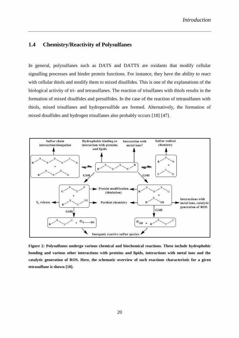
1.4 Chemistry/Reactivity of Polysulfanes
In general, polysulfanes such as DATS and DATTS are oxidants that modify cellular
signalling processes and hinder protein functions. For instance, they have the ability to react
with cellular thiols and modify them to mixed disulfides. This is one of the explanations of the
biological activity of tri- and tetrasulfanes. The reaction of trisulfanes with thiols results in the
formation of mixed disulfides and persulfides. In the case of the reaction of tetrasulfanes with
thiols, mixed trisulfanes and hydropersulfide are formed. Alternatively, the formation of
mixed disulfides and hydrogen trisulfanes also probably occurs [18] [47].
Figure 2: Polysulfanes undergo various chemical and biochemical reactions. These include hydrophobic
bonding and various other interactions with proteins and lipids, interactions with metal ions and the
catalytic generation of ROS. Here, the schematic overview of such reactions characteristic for a given
tetrasulfane is shown [18].

Introduction
21
Disulfides react with cysteine residues in proteins to undergo thiol/disulfide reactions. One of
the best examples is the thiolation behaviour of disulfides such as glutathione disulphide
(GSSG) [18].
Nonetheless, it will not be sufficient to explain the diversified biological activities of
polysulfanes such as antibacterial, antifungal and anti-carcinogenic effects, by just
considering thiolation. In 2006, Jacob et al. proposed several possibilities for the reactions
characteristic of polysulfanes based on their known chemical properties. Chemical reactions
of polysulfanes endow them with unique biological activities. The foremost important
reaction, the thiolation reaction has been discussed above (Figure 2) [18] [48] [49].
Other than the thiolation reactions, polysulfanes may undergo homolytic S-S bond cleavage.
This bond cleavage results in the formation of perthiyl radicals. Polysulfanes also undergo
sulfur-transfer reactions. This reaction involves the transfer of S2 or S3 subunits to molecules
containing one or two conjugated double bonds. Another important reaction of polysulfanes is
their interaction with metal ions. Polysulfanes can probably form metal complexes due to
their ability to coordinate with several sulfur atoms at a time. Other aspects of polysulfane
chemistry are the polysulfane-perthiol-perthiyl radical chemistry and hydrogen sulfide release
mechanisms [50] [51] [52] [53] [52].
Introduction
22
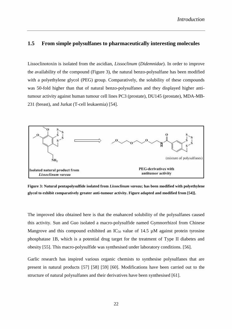
1.5 From simple polysulfanes to pharmaceutically interesting molecules
Lissoclinotoxin is isolated from the ascidian, Lissoclinum (Didemnidae). In order to improve
the availability of the compound (Figure 3), the natural benzo-polysulfane has been modified
with a polyethylene glycol (PEG) group. Comparatively, the solubility of these compounds
was 50-fold higher than that of natural benzo-polysulfanes and they displayed higher anti-
tumour activity against human tumour cell lines PC3 (prostate), DU145 (prostate), MDA-MB-
231 (breast), and Jurkat (T-cell leukaemia) [54].
Figure 3: Natural pentapolysulfide isolated from Lissoclinum vareau; has been modified with polyethylene
glycol to exhibit comparatively greater anti-tumour activity. Figure adapted and modified from [54]].
The improved idea obtained here is that the enahanced solubility of the polysulfanes caused
this activity. Sun and Guo isolated a macro-polysulfide named Gymnorrhizol from Chinese
Mangrove and this compound exhibited an IC50 value of 14.5 µM against protein tyrosine
phosphatase 1B, which is a potential drug target for the treatment of Type II diabetes and
obesity [55]. This macro-polysulfide was synthesised under laboratory conditions. [56].
Garlic research has inspired various organic chemists to synthesise polysulfanes that are
present in natural products [57] [58] [59] [60]. Modifications have been carried out to the
structure of natural polysulfanes and their derivatives have been synthesised [61].

Introduction
23
Figure 4: Pictorial representation of self-assembly, a unique property of resorcinarene tetrasulfane
adsorbates on gold monolayers. Figure adapted from [62].
The resorcinarene tetrasulfanes have been synthesised such that the resorcinarene acts as a
head group and tetrasulfanes as the tail group, which can easily self-assemble [63] [64] [65]
on gold substrate (Figure 4) [55].

Objectives
24
2 Objectives
Diallyltetrasulfanes and dipropyltetrasulfanes from the Allium species of garlic and onion,
respectively, possesses high biological activity. These naturally occurring tetrasulfanes have
been therefore chosen and modified.
In the present project, a series of unsymmetrical tetrasulfanes have been synthesised, with a
decent stability and solubility. The novel unsymmetrical tetrasulfanes have been found to be
odourless. These tetrasulfanes exhibit amphiphilic properties that improve the permeability
and absorption through biological membranes. These compounds were analysed for various
physico-chemical properties using methods such as electrochemistry, sita/ring tensiometry
and densitometry. In-depth biological studies have been performed using different
antimicrobial assays and in different cell culture assays.
Results and Discussion
25
3. Results and Discussion
3.1 Synthesis of unsymmetrical tetrasulfanes
For many years, trisulfanes and tetrasulfanes have been under exploration to identify their
effective physico-chemical and biological activities. Diallyltetrasulfane (DATTS) is one such
active compound that is present in garlic. With regard to DATTS, attempts have been
undertaken to synthesise its derivatives. The research has been mainly concentrated on
aliphatic symmetrical tri- and tetrasulfanes. For the first time in Prof Jacob’s group, and as
part of this thesis, studies have been carried out to synthesise tailor-made unsymmetrical
tetrasulfanes.
The chemical compounds such as, 3-thiopropionic acid, 1-propanethiol, benzylthiol and
thiophenol have been used in the modified Derbesy and Harpp method [66]. Different
combinations of these compounds have been used to design the novel unsymmetrical
tetrasulfanes. Novel compounds such as DPhTTS (7) BnTTSPs (8), PrTTSPs (9) BnTTSPh
(10) and PhTTSPs (11) PrTTSBn (12) PrTTSPh (13) have been synthesised. Diploma
students, Sher Ali and Ahsan Raza, from the group of Prof. Jacob have been also a part of this
project.
Results and Discussion
26
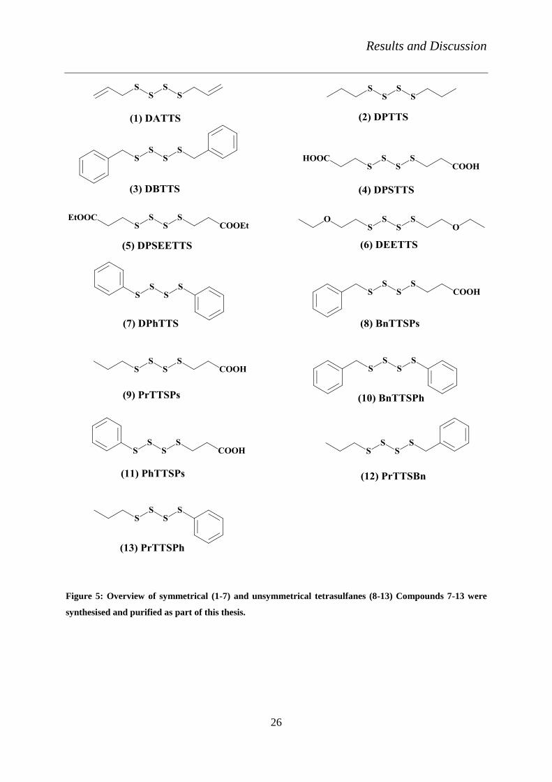
Figure 5: Overview of symmetrical (1-7) and unsymmetrical tetrasulfanes (8-13) Compounds 7-13 were
synthesised and purified as part of this thesis.
Results and Discussion
27
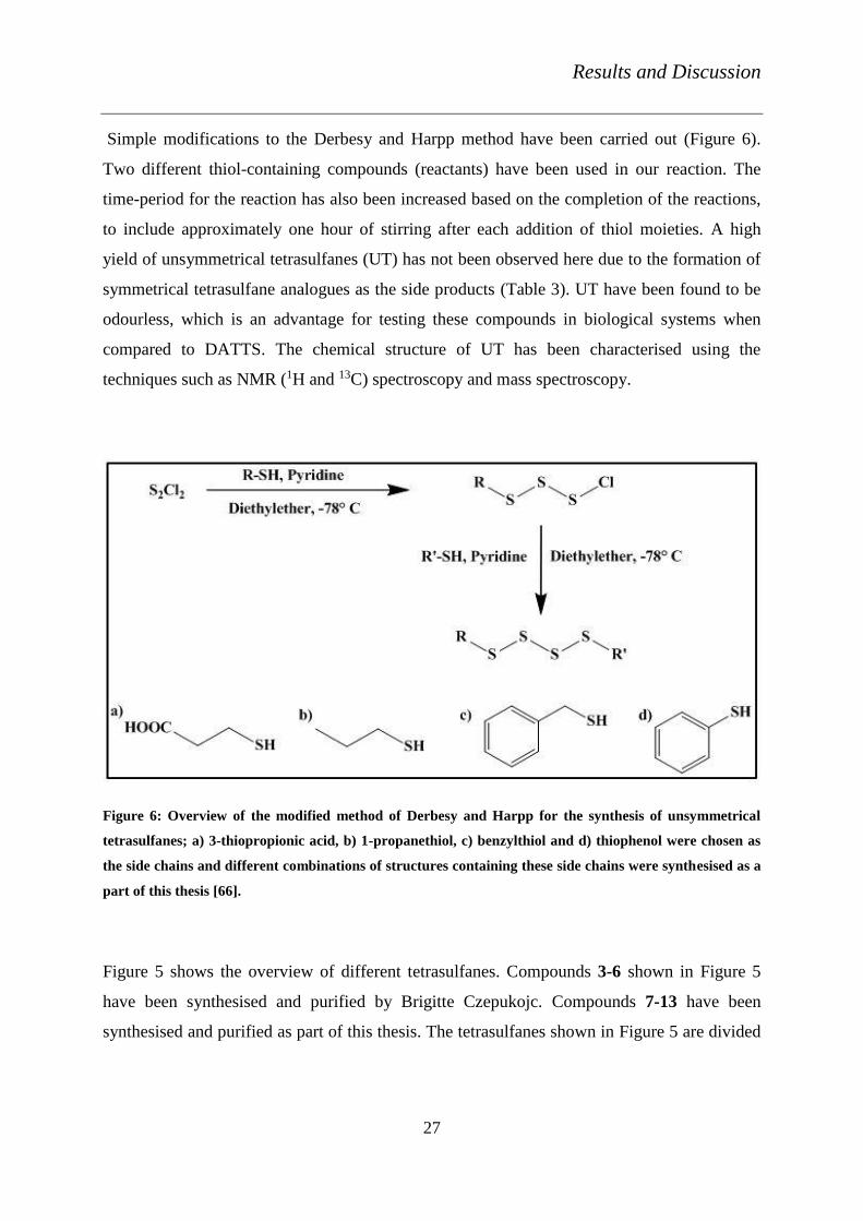
Simple modifications to the Derbesy and Harpp method have been carried out (Figure 6).
Two different thiol-containing compounds (reactants) have been used in our reaction. The
time-period for the reaction has also been increased based on the completion of the reactions,
to include approximately one hour of stirring after each addition of thiol moieties. A high
yield of unsymmetrical tetrasulfanes (UT) has not been observed here due to the formation of
symmetrical tetrasulfane analogues as the side products (Table 3). UT have been found to be
odourless, which is an advantage for testing these compounds in biological systems when
compared to DATTS. The chemical structure of UT has been characterised using the
techniques such as NMR (1H and 13C) spectroscopy and mass spectroscopy.
Figure 6: Overview of the modified method of Derbesy and Harpp for the synthesis of unsymmetrical
tetrasulfanes; a) 3-thiopropionic acid, b) 1-propanethiol, c) benzylthiol and d) thiophenol were chosen as
the side chains and different combinations of structures containing these side chains were synthesised as a
part of this thesis [66].
Figure 5 shows the overview of different tetrasulfanes. Compounds 3-6 shown in Figure 5
have been synthesised and purified by Brigitte Czepukojc. Compounds 7-13 have been
synthesised and purified as part of this thesis. The tetrasulfanes shown in Figure 5 are divided

Results and Discussion
28
into two groups such as symmetrical (1-7) and unsymmetrical tetrasulfanes (8-13), for the
sake of comparative physico-chemical and biological studies.
Table 3: The tetrasulfanes synthesised as part of this thesis with their clogP and respective yields.
Compounds clogP Yield (%)
DPhTTS (7) 5.04 90
BnTTSPs (8) 2.76 75
PrTTSPs (9) 2.74 19
BnTTSPh (10) 5.37 49
PhTTSPs (11) 3.12 72
PrTTSBn (12) 5.00 59
PrTTSPh (13) 4.66 62

Results and Discussion
29
3.2 Physico-chemical properties
Physico-chemical properties of tetrasulfanes have been analysed. Surface tension analysis of
the compounds has been performed using the techniques such as ring tensiometry and sita
tensiometry. The surface tension activity has also been confirmed with densitometry
experiments. This activity of the compounds in turn reveals their amphiphilic nature. The
redox properties of the compounds have been studied using Cyclic Voltammetry. The results
and discussion of each experiment is discussed individually under different sections. Figure 7
illustrates the overview of the experiments performed with the compounds and their aims.
Figure 7: Overview of the physico-chemical measurements performed with tetrasulfanes and Se- and Te-
containing compounds

Results and Discussion
30
3.2.1 Electrochemistry
In the case of polysulfanes redox activity is considered to be key to their biological activity
[67] [68]. Hence, electrochemical analysis of compounds DATTS (1), DBTTS (3), DPSTTS
(4), DPSEETTS (5), DEETTS (6), DPhTTS (7), BnTTSPs (8), PrTTSPs (9), PhTTSBn (10)
and PhTTSPs (11) has been performed using Cyclic Voltammetry in conjunction with a
dropping mercury electrode [69] [70] [71]. The cathodic reduction potentials (Epc) and the
anodic oxidation potentials (Epa) of the compounds have been compared with the ones of
DATTS (1) (Table 4). The ST and UT were found to be redox active and the obtained E1/2
values of these compounds were similar to the value of DATTS. Cyclic voltammogram of
BnTTSPs (9) and PrTTSPs (8) in a dropping mercury electrode experiment is shown in Figure
8.
Table 4: Overview of Epc, Epa and E1/2 of tetrasulfanes.
Compound Epa (mV) Epc (mV) E1/2 (mV)
DATTS (1) -603 -680 -642
DPTTS (2) -597 -682 -640
DBTTS (3) -594 -645 -619
DPSTTS (4) -614 -688 -651
DPSEETTS (5) -602 -679 -641
BnTTSPs (8) -533 -840 -687
PrTTSPs (9) -560 -786 -674
PhTTSBn (10) -559 -652 -606
PhTTSPs (11) -566 -748 -658

Results and Discussion
31
Figure 8: Cyclic voltammogram of BnTTSPs (9) and PrTTSPs (8) in a dropping mercury electrode
experiment.
3.2.2 Sita tensiometry measurements
The unsymmetrical tetrasulfanes with propionic acid side chains have been expected to
exhibit amphiphilic properties. This experiment has been performed to measure the changes in
surface tension of the compounds with the change in their concentration using the bubble
pressure method [72] [73] [74]. The identification of micelle formation could be carried out
by calculating the CMC of the compounds [75] [76]. SDS (14), a surface-active molecule, has
been used as a control. These compounds have been analysed along with DPSTTS (4) at
different concentrations from 1 µM to 3 mM in distilled water with 1 % DMSO. Surprisingly,
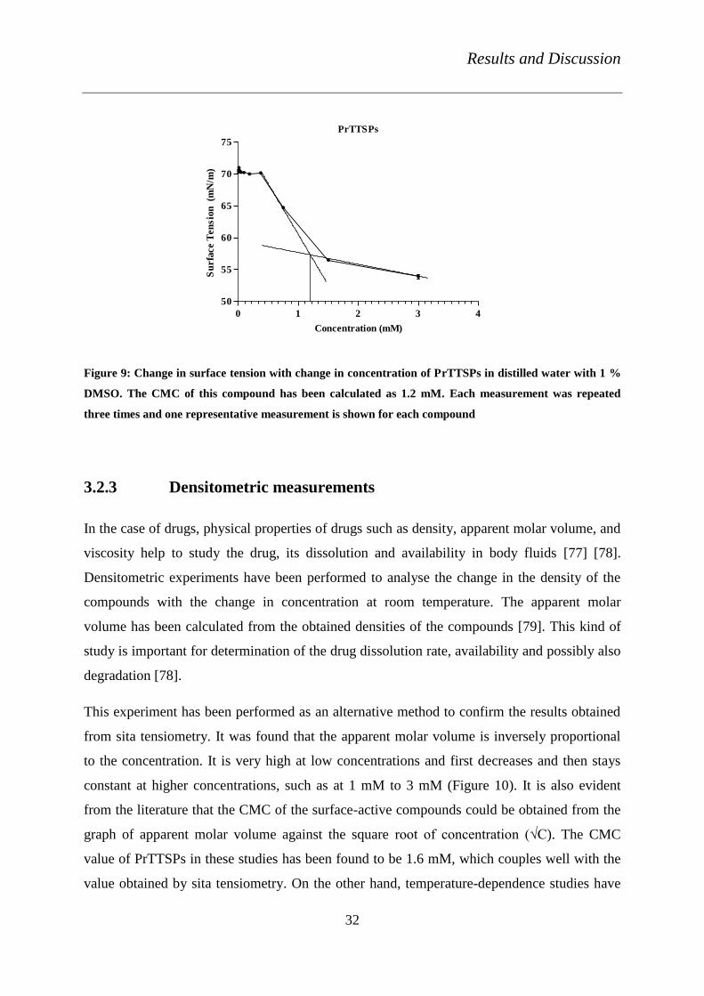
PrTTSPs (9) exhibited a CMC of 1.2 mM (Figure 9). This initial experiment supports the idea
of micelle formation of tetrasulfanes. PhTTSPs (11) behaved differently in this experiment.
The surface tension of the compound dropped at 2.6 mM (data not shown). This concentration
could be the CMC of the compound, but it could also be due to the high ionic concentration of
the liquid medium. The CMC also varies with temperature and the solvent used in the
experiment. The other tetrasulfanes tested did not show any surface activity under the
experimental conditions used.
Results and Discussion
32
PrTTSPs
0 1 2 3 4
50
55
60
65
70
75
Concentration (mM)
Su
rfa
ce T
en
sio
n (m
N/m
)
Figure 9: Change in surface tension with change in concentration of PrTTSPs in distilled water with 1 %
DMSO. The CMC of this compound has been calculated as 1.2 mM. Each measurement was repeated
three times and one representative measurement is shown for each compound
3.2.3 Densitometric measurements
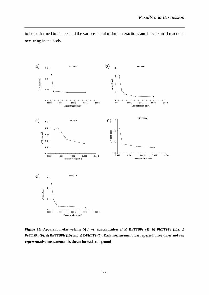
In the case of drugs, physical properties of drugs such as density, apparent molar volume, and
viscosity help to study the drug, its dissolution and availability in body fluids [77] [78].
Densitometric experiments have been performed to analyse the change in the density of the
compounds with the change in concentration at room temperature. The apparent molar
volume has been calculated from the obtained densities of the compounds [79]. This kind of
study is important for determination of the drug dissolution rate, availability and possibly also
degradation [78].
This experiment has been performed as an alternative method to confirm the results obtained
from sita tensiometry. It was found that the apparent molar volume is inversely proportional
to the concentration. It is very high at low concentrations and first decreases and then stays
constant at higher concentrations, such as at 1 mM to 3 mM (Figure 10). It is also evident
from the literature that the CMC of the surface-active compounds could be obtained from the
graph of apparent molar volume against the square root of concentration (√C). The CMC
value of PrTTSPs in these studies has been found to be 1.6 mM, which couples well with the
value obtained by sita tensiometry. On the other hand, temperature-dependence studies have
Results and Discussion
33
to be performed to understand the various cellular-drug interactions and biochemical reactions
occurring in the body.
BnTTSPs
0.000 0.001 0.002 0.003 0.004
0.0
0.5
1.0
1.5
V
(d
m3/m
ol)
Concentration (mol/l)
PhTTSPs
0.000 0.001 0.002 0.003 0.004
0
1
2
3
4
Concentration (mol/l)
V
(d
m3/m
ol)
PrTTSPs
0.000 0.001 0.002 0.003 0.004
0.0
0.1
0.2
0.3
0.4
0.5
Concentration (mol/l)
V
(d
m3/m
ol)
PhTTSBn
0.000 0.001 0.002 0.003 0.004
0.0
0.5
1.0
1.5
Concentration (mol/l)
V
(d
m3/m
ol)
DPhTTS
0.000 0.001 0.002 0.003 0.004
0
1
2
3
Concentration (mol/l)
V
(d
m3/m
ol)
a) b)
c) d)
e)
Figure 10: Apparent molar volume (ɸV) vs. concentration of a) BnTTSPs (8), b) PhTTSPs (11), c)
PrTTSPs (9), d) BnTTSPh (10) and e) DPhTTS (7). Each measurement was repeated three times and one
representative measurement is shown for each compound

Results and Discussion
34
3.3 Molecular Non-Covalent Interaction Studies
3.3.1 Haemolytic assay
This assay has been performed to measure the non-covalent molecular interactions of the
tetrasulfanes with Red Blood Cells (RBCs) [49]. In the following study, the results of all the
compounds have been compared to DPTTS (2). PrTTSPs (9) exhibited nearly 60 % of
haemolysis and an EC50 value of 264.2 µM, when compared to DPTTS (2), which exhibited
nearly 42 % of haemolysis and an EC50 value of 724.2 µM (Figure 11). The compounds
DPhTTS (7) and BnTTSPh (10), however, presented an EC50 value of 385.6 µM and 266.2
µM, respectively. These compounds also indicated the haemolysis almost equal to that of
DPTTS (2). BnTTSPs (8) also exhibited haemolysis of nearly 46 % (data not shown), which
is in close accordance with DATTS (1). On the other hand, PhTTSPs (11) did not show
significant haemolysis of RBCs.

Results and Discussion
35
Figure 11: a) Haemolysis of RBCs incubated with the compounds at 1 mM concentration. b)
Concentration-dependence studies of DPTTS (2), PrTTSPs (9), DPhTTS (7), and PhTTSBn (10) on
haemolysis of RBCs. Absorbance was recorded in UV-visible spectrophotometer at 540nm. Data
presented as haemolysis percentage ± SEM. ANOVA test: p < 0.05 (*), p < 0.01 (**) or p < 0.001 (***) in
relation to the relevant values for the particular control DPTTS.
10 100 250 500 750 1000 DMSO0
20
40
60
80
n.sn.s
n.s
***
n.sn.s ***
**
***
***
n.s
n.s
**
n.s
n.s
n.s
DPTTS
PrTTSPs
DPhTTS
PhTTSBn
Concentration (M)
Hem
oly
sis
%
b)
DPTTS DPhTTS PrTTSPs PhTTSBn PhTTSPs 1%DMSO0
20
40
60
80DPTTS
DPhTTS
PrTTSPs
PhTTSBn
PhTTSPs
1%DMSO
nsns
ns
*
ns
Concentration (1mM)
Hem
oly
sis
%
a)

Results and Discussion
36
3.3.2 FRET measurements
The Fluorescence Resonance Energy Transfer (FRET) mechanism involves two chemical
species, a fluorophore, that when excited emits fluorescence, and a quencher that reduces the
fluorescence of the fluorophore [80]. When the FRET mechanism is applied to optical
microscopy the spatial approach between two molecules, within several nanometers, can be
studied [81]. The combination of FRET and optical microscopy also reveals information
about the binding to and interaction with proteins, lipids, enzymes, DNA and RNA [82] [83].
The fluorescence quenching can be measured quantitatively and analysed using the Stern-
Volmer equation.
Fo / F[Q] = 1 + Ksv [Q] or Ksv = (Fo / F[Q] - 1) / [Q]
Equation 2: The Ster-Volmer equation.
In equation 2, [Q] represents the concentration of the quencher; Fo represents the fluorescence
intensity measured in the absence of a quencher, F[Q] represents the fluorescence intensity
measured in the presence of a quencher, and Ksv represents the Stern-Volmer constant. A
slope can be obtained from the plot of Fo / F[Q] against [Q]. This slope value represents the
Stern-Volmer constant. In general, a more sensitive system results in a steeper slope value,
which in turn represents a higher Ksv.
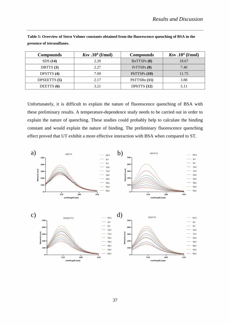
The FRET measurements of compounds 3-11 have been performed with BSA. A shift in the
intensity of the fluorescence of BSA has been observed in the presence of tetrasulfanes. This
effect is also visible in Figure 12. SDS (14) was used as a control and showed a Ksv value of
2.20 .104 l/mol. UT exhibited higher values of Ksv when compared to the ones of ST.
PhTTSPs (11) and BnTTSPs (8) exhibit the greatest fluorescence quenching effect among all
the tetrasulfanes. PrTTSPs (9) (UT) exhibited a similar effect to that of DPSTTS (4) (ST). The
Ksv values of tetrasulfanes are summarised in Table 5.
Results and Discussion
37
Table 5: Overview of Stern Volmer constants obtained from the fluorescence quenching of BSA in the
presence of tetrasulfanes.
Compounds Ksv .104 (l/mol) Compounds Ksv .104 (l/mol)
SDS (14) 2.20 BnTTSPs (8) 18.67
DBTTS (3) 2.27 PrTTSPs (9) 7.40
DPSTTS (4) 7.00 PhTTSPs (10) 11.75
DPSEETTS (5) 2.17 PhTTSBn (11) 3.88
DEETTS (6) 3.21 DPhTTS (12) 5.11
Unfortunately, it is difficult to explain the nature of fluorescence quenching of BSA with
these preliminary results. A temperature-dependence study needs to be carried out in order to
explain the nature of quenching. These studies could probably help to calculate the binding
constant and would explain the nature of binding. The preliminary fluorescence quenching
effect proved that UT exhibit a more effective interaction with BSA when compared to ST.
350 400 450
0
100
200
300
400
500
BSA
2l
5l
10l
15l
20l
30l
40l
50l
60l
DBTTS
wavelength (nm)
Inte
nsit
y (
a.u
)
350 400 450
0
100
200
300
400
500
BSA
2l
5l
10l
15l
20l
30l
40l
50l
60l
DPSTTS
wavelength (nm)
Inte
nsit
y (
a.u
)
350 400 450
0
100
200
300
400
500
BSA
2l
5l
10l
15l
20l
30l
40l
50l
60l
DPSEETTS
wavelength (nm)
Inte
nsit
y (
a.u
)
350 400 450
0
100
200
300
400
500
BSA
2l
5l
10l
15l
20l
30l
40l
50l
60l
DEETTS
wavelength (nm)
Inte
nsit
y (
a.u
)
a) b)
c) d)

Results and Discussion
38
350 400 450
0
100
200
300
400BSA
2µl
5µl
10µl
15µl
20µl
30µl
40µl
50µl
60µl
BnTTSPs
wavelength (nm)
Inte
nsit
y (
a.u
)
350 400 450
0
100
200
300
400BSA
2µl
5µl
10µl
15µl
20µl
30µl
40µl
50µl
60µl
PrTTSPs
wavelength (nm)
Inte
nsit
y (
a.u
)
350 400 450
0
100
200
300
400BSA
2µl
5µl
10µl
15µl
20µl
30µl
40µl
50µl
60µl
PhTTSPs
wavelength (nm)
Inte
nsit
y (
a.u
)
350 400 450
0
100
200
300
400BSA
2µl
5µl
10µl
15µl
20µl
30µl
40µl
50µl
60µl
PhTTSBn
wavelength (nm)
Inte
nsit
y (
a.u
)
350 400 450
0
100
200
300
400BSA
2µl
5µl
10µl
15µl
20µl
30µl
40µl
50µl
60µl
DPhTTS
wavelength (nm)
Inte
nsit
y (
a.u
)
e) f)
g) h)
i)
Figure 12: The quenching of the fluorescence bovine serum albumin with different concentrations of a)
DBTTS (3), b) DPSTTS (4), c) DPSEETTS (5), d) DEETTS (6), e) BnTTSPs (8), f) PrTTSPs (9), g)
PhTTSPs (11), h) PhTTSBn (10) and i) DPhTTS (7) at an excitation wavelength of 295 nm. Each
measurement was repeated three times and one representative measurement is shown for each compound.

Results and Discussion
39
3.3.3 Circular dichroism measurements
This experiment has been performed to study the interaction of compound PrTTSPs (9) with
the protein, haemoglobin. This experiment was an alternative method to study the non-
covalent membrane interactions of tetrasulfanes. PrTTSPs (9) showed a maximum haemolysis
in our experiments and therefore was chosen for circular dichroism (CD) [84] [85]
measurements, at various concentrations of 50 µM, 100 µM and 200 µM. A significant
change in the shift of the wavelength of absorption of haemoglobin was not observed. It could
be also possible that 200 µM of PrTTSPs (9) was a very low concentration to produce a
significant shift in the wavelength. In the future, CD experiment needs to be performed with
higher concentrations of tetrasulfanes.

Results and Discussion
40
3.4 Biological activities of the compounds
The biological activities of compounds 1-11 have been studied. First of all, detailed
antimicrobial analyses have been carried out. Experiments have been performed with
eukaryotic cell lines such as MH7A and neuronal cell lines of rats. An overview of the
biological assays performed with the compounds is shown in Figure 13
Figure 13: Overview of the biological assays performed with various tetrasulfanes and Se- and Te-
containing compounds against different microorganisms and in different cell lines.

Results and Discussion
41
3.4.1 Activity against microbes and small organisms
3.4.1.1 Nematode assay
As a pre-screening of drugs or to study the important metabolites and their actions, model
organisms such as the common yeast strain, Saccharomyces cerevisiae, fruit flies or
nematodes are being widely used by scientists primarily due to ethical considerations [48].
These organisms also show easy reproduction, enabling repeated studies.
Nematodes are genetically tractable hermaphrodites, transparent organisms visible under the
microscope. The co-ordination, reproduction and chemotaxis could be studied in these
organisms; thus, they are ideal for the pre-screening of our compounds before any complex
analysis [86]. They may also reveal certain possible agricultural applications of our
compounds.
DMSO 10 50 100 250 500 10000
20
40
60
80
100
120
0 hour
6 hour
24 hour
***
*
***
**
***
***
*** ***
******
***
Concentration (M)
viabil
ity i
n %
a)

Results and Discussion
42
DMSO 25 50 100 250 500 10000
20
40
60
80
100
1200 hour
6 hour
24 hour
***
**
***
*
***
***
***
***
***
***
***
***
Concentration (M)
Via
bil
ity %
b)
DMSO 25 50 100 250 500 10000
20
40
60
80
100
120
0 hour
6 hour
24 hour
n.s
***
**
***
**
***
***
***
***
***
***
***
Concentration (M)
Via
bil
ity %
c)
Results and Discussion
43
DMSO 25 50 100 250 500 10000
20
40
60
80
100
120
0 hour
6 hour
24 hour
ns
***
ns
***
**
***
**
******
***
***
Concentration (M)
Via
bil
ity i
n %
d)
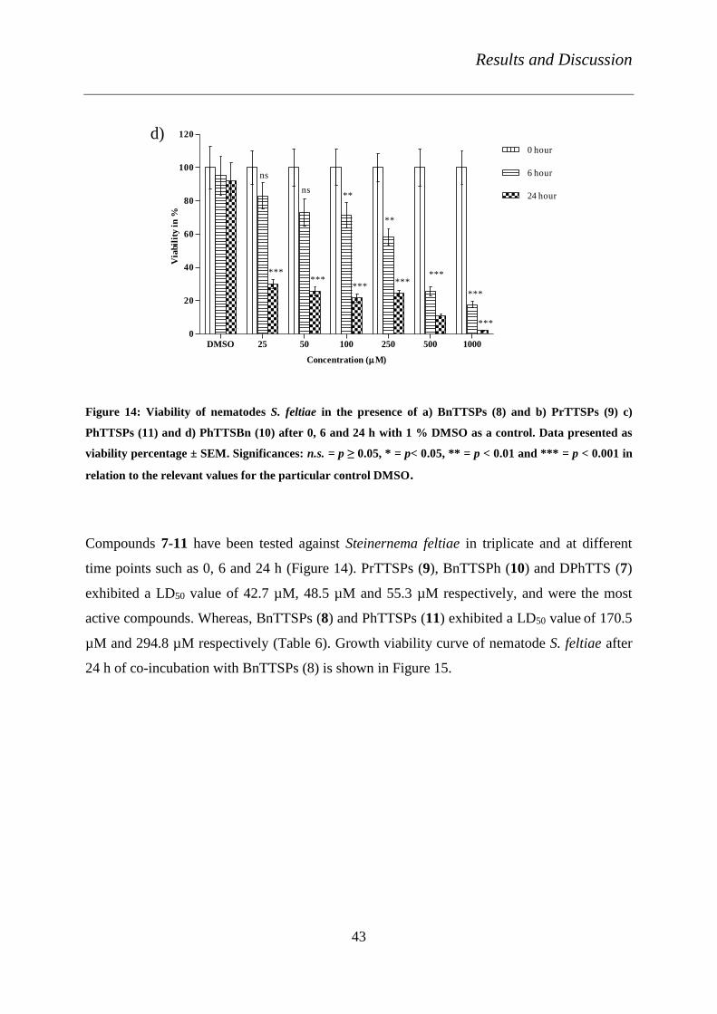
Figure 14: Viability of nematodes S. feltiae in the presence of a) BnTTSPs (8) and b) PrTTSPs (9) c)
PhTTSPs (11) and d) PhTTSBn (10) after 0, 6 and 24 h with 1 % DMSO as a control. Data presented as
viability percentage ± SEM. Significances: n.s. = p ≥ 0.05, * = p< 0.05, ** = p < 0.01 and *** = p < 0.001 in
relation to the relevant values for the particular control DMSO.
Compounds 7-11 have been tested against Steinernema feltiae in triplicate and at different
time points such as 0, 6 and 24 h (Figure 14). PrTTSPs (9), BnTTSPh (10) and DPhTTS (7)
exhibited a LD50 value of 42.7 µM, 48.5 µM and 55.3 µM respectively, and were the most
active compounds. Whereas, BnTTSPs (8) and PhTTSPs (11) exhibited a LD50 value of 170.5
µM and 294.8 µM respectively (Table 6). Growth viability curve of nematode S. feltiae after
24 h of co-incubation with BnTTSPs (8) is shown in Figure 15.

Results and Discussion
44
Table 6: Overview of the compounds tested against nematode S. feltiae and the LD50 values
Compounds LD50 (µM)
DATTS (1) 150.0
DPhTTS (7) 55.3
BnTTSPs (8) 170.5
PrTTSPs (9) 42.7
BnTTSPh (10) 48.5
PhTTSPs (11) 294.8
10 1 .0 10 1 .5 10 2 .0 10 2 .5 10 3 .0
50
100
150
Concentration (M)
Via
bil
ity %
Figure 15: Growth viability curve of nematode S. feltiae after 24 h of co-incubation with BnTTSPs (8).
Data presented as viability percentage ± SEM.
3.4.1.2 Toxicity against different yeast strains
Agar well-diffusion method has been used to study the activity of tetrasulfanes against
different strains of yeast. Here, four different strains of yeast have been used, namely
Debariomyces hansenii, Saccharomyces cerevisiae, Candida guilliermondii and Candida
albicans 174.

Results and Discussion
45
The tetrasulfanes DBTTS (3), DPSTTS (4), DPSEETTS (5), DEETTS (6), DPhTTS (7),
BnTTSPs (8), PrTTSPs (9), BnTTSPh (10) and PhTTSPs (11) have been pre-screened against
the different strains of yeast (Table 7). ST did not exhibit any significant activity against the
strains of yeast except DEETTS (6). Table 7 shows that the compounds DBTTS (3) and
DPSTTS (4) did not exhibit any activity against D. hansenii, but these compounds presented a
minimal inhibitory zone against the other three strains of yeast. DPSEETTS (5) exhibits a
moderate activity against all the four different strains of yeast. DEETTS (6) is the most active
compound among the ST. It shows the zonal diameter of 17.5 ± 4 mm, 19 ± 1 mm, 17.5 ± 4
mm and 23 ± 1 mm against D. hansenii, S. cerevisiae, C. guilliermondii, and C. albicans,
respectively. Thus, DEETTS (6) has been chosen for further concentration-dependence
studies.
Table 7: Pre-screening of compounds 3-11 and 14 at a concentration of 3 mM against four different
strains of yeast, namely D. hansenii, S. cerevisiae, C. guilliermondii and C. albicans 174. A diameter of 12
mm is considered as the minimum inhibitory zone. 100 % of DMSO has been used as control. The values
represent the mean ± SEM of three independent experiments.
Compounds D. hansenii S. cerevisiae C. guilliermondii C. albicans 174
DBTTS (3) - 11.5 ± 0 12.0 ± 0 14.5 ± 2
DPSTTS (4) - 14.5 ± 0 12.0 ± 1 14.5 ± 1
DPSEETTS
(5) 12.0 ± 0 15.5 ± 0 13.5 ± 2 17.5 ± 1
DEETTS (6) 17.5 ± 4 19.0 ± 1 17.5 ± 4 23.0 ± 1
DPhTTS (7) - 13.3 ± 1 17.5 ± 1 15.5 ± 4
BnTTSPs (8) - 14.5 ± 2 16.0 ± 0 14.5 ± 4
PrTTSPs (9) 12.5 ± 1 19.5 ± 7 14.3 ± 0 17.0 ± 4
BnTTSPh (10) - 12.5 ± 1 - 15.0 ± 3
PhTTSPs (11) - 20.0 ± 4 16.0 ± 1 16.3 ± 1
SDS (14) 16.3 ± 0 11.5 ± 1 14.3 ± 0 14.5 ± 1
The activities of all of the compounds have been compared to the activity of SDS (14). SDS
exhibited a minimum inhibitory zone of 16.25 ± 0 mm, 11.5 ± 1 mm, 14.25 ± 0 mm and 14.5
± 1 mm against D. hansenii, S. cerevisiae, C. guilliermondii and C. albicans 174 respectively.
DEETTS (6) did not exhibit a similar activity of SDS (14) against D. hansenii, whereas a

Results and Discussion
46
significant difference was found against the other three yeast strains (Figure 16). DPSEETTS
(5) also exhibited a significant difference in the activity from that of SDS (14), but the activity
was very low against D. hansenii (14), whereas the activity against S. cerevisiae and C.
albicans was considerably higher. The concentration-dependence studies of DEETTS (6) also
exhibited a similar activity as that of SDS (14), against D. hansenii. The respective percentage
inhibition curves are illustrated in Figure 16.
Figure 16: Growth inhibition curve of yeast strains treated with compounds, a) DEETTS against S.
cerevisiae and b) DEETTS against D. hansenii. SDS is used as a particular control. Data presented as
percentage inhibition ± SEM.
As evident from the pre-screening test, tetrasulfanes did not show any significant effect
against S. cerevisiae and SDS did not exhibit a concentration-dependent activity either. The
activities among the compounds themselves were compared to each other. It was found that
PrTTSPs (9) and PhTTSPs (11) at a concentration of 3 mM were considerably more active
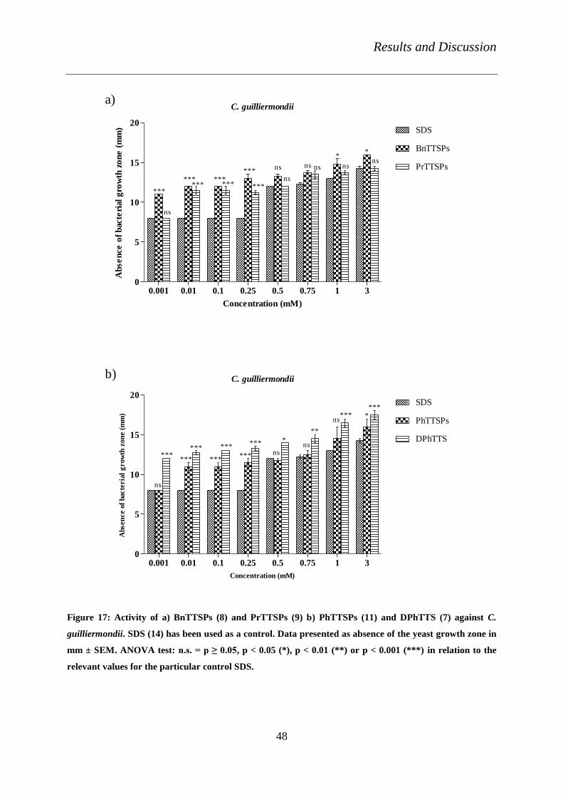
against S. cerevisiae, while other compounds failed to show any significant activity.
The percentage inhibition of PhTTSPs and SDS against C. guilliermondii was analysed.
Compounds BnTTSPs (8), PrTTSPs (9), PhTTSPs (11) and DPhTTS (7) presented
significantly higher activities against C. guilliermondii than the activity of SDS (14) (Figure
17). BnTTSPs (8) exhibited a minimum inhibition at a concentration of 1 µM and PrTTSPs
(9) at a concentration of 10 µM, whereas SDS exhibited a minimum inhibition only at a
10 2 .5 10 3 .0 10 3 .5
20
40
60
80
100DEETTS
S. cerviciae
SDS
Concentration (M)
% I
nh
ibit
ion
a)
10 2 .0 10 2 .5 10 3 .0 10 3 .5 10 4 .0
20
40
60
80
100
SDS
D. hansenii
DEETTS
Concentration (M)
% I
nh
ibit
ion
b)

Results and Discussion
47
concentration of 500 µM. DPhTTS (7), which did not show any activity against D. hansenii,
was effective against C. guilliermondii at very low concentrations (1 µM).
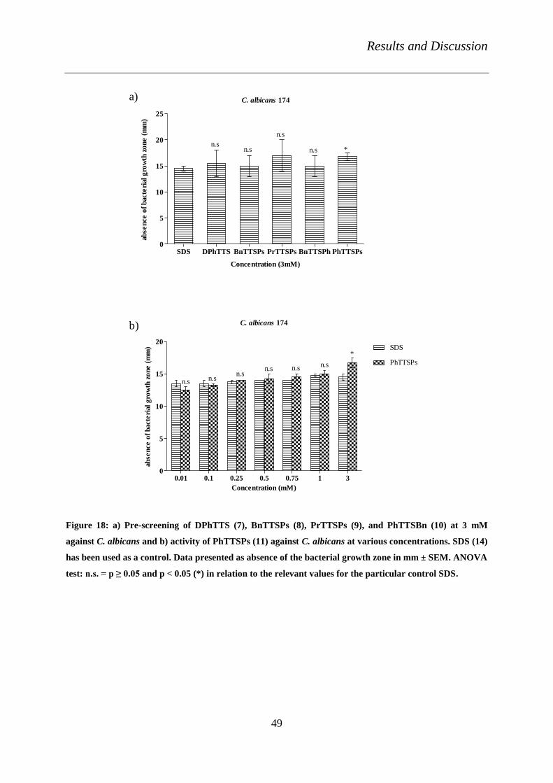
C. albicans 174 is a semi-pathogenic strain. Compounds DPhTTS (7), BnTTSPs (8), PrTTSPs
(9), BnTTSPh (10) and PhTTSPs (11) exhibited an activity against C. albicans 174.
Nevertheless, the activity of DPhTTS (7), BnTTSPs (8) and BnTTSPh (10) could be probably
due to solvent (DMSO) influence but, importantly, PhTTSPs (11) exhibited a complete
inhibitory activity against C. albicans 174 with a MIC of 16.1 µM (Figure 18) (Table 8).
DMSO exhibited a static effect against C. albicans, thus, it is important to note that the
activity of some of the compounds tested is probably due to the influence of DMSO.
Results and Discussion
48
C. guilliermondii
0.001 0.01 0.1 0.25 0.5 0.75 1 30
5
10
15
20SDS
BnTTSPs
PrTTSPs
***
*** ******
ns ns
**
ns
ns*** *** ***
ns nsns
Concentration (mM)
Ab
se
nce
of
bacte
rial
gro
wth
zo
ne
(m
m)
C. guilliermondii
0.001 0.01 0.1 0.25 0.5 0.75 1 30
5
10
15
20SDS
PhTTSPs
DPhTTS
*
ns
*** ****** ns
ns
ns
******
******
*
**
******
Concentration (mM)
Absen
ce o
f bacte
ria
l g
row
th z
on
e (
mm
)
a)
b)
Figure 17: Activity of a) BnTTSPs (8) and PrTTSPs (9) b) PhTTSPs (11) and DPhTTS (7) against C.
guilliermondii. SDS (14) has been used as a control. Data presented as absence of the yeast growth zone in
mm ± SEM. ANOVA test: n.s. = p ≥ 0.05, p < 0.05 (*), p < 0.01 (**) or p < 0.001 (***) in relation to the
relevant values for the particular control SDS.
Results and Discussion
49
C. albicans 174
SDS DPhTTS BnTTSPs PrTTSPs BnTTSPh PhTTSPs0
5
10
15
20
25
n.sn.s
n.s
n.s *
Concentration (3mM)
ab
se
nce
of
bacte
rial
gro
wth
zo
ne
(m
m)
C. albicans 174
0.01 0.1 0.25 0.5 0.75 1 30
5
10
15
20SDS
PhTTSPs
n.s n.sn.s
n.s n.s n.s
*
Concentration (mM)
ab
se
nce
of
bacte
rial
gro
wth
zo
ne
(m
m)
a)
b)
Figure 18: a) Pre-screening of DPhTTS (7), BnTTSPs (8), PrTTSPs (9), and PhTTSBn (10) at 3 mM
against C. albicans and b) activity of PhTTSPs (11) against C. albicans at various concentrations. SDS (14)
has been used as a control. Data presented as absence of the bacterial growth zone in mm ± SEM. ANOVA
test: n.s. = p ≥ 0.05 and p < 0.05 (*) in relation to the relevant values for the particular control SDS.

Results and Discussion
50
Table 8: Overview of the tetrasulfanes tested against corresponding yeast strains with relevant EC50
values.
Compounds Fungi
D. hansenii C. albicans174 C.guillirmondii S. cerviciae
DEETTS (6) 612.5 n.a n.a 860.6
DPhTTS (7) n.a n.a 12.9 n.a
BnTTSPs (8) n.a n.a 15.8 n.a
PrTTSPs (9) n.a n.a 41.1 625.2
PhTTSPs (11) n.a 16.1 334.7 836.5
SDS (14) 230.0 671.9 501.8 n.a
It should be noted here that the commercially available sulfur-containing antibiotic,
sulfadimethoxin was also tested against the different strains of yeast. It is essential to note that
at a concentration of 3 mM, this antimicrobial agent did not show any activity within 24
hours. However, after 48 hours, it exhibited an average inhibitory zone of 18 mm against C.
guilliermondii. The mode of action of Sulfadimethoxin is by inhibiting the formation of folic
acid and this antibiotic is used as a treatment against infections in the respiratory tract, skin or
soft tissue. Nevertheless, compounds DPhTTS, BnTTSPs and PrTTSPs have been
considerably more active against C. guilliermondii, even at very low concentration and within
24 hours.
3.4.1.3 Activity against different filamentous fungi
This assay has been performed to study the activity of the tetrasulfanes against filamentous
fungi such as Trichoderma viride, Aspergillus flavus, Geotrichum candidum, Penicillium
aeruginosa and Mucor plumbeus, using the agar dilution method. The growth of fungi was
monitored for 8-10 days. Initially compounds 3-11, at a concentration of 3 mM were pre-
screened against the fungal strains. The activity of the compounds is tabularised below (Table

Results and Discussion
51
9). Among ST, only DPSEETTS (5) was considerably active against four different strains of
fungi and statically on T. viride. DPSTTS (4) and DEETTS (6) exhibited only a temporary
effect against P. aeruginosa and A. flavus, respectively, and failed to stop the complete
growth of the fungi.
Table 9: Overview of the activity of compounds 3-11, against T. viride, A. flavus, G. candidum, M.
plumbeus and P. aeruginosa, with DMSO as a control. + indicates the complete inhibition of the growth of
fungus, - indicates the absence of any activity of compounds on the fungus and ± indicates the static effect
of the compounds.
Compounds Trichoderma
viride
Aspergillus
flavus
Mucor
plumbeus
Geotrichum
candidum
Penicillium
aeruginosa
DBTTS (3) - - n.a - -
DPSTTS (4) - - n.a - Prolonged the
growth until 8 days
DPSEETTS
(5) ± - n.a - -
DEETTS (6) ± Prolonged the
growth until 6 days n.a + +
BnTTSPs (8) + ± - + +
PrTTSPs (9) + + + + +
PhTTSBn (10) - - - - -
PhTTSPs (11) + + + + +
DMSO - - - - -
UT were the most active compounds against the fungal strains in this experiment. BnTTSPs
(8), PrTTSPs (9), and PhTTSPs (11) were active against all five strains of the filamentous
fungi tested, except for BnTTSPs (8) against M. plumbeus. These results clearly show that the
ST did not exhibit any activity against these strains. The UT (PrTTSPs (9), and PhTTSPs
(11)) stood out among all the other compounds in this experiment. BnTTSPs (8), however,
exhibited only a static activity against A. flavus. PhTTSBn (10) did not exhibit any activity
against the fungal strains. There was also no solvent (DMSO) effect against these strains.
The concentration-dependence studies have also been carried out in the presence of UT
against T. viride. The MIC of compounds BnTTSPs (8), PrTTSPs (9) and PhTTSPs (11) was
determined as ≤ 1 mM, ≤ 2 mM and ≤ 2 mM, respectively. Further in-depth studies have to be
conducted to reveal the mode of action of tetrasulfanes against the different strains of fungi.
Results and Discussion
52
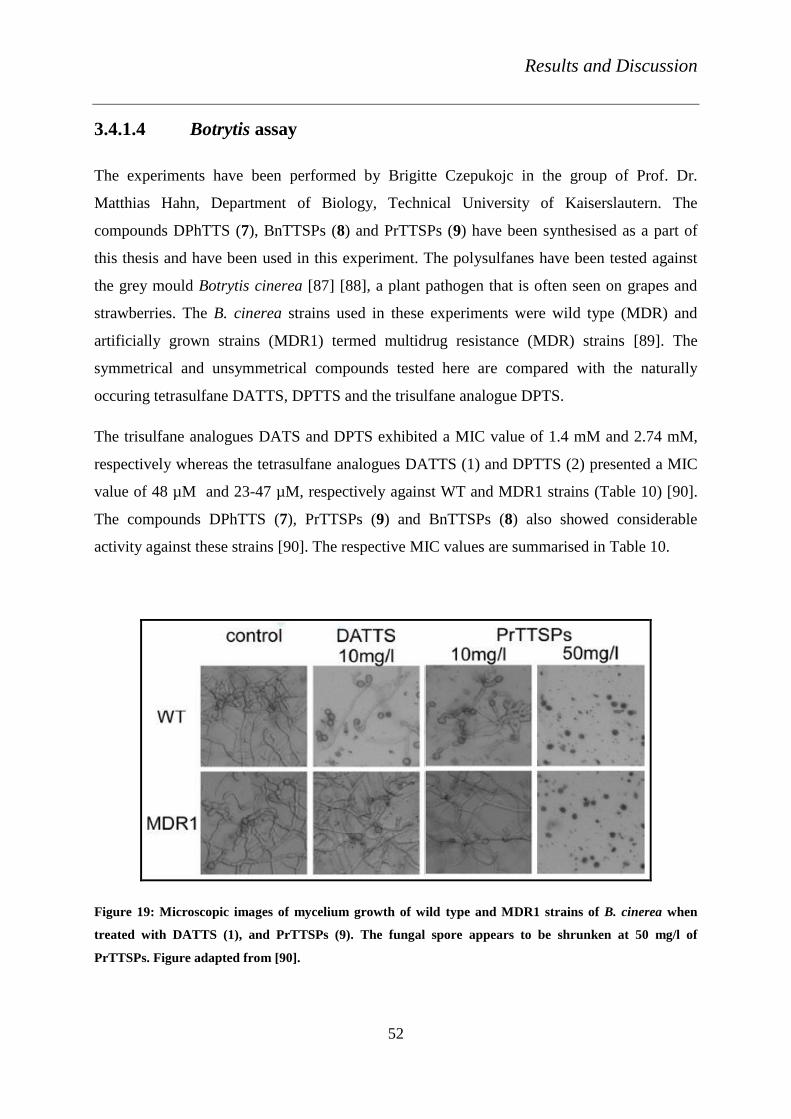
3.4.1.4 Botrytis assay
The experiments have been performed by Brigitte Czepukojc in the group of Prof. Dr.
Matthias Hahn, Department of Biology, Technical University of Kaiserslautern. The
compounds DPhTTS (7), BnTTSPs (8) and PrTTSPs (9) have been synthesised as a part of
this thesis and have been used in this experiment. The polysulfanes have been tested against
the grey mould Botrytis cinerea [87] [88], a plant pathogen that is often seen on grapes and
strawberries. The B. cinerea strains used in these experiments were wild type (MDR) and
artificially grown strains (MDR1) termed multidrug resistance (MDR) strains [89]. The
symmetrical and unsymmetrical compounds tested here are compared with the naturally
occuring tetrasulfane DATTS, DPTTS and the trisulfane analogue DPTS.
The trisulfane analogues DATS and DPTS exhibited a MIC value of 1.4 mM and 2.74 mM,
respectively whereas the tetrasulfane analogues DATTS (1) and DPTTS (2) presented a MIC
value of 48 µM and 23-47 µM, respectively against WT and MDR1 strains (Table 10) [90].
The compounds DPhTTS (7), PrTTSPs (9) and BnTTSPs (8) also showed considerable
activity against these strains [90]. The respective MIC values are summarised in Table 10.
Figure 19: Microscopic images of mycelium growth of wild type and MDR1 strains of B. cinerea when
treated with DATTS (1), and PrTTSPs (9). The fungal spore appears to be shrunken at 50 mg/l of
PrTTSPs. Figure adapted from [90].

Results and Discussion
53
Table 10: Overview of the polysulfanes tested against the wild type and MDR1 B. cinerea strains. The
MIC values in WT strains, resistance factor of MDR1 strains and the strength of AtrB induction are
tabulated below. Table adapted from [90].
Compounds WT MIC
[mg/L] [µM]
Resistance factor
of MDR1
DATTS (1) 10 48 >100x*
DPTTS (2) 5-10 23-47 >100x*
DPhTTS (7) 50 177 2x
BnTTSPs (8) 50 171 1x
PrTTSPs (9) 50 205 1x
DPTS 500 2740 > 2x*
0 no induction or only slight induction at 100 mg/1; + Strong induction only at 100 mg/1 (no induction at 1 mg/1); ++
Medium induction already at 1 mg/1. *the compounds had less solubility at higher concentrations and hence were not
analysed at this concentration.
The compounds have been treated against the mycelium of B. cinerea and their microscopic
images were captured. The fungal spore appears to be shrunken at 50 mg/l of PrTTSPs (9) and
confirmed the apoptosis of the cells (Figure 19). DATTS (1) was found to be considerably
active against the strain of WT compared to the MDR1 strain. Compounds BnTTSPs (8),
PrTTSPs (9) and DPhTTS (7) exhibited effective activity against both the strains of WT type
and MDR1. These compounds could therefore be toxic against the strain of B. cinerea. This
strain, however, may induce resistance mechanism.
3.4.1.5 Antibacterial assay
The inhibitory activity against bacteria has been established by the agar well-diffusion method
as stated in [91]. This assay was performed in order to evaluate the growth inhibitory effect
(bactericide/bacteriostatic) of the compounds implemented. The compounds have been
investigated using a series of concentrations and the EC50 values have been calculated
accordingly.
Compounds 3-11 have been tested against eight different strains of bacteria, namely
Staphylococcus aureus WDCM 5233, Escherichia coli VKPM-M17, Micrococcus luteus,
Bacillus subtilis A1 WT, Bacillus licheniformis WT, Salmonella typhimurium 1754,
Staphylococcus roseus, and Pseudomonas aeruginosa. A pre-screening of the compounds at a
concentration of 3 mM is shown in Table 11, which represents the inhibitory zone diameters
as the mean ± SEM [22].

Results and Discussion
54
Table 11: Pre-screening of compounds 4-6, 8, 9, 11 and 14 at a concentration of 3 mM against five
different bacterial strains: S. aureus, M. luteus, B. subtilis, B. licheniformis and S. roseus. 12 mm of
diameter is considered as the minimum inhibitory zone. 100 % DMSO has been used as a control. The
values represent the mean ± SEM of three independent experiments.
Compounds S. aureus M. luteus B. subtilis B. licheniformis S. roseus
DPSTTS (4) - - - - 12.0 ± 1
DPSEETTS (5) 19.0 ± 1 15.5 ± 1 14.0 ± 1 - 14.8 ± 0
DEETTS (6) 19.5 ± 1 13.0 ± 1 - - -
BnTTSPs (8) 19.7 ± 1 19.0 ± 1 - 25.8 ± 0 28.5 ± 2
PrTTSPs (9) 15.0 ± 2 23.3± 0 20.5 ± 1 25.3 ± 1 25.0 ± 1
PhTTSPs (11) 17.5 ± 1 18.5 ± 2 20.0 ± 1 24.5± 2 23.0 ± 1
SDS (14) 22.5 ± 1 n.a - n.a n.a
Compounds 3-7, being the ST, were active only against four different strains of bacteria.
DPSEETTS (5) is one of those compounds, which showed an inhibitory diameter of 19 ± 1
mm against S. aureus; this is comparable to BnTTSPs (8), which shows a similar inhibitory
zone. In addition, DPSEETTS (5) exhibited an activity against B. subtilis, M. luteus and S.
roseus. DBTTS (3), DPhTTS (7) and BnTTSPh (10) did not show any activity against any of
the strains of bacteria. None of the compounds exhibited an activity against E. coli, S.
typhimurium or P. aeruginosa.
Compounds BnTTSPs (8), PrTTSPs (9) and PhTTSPs (11) exhibited a very high activity
against S. aureus, M. luteus, B. licheniformis and S. roseus. Surprisingly, only PhTTSPs (11)
showed an activity against the sporulating B. subtilis strain, with an inhibitory zone of 17.5 ±
1 mm. These results indicates the effectiveness of the activity of the compounds with a
propionic acid as a side chain, which is expected to be amphiphilic in nature. Nevertheless,
none of the compounds was active against the Gram-negative bacteria, E. coli. In other words,
this could be a simple hint of the mode of action of tetrasulfanes, as Gram-positive and Gram-
negative bacteria have different structures of cell walls. From the above results, it is evident
that UT exhibited greater activity against the strains of bacteria when compared to ST.
In order to perform the concentration-dependence studies with ST and UT S. aureus strain has
been selected among the eight different strains of bacteria. S. aureus is a Gram-positive
Results and Discussion
55
bacterium, that is found in the human respiratory system and on the skin. This species can
cause serious skin infections, respiratory diseases and food poisoning. It also has the ability to
rapidly gain resistance against the antibiotics. Thus, for the concentration-dependence studies,
S. aureus was the primary choice and most of the tetrasulfanes were active against this strain
during the pre-screening (Table 11). A series of concentrations of the compounds from 1 µM
to 3 mM was prepared in DMSO and the relevant EC50 values were calculated. DMSO and
SDS (14) have been used as negative and positive controls, respectively.
None of the tetrasulfanes exhibited an activity against E. coli or S. typhimurium, which are
Gram-negative bacteria. SDS (14), however, exhibited an EC50 value of 773.1 µM. against E.
coli. The B. subtilis strain used in this experiment was a unique one that possess spore-
forming ability. Only compounds DPSEETTS (5) and PhTTSPs (11) exhibited activity
against B. subtilis in this experiment.
Among ST, the concentration dependence studies were only performed with DEETTS (6)
against S. aureus. This compound exhibited a significant activity at a concentration of 0.25
mM when compared to SDS. The EC50 value of DEETTS against S. aureus was calculated as
667.6 µM and that of SDS as 772.9 µM (Table 12). Only DPSEETTS (5) was active against
B. subtilis, whilst SDS (14) did not show any inhibitory effect against this strain even at a
concentration of 3 mM. DPSEETTS was also active against S. aureus, but did not exhibit a
concentration-dependent activity. The bar graphs without the standard deviation show that the
same zonal diameters were obtained in three independent experiments.
BnTTSPs (8), PrTTSPs (9) and PhTTSPs (11) were found to show inhibitory zones with
diameters of 19.66 ± 1 mm, 15 ± 2 mm and 17.5 ± 1 mm, respectively, against S. aureus at a
concentration of 3mM. Concentration-dependence studies have been carried out (Figure 20).
BnTTSPs (8) exhibited a significant activity at very low concentration of 0.1 mM, against S.
aureus, when compared to SDS. PrTTSPs (9), PhTTSPs (11) and SDS (14) did not show any
activity against S. aureus at low concentrations (below 0.5 mM). PhTTSPs (11) was the only
compound that exhibited a significant effect against B. subtilis with an inhibitory percentage
of 85-90 % at a concentration of 3 mM and an EC50 value of 646.6 µM. This compound did
not show any effect against Gram-negative bacteria (E. coli and S. typhimurium).
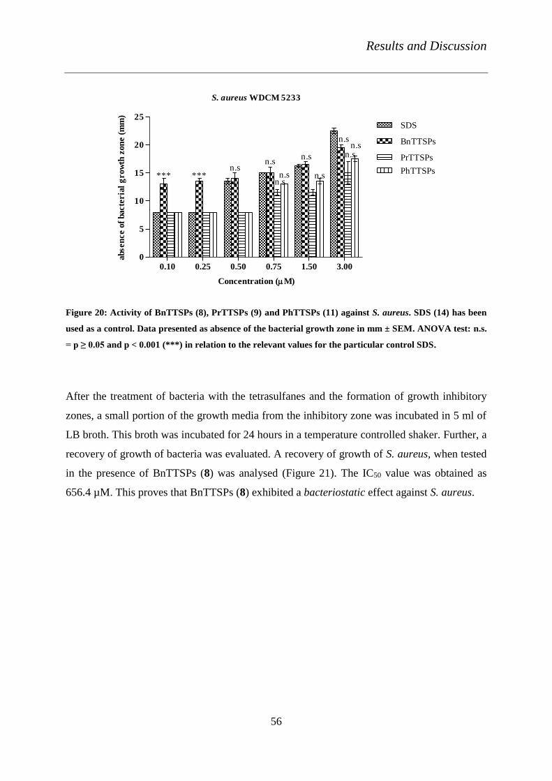
Results and Discussion
56
S. aureus WDCM 5233
0.10 0.25 0.50 0.75 1.50 3.000
5
10
15
20
25SDS
BnTTSPs
PrTTSPs
PhTTSPsn.sn.s
n.s
n.s
n.sn.sn.s*** ***
n.sn.s
Concentration (M)
absen
ce o
f bacte
ria
l g
row
th z
on
e (
mm
)
Figure 20: Activity of BnTTSPs (8), PrTTSPs (9) and PhTTSPs (11) against S. aureus. SDS (14) has been
used as a control. Data presented as absence of the bacterial growth zone in mm ± SEM. ANOVA test: n.s.
= p ≥ 0.05 and p < 0.001 (***) in relation to the relevant values for the particular control SDS.
After the treatment of bacteria with the tetrasulfanes and the formation of growth inhibitory
zones, a small portion of the growth media from the inhibitory zone was incubated in 5 ml of
LB broth. This broth was incubated for 24 hours in a temperature controlled shaker. Further, a
recovery of growth of bacteria was evaluated. A recovery of growth of S. aureus, when tested
in the presence of BnTTSPs (8) was analysed (Figure 21). The IC50 value was obtained as
656.4 µM. This proves that BnTTSPs (8) exhibited a bacteriostatic effect against S. aureus.

Results and Discussion
57
10 1 10 2 10 3 10 4
0
20
40
60
80
100
Concentration (M)
viabil
ity i
n %
Figure 21: a) Recovery of viability curve of S. aureus, after 24 h of treatment with BnTTSPs (8). Data
provided as viability percentage ± SEM. DMSO has been used as a control
Table 12: Overview of the tetrasulfanes tested against different bacterial strains with relevant EC50 values
of growth inhibition.
Compounds
EC50 of Inhibition
of S. aureus (µM)
DEETTS (6) 667.6
BnTTSPs (8) 259.4
PrTTSPs (9) 747.0
PhTTSPs (11) 775.9
SDS (14) 772.9
Different sulfur-containing compounds and their MIC values against different microbial
strains, collected from different experiments are tabulated in Table 13. Although it is not
appropriate method, a small comparative study has been performed. It is evident that DATTS
is more active than ST. UT, however exhibit greater activity than DATTS. The MIC value of
DATTS against S. aureus has been obtained as 0.5 mg/ml, whereas the EC50 value of UT is in

Results and Discussion
58
the micromolar range. Importantly, DATTS shows an effect against P. aeruginosa, which is
not found for any of the tetrasulfanes in our experiments [13].
Table 13: The minimum inhibitory concentrations of sulfur-containing compounds against different
microbial strains are tabulated. These are the results from different publications, in order to compare the
activity of different compounds. The MIC value is provided in g/ml.
Microorganism Allicin DAS DADS DATS DATTS Reference
S. aureus 15 20 4 2 0.5
[13] P. aeruginosa 15 88 72 36 16
C. albicans 0.8 32 4 1 0.5
A. niger 32 40 8 2 1
3.4.1.6 Anti-bacteriophage assay
The anti-bacteriophage assay was performed using the classical agar overlay method [92].
The structure of T4 phage consists of a head part, a tail and a base plate. The head part of the
phage contains the DNA. The base plate is attached to both long and short tail fibres. Using
the long and short tail fibres the phage binds with the host cell organism and bacteria
respectively. Followed by the binding, the phage transfers its genome into the host cell
organism. [93]. The T-4 phage was bound with the strain of E. coli C-T4 in our experiments.
Tetrasulfanes, 3-11, have been screened at specific concentrations of 0.2 mM, 0.5 mM, 0.75
mM and 1.5 mM and with different dilutions of phage solutions (T4 phage in 0.96 % NaCl).
DMSO has been used as a solvent control and the values of viability obtained for DMSO have
been normalised to 100 % of viability.
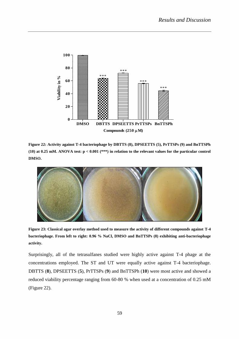
Results and Discussion
59
DMSO DBTTS DPSEETTS PrTTSPs BnTTSPh0
20
40
60
80
100
***
***
***
***
Compounds (250 M)
Via
bil
ity i
n %
Figure 22: Activity against T-4 bacteriophage by DBTTS (8), DPSEETTS (5), PrTTSPs (9) and BnTTSPh
(10) at 0.25 mM. ANOVA test: p < 0.001 (***) in relation to the relevant values for the particular control
DMSO.
Figure 23: Classical agar overlay method used to measure the activity of different compounds against T-4
bacteriophage. From left to right: 0.96 % NaCl, DMSO and BnTTSPs (8) exhibiting anti-bacteriophage
activity.
Surprisingly, all of the tetrasulfanes studied were highly active against T-4 phage at the
concentrations employed. The ST and UT were equally active against T-4 bacteriophage.
DBTTS (8), DPSEETTS (5), PrTTSPs (9) and BnTTSPh (10) were most active and showed a
reduced viability percentage ranging from 60-80 % when used at a concentration of 0.25 mM
(Figure 22).

Results and Discussion
60
Table 14: IC50 values of compounds DBTTS (8), DPSTTS (4), DPSEETTS (5), DEETTS (6), and PrTTSPs
(9) with respect to T4- bacteriophage after 24 h of incubation.
Compounds IC50 (mM)
DBTTS (3) ≤ 0.25
DPSTTS (4) ≤ 0.75
DPSEETTS (5) ≤ 0.30
DEETTS (6) ≤ 0.75
PrTTSPs (9) ≤ 0.25
Application of BnTTSPs (8), PhTTSPs (11) and DPhTTS (7) resulted in 100 % inhibition of
growth of T-4 phage at a concentration of 0.25 mM of tetrasulfanes. Further concentration-
dependence studies are now required for compounds 8, 11 and 7 in order to determine their
respective MIC values. Figure 23 shows the reduced number of bacteriophage when treated
with BnTTSPs (8) in comparison to the number of bacteriophages in 0.96 % NaCl
physiological solution and DMSO as solvent control. From these preliminary studies, the IC50
values of the tetrasulfanes were identified (Table 14). Further in-depth analyses must be
conducted to reveal the mode of action and specific interactions of the compounds.

Results and Discussion
61
3.5 Activity in Primary Cell lines
3.5.1 Activity in human synovial fibroblast cell lines
Several natural and ancient therapies involve natural products that contain sulfur or sulfur-
containing compounds. Inflammatory or allergic reactions, arthritis and other joint disorders,
as well as control of tumour growth are some of the cases where these compounds have
shown a considerable impact. Therefore, a comparative study of the known compound
DBTTS (3), together with its structural analogues DPhTTS (7) and BnTTSPh (10)
(synthesised as part of this thesis) on human synovial fibroblast cell lines (MH7A) has been
performed. These experiments have been performed in the group of our co-operation partner
Dr Burkhard Kloesch, at the Ludwig Boltzmann Institute for Rheumatology and Balneology,
Vienna, Austria.
The apoptotic and anti-inflammatory properties have been studied with tetrasulfanes
containing aromatic benzyl and phenyl side chains. The cytotoxic effects of these cell lines
have been investigated by measuring the lactate dehydrogenase (LDH activity). The cell lines
have been co-incubated with the compounds at different concentrations for 24 h and the
increase in LDH activity in the supernatants of the cell culture was determined using a
commercially available kit (Roche). DBTTS (3) considerably increased LDH activity, which
confirms the high cytotoxic effect that interestingly disappeared when the compound has been
co-incubated with GSH. BnTTSPh (10) had a reduced effect and DPhTTS (7) did not show
any cytotoxic effect as well.
The pro-apoptotic impact of the compounds in these cell lines has been studied by Annexin-
V/7-AAD. In comparison to other compounds, DBTTS (3) triggered an increase in apoptosis,
when applied in concentrations of less than 2 µM. The increase in caspase 3/7 activity has
been measured with a commercially available kit.
The inhibition of IL-1β induced secretion of IL-6 of the MH7A cell lines have been studied.
The compounds have been incubated with MH7A cell lines at different concentrations. These
studies were performed with the enzyme-linked immunosorbent assay (ELISA). DPhTTS (7)

Results and Discussion
62
and BnTTSPh (10) did not show significant inhibition of IL-6 secretion, whereas DBTTS (3)
exhibited a dose-dependent inhibition of IL-6 secretion additionally changed the morphology
of the cells.
The adhesiveness of the MH7A cell lines was studied in the presence of tetrasulfanes. The
experiments were performed by trypsinising the cells at different time points and counting the
cells using a cell analyser. The studies were confirmed by Crystal Violet staining techniques.
All three compounds inhibited the adherence of the cells, but at different concentrations.
DPhTTS (7) showed the inhibition of adherence only when employed at higher
concentrations, such as 5 and 10 µM. DBTTS (3) and BnTTSPh (10), in contrast, inhibited
the adherence of the cells in a concentration-dependent manner and DBTTS (3) in the most
effective way at very low concentrations, starting at 0.5 µM.
These results are, however, preliminary. Further studies are ongoing to confirm these
preliminary results and potential activity of tetrasulfanes in MH7A cell lines needs to be
understood completely.
3.5.2 Activity in neuronal cell lines of rats
The propidium iodide-calcein AM (live and dead) assay has been performed to analyse the
cytotoxic effect of the tetrasulfanes, in the neuronal 2A cell lines of rats. Dr Khairan Khairan
performed the experiments in the group of Prof. Dr. med. Karl-Herbert Schaefer (at the
University of Applied Sciences, Zweibruecken). A suitable control with water has been used
and the results have been normalised to 100 %. Calcein AM and propidium iodide were
chosen according to their nature of staining in cells. The former is highly lipophilic and
permeable through the cell membrane and differentiates it with a green fluorescence, unlike
the latter, which intercalates with the DNA of the nucleus to give red fluorescence. Thus, live
and dead cells could be determined by this assay [94].
PrTTSPs (9), PhTTSPs (11), BnTTSPs (8) and BnTTSPh (10) did not show any considerable
cytotoxic effects. DPhTTS (7) exhibited a significant activity by decreasing the viability of
the cells by 28 % at a concentration of 100 µM.

Summary and Outlook
63
4. Summary and Outlook
The aim of the present study was to synthesise and characterise novel unsymmetrical redox-
modulating tetrasulfanes. The activity of these amphiphilic tetrasulfanes was investigated in
various biological systems.
Novel amphiphilic unsymmetrical tetrasulfanes were the foremost focus and the compounds
DPhTTS (7), BnTTSPs (8), PrTTSPs (9), BnTTSPh (10), PhTTSPs (11), PrTTSBn (12) and
PrTTSPh (13) were synthesised with simple modifications to the method of Derbesy and
Harpp. The characterisation of the compounds was performed by NMR spectroscopy and
HRMS analysis. These compounds were studied in different assays, together with the
symmetrical counterparts DBTTS (3), DPSTTS (4), DPSEETTS (5) and DEETTS (6), which
were synthesised and purified by a colleague, Brigitte Czepukojc.
Physico-chemical and biological studies were performed and comparative correlations and the
significance of symmetrical (ST) and unsymmetrical (UT) tetrasulfanes was elucidated.
Redox activity of ST and UT was confirmed and found to be similar to the one of DATTS.
In order to analyse the amphiphilic nature of the tailor-made UT, surface tension analysis was
performed with a ring and sita tensiometer, using the compounds BnTTSPs (8), PrTTSPs (9)
and PhTTSPs (11). Remarkable results were obtained for PrTTSPs (9), at a CMC of 1.2 mM.
Densitometry was performed with UT and the apparent molar volume was calculated using
the change in density of the compounds with respect to the concentrations. This experiment
was used as an alternative method to confirm the CMC of PrTTSPs (9).
The haemolysis assay was performed in order to study possible membrane/protein
interactions. The haemolysis percentage was calculated for tetrasulfanes treated with RBCs
and was compared to DPTTS (2). Here PrTTSPs (9) had a significantly higher activity than
DPTTS (2). In order to investigate possible interactions with proteins circular dichroism
measurements were performed with haemoglobin and FRET measurements with BSA.
Overall BnTTSPs (8), PrTTSPs (9) and PhTTSPs (11) seemed to bind stronger with the
proteins when compared to their analogues.
Summary and Outlook
64
In the nematode assay, BnTTSPs and PrTTSPs (UT) and DPhTTS (ST) were effective. These
compounds could be potential candidates for agricultural applications in future.
Detailed microbiological studies were performed with tetrasulfanes 3-11, in eight different
bacterial strains, four different yeast strains, and five different fungal spores and with the T-4
bacteriophage. ST were not widely active against these strains when compared to UT.
BnTTSPs (8), PrTTSPs (9) and PhTTSPs (11) were active against strains such as S. aureus,
M. luteus and B. subtilis. In contrast, both ST and UT were inactive against Gram-negative
bacterial strains (E. coli and S. typhimurium). This implies that the mode of action of these
compounds might be related to differences in the cell wall structures of the Gram-positive and
Gram-negative bacteria. Similar results were obtained in the case of yeasts. DPhTTS (7) and
DEETTS (6) from ST were active. DPhTTS (7) had an outstanding activity against C.
guilliermondii, whereas DEETTS had greater effect against S. cerevisiae and D. hansenii.
BnTTSPs (8), PrTTSPs (9) and PhTTSPs (11) (UT) however, showed significant activities
against yeasts. The activities of UT are in good agreement with the results in bacteria.
BnTTSPh (10), however, did not show any effective activity against bacteria or yeast strains.
This, in turn, underlines the structural importance of the propionic acid side chain and
possibly benefits of an amphiphilic nature.
The activity of ST and UT was tested against filamentous fungal spores like Trichoderma
viride, Aspergillus flavus, Geotrichum candidum, Penicillium aeruginosa and Mucor
plumbeus. DPSEETTS (5) and DEETTS (6) exhibited only fungistatic activity against T.
viride. Apart from these effects, ST (3-6) did not show any specific activity against
filamentous fungi. BnTTSPs (8), PrTTSPs (9) and PhTTSPs (11), however, were able to
completely inhibit the growth of all five types of spores. The compounds were also tested
against the specific wild type and multi-drug resistant fungal strains of Botrytis cinerea.
DPhTTS (7), BnTTSPs (8) and PrTTSPs (9) achieved MIC values higher than the one of
DATTS (1). PrTTSPs (9) displayed an apoptotic effect against these fungal spores. Activity
against the T-4 bacteriophage shows that UT can be extremely harmful in very low
concentrations and ST were also active. This is in accordance with the results obtained against
bacteria and fungi. Antimicrobial activities reveal the diverse actions of these compounds and
their mode of action is indeed an interesting topic for future study.

Summary and Outlook
65
Overall, BnTTSPs (8), PrTTSPs (9) and PhTTSPs (11) were found to be positive in various
experiments. Among these compounds, PrTTSPs (9) is generally the best. This compound is
amphiphilic, has a CMC (hence forms micelles) and seems to interact strongly with biological
molecules in vitro. PrTTSPs (9) stood out in all the assays performed in this present study.
Further analyses need to be performed to study the structure-activity relationships of both ST
and UT. Attempts are currently underway to study the biochemical pathways and dose-
dependent absorption of such polysulfane compounds in vivo and in vitro. In the future, it
would also be interesting to study the impact of these compounds in tumour-encoding genes.
Part II
66
PART II
From sulfur to selenium and tellurium containing
compounds
Introduction
67
1. Introduction
The compound PrTTSPs (9) turned out to be amphiphilic and exhibited interesting redox
properties. This compound also showed considerable biological activities among all the
tetrasulfanes tested, in the first part of this thesis. The results obtained for compound PrTTSPs
(9), therefore, provided some hints about the possible physico-chemical properties and
biological activities and stimulated our interest in more potent amphiphilic redox modulators.
The most obvious idea was therefore to move down in Group 16 of the Periodic Table from
sulfur to selenium and on to tellurium [95] [96]. According to our previous studies, Se- and
Te-containing compounds should exhibit enhanced biological activity and also more stability
[97] [98] [99]. The amphiphilicity of the compounds could possibly enable them to cross the
cell membranes easily and to interact with cellular components even stronger than PrTTSPs
(9) [48] [100]. The Se- and Te-containing compounds should also strongly interact with
membrane proteins.
Remarkably, only a few redox modulating amphiphilic agents have been reported so far [101].
Extensive biological studies with such amphiphilic agents are also not found in the literature.
Therefore, in the group of Prof. Jacob, a series of amphiphilic Se- and Te-containing
compounds with an anionic head group and hydrocarbon tails of different tail lengths was
synthesised. A PhD student of the group at the time, Peng Du., performed the synthesis and
purification of the compounds. The Se- and Te-containing compounds have been found to be
fairly soluble in water, more stable than most polysulfanes, odourless and easy to handle.
These compounds are also expected to show a selective biological activity against different
cancer cell lines [75] [76].

Objectives
68
2 Objectives
In the second part of this thesis, studies have therefore been performed with novel amphiphilic
selenium- and tellurium-containing compounds. The physico-chemical analyses have been
carried out as described previously for the polysulfanes, including ring tensiometry and sita
tensiometry. A study of the non-covalent interactions of the compounds with the protein BSA
has been performed using FRET measurements.
Subsequently, the novel Se- and Te-containing compounds have been tested in various
biological systems. The activity of these compounds against different types of yeast has been
studied. An investigation of the activities of the compounds against various strains of bacteria
has also been performed. Figure 24 provides an overview of the structure of Se- and Te-
containing compounds 15-27 which have been employed as a part of this study and have been
synthesised and purified by Peng Du.

Objectives
69
Figure 24: Overview of novel selenium and tellurium compounds synthesised and purified by Peng Du;
these were analysed in various systems. The chemical names of compounds are mentioned in page 124.

Results and Discussion
70
3. Results and Discussion of Se- and Te-containing compounds
3.1 Physico- Chemical Properties
3.1.1 Ring and Sita Tensiometry
Ring tensiometric and sita tensiometric experiments have been used to calculate the CMC of
the Se- and Te-containing compounds [72] [73] [74]. The experiments were performed with
compounds 14-24. In essence, the results illustrated in Figure 25 and table 15 confirm the
amphiphilic properties of the compounds tested. These compounds exhibit a CMC in water in
the millimolar range. The CMC of the compounds generally decreases with the increasing
hydrocarbon chain length. SDS (14) was used as a control and showed a CMC of 0.9 mM.
DP71 (22) exhibited the same CMC as the one of SDS (14). DP80 (19), DP73 (23) and DP72
(24), however, showed the lowest CMC values of 0.7 mM, 0.5 mM and 0.3 mM, respectively
(Table 15). These results prove that the novel Se- and Te-containing compounds are surface-
active amphiphilic molecules as may have been expected from their molecular structure.
Table 15: Overview of CMC values of selenium- and tellurium-containing compounds. As measured by
ring tensiometry, ‘n’ represents the carbon chain length.
Compound Element present n CMC (mM)
SDS (14) - - 0.9
DP69 (15) Se 8 2.6
DP 49 (16) Se 9 2.5
DP 70 (17) Se 10 1.2
DP 02 (18) Se 11 1.1
DP 80 (19) Se 12 0.7
DP 67 (20) Te 8 5.0
DP 68 (21) Te 9 4.8
DP 71 (22) Te 10 0.9
DP 73 (23) Te 11 0.5
DP 72 (24) Te 12 0.3

Results and Discussion
71
DP02
0 5 10
0
20
40
60
80
Concentration (mM)
Su
rfa
ce T
en
sio
n
(mN
/m)
DP80
0 5 10
40
50
60
70
80
Concentration (mM)
Su
rfa
ce T
en
sio
n
(mN
/m)
DP69
0 5 10
40
50
60
70
80
Concentration (mM)
Su
rfa
ce T
en
sio
n
(mN
/m)
DP72
0 2 4 6 8 10 12
30
40
50
60
70
Concentration (mM)
Su
rface
Ten
sion
(m
N/m
)
DP67
0 5 10
40
50
60
70
80
Concentration (mM)
Su
rface
Ten
sion
(m
N/m
)
DP49
0 5 10
30
40
50
60
70
80
Concentration (mM)
Su
rfa
ce T
en
sio
n
(mN
/m)
Figure 25: Concentration-dependent changes in surface tension for compounds of DP02 (18), DP49 (16),
DP67 (20), DP80 (19), DP69 (15), DP72 (24), in distilled water as measured by ring tensiometry. Each
measurement was repeated three times and one representative measurement is shown for each compound.

Results and Discussion
72
Table 16: Comparison of CMC values calculated by Sita tensiometric (bubble pressure method) and ring
tensiometric method for the compounds with the shortest and longest, hydrocarbon tails, respectively.
Compound CMC (mM) from Sita
tensiometer
CMC (mM) from Ring
tensiometer
DP80 (19) 0.4 0.7
DP67 (20) 0.5 5.0
DP72 (24) 0.5 0.3
DP80
0 1 2 3 4
50
60
70
80
90
Concentration (mM)
Su
rfa
ce T
en
sio
n (m
N/m
)
DP69
0 1 2 3 4
55
60
65
70
75
80
Concentration (mM)
Su
rfa
ce T
en
sio
n (m
N/m
)
DP72
0 1 2 3 4
40
60
80
100
Concentration (mM)
Su
rfa
ce T
en
sio
n (m
N/m
)
DP67
0 1 2 3 4
60
62
64
66
68
70
72
74
76
Concentration (mM)
Su
rfa
ce T
en
sio
n (m
N/m
)
a) b)
d)c)
Figure 26: Concentration-dependent changes in surface tension for compounds as measured by the bubble
pressure method. a) DP69 (15) b) DP80 (19) c) DP67 (20) d) DP72 (24) in distilled water with 1 % DMSO.
Each measurement was repeated three times and one representative measurement is shown for each
compound.
A comparison of the CMC values obtained by both ring tensiometric and sita tensiometric
experiments (Figure 26) is shown in Table 16. The difference in the values of CMC obtained
is probably due to the different solvent systems used in both the experiments.

Results and Discussion
73
3.2 Non-Covalent Membrane Interaction Studies
3.2.1 FRET Measurements
Quenching of the fluorescence of BSA [80] [82] [83] was performed with the compounds
SDS (14), DP69 (15), DP80 (19), DP67 (20), DP70 (24), DP56 (25) and DP53 (26). SDS (14)
was used as a control and showed a Stern-Volmer constant of 2.20 .104 l/mol (Table 17). The
Se- and Te-containing compounds analysed point towards a certain correlation between the
hydrocarbon chain length of the compounds and their intensity of quenching. The compounds
with the shortest carbon chain lengths showed the lowest Stern-Volmer constants, whereas the
longest carbon chain lengths compound resulted in the highest values. Te-containing
compound DP72 (24), with the largest carbon chain length (12), showed the maximum Stern
Volmer value of 56.76 .104 l/mol. Thus, the longest carbon-chain containing compounds have
the maximum quenching of fluorescence of BSA (Figure 27). This is not entirely unexpected
as a longer hydrocarbon chain also implies a stronger hydrophobic interaction with-parts of-
the protein.
Table 17: Stern Volmer constants calculated from the quenching effect of bovine serum albumin
fluorescence with various selenium- and tellurium-containing compounds.
Compounds n Ksv .104 (l/mol)
SDS (14) - 2.20
DP69 (15) 8 6.99
DP80 (19) 12 28.70
DP67 (20) 8 2.10
DP72 (24) 12 56.76
DP56 (25) 8 1.90
DP53 (26) 10 4.53

Results and Discussion
74
350 400 450
0
100
200
300
400
BSA
2l
5l
10l
15l
20l
30l
40l
50l
60l
DP56
wavelength (nm)
Inte
nsit
y (
a.u
)
350 400 450
0
100
200
300
400
500
BSA
2l
5l
10l
15l
20l
30l
40l
50l
60l
DP53
wavelength (nm)
Inte
nsit
y (
a.u
)
350 400 450
0
100
200
300
400
BSA
2l
5l
10l
15l
20l
30l
40l
50l
60l
DP69
wavelength (nm)
Inte
nsit
y (
a.u
)
350 400 450
0
100
200
300
400
BSA
2l
5l
10l
15l
20l
30l
40l
50l
60l
DP80
wavelength (nm)
Inte
nsit
y (
a.u
)
350 400 450
0
100
200
300
400
BSA
2l
5l
10l
15l
20l
30l
40l
50l
60l
DP67
wavelength (nm)
Inte
nsit
y (
a.u
)
350 400 450
0
100
200
300
400
BSA
2l
5l
10l
15l
20l
30l
40l
50l
60l
DP72
wavelength (nm)
Inte
nsit
y (
a.u
)
a) b)
c) d)
e) f)
Figure 27: Quenching of bovine serum albumin fluorescence in the presence of different concentrations of
a) DP56 (25), b) DP53 (26), c) DP69 (15), d) DP80 (19), e) DP67 (20) and f) DP72 (24). Spectra were
obtained using UV-light with an excitation wavelength of 295 nm.
Results and Discussion
75
3.3 Biological activities of the selenium and tellurium compounds
3.3.1 Toxicity against yeast strains
As already mentioned a change from sulfur to selenium and especially also to tellurium is
expected to increase the biological-cytotoxic-activity considerably. In order to test this
hypothesis, the rather simple, fast and straight forward yeast assay has been performed first.
The Se- and Te-containing compounds were tested against D. hansenii, S. cerevisiae, C.
guilliermondii and C. albicans. A Pre-screening of compounds 14, 15, 19, 20 and 24-27 at a
concentration of 3 mM was carried out against the different strains of yeast. Furthermore,
concentration-dependence studies were also performed and the relevant EC50 value was
calculated. The results of the pre-screening experiments reveal that only the Te-containing
compounds DP67 (20) and DP72 (24) were active against D. hansenii. Both the Se- and Te-
containing compounds, however, were active against S. cerevisiae, C. guilliermondii and C.
albicans 174.
Remarkably, DP72 (24) exhibited an inhibitory zone diameter of 21 ± 1 mm against S.
cerevisiae when compared to SDS (14), which showed a diameter of just 11.25 ± 0 mm
(Table 18). DP72 (24) also exhibited greater activity against the strain of C. albicans than
SDS. Unfortunately, DMSO also showed a minimum inhibition against this strain. The
inhibition caused by this particular solvent was however, only a static effect. DP72 (24) was
the most active compound against the various strains of yeast when compared to its short tail
analogue DP67 (20).
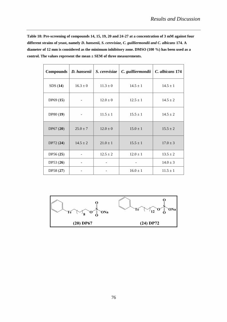
Results and Discussion
76
Table 18: Pre-screening of compounds 14, 15, 19, 20 and 24-27 at a concentration of 3 mM against four
different strains of yeast, namely D. hansenii, S. cerevisiae, C. guilliermondii and C. albicans 174. A
diameter of 12 mm is considered as the minimum inhibitory zone. DMSO (100 %) has been used as a
control. The values represent the mean ± SEM of three measurements.
Compounds D. hansenii S. cerevisiae C. guilliermondii C. albicans 174
SDS (14) 16.3 ± 0 11.3 ± 0 14.5 ± 1 14.5 ± 1
DP69 (15) - 12.0 ± 0 12.5 ± 1 14.5 ± 2
DP80 (19) - 11.5 ± 1 15.5 ± 1 14.5 ± 2
DP67 (20) 25.0 ± 7 12.0 ± 0 15.0 ± 1 15.5 ± 2
DP72 (24) 14.5 ± 2 21.0 ± 1 15.5 ± 1 17.0 ± 3
DP56 (25) - 12.5 ± 2 12.0 ± 1 13.5 ± 2
DP53 (26) - - - 14.0 ± 3
DP58 (27) - - 16.0 ± 1 11.5 ± 1

Results and Discussion
77
C. guilliermondii
0.001 0.01 0.1 0.25 0.5 0.75 1 30
5
10
15
20
SDS
DP80
******
******
ns
ns*
*
Concentration (mM)
Absen
ce o
f bacte
ria
l g
row
th z
on
e (
mm
)
C. guilliermondii
0.001 0.01 0.1 0.25 0.5 0.75 1 30
5
10
15
20
SDS
DP67
DP72*** ***
******
ns ** *
ns
ns
****** ***
nsns
ns
ns
Concentration (mM)
Absen
ce o
f bacte
ria
l g
row
th z
on
e (
mm
)
a)
b)
Figure 28: Activity of a) DP80 (19), b) DP67 (20) and DP72 (24) against C. guilliermondii. SDS (14) has
been used as a control. Data presented as absence of the bacterial growth zone in mm ± SEM. ANOVA
test: n.s. = p ≥ 0.05, p < 0.05 (*), p < 0.01 (**) or p < 0.001 (***) in relation to the relevant values for the
particular control SDS.

Results and Discussion
78
Compounds DP80 (19), DP67 (20) and DP72 (24) exhibited significantly higher activity
against C. guilliermondii; hence, concentration-dependence studies of these compounds were
performed and compared with the results obtained for SDS (14) (which also serve as ‘archive’
for the statistical evaluation). DP80 (19) and DP67 (20) showed an inhibitory effect at very
low concentrations (1 µM) (Figure 28). Surprisingly, DP72 (24), exhibited a minimum
inhibitory effect at a concentration of 10 µM against C. guilliermondii. SDS (14) exhibited a
minimum inhibition against this strain only at a concentration of 500 µM. This confirms once
more that the presence of tellurium, in addition to amphiphilicity, is important for biological
activity.
As evident from the pre-screening experiments, all the compounds including SDS (14)
showed an inhibitory effect against S. cerevisiae. Interestingly, DP72 (24) was the most active
compound against this strain. All of the compounds exhibited an inhibitory activity against C.
albicans. Yet again, DP72 (24) was the most active compound against C. albicans and the
EC50 of growth inhibition was calculated as just 33.17 µM (Figure 29). DP72 (24) was clearly
the best compound employed in this assay, that exhibited an inhibitory effect against all the
strains of yeast studied. Various compounds and the EC50 of growth inhibition against
different strains of yeast are summarised in Table 19.

Results and Discussion
79
SDS DP56 DP53 DP58 DP69 DP80 DP67 DP720
5
10
15
20
C.albicans 174
nsns
ns
ns nsns
***
Concentration (3mM)
Absen
ce o
f bacte
ria
l g
row
th z
on
e (
mm
)
C. albicans 174
0.001 0.01 0.1 0.25 0.5 0.75 1 30
5
10
15
20
DP72
SDS
*
***
*****
******
******
Concentration (mM)
Absen
ce o
f bacte
ria
l g
row
th z
on
e (
mm
)
a)
b)
Figure 29: a) Pre-screening of DP56 (25), DP53 (26), DP58 (27), DP69 (15), DP80 (19), DP67 (20), DP72
(24) and SDS (14) against C. albicans 174 at a concentration of 3 mM, b) Activity of DP72 (24) against C.
albicans 174. SDS (14) has been used as a control. Data presented as absence of the bacterial growth zone
in mm ± SEM. ANOVA test: n.s. = p ≥ 0.05, p < 0.05 (*), p < 0.01 (**) or p < 0.001 (***) in relation to the
relevant values for the particular control SDS
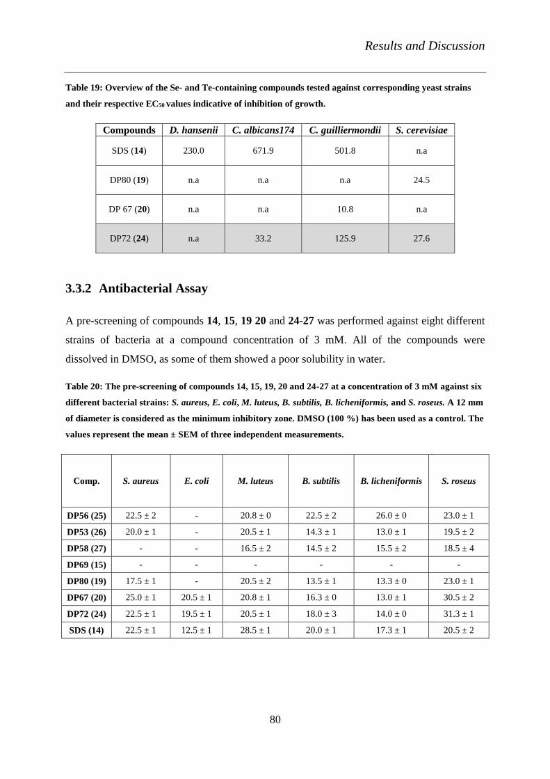
Results and Discussion
80
Table 19: Overview of the Se- and Te-containing compounds tested against corresponding yeast strains
and their respective EC50 values indicative of inhibition of growth.
Compounds D. hansenii C. albicans174 C. guilliermondii S. cerevisiae
SDS (14) 230.0 671.9 501.8 n.a
DP80 (19) n.a n.a n.a 24.5
DP 67 (20) n.a n.a 10.8 n.a
DP72 (24) n.a 33.2 125.9 27.6
3.3.2 Antibacterial Assay
A pre-screening of compounds 14, 15, 19 20 and 24-27 was performed against eight different
strains of bacteria at a compound concentration of 3 mM. All of the compounds were
dissolved in DMSO, as some of them showed a poor solubility in water.
Table 20: The pre-screening of compounds 14, 15, 19, 20 and 24-27 at a concentration of 3 mM against six
different bacterial strains: S. aureus, E. coli, M. luteus, B. subtilis, B. licheniformis, and S. roseus. A 12 mm
of diameter is considered as the minimum inhibitory zone. DMSO (100 %) has been used as a control. The
values represent the mean ± SEM of three independent measurements.
Comp. S. aureus E. coli M. luteus B. subtilis B. licheniformis S. roseus
DP56 (25) 22.5 ± 2 - 20.8 ± 0 22.5 ± 2 26.0 ± 0 23.0 ± 1
DP53 (26) 20.0 ± 1 - 20.5 ± 1 14.3 ± 1 13.0 ± 1 19.5 ± 2
DP58 (27) - - 16.5 ± 2 14.5 ± 2 15.5 ± 2 18.5 ± 4
DP69 (15) - - - - - -
DP80 (19) 17.5 ± 1 - 20.5 ± 2 13.5 ± 1 13.3 ± 0 23.0 ± 1
DP67 (20) 25.0 ± 1 20.5 ± 1 20.8 ± 1 16.3 ± 0 13.0 ± 1 30.5 ± 2
DP72 (24) 22.5 ± 1 19.5 ± 1 20.5 ± 1 18.0 ± 3 14.0 ± 0 31.3 ± 1
SDS (14) 22.5 ± 1 12.5 ± 1 28.5 ± 1 20.0 ± 1 17.3 ± 1 20.5 ± 2

Results and Discussion
81
The Se- and Te-containing compounds showed an activity against most of the strains tested,
except P. aeruginosa. DP56 (25) and DP53 (26) exhibited inhibitory zone diameters of 22.5 ±
2 mm and 20 ± 1 mm, respectively, against S. aureus (Table 20). DP58 (27), with the longest
hydrocarbon chain, when compared to DP56 (25), did not show any major toxicity effect
against S. aureus. DP58 (27), however, exhibited a minimum inhibitory activity against the
strains such as M. luteus, B. subtilis, B. licheniformis and S. roseus. DP80 (19), with the
longest hydrocarbon chain length showed significant activity against more than five different
strains when compared to its ‘short tail analogue’ DP69 (15).
The Te-containing compounds DP67 (20) and DP72 (24) exhibited the best results and
showed activity against both Gram-positive (S. aureus) and Gram-negative bacteria (E. coli
and S. typhimurium) (Figure 30). DP67 (20), DP72 (24) and SDS (14) exhibited a respective
diameter of the inhibitory zone of 14 ± 1 mm, 13 ± 0 mm and 12.5 ± 1 mm against S.
typhimurium. None of the other compounds in our studies exhibited an activity against this
bacterium. The concentration-dependence studies of the active compounds were performed
against S. aureus and E. coli. The EC50 values of growth inhibition for the various compounds
were calculated and are tabularised in Table 21.

Results and Discussion
82
Figure 30: Activity of a) DP72 (24) against S. aureus and b) DP67 (20) and DP72 (24) against E. coli. SDS
(14) has been used as a control. Data presented as absence of the bacterial growth zone in mm ± SEM.
ANOVA test: p < 0.05 (*) or p < 0.001 (***) in relation to the relevant values for the particular control
SDS.
E. coli VKPM-M17
0.1mM 0.25mM 0.5mM 0.75mM 1mM 3mM0
5
10
15
20
25SDS
DP67
DP72
*** ******
*** ****** ***
******
***
******
Concentration (mM)
Absen
ce o
f bacte
ria
l g
row
th z
on
e (
mm
)
b)
S. aureus WDCM 5233
0.1 0.25 0.5 0.75 1 30
10
20
30 SDS
DP72
***
******
*** ***
*
Concentration (mM)
Absen
ce o
f bacte
ria
l g
row
th z
on
e (
mm
)
a)

Results and Discussion
83
Figure 31: a) Comparative study of the activity of DP72 (24) against S. aureus and E. coli and b) DP80 (19)
against S. aureus. The absence of growth zone is represented as mm ± SEM. SDS (14) has been used as
control. ANOVA test: n.s. = p ≥ 0.05, p < 0.05 (*), p < 0.01 (**) or p < 0.001 (***) in relation to the relevant
values for the particular control SDS
DP72
0.1 0.25 0.5 0.75 1 30
5
10
15
20
25
E. coli VKPM-M17
S. aureus WDCM 5233
*
*****
** **
***
***
*
*****
** **
Concentration (mM)
Absen
ce o
f bacte
ria
l g
row
th z
on
e (
mm
)
a)
S. aureus WDCM 5233
0.1 0.25 0.5 0.75 1 30
5
10
15
20
25SDS
DP80
***
*** *** n.s n.s
Concentration (mM)
Absen
ce o
f bacte
ria
l g
row
th z
on
e (
mm
)
b)

Results and Discussion
84
The study concentration-dependence of activity for compound DP72 (24) was performed
against S. aureus and E. coli. The results of all of the concentration dependence studies were
compared with the one for SDS (14). DP72 (24) exhibited the most significant activity against
S. aureus at a concentration of 100 µM, when compared with that of SDS (14) (Figure 30).
The EC50 values of growth inhibition of DP72 (24) and SDS (14) against S. aureus were
calculated as 116.2 µM and 772.9 µM, respectively (Table 21).
Only the Te-containing compounds DP67 (20) and DP72 (24) exhibited activity against the
Gram-negative bacterium, E. coli. (Figure 30). Both of the compounds exhibited a significant
activity at a concentration of 100 µM when compared to SDS (14). DP72 (24) exhibited a
more significant activity against S. aureus when compared to its activity against E. coli
(Figure 31). The Se- and Te-containing compounds tested against different bacterial strains,
their respective EC50 values of growth inhibition and IC50 values of recovery of viability is
summarised in Table 21.
Figure 32: The recovery of viability curve of bacterial strains after 24 h of treatment with various
compounds. Figure a) Activity of DP67 (20) and DP72 (24) against E. coli. Figure b) DP72 (24) against S.
aureus. Data presented as viability percentage ± SEM.
After the treatment of bacteria with the Se- and Te-containing compounds and the formation
of growth inhibitory zones, a small portion of the growth media from the inhibitory zone was
incubated in 5 ml of LB broth. This broth was incubated for 24 hours in a temperature-
controlled shaker. The recovery of growth of the bacteria was subsequently evaluated (Figure
32).
10 1 10 2 10 3 10 4
0
50
100
150
DP67
DP72
SDS
E. coli VKPM-M17
Concentration (M)
viabil
ity i
n %
10 1 10 2 10 3 10 4
0
20
40
60
80
100
DP72 in S. aureus WDCM 5233
Concentration (M)
viabil
ity i
n %

Results and Discussion
85
S. aureus WDCM 5233
0.1 0.25 0.5 0.75 1 30
5
10
15
20
25 SDS
DP56
DP53
***
*** **** ns
ns
***
***
***
*** ***
ns
Concentration (mM)
Abs
ence
of
bact
eria
l g
row
th z
one
(mm
)a)
Figure 33: a) The activity of compounds DP56 (25) and DP53 (26) against S. aureus. SDS (14) has been
used as a control. The absence of the bacterial growth zone is represented in mm ± SEM. ANOVA test:
n.s. = p ≥ 0.05, p < 0.01 (**) or p < 0.001 (***) in relation to the relevant values for the particular control
SDS.
Figure 32 shows the recovery of viability curve for E. coli in the case of DP67 (20) and DP72
(24) and S. aureus in the case of DP72 (24). Subsequently, the IC50 of the recovery of growth
of the strains of bacteria was calculated. DP56 (25) and DP53 (26) exhibited a significant
activity against S. aureus at a concentration of 100 µM when compared to the activity of SDS
(14) (Figure 33). Unlike the Te-containing compounds, DP56 (25) and DP53 (26) did not
show the typical structure-activity relationship against S. aureus. For instance, DP56 (25),
with a shorter hydrocarbon chain length, exhibited a higher activity than DP53 (26).
Compounds DP56 (25) and DP53 (26) exhibited an IC50 of recovery of viability as 787.3 µM
and 334.3 µM respectively, against S. aureus. The recovery of viability of S. aureus in the
presence of SDS was unable to be calculated due to the effective growth of this bacterium
even at very low concentrations starting from 10 µM.
Overall, Te-containing compounds DP 67 (20) and its ‘long-tail analogue’, DP72 (24) were
the most active compounds tested against the bacteria. The Se-containing compound, DP53
(26) was also highly active against S. aureus.

Results and Discussion
86
Table 21: Se- and Te-containing compounds tested against various strains of bacteria. EC50 values of
inhibition and IC50 value of recovery of viability are summarised.
Compounds Bacterial
Strains
EC50 of Inhibition
(µM)
IC50 of Viability
(µM)
SDS (14)
S. aureus 772.9 n.a
E. coli 773.1 n.a
DP80 (19) S. aureus 39.6 n.a
DP67 (20) E. coli 336.7 546.4
DP72 (24)
E. coli 174.8 ambiguous
S. aureus 116.2 ambiguous
DP56 (25) S. aureus 172.5 787.3
DP53 (26) S. aureus 83.3 334.3
A commercially available antibiotic, sulfadimethoxin, was also tested against S. aureus, B.
subtilis, E. coli and S. typhimurium as a particular control. This antibiotic exhibits a growth
inhibitory zone of 20 mm, on an average, against S. aureus and E. coli, but only at a
concentration of 100 mM, which is extremely high when compared to the concentrations
considered for the Se- and Te-containing compounds. Sulfadimethoxin also shows a static
effect against B. subtilis.

Summary and Outlook
87
4. Summary and Outlook
In the second part of this thesis, more sophisticated compounds have been derived based on
the idea of amphiphilicity and chalcogen (bio-) chemistry. Se- and Te-containing amphiphilic
compounds were produced and subsequently analysed physico-chemically and biologically.
The surface tension analysis was performed by ring tensiometric and sita tensiometric
experiments. The CMC value decreases as the length of the hydrocarbon chain increases in
the compounds. FRET experiments were performed to study the non-covalent interactions of
the compounds with BSA. The quenching of the fluorescence of BSA was studied and the
Stern-Volmer constants were calculated. As may be expected the value of the Stern-Volmer
constants increases with the increase in the hydrocarbon chain length in the compounds.
The anti-microbial activities of the Se- and Te-containing compounds were studied. The
compounds were tested against four different strains of yeast and eight different strains of
bacteria. All the compounds tested exhibited activity against strains such as S. aureus, M.
luteus, B. subtilis, B. licheniformis and S. roseus, except for DP69 (15). DP67 (20) and DP72
(24) exhibited activity against both Gram-positive and Gram-negative bacteria. These
compounds were even more active than SDS. The compounds containing the longest
hydrocarbon chains exhibited lower EC50 values of growth inhibition. Similar results were
obtained for the Se- and Te-containing compounds against different strains of yeast. Se-
containing compounds exhibited an effective inhibitory activity against C. guilliermondii and
the semi-pathogenic strain C. albicans. Undoubtedly, the Te-containing compounds, DP67
(20) and DP72 (24), were the most active ones against all the four strains of yeast. DP72 (24)
also showed a complete inhibitory activity against C. albicans.
As expected, the idea to move from the tetrasulfane PrTTSPs (9) to other amphiphilic agents,
but this time with the more stable yet also more reactive Se- and Te-containing compounds
has worked rather well. Se- and Te-containing compounds were highly active in all our
biological assays when compared to the activity of PrTTSPs (9). DP67 (20) and DP72 (24)
should be considered in the future for potential applications as fungicides or bactericides.

Experimental Part
88
5. Experimental Part
5.1 Materials and Methods
All of the chemical reagents used for the synthesis of tetrasulfanes were purchased from
Sigma-Aldrich, including thionylchloride, pyridine, thiophenol, mercaptobenzene,
thiopropionic acid and used without further purification unless stated. All of the reactions
were carried out under an Argon (4.6) atmosphere, at room temperature and with deionised
MilliQ water (resistance ≥18 MP.cm). The completion of the reactions were monitored by
thin layer chromatography technique with Merck aluminium-backed plates (Kieselgel 60
F254, Darmstadt, Germany) and visualised under ultraviolet light. Column Chromatography
technique was used to purify the compounds with Silica gel 60 (40-63 Lm, Fluka) and under
mild nitrogen pressure.
5.2 NMR Spectroscopy
All NMR spectra were recorded on Bruker (Rheinstetten) type Avance 500, at the Institute of
Pharmaceutical Chemistry. 1H and 13C NMR spectra were recorded at 500 and 125 MHz,
respectively. Chemical shifts recorded in CDCl3 solvent were expressed in δ (ppm) relative to
the signal at 7.26 ppm for 1H NMR and at 77.0 ppm for 13C NMR.
5.3 Mass spectroscopy
Spectra were recorded in QTRAPR LC/MS/MS System by AB Sciex Del Optiplex 960.
HPLC grade methanol was used as the solvent.

Experimental Part
89
5.4 Calculation of clogP
The calculation of the clogP was performed using the ALOGPS 2.1 Software and
ChemBioDraw Ultra 12.0.
5.5 Synthesis of unsymmetrical tetrasulfanes
The chemicals required for the synthesis of unsymmetrical tetrasulfanes were thionyl
chloride, pyridine, and other thiol compounds such as propanethiol, mercaptopropionic acid,
thiophenol, and mercaptobenzene.
5.5.1 General procedure for the synthesis of tetrasulfanes (I)
To a solution of S2Cl2 (10 mmol) in diethyl ether (25 ml), an equimolar volume of respective
thiol-containing compound (10 mmol) and pyridine (10 mmol) in diethyl ether (10 ml) was
added drop wise for 30 minutes at a temperature of -78°C and was stirred for another 30
minutes at the same temperature. Then, another equimolar volume of the same thiol-
containing compound (10 mmol) and pyridine (10 mmol) in diethyl ether (10 ml) was added
drop wise for another 30 minutes and again stirred at the same temperature for an additional
30 minutes before the reaction mixture was allowed to return to room temperature. After the
completion of the reaction, the organic layer of the reaction mixture was washed with distilled
water (3 times). The organic layer was dried over MgSO4. The solvent was evaporated and the
product was purified by column chromatography.
5.5.2 General procedure for the synthesis of UT (II)
The general procedure I was modified in the following way to obtain the unsymmetrical
polysulfanes. To a solution of S2Cl2 (2.70 g, 20 mmol) in diethyl ether (50 ml), an equimolar
volume of the first respective thiol-containing compound (20 mmol) and pyridine (1.58 g, 10
mmol) in diethyl ether (25 ml) was added drop wise for 1 hour at -78 °C and stirred for
another 45 minutes at the same temperature. Then, another equimolar volume of the
respective second thiol-containing compound (20 mmol) and pyridine (20 mmol) in diethyl
Experimental Part
90
ether (25 ml) was added drop wise for 1 hour and continued to stir at the same temperature for
another 1 hour, before the reaction mixture was allowed to come to room temperature.
Subsequently, the organic layer was washed with distilled water. This layer was dried over
MgSO4. The solvent was evaporated and the crude compound obtained was further purified by
column chromatography.
5.5.2.1 1, 4-diphenyltetrasulfane (DPhTTS)
DPhTTS (7) was synthesised by the general procedure for the synthesis of tetrasulfanes (I)
(refer section 5.5.1). Thiophenol was used as the respective thiol-containing compound.
DPhTTS (7) was purified by column chromatography using the solvent petroleum benzene.
The compound was obtained as yellow oil with a yield of 90 %. TLC: Rf: 0.36,
(Petroleumbenzene). clogP: 5.04.
1H NMR (500 MHz, CDCl3): δ 7.283- 7.345 (complex, 3H), 7.546- 7.571 (m, 2H), 13C NMR
(CDCl3): 128.341, 129.135, 130.165, 136.194. HRMS (ESI) m/z = 281.9673
5.5.2.2 3-(propyltetrasulfanyl)-propanoic acid (PrTTSPs)
PrTTSPs (9) was synthesised by the general procedure for the synthesis of unsymmetrical
polysulfanes (II) (refer section 5.5.2). The first thiol-containing compound used was
mercaptopropane and the second thiol-containing compound used was mercaptopropionic
acid. PrTTSPs (9) was purified by column chromatography using the solvent
dichloromethane. The compound was obtained as pale yellow oil with a yield of 71 %. TLC:
Rf: 0.20, (CH2Cl2). clogP: 2.74.
1H NMR (500 MHz, CDCl3): δ 1.010- 1.040 (t, 3H), 1.763-1.836 (m, 2H), 2.905-2.948 (q,
4H), 3.175- 3.204 (t, 2H), 13C NMR (CDCl3): δ 13.048, 22.359, 33.294, 33.833, 41.452,
176.883. m/z = 266.9614

Experimental Part
91
5.5.2.3 3-(benzyltetrasulfanyl)-propanoic acid (BnTTSPs)
BnTTSPs (8) was synthesised by the general procedure for the synthesis of unsymmetrical
polysulfanes (II) (refer section 5.5.2). The first thiol-containing compound used was
benzylmercaptan and the second thiol-containing compound used was mercaptopropionic
acid. BnTTSPs (8) was purified by column chromatography using the solvents
dichloromethane:methanol (9:1). The compound was obtained as yellow oil with a yield of 75
%. TLC: Rf: 0.27 (CH2Cl2: CH3OH-9:1). clogP: 2.76.
1H NMR (CDCl3, 500 MHz): δ 1.230-1.258 (t, 1H), 2.171 (s, 3H), 2.852-2.881 (t, 2H), 3.142-
3.171 (t, 1H), 7.307- 7.345 (m, 4H), 13C NMR (CDCl3): δ 30.910, 33.272, 33.582, 43.748,
53.409, 127.799, 128.703, 129.497, 129.544, 136.082, 175.650, m/z = 314.961
5.5.2.4 3-(phenyltetrasulfanyl)-propanoic acid (PhTTSPs)
PhTTSPs (11) was synthesised by the general procedure for the synthesis of unsymmetrical
polysulfanes (II) (refer section 5.5.2). The first thiol-containing compound used was
thiophenol and the second thiol-containing compound used was mercaptopropionic acid.
PhTTSPs (11) was purified by column chromatography using the solvents
dichloromethane:methanol (9.5:0.5). The compound was obtained as light yellow powder
with a yield of 84 %. TLC: Rf: 0.32 (CH2Cl2: CH3OH-9.5:0.5). clogP: 3.12.
1H NMR (CDCl3, 500 MHz): δ 2.731-2.760 (t, 2H), 3.115-3.144 (t, 2H), 7.233-7.293 (m, 3H),
7.482-7.501 (m, 2H), 13C NMR (CDCl3): 33.634, 33.715, 128.038, 128.805, 129.714,
135.051, 172.943, m/z = 277.9
5.5.2.5 1-benzyl-4-phenyltetrasulfane (BnTTSPh)
BnTTSPh (10) was synthesised by the general procedure for the synthesis of unsymmetrical
polysulfanes (II) (refer section 5.5.2). The first thiol-containing compound used was
thiophenol and the second thiol-containing compound used was benzylmercaptan. BnTTSPh

Experimental Part
92
(10) was purified by column chromatography using the solvent n-Hexane. The compound was
obtained as yellow powder with a yield of 49 %. TLC: Rf: 0.23 (n-Hexane). clogP: 5.37.
1H NMR (CDCl3, 500 MHz): δ 4.068 (s, 2H), 7.202-7.251 (complex, 5H), 13C NMR (CDCl3):
33.634, 33.71, 128.038, 128.805, 129.71, 135.65, 172.94, m/z = 296.8
5.5.2.6 1-propyl-4-benzyltetrasulfane (PrTTSBn)
PrTTSBn (12) was synthesised by the general procedure for the synthesis of unsymmetrical
polysulfanes (II) (refer section 5.5.2). The first thiol-containing compound used was
benzylmercaptan and the second thiol-containing compound was thiopropane. PrTTSBn (12)
was purified by column chromatography using the solvent n-Hexane. The compound was
obtained as yellow powder, with a yield of 59 %. TLC: Rf: 0.465 (n-Hexane). clogP: 5.
1H NMR (CDCl3, 500 MHz): δ 3.577-3.594 (m, 5H), 5.214-5.276 (complex, 5H), 5.847-5.930
(m, 3H), 13C NMR (CDCl3):42.053, 119.569, 132.478, m/z = 261.2
5.5.2.7 1-propyl-4-phenyltetrasulfane (PrTTSPh)
PrTTSPh (13) was synthesised by the general procedure for the synthesis of unsymmetrical
polysulfanes (II) (refer section 5.5.2). The first thiol-containing compound used was
thiophenol and the second thiol-containing compound used was thiopropane. PrTTSPh (13)
was purified by column chromatography using the solvents dichloromethane:methanol (9:1).
The compound obtained was obtained as pale yellow oil with a yield of 62 %. TLC: Rf: 0.340
(CH2Cl2: CH3OH-9:1).clogP: 4.66.
1H NMR (CDCl3, 500 MHz): δ 0.967-0.997 (t, 2H), 1.150-1.178 (t, 1H), 1.664-1.796 (m, 2H),
2.730-2.901 (t, 2H), 3.413-3.455 (q, 1H), 7.211-7.319 (complex, 3H), 7.502- 7.521 (m, 1H),
13C NMR (CDCl3): 13.054, 15.252, 22.358, 41.365, 65.821, 128.314, 129.111, 130.136,
136.169, m/z = 248.6

Experimental Part
93
5.6 Physico-Chemical Measurements
5.6.1 Electrochemistry
The redox behaviour of the symmetrical, unsymmetrical tetrasulfanes and other selenium and
tellurium containing compounds was studied at a BAS 100 W electrochemical workstation by
recording their cyclic voltammograms. During the analysis of tetrasulfanes, a dropping
mercury electrode was used as the working electrode and a glassy carbon electrode for the
analysis of Se and Te containing compounds. In both the experiments, the Ag/AgCl electrode
(SSE) was used as the reference electrode and a platinum spiral was used as the counter
electrode. Four full cycles were recorded, the first of which was used to record the peak
potentials. The parameters were set and the potential range varied between -1200 to 0 mV for
tetrasulfanes and -1200 to +1200 mV for Se and Te containing compounds; the scan rate was
fixed as 250 mV/s. The stock solutions of the compounds were prepared in methanol. The
compounds were dissolved in a cuvette containing 33 % methanol in 50 mM potassium
phosphate buffer at pH 7.4 to obtain the final concentration of the compound as 250 µM.
Before each measurement, the electrolysis cell was purged with nitrogen and stirred well. All
of the measurements were performed in triplicate at room temperature.
5.6.2 Ring tensiometry
The instrument KRÜSS Tensiometer K6 (KRÜSS GmbH, Hamburg) was used at the Institute
of Pharmaceutical Nanotechnology, Department of Pharmacy, University of Saarland. All the
compounds were dissolved in distilled water at different concentrations, such as 1, 3, 5, 10, 15
and 30 mM and all of the measurements were performed at room temperature. 10 ml of each
concentration of compound was prepared in a glass vial. The vial was placed on the sample
holder, which was levelled using the set screw and the micrometre screw. The ring was
immersed into the vial such that it was very close to the surface of the liquid. The balance
beam, on which the ring was hanging, was set at the zero reference mark. With the help of a
wheel, the tension was increased slowly from 0 mN/m until the ring came out of the liquid.

Experimental Part
94
The tension at which the ring came out of the liquid was recorded as the surface tension of the
liquid. The experiment was repeated five times with each concentration of a compound. A
control experiment was performed with SDS (14). The critical micelle concentration of the
compounds was obtained by plotting a graph of different concentrations of the compounds
against their values of surface tension.
5.6.3 Sita tensiometry
The surface tension analysis of the compounds was performed with SITA science line t60
using the bubble pressure method. The range of the measurement of surface tension was 10-
100 mN/m and the resolution was 0.1 mN. The measuring range of bubble frequency was 30
ms-60 s and the resolution was 1 ms. All the compounds were dissolved in 2 ml of distilled
water, containing less than 1 % DMSO, at various concentrations such as 5 µM to 3 mM. The
capillary was inserted and air was pumped into the solutions of the compounds. The bubble
pressure developed in the solution is proportional to its surface tension. The surface tension in
mN/m was recorded electronically by the computer. All of the experiments were performed in
triplicate at room temperature. SDS (14) was used as a control. In order to obtain the CMC
value of a compound, a graph was plotted with different concentrations of the compound
against the values of surface tension.
5.6.4 Densitometry
All of the compounds were dissolved in ethanol containing less than 1 % of DMSO. Different
concentrations of compounds were prepared such as, from 188.7 µM to 3 mM. The density
measurements were performed using the instrument, Anton Paar DMA 4500, Density meter.
Before measuring the densities of different concentrations of the compounds, the instrument
was calibrated with distilled water. The densities were measured for each concentration of the
compounds and their corresponding apparent molar volumes were calculated. Graphs were
plotted with apparent molar volumes of the compounds against their concentrations.

Experimental Part
95
5.7 Molecular Non-Covalent Interaction Studies
5.7.1 Haemolysis assay
5.7.1.1 Preparation of buffer
HPS buffer A was prepared by mixing 145 mM NaCl, 7.5 mM KCl, 10 mM glucose and 10
mM HEPES at a pH 7.4. This buffer was used for washing human blood.
5.7.1.2 Preparation of human blood
All of the blood samples were obtained from the Institute of Clinical Haematology and
Transfusion Medicine, Saarland University hospital (Homburg). Human venous blood was
used for the experiments and was collected from healthy donors in Na-EDTA anticoagulant-
coated test tubes, which were stored at 4 °C for a maximum of two days. Fresh blood samples
were used before each experiment.
5.7.1.3 Preparation of red blood cells
The whole blood from the anticoagulant-coated test tubes was aliquot into eppendorfs. These
aliquots were centrifuged at 2000 g for 5 minutes at room temperature, and the supernatant
was removed by aspiration. Then, the blood cells were suspended in HPS buffer A and
vortexed well. Similarly, the washing steps were repeated three times under the same
conditions. After washing, the cells were re-suspended again in HPS buffer A. Cells were
centrifuged at 16000 g for 10 s in order to compact them. These compact cells were used for
further experiments.
5.7.1.4 Procedure
Drabkin’s reagent, purchased from Sigma Aldrich was used for this assay. This reagent
determines the haemoglobin content at an absorption of 540 nm. Different stock solutions of
the compounds, such as 1, 5, 25, 50, 75 and 100 mM, were prepared in DMSO. The RBCs
were suspended in 990 µl of HPS buffer A, to make 2.5 % haematocrit. Then, 10 µl of the

Experimental Part
96
stock of each concentration, of every compound was added to the RBC suspension to make
the final concentrations of 10, 50, 250, 500, 750 and 1000 µM of the compounds with 1 % of
DMSO in each of them. A control experiment was performed in a similar way with only 10 µl
of DMSO in 990 µl of RBC suspension. All of the above mixtures were prepared in triplicate
and were incubated in a water bath at 37 °C for 30 minutes. Meanwhile, 2 ml of Drabkin’s
reagent was prepared in 3 ml cuvettes. After incubation, all of the samples along with the
DMSO control were centrifuged at 12,000 g for 30 seconds. Then, 500 µl of supernatant was
transferred to the 2 ml of Drabkin’s reagent that was prepared in the cuvettes. A control for
the completely haemolysed RBCs was prepared by adding 2.5 % haematocrit in 500 µl of a
detergent solution in 2 ml of Drabkin’s reagent. All of the samples with Drabkin’s reagent
were incubated in the dark for 30 minutes. After incubation, the absorption was measured at
540 nm in a UV-Vis spectrophotometer (UV mini 1240, UV-Vis Spectrophotometer,
Shimadzu), with a blank of 500 µl HPS buffer A in 2 ml Drabkin’s reagent.
5.7.2 FRET measurements
All the experiments were performed in a Variance Cary Eclipse fluorescent spectrometer and
the obtained results were analysed using Cary Eclipse software. BSA was purchased from
Sigma Aldrich and a stock solution was prepared at a concentration of 1 .10-6 M, which was
later diluted to 1 .10-7 M in 3ml of sodium citrate buffer solution. The stock solution of the
compounds was prepared in ethanol at 3 mM with less than 1 % of DMSO. The excitation
wavelength was set at 280 nm and the fluorescence intensity was recorded between 300 and
500 nm. The excitation slit value was set at 5 and the emission slit value was set at 10. The
graphs were smoothed by setting the smoothing factor at 11. Initially the fluorescence
intensity of BSA was recorded. Then, 2 µl of the compound was added to 3 ml of 1 .10-7 M
BSA solution and quenching of the fluorescence of BSA was recorded. The spectra were then
recorded with 5 µl of compound, and then for each 5 µl addition of the compound until a total
volume of 60 µl of compound had been added to BSA solution. Fluorescence intensity of the
BSA solution, in the presence of different concentrations of the compounds was measured. A
control experiment was performed with only the solvent ethanol and 1 % DMSO.

Experimental Part
97
5.8 Biological Assays
5.8.1 Nematode assay
Steinernema feltiae nematode sample, in the form of a soft cake, was purchased from Sautter
& Stepper GmbH (Ammerbruch, Germany) and stored in the dark at 4 ºC. S. feltiae is the
commercially available non-pathogenic model organism for agricultural crops and hence the
best for pre-screening of the compounds. Fresh samples were ordered before each experiment
and not used for more than a week. A 1.5 g sample of the nematode soft cake was dissolved in
250 ml of distilled water at 27 °C. This solution was allowed to stay at room temperature and
in moderate light for nearly 30 minutes by stirring at regular intervals in order to resuscitate
the nematodes. The viability of the suspension of nematodes was calculated under the
microscope (TR 200, VWR International, Belgium) at a four-fold magnification. The viability
of more than 80 % was considered optimal and the samples were then subjected to the
experiment. Stock solutions of all of the compounds were prepared in DMSO and further
diluted in distilled water at concentrations ranging from 10 µM to 1 mM, such that final
concentration of DMSO was less than 1 % in each case. Then, 100 µl of each concentration of
the compound and 100 µl of the nematode suspension were added to 96-well plates in
triplicate. A control experiment was performed with 1 % DMSO. The live and dead
nematodes were counted under the microscope immediately after 30 minutes, 6 hours and 24
hours of incubation. LD50 value was calculated for each compound and the whole experiment
was repeated three times.

Experimental Part
98
5.8.2 Anti-yeast experiment
5.8.2.1 Preparation of Saburo broth and media
Saburo broth was prepared by adding glucose (40 g/l) peptone (10 g/l), and yeast extract (5
g/l) in distilled water. Saburo media was prepared using the same constituents along with 0.9
% agar.
5.8.2.2 Re-cultivation of yeast
Using a microbial inoculation needle, a small inoculum of strain of yeast, from the yeast
culture deposits (YSU), was removed and dispersed well in 5 ml of broth of Saburo in a test
tube. The test tube was stored overnight in a temperature-controlled shaker. After the
incubation period the culture of yeast measuring an optical density of 0.5 a.u turbidity in a
UV-spectrometer, was used for the study of the activity of compounds against yeast.
5.8.2.3 Procedure
The procedure of the yeast assay was followed according to [91]. The experiments were
performed in Yerevan State University, Armenia. Different strains of yeast namely;
Debariomyces hansenii, Candida guilliermondii, Saccharomyces cerevisiae and Candida
albicans 174 were used for the experiments. All of the chemical compounds were dissolved in
DMSO (100 %) to prepare the following concentrations: 0.001, 0.01, 0.1, 0.25, 0.5, 0.75, 1
and 3 mM. Concentration-dependence studies of all the compounds were performed against
the strains of yeast using the agar well-diffusion method. DMSO (100 %) was used as a
control. The test-organisms were added on sterile petridishes. Then, approximately 20 ml of
the growth media (Saburo) for yeast, was added to the petridishes with the microorganisms.
The petridishes were shaken well such that the microorganisms had spread throughout. Small
wells of 8 mm in diameter were punched in the agar plates (petridishes) using a sterile
stainless steel borer. Then, 100 µl of each concentration of a compound was transferred to the
wells. The plates were incubated micro-aerophilically at a temperature of 28 °C for 24 hours.
After the incubation, the diameters of the inhibitory zones were measured in mm.
Experimental Part
99
5.8.3 Antifungal experiment
5.8.3.1 Preparation of Wort broth and media
A 50 g wort extract was mixed in 200 ml of distilled water at a temperature of 45°C and this
temperature was maintained for 30 minutes. The temperature of the water bath was slowly
increased such as 1 °C every minute and continued for 25 minutes up to a final temperature of
70 °C. Then, 100 ml of distilled water (70 °C) was added to the mixture. The temperature of
the solution was maintained at 70 °C for 55 minutes, before the solution was cooled down to
room temperature. Then, the solution was weighed and made up to 450 g by adding the
required amount of distilled water. The broth was filtered and the required amount of agar
was added. The media was sterilised for further use.
5.8.3.2 Spore collection
0.96 % NaCl physiological solution was prepared and sterilised. The fungi were grown in
petridishes for 10 days. Using 2-3 ml of the 0.96 % NaCl solution, the spores were collected
into eppendorfs. The spores were counted on square grid under the microscope and spore
count was adjusted to 1 .107 spores in the Petri dish for each experiment. Spores were
collected from Trichoderma viride, Aspergillus flavus, Penicillium aeruginosa, Fusarium CB
1853, Mucor plumbeus and Geotrichum candidum.
5.8.3.3 Procedure
Sterile plastic petridishes with a diameter of 35 mm were used for all of the experiments. All
of the compounds were dissolved in DMSO (100 %). Compounds were added to the petri
dishes and then wort media was added to make a final concentration of 3 mM for each
compound. Subsequently, spores were added to the plates to make a final number of 1 .107
spores. The plates were incubated at 27 °C for 1 week.

Experimental Part
100
5.8.4 Antibacterial experiment
5.8.4.1 Preparation of LB broth and media
LB broth was prepared by adding peptone (10 g/l), yeast extract (5 g/l), NaCl (10 g/l), sucrose
(5 g/l) and MgSO4 (0.5 g/l) in distilled water. LB media was prepared with the same
constituents using 0.9 % agar.
4.8.4.2 Re-cultivation of bacteria
Using a microbial inoculation needle, a small inoculum of strain of bacteria, from the
bacterial-culture-deposits (YSU), was removed and dispersed well in 5 ml of broth of LB in a
test tube. The test tube was incubated in a temperature (37 °C) controlled shaker (100 rpm)
overnight. The culture measuring an optical density of 0.5 a.u turbidity was used for the
experiment.
4.8.4.3 Procedure
The antibacterial experiments were performed under the Chair of Microbiology, Plants and
Microbes Biotechnology, Department of Biology in Yerevan State University. The procedure
of the experiment was followed according to the [91]. Staphylococcus aureus WDCM 5233
and S. typhimurium 1754 were obtained from the Microbiological Depository Centre,
Armenia, and E. coli M-17 VKPM-B8208 from the Russian National Collection of Industrial
Microorganisms. M. luteus, B. subtilis A1, B. licheniformis WT, S. roseus, P. aeruginosa
were the wild isolates. All of the compounds were dissolved in DMSO (100 %) to prepare the
following concentrations: 0.001, 0.01, 0.1, 0.25, 0.5, 0.75, 1 and 3mM. A concentration-
dependence study of all of the compounds was performed on S. aureus WDCM 5233 and E.
coli M17 using the agar well-diffusion method. DMSO (100 %) was used as a control. The
test organism was placed on the agar plate. Wells of 8 mm in diameter were punched in the
agar plates using a sterile stainless steel borer. Then, 100 µl of each concentration of a
compound was transferred to the wells. The plates were incubated in a thermostat micro-
Experimental Part
101
aerophilically at 37 °C for 24 hours. The diameters of the inhibitory zones were measured in
mm.
5.8.5 Anti-bacteriophage experiment
5.8.5.1 Preparation of LB broth and media
The sterile LB broth was prepared with the same constituents as described above in the
section 4.7.1. Sterile LB media was prepared with 1.8 % and 0.8 % agar.
5.8.5.2 Re-cultivation of E. coli C-T4
Using a microbial inoculation needle, a small inoculum of strain of E. coli C-T4, from the
bacterial-culture deposits (YSU), was removed and cultivated on a nutrient slant agar media
(commercially available).
5.8.5.3 Isolation of T4-bacteriophage
A petridish with LB media (1.8 % agar) was prepared. 1 .108 colonies of T4 bacteriophage
and 1 .108 cells of E. coli C-T4 were mixed in a falcon tube. This solution was further diluted
with 7 ml of 0.8 % LB agar and was poured onto the 1.8 % agar plate. The plate was
incubated overnight in an incubator at 37 °C. Then, 0.96 % NaCl physiological solution was
used to isolate the T4 phage from the agar plates. The phage solution was centrifuged at
12000 g for 5 minutes. The supernatant was collected and filtered through a bacterial filter of
diameter of 0.2 nm. The purified T4 phage was stored at -30 °C for further use.
5.8.5.4 Procedure
The procedure of the experiment was followed according to [92]. All of the experiments were
performed in the Faculty of Biology, YSU, Armenia. All of the chemicals were purchased
from Sigma-Aldrich. A stock solution of T4-bacteriophage with 108 cells was used. Here, 30
µl of T4 bacteriophage and 30 µl of different concentrations of compounds such as 0.25, 0.75,
1.5, and 3mM were incubated for 90 minutes. Then, the petridishes containing LB media with
Experimental Part
102
1.8 % agar were prepared and dried under UV for 20 minutes for sterilisation. 102, 104, 106
and 108 dilutions of T4 phage along with the compounds were prepared using 5 ml of sterile
LB broth. The test tubes were shaken well for 5 minutes, to allow the phage to completely
diffuse. 108 cells of E. coli C-T4 was prepared using the E. coli C-T4 slant agar culture. The
108 cells of E. coli C-T4 culture was prepared in 5 ml LB broth with an optical density of
0.4/0.5 a.u. 1ml of each dilution of the compounds with 500 µl of E. coli C-T4 culture were
mixed with 7 ml of LB media (0.8 % agar) and poured on to the petridish containing LB
media with1.8 % of agar. The petridishes were incubated in a thermostat at 37 °C for 24
hours. The viable colonies of bacteriophage were counted by naked eye.
References
103
6. References
1. Lill, R., et al., The essential role of mitochondria in the biogenesis of cellular iron-
sulfur proteins. Journal of Biological Chemistry, 1999. 380(10): p. 1157-66.
2. Devillanova, F.A., Front matter, in Handbook of Chalcogen Chemistry: New
Perspectives in Sulfur, Selenium and Tellurium. 2006, The Royal Society of
Chemistry. p. i-iv.
3. Muoio, R., P. Casoria, and B. Menale, A Comparative Study of Sulphur Content of
Some Allium L. Species. Economic Botany, 2004. 58(2): p. 227-230.
4. Block, E., The Organosulfur Chemistry of the Genus Allium – Implications for the
Organic Chemistry of Sulfur. Angewandte Chemie International Edition in English,
1992. 31(9): p. 1135-1178.
5. Nimni, M., B. Han, and F. Cordoba, Are we getting enough sulfur in our diet?
Nutrition & Metabolism, 2007. 4(1): p. 24.
6. Fritsch, R.M. and N. Friesen, 1 Evolution, Domestication and Taxonomy. Allium Crop
Science: Recent Advances, ed. H.D.a.C. Rabinowitch, L.2002.
7. Sinha, N.K., et al., Handbook of Vegetables and Vegetable Processing, ed. N.K.
Sinha2010: Wiley-Blackwell.
8. Ricroch, A., et al., Evolution of genome size across some cultivated Allium species.
Genome, 2005. 48(3): p. 511-20.
9. Benavides, G.A., et al., Hydrogen sulfide mediates the vasoactivity of garlic.
Proceedings of the National Academy of Sciences, 2007. 104(46): p. 17977-17982.
10. Huang, Y.-T., et al., Diallyl trisulfide and diallyl disulfide ameliorate cardiac
dysfunction by suppressing apoptotic and enhancing survival pathways in
experimental diabetic rats. Journal of Applied Physiology, 2013. 114(3): p. 402-410.
11. Jacob, C., A scent of therapy: pharmacological implications of natural products
containing redox-active sulfur atoms. Natural Product Reports, 2006. 23 p. 851-863.
12. Petrovska, B.B. and S. Cekovska, Extracts from the history and medical properties of
garlic. Pharmacognosy Reviews, 2010. 4(7): p. 106-110.
13. Rahman, K. and G.M. Lowe, Garlic and Cardiovascular Disease: A Critical Review.
The Journal of Nutrition, 2006. 136(3): p. 736S-740S.

References
104
14. Pinto, J.T. and R.S. Rivlin, Antiproliferative Effects of Allium Derivatives from Garlic.
The Journal of Nutrition, 2001. 131(3): p. 1058S-1060S.
15. Elodie, V., et al., Antiproliferative effect of natural tetrasulfides in human breast
cancer cells is mediated through the inhibition of the cell division cycle 25
phosphatases International Journal of Oncology, 2011. 38(4): p. 1103-1111.
16. Busch, C., et al., Diallylpolysulfides induce growth arrest and apoptosis. International
Journal of Oncology, 2010. 36(3): p. 743-9.
17. Balandrin, M.F., Biologically active volatile organosulfur compounds from seeds of
the neem tree, Azadirachta indica (Meliaceae). Journal of Agriculture and Food
Chemistry, 1988. 36(5): p. 1048-1054.
18. Munchberg, U., et al., Polysulfides as biologically active ingredients of garlic.
Organic & Biomolecular Chemistry, 2007. 5(10): p. 1505-1518.
19. Steudel, R., The Chemistry of Organic Polysulfanes R−Sn−R (n > 2). Chemical
Reviews, 2002. 102: p. 3905−3945.
20. Elnima, E., et al., The antimicrobial activity of garlic and onion extracts. Pharmazie,
1983 38(11): p. 747-8.
21. Fujisawa, H., et al., Antibacterial potential of garlic-derived allicin and its
cancellation by. Bioscience, Biotechnology, and Biochemistry, 2009. 73(9): p. 1948-
55.
22. Fani, M., J. Kohanteb, and M. Dayaghi, Inhibitory activity of garlic (Allium sativum)
extract on multidrug-resistant Streptococcus mutans. Vol. 25. 2007. 164-168.
23. Durairaj, S., S. Srinivasan, and P. Lakshmanaperumalsamy, In vitro Antibacterial
Activity and Stability of Garlic Extract at Different pH and Temperature. Electronic
Journal of Biology, 2009. 5(1): p. 5-10.
24. Robinson, P.A., Studies Towards the Synthesis of Marine Polysulfide Natural
Products, in Loughborough’s Institutional Repository. 2010, Loughborough
University. p. 164.
25. Olobatoke, R.Y., Effect of dietary garlic powder on layer performance, fecal bacterial
load, and egg quality. Poultry Science, 2011. 90(3): p. 665-70.
26. Ankri, S. and D. Mirelman, Antimicrobial properties of allicin from garlic. Microbes
and Infection, 1999. 2: p. 125−129.
27. Block, E., The chemistry of alkyl thiosulfate esters. 8. (E,Z)-Ajoene: a potent
antithrombotic agent from garlic. Journal of American Chemical Society, 1984.
106(26): p. 8295-8296.

References
105
28. Yin, M.-c. and W.-s. Cheng, Antioxidant and antimicrobial effects of four garlic-
derived organosulfur compounds in ground beef. Meat Science, 2003. 63(1): p. 23-28.
29. Jones, M.G., The Biochemical and Physiological Genesis of Alliin in Garlic.
Medicinal and Aromatic Plant Science and Biotechnology, 2007. 1: p. 21-24.
30. Iwalokun, B.A., et al., In Vitro Antimicrobial Properties of Aqueous Garlic Extract
Against Multidrug-Resistant Bacteria and Candida Species from Nigeria. Journal of
Medicinal Food, 2004. 7 (3): p. 327-333.
31. Samuel, J.K., B. Andrews, and H.S. Jebashree, In vitro evaluation of the antifungal
activity of Allium sativum bulb extract against Trichophyton rubrum, a human skin
pathogen. World Journal of Microbiology and Biotechnology, 2000. 16(7): p. 617-
620.
32. Rattanachaikunsopon, P. and P. Phumkhachorn, Diallyl Sulfide Content and
Antimicrobial Activity against Food-Borne Pathogenic Bacteria of Chives (Allium
schoenoprasum). Japan Society for Bioscience, Biotechnology and Agrochemistry
2008. 72(11): p. 2987-2991.
33. O'Gara, E.A., D.J. Hill, and D.J. Maslin, Activities of Garlic Oil, Garlic Powder, and
Their Diallyl Constituents against Helicobacter pylori. Applied and Environmental
Microbiology, 2000. 66(5): p. 2269-2273.
34. Rios, J.L. and M.C. Recio, Medicinal plants and antimicrobial activity. Journal of
Ethnopharmacology, 2005. 100(1-2): p. 80-84.
35. Kim, J.W., et al., Antimicrobial Activity of Alk(en)yl Sulfides Found in Essential Oils
of Garlic and Onion. Food Science and Biotechnology, 2004. 13(2): p. 235-239.
36. Alorainy, M.S., Evaluation of Antimicrobial Activity of Garlic (Allium sativum)
Against E. coli O157:H7. Journal of Agricultural and Veterinary Sciences, 2011. 4(2):
p. 149-157.
37. Elgayyar, M., et al., Antimicrobial Activity of Essential Oils from Plants against
Selected Pathogenic and Saprophytic Microorganisms. Journal of Food Protection,
2001. 7(6): p. 927-1084.
38. Seki, T., et al., Anticancer Property of Allyl Sulfides Derived from Garlic. Journal of
Food and Drug Analysis, 2012. 20(1): p. 309-312.
39. Rivlin, R.S., M. Budoff, and H. Amagase, Significance of Garlic and Its Constituents
in Cancer and Cardiovascular Disease. American Society for Nutrition, 2006. 136(3).
40. Riboli, E., et al., European Prospective Investigation into Cancer and Nutrition
(EPIC): study populations and data collection. Public Health Nutrition 2002. 5(6B): p.
1113-24.

References
106
41. van Duijnhoven, F.J., et al., Fruit, vegetables, and colorectal cancer risk: the
European Prospective Investigation into Cancer and Nutrition. The American Journal
of Clinical Nutrition, 2009. 89(5): p. 1441-52.
42. Munday, R., J.S. Munday, and C.M. Munday, Comparative effects of mono-, di-, tri-,
and tetrasulfides derived from plants of the Allium family: redox cycling in vitro and
hemolytic activity and Phase 2 enzyme induction in vivo. Free Radical Biology and
Medicine, 2003. 34(9): p. 1200-1211.
43. Malki, A., M. El-Saadani, and A.S. Sultan, Garlic constituent diallyl trisulfide induced
apoptosis in MCF7 human breast cancer cells. Cancer Biology Therapy, 2009.
22(1555-8576 ): p. 2175-85.
44. Abu-Yousef, I.A., A.Z. Rys, and N.D. Harpp, Preparation and reactivity of
unsymmetrical di- and trisulfides. Journal of Sulfur Chemistry, 2006. 27(1): p. 15-24.
45. Thannickal, V.J. and B.L. Fanburg, Reactive oxygen species in cell signaling.
American Journal Of Physiology - Lung Cellular And Molecular Physiology, 2010.
17(1): p. 78-84.
46. Pari, L. and P. Murugavel, Diallyl tetrasulfide improves cadmium induced alterations
of acetylcholinesterase, ATPases and oxidative stress in brain of rats. Toxicology,
2007. 234: p. 44-50.
47. Giles, G.I. and C. Jacob, Reactive sulfur species: an emerging concept in oxidative
stress. Biological Chemistry, 2002. 383(3-4): p. 375-88.
48. Viswanathan, U.M., et al., Tetrasulfanes as Selective Modulators of the Cellular
Thiolstat. Phosphorus, Sulfur, and Silicon and the Related Elements, 2013. 188(4): p.
446-453.
49. Schneider, T., et al., Interactions of polysulfanes with components of red blood cells.
Medicinal Chemistry Communication, 2011. 2: p. 196-200.
50. Zhao, Y., et al., Controllable Hydrogen Sulfide Donors and Their Activity against
Myocardial Ischemia-Reperfusion Injury. ACS Chemical Biology, 2013. 8(6): p.
1283-1290.
51. Predmore, B.l., et al., The polysulfide diallyl trisulfide protects the ischemic
myocardium by preservation of endogenous hydrogen sulfide and increasing nitric
oxide bioavailability. American journal of physiology. Heart and circulatory
physiology, 2012. 302(11): p. H2410-H2418.
52. Kabil, O. and R. Banerjee, Redox Biochemistry of Hydrogen Sulfide. Journal of
Biological Chemistry, 2010. 285(29): p. 21903-21907.

References
107
53. Everett, S.A., Folkes, L. K.,Wardman, P.,Asmus, K. D., Free-radical repair by a
novel perthiol: reversible hydrogen transfer and perthiyl radical formation. Free
Radical Research, 1994. 20(6): p. 387-400.
54. Mahendran, A., et al., Synthesis, Characterization, Mechanism of Decomposition, and
Antiproliferative Activity of a Class of PEGylated Benzopolysulfanes Structurally
Similar to the Natural Product Varacin. The Journal of Organic Chemistry, 2010.
75(16): p. 5549-5557.
55. Gong, J.-X., Total Synthesis of Gymnorrhizol, an Unprecedented 15-Membered
Macrocyclic Polysulfide from the Chinese Mangrove Bruguiera gymnorrhiza.
American Chemical Society (Org Lett), 2007. 9(9).
56. Schneider, T., et al., Selective antimicrobial activity associated with sulfur
nanoparticles. Journal of Biomedical Nanotechnology, 2011. 7(1550-7033 (Print)).
57. Rauh, R.D., et al., Formation of lithium polysulfides in aprotic media. Journal of
Inorganic and Nuclear Chemistry, 1977. 39(10): p. 1761-1766.
58. Chung, C., Spontaneously Adsorbed Monolayer Films; Fabrication, Characerisation
and Apllication of Monolayers of Alkanethiol and Sulfur-Bearing Cyclodextrin
Derivatives, 1990, Iowa State University: Ames.
59. Baer, J.E. and M. Carmack, The Ultraviolet Absorption Spectra of Aliphatic Sulfides
and Polysulfides. Journal of the American Chemical Society, 1949. 71(4): p. 1215-
1218.
60. Rao, M.V., C.B. Reese, and Z. Zhao, Dibenzoyl tetrasulphide - A rapid sulphur
transfer agent in the synthesis of phosphorothioate analogues of oligonucleotides.
Tetrahedron Letters, 1992. 33(33): p. 4839-4842.
61. Ruysink, A.F.J., et al., Preparation, structure and properties of triniobium
tetrasulphide, Nb3S4. Acta Crystallographica Section B, 1968. 24(12): p. 1614-1619.
62. Thoden van Velzen, E.U., J.F.J. Engbersen, and D.N. Reinhoudt, Synthesis of self-
assembling resorcin[4]arene tetrasulfide adsorbates. Synthesis, 1995. 1995(8): p.
989-997.
63. He, P., et al., Organized self-assembled lipoyl-β-cyclodextrin derivative monolayer on
a gold electrode. Electroanalysis, 1997. 9(1): p. 68-73.
64. Weisbecker, C.S., M.V. Merritt, and W.G. M., Molecular Self-Assembly of Aliphatic
Thiols on Gold Colloids. Langmuir, 1996. 12: p. 3763-3772.
65. Ju, X., et al., Preparation and Stability of Diallyl Trisulfide Self-Assembled Micellar
Injection. PDA Journal of Pharmaceutical Science and Technology 2010. 64(2): p. 92-
96.

References
108
66. Derbesy, G. and D.N. Harpp, A Simple Method to Prepare Unsymmetrical Disulfide
Trisulfide and Tetrasulfide Tetrahedron Letters, 1994. 35(30): p. 5381-5384.
67. Álvarez, E.D., et al., Bio-Electrochemistry and Chalcogens, in Applications of
Electrochemistry in Medicine, M. Schlesinger, Editor 2013, Springer.
68. Viswanathan, U.M., T. Burkholz, and C. Jacob, Electrochemistry at the Edge of
Reason: Chalcogen-Based Redox Systems in Biochemistry and Drug Design.
Zeitschrift für Physikalische Chemie, 2013. 227(5): p. 691-706.
69. Kissinger, P.T. and W.R. Heineman, Cyclic voltammetry. Journal of Chemical
Education, 1983. 60(9): p. 702.
70. Giles, N.M., et al., Metal and Redox Modulation of Cysteine Protein Function.
Chemistry & biology, 2003. 10(8): p. 677-693.
71. Giles, N.M., et al., Targeting oxidative stress-related diseases: organochalcogen
catalysts as redox sensitizers. Biochemical Pharmacology, 2003. 66(10): p. 2021-
2028.
72. Fainerman, V., et al., Application of the maximum bubble pressure technique for
dynamic surface tension. Journal of colloid and interface science, 2006. 304(1): p.
222-5.
73. Mysels, K.J., The maximum bubble pressure method of measuring surface tension,
revisited. Colloids and Surfaces, 1990. 43(2): p. 241-262.
74. Christov, N.C., et al., Maximum Bubble Pressure Method: Universal Surface Age and
Transport Mechanisms in Surfactant Solutions. Langmuir, 2006. 22: p. 7528-7542.
75. Du, P., et al., Synthesis of amphiphilic, chalcogen-based redox modulators with in
vitro cytotoxic activity against cancer cells, macrophages and microbes. Medicinal
Chemistry Communications, 2014. 5: p. 25-31.
76. Du, P., et al., Synthesis of amphiphilic seleninic acid derivatives with considerable
activity against cellular membranes and certain pathogenic microbes. Journal of
Hazardous Materials, 2014. 269(0): p. 74-82.
77. Jayamadhuri, N., et al., Ultrasonic Velocity, Density and Viscosity Studies in the
Aqueous Solutions of Hydrate Compounds. Research Journal of Pharmaceutical,
Biological and Chemical Sciences, 2012. 3(2): p. 861.
78. Nikumbh, A.B., G.K. Kulkarni, and R.C. Bhujbal, Study of flow and interaction
parameters of Aspirin in NS, DNS, RL, and pure water system at 298.15K, 303.15K,
308.15K, 310.15K and 313.15K. International Journal of Innovative Research in
Science, Engineering and Technology, 2013. 2(9): p. 5056-5066.

References
109
79. Attwood, D., et al., Apparent molar volumes and adiabatic compressibilities of
aqueous solutions of amphiphilic drugs. Colloid & Polymer Science, 1994. 272(1): p.
108-114.
80. Rahman, M.M., An Introduction to Fluorescence Resonance Energy Transfer (FRET).
Science Journal of Physics, 2012. 2012.
81. Kandagal, P.B., et al., Study of the interaction of an anticancer drug with human and
bovine serum albumin: Spectroscopic approach. Journal of Pharmaceutical and
Biomedical Analysis, 2006. 41(2): p. 393-399.
82. Selvin, P.R. and S. Kenneth, [13] Fluorescence resonance energy transfer, in
Methods in Enzymology1995, Academic Press. p. 300-334.
83. Selvin, P.R., The renaissance of fluorescence resonance energy transfer. Nature
Structural Biology 2000. 7(9): p. 730-734.
84. Holzwarth, G. and P. Doty, The Ultraviolet Circular Dichroism of Polypeptides1.
Journal of the American Chemical Society, 1965. 87(2): p. 218-228.
85. Greenfield, N.J. and G.D. Fasman, Computed circular dichroism spectra for the
evaluation of protein conformation. Biochemistry, 1969. 8(10): p. 4108-4116.
86. Artal-Sanz, M., L.d. Jong, and N. Tavernarakis, Caenorhabditis elegans: A versatile
platform for drug discovery. Journal of Biotechnology, 2006. 1: p. 1405-1418.
87. Hansen, H.N. and R.E. Smith, The mechanism of variation in imperfect fungi: Botrytis
cinerea. Journal Phytopathology, 1932. 22(12): p. 953-964.
88. Rosslenbroich, H.-J. and D. Stuebler, Botrytis cinerea - history of chemical control
and novel fungicides for its management. Crop Protection, 2000. 19(8-10): p. 557-561.
89. Kretschmer, M., et al., Fungicide-Driven Evolution and Molecular Basis of Multidrug
Resistance in Field Populations of the Grey Mould Fungus Botrytis cinerea. PLoS
Pathogens, 2009. 5(12).
90. Czepukojc, B., et al., Synthetic polysulfane derivatives induce cell cycle arrest and
apoptotic cell death in human hematopoietic cancer cells. Food and Chemical
Toxicology, 2014. 64: p. 249–257.
91. Burns, J.L., et al., Comparison of Agar Diffusion Methodologies for Antimicrobial
Susceptibility Testing of Pseudomonas aeruginosa Isolates from Cystic Fibrosis
Patients. Journal of Clinical Microbiology, 2000. 38(5): p. 1818-1822.
92. Vipra, A., et al., Determining the Minimum Inhibitory Concentration of
Bacteriophages: Potential Advantages. Advances in Microbiology, 2013. 3: p. 181-
190.
References
110
93. Rabbani, I., et al., The Dynamics of Bacteriophage T4 Binding to Escherichia coli.
Journal of Experimental Microbiology and Immunology, 2004. 5: p. 61-64.
94. Khairan, K., Synthesis of natural products, their synthetic analogues and evaluation of
various associated biological activities in Naturwissenschaftlich-Technischen Fakultät
III, Pharmazie2013, Universitaet des Saarlandes: Saarbrücken p. 1-131.
95. Jacob, C., W. Maret, and B.L. Vallee, Selenium redox biochemistry of zinc-sulfur
coordination sites in proteins and enzymes. Proceedings of the National Academy of
Sciences of the United States of America, 1999. 96(5): p. 1910-1914.
96. Jacob, C., et al., Water-soluble organotellurium compounds: catalytic protection
against. Chemical Research in Toxicology, 2000. 13(1): p. 3-9.
97. Doering, M., et al., Synthesis and Selective Anticancer Activity of Organochalcogen
Based Redox Catalysts. Journal of Medicinal Chemistry, 2010. 53(19): p. 6954-6963.
98. Jamier, V., A.B. Lalla, and C. Jacob, Selenium- and tellurium-containing
multifunctional redox agents as biochemical redox modulators with selective
cytotoxicity. Chemistry A European Journal, 2010. 16(1521-3765 (Electronic)): p.
10920-10928.
99. Aparecida, I. and S. Pimentel, In Vitro and In Vivo Activity of an Organic Tellurium
Compound on Leishmania (Leishmania) chagasi. Plos One, 2012. 7(11): p. 1-7.
100. Du, P., et al., Emerging Biological Applications of Chalcogens: Amphiphilic Selenic
Acids with Multiple Impact on Living Cells. Proceedings of the 3rd International
Conference on Research Frontiers in Chalcogen Cycle Science & Technology, 2013:
p. 59-68.
101. Doering, M., et al., Selenium- and tellurium-containing redox modulators with distinct
activity against macrophages: possible implications for the treatment of inflammatory
diseases. Tetrahedron, 2012. 68(51): p. 10577-10585.

List of own publications
111
List of own publications
P. Du, U.M. Viswanathan, Z. Xu, H. Ebrahimnejad, B. Hanf, T. Burkholz, M. Schneider, I.
Bernhardt, G. Kirsch, M. Montenarh, C. Jacob, Synthesis and biological activity of
amphiphilic seleninic acid derivatives with considerable activity against membranes, cancer
cells and certain pathogenic microbes, Journal of Hazardous Materials, 269, 74-82 (2014)
P. Du, U.M. Viswanathan, K. Khairan, T. Buric, N.E.B. Saidu, Z. Xu, B. Hanf, I. Bazukyan,
A. Trchounian, F. Hannemann, I. Bernhardt, T. Burkholz, B. Diesel, A.K. Kiemer, K.-H.
Schaefer, M. Montenarh, G. Kirsch, C. Jacob, Synthesis of amphiphilic, chalcogen-based
redox modulators with in vitro cytotoxic activity against cancer cells, macrophages and
microbes, Med. Chem. Commun., 5, 25-31 (2014).
B. Czepukojc, A.K. Baltes, M. Kelkel, C. Cerella, U.M. Viswanathan, F. Salm, T. Burkholz,
C. Schneider, M. Montenarh, M. Diederich, C. Jacob, Towards pharmaceutically useful,
redox-modulating polysulfanes based on naturally occurring garlic compounds, Food and
Chemical Toxicology, 64, 249–257 (2014).
Uma M. Viswanathan, B. Czepukojc, A. Raza, S. Ali, T. Burkholz, C Jacob, Tetrasulfanes
as Selective Modulators of the Cellular Thiolstat, Phosphorus, Sulfur, and Silicon and the
related elements, 188(4), 446-453 (2013)
Uma M. Viswanathan, T. Burkholz and C. Jacob, Electrochemistry at the edge of reason:
Chalcogen-based redox systems in biochemistry and drug design, Zeitschrift für
Physikalische Chemie, DOI: 10.1524/zpch.2013.0321 (2013)
E. Domínguez Álvarez, Uma M. Viswanathan, T. Burkholz, K. Khairan, C. Jacob, Bio-
Electrochemistry and Chalcogens, Chapter 7, in: Modern Aspects of Electrochemistry,
Volume 56, 249-282 (2013)
B. Czepukojc, M. Leroch, F. Salm, U.M. Viswanathan, T. Burkholz, M. Hahn, C. Jacob,
Antifungal activity of tetrasulfanes against Botrytis cinerea, Natural Product
Communications, 8(11), 1599-1603 (2013).
M. Doering, B. Diesel, M.C.H. Gruhlke, Uma M. Viswanathan, T. Burkholz, A. Slusarenko,
A.K. Kiemer and C. Jacob, Selenium- and Tellurium-Containing Redox Modulators with
Distinct Activity against Macrophages: Possible Implications for the Treatment of
Inflammatory Diseases, Tetrahedron, 68, 10577-10585 (2012)
P. Du, U.M. Viswanathan, Z. Xu, H. Ebrahimnejad, B. Hanf, T. Burkholz, M. Schneider, I.
Bernhardt, G. Kirsch, M. Montenarh, C. Jacob, Emerging Biological Applications of
Chalcogens: Amphiphilic Selenic Acids with Multiple Impact on Living Cells. in E.R. Rene, P.
Kijjanapanich, P.N.L. Lens (eds.), Proceedings of the 3rd International Conference on
Research Frontiers in Chalcogen Cycle Science & Technology, Delft, The Netherlands, May
27-28, 59-68 (2013).

List of own publications
112
Poster Presentation
Uma M. Viswanathan, Brigitte Czepukojc, Thomas Schneider, Torsten Burkholz, Claus
Jacob, Shiraz Markarian, Reactive Sulfur Species: From natural product research to
applications in Medicine and Agriculture, International conference on Frontiers in
Chemistry,Armenia, 25-29 August 2013.
Uma M. Viswanathan, Lisa Faulstich, Torsten Burkholz, Claus Jacob: From Intracellular
Redox Signalling to the Development of Sensor / Effector Agents and Green Pesticides,
International Conference in Metabolism 2014 - Alterations of metabolic pathways as
therapeutic targets Luxemberg, 29-30 January 2014.

Appendix
113
Appendix
1H NMR spectrum of PrTTSPs
13C NMR spectrum of PrTTSPs

Appendix
114
1H NMR spectrum of BnTTSPs
13C NMR spectrum of BnTTSPs
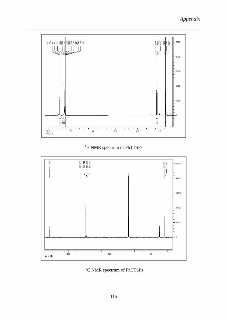
Appendix
115
1H NMR spectrum of PhTTSPs
13C NMR spectrum of PhTTSPs
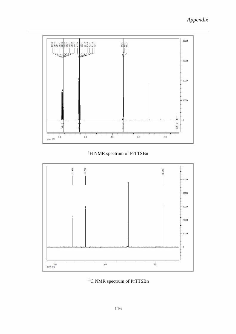
Appendix
116
1H NMR spectrum of PrTTSBn
13C NMR spectrum of PrTTSBn
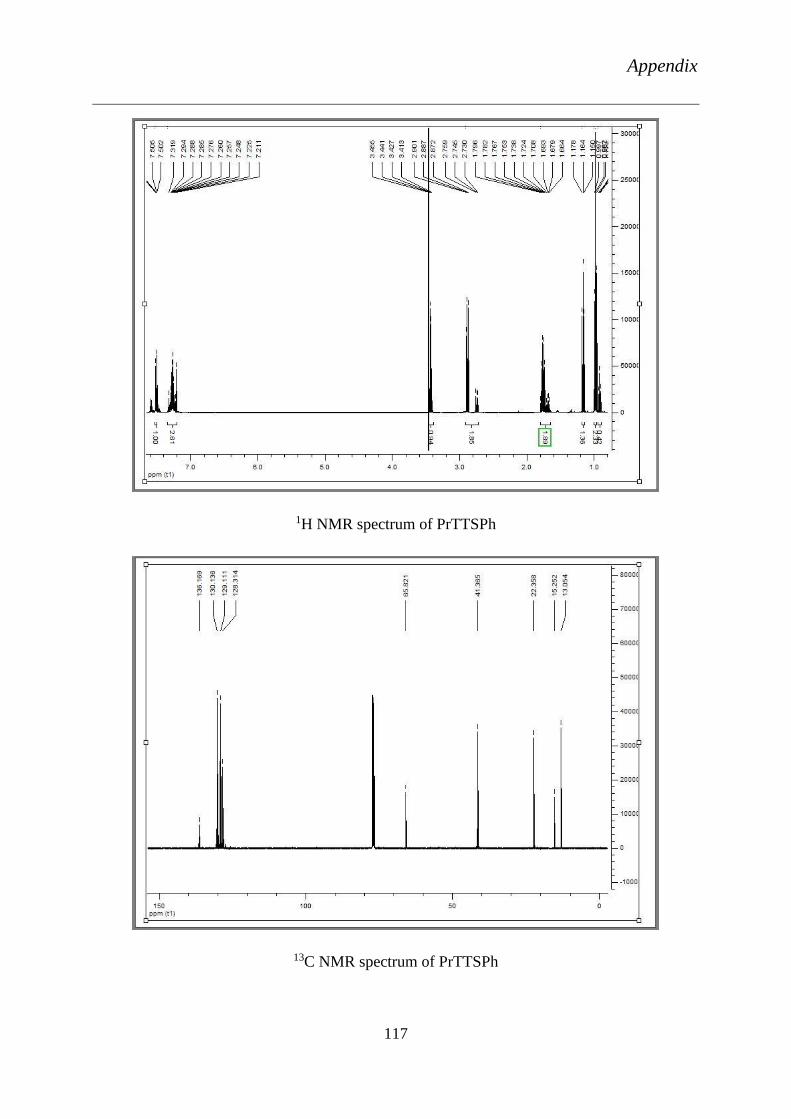
Appendix
117
1H NMR spectrum of PrTTSPh
13C NMR spectrum of PrTTSPh

Appendix
118
D.hansenii S.cerviciae C.gullirmondii C.albicans0
10
20
30SDS
DBTTS
DPSTTS
DPSEETTS
DEETTS
n.s n.s
n.s
n.sn.s
*****
***
*** **n.s
*
*
***
******
Concentration (3mM)
absen
ce o
f bacte
ria
l g
row
th z
on
e (
mm
)
D. hansenii
0.25mM 0.5mM 0.75mM 1mM 3mM0
5
10
15
20
25SDS
DEETTS
n.sn.s
n.s
n.sn.s
Concentration (mM)
absen
ce o
f bacte
ria
l g
row
th z
on
e (
mm
)
Pre-screening of DBTTS (3), DPSTTS (4), DPSEETTS (5) and DEETTS (6) at a concentration of 3 mM
against four different strains of yeast: D. hansenii, S. cerevisiae, C. guilliermondii and C. albicans. SDS (14)
has been used as a control. Data presented as absence of the bacterial growth zone in mm ± SEM of three
independent experiments. ANOVA test: p < 0.05 (*), p < 0.01 (**) or p < 0.001 (***) in relation to the
relevant values for the particular control SDS.
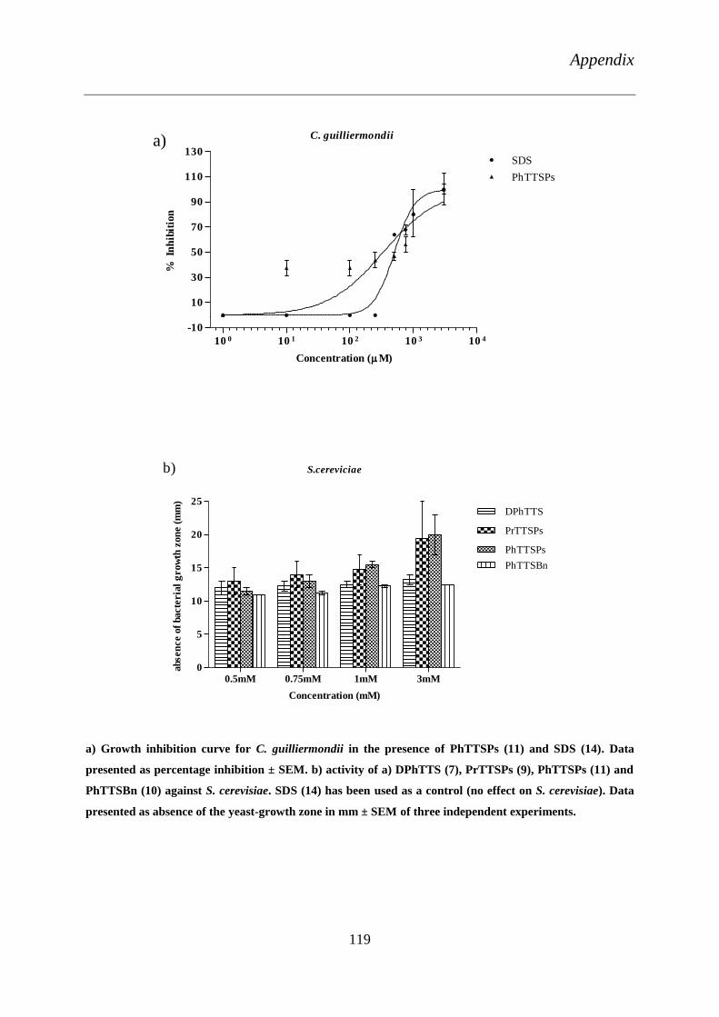
Appendix
119
10 0 10 1 10 2 10 3 10 4
-10
10
30
50
70
90
110
130SDS
C. guilliermondii
PhTTSPs
Concentration (M)
% I
nh
ibit
ion
a)
S.cereviciae
0.5mM 0.75mM 1mM 3mM0
5
10
15
20
25DPhTTS
PrTTSPs
PhTTSPs
PhTTSBn
Concentration (mM)
absen
ce o
f bacte
ria
l g
row
th z
on
e (
mm
)
b)
a) Growth inhibition curve for C. guilliermondii in the presence of PhTTSPs (11) and SDS (14). Data
presented as percentage inhibition ± SEM. b) activity of a) DPhTTS (7), PrTTSPs (9), PhTTSPs (11) and
PhTTSBn (10) against S. cerevisiae. SDS (14) has been used as a control (no effect on S. cerevisiae). Data
presented as absence of the yeast-growth zone in mm ± SEM of three independent experiments.

Appendix
120
Activity of a) DEETTS (6) against S. aureus and b) DPSEETTS (5) against B. subtilis. SDS (14) has been
used as a control. SDS (14) did not show any activity against B. subtilis. Data presented as absence of the
bacterial growth zone in mm ± SEM of three independent experiments. ANOVA test: n.s. = p ≥ 0.05 or p <
0.001 (***) in relation to the relevant values for the particular control SDS.
S. aureus WDCM 5233
0.25mM 0.5mM 0.75mM 1mM 3mM0
5
10
15
20
25SDS
DEETTS
***
ns
nsns
ns
Concentration (M)
absen
ce o
f bacte
ria
l g
row
th z
on
e (
mm
)
a)
B. subtilis A1
0.5mM 0.75mM 1mM 3mM0
5
10
15
20DPSEETTS
nsns
nsns
Concentration (mM)
absen
ce o
f bacte
ria
l g
row
th z
on
e (
mm
)
b)

Appendix
121
Growth inhibition curve for bacterial strains treated in the presence of compounds. a) DEETTS (6)
against S. aureus and b) DPSEETTS against B. subtilis. Data presented as percentage inhibition ± SEM.
Growth inhibition curve for different strains of bacteria treated with: a) BnTTSPs (8), PrTTSPs (9),
PhTTSPs (11) and SDS (14) against S. aureus and b) PhTTSPs (11) against B. subtilis. Data presented as
percentage inhibition ± SEM.
10 2 .5 10 3 .0 10 3 .5
40
60
80
100PhTTSPs
B. subtilis A1
Concentration (M)
% I
nh
ibit
ion
b)
10 2 .0 10 2 .5 10 3 .0 10 3 .5 10 4 .0
-5
15
35
55
75
95
115 SDS
BnTTSPs
PrTTSPs
PhTTSPs
S. aureus WDCM 5233
Concentration (M)
% I
nh
ibit
ion
a)
10 2 .5 10 3 .0 10 3 .5
0
20
40
60
80
100SDS
DEETTS
S. aureus WDCM 5233
Concentration (M)
% I
nh
ibit
ion
a)
10 2 .6 10 2 .8 10 3 .0 10 3 .2 10 3 .4 10 3 .6
40
60
80
100
120
B. subtilis A1
DPSEETTS
Concentration (M)
% I
nh
ibit
ion
b)

Appendix
122
Growth inhibition curve for different strains of yeast treated in the presence of compounds: a) DP80 (19),
DP67 (20) and DP72 (24) against C. guilliermondii and b) DP72 against C. albicans. Data presented as
percentage inhibition ± SEM.
S. cerviciae
0.25mM 0.5mM 0.75mM 1mM 3mM0
5
10
15
20DP72
nsns
nsns
ns
Concentration (mM)
absen
ce o
f bacte
ria
l g
row
th z
on
e (
mm
)
a)
Activity of a) DP72 (24) against S. cerevisiae. Data presented as absence of the bacterial growth zone in
mm ± SEM of three independent experiments. SDS (14) did not show any effect against S. cerevisiae.
ANOVA test: n.s. = p ≥ 0.05 in relation to the relevant values for the particular control SDS.
10 0 10 1 10 2 10 3 10 4
0
20
40
60
80
100
C. guilliermondii
DP80
DP67
DP72
Concentration (M)
% I
nh
ibit
ion
a)
10 0 10 1 10 2 10 3 10 4
0
20
40
60
80
100
120
C. albicans 174
SDS
DP72
Concentration (M)
% I
nh
ibit
ion
b)

Appendix
123
Growth inhibition curve for different bacterial strains treated in the presence of compounds: a) DP56 (25)
and DP53 (26) against S. aureus, and b) SDS (14), DP67 (20) and DP72 (24) against E. coli; c) a
comparison of DP72 (24) against S. aureus and E. coli and d) SDS (14) against E. coli control is shown
above. Data presented as percentage inhibition ± SEM.
10 2 .0 10 2 .5 10 3 .0 10 3 .5
0
40
80
120DP56
S. aureus WDCM 5233
DP53
Concentration (M)
% I
nh
ibit
ion
a)
10 2 10 3 10 4
0
20
40
60
80
100
E. coli VKPM-M17
SDS
Concentration (M)
% I
nh
ibit
ion
d)
10 2 .0 10 2 .5 10 3 .0 10 3 .5
0
20
40
60
80
100
120SDS
E. coli
DP72
S. aureus
Concentration (M)
% I
nh
ibit
ion
c)
10 2 10 3 10 4
40
60
80
100SDS
DP67
DP72
E. coli VKPM-M17
Concentration (M)
% I
nh
ibit
ion
b)

Appendix
124
Compound Chemical Name
SDS (14) Sodium dodecyl sulfate
DP69 (15) Sodium 8-(phenylselanyl)octyl sulfate
DP 49 (16) Sodium 9-(phenylselanyl)nonyl sulfate
DP 70 (17) Sodium 10-(phenylselanyl)decyl sulfate
DP 02 (18) Sodium 11-(phenylselanyl)undecyl sulfate
DP 80 (19) Sodium 12-(phenylselanyl)dodecyl sulfate
DP 67 (20) Sodium 8-(phenyltellanyl)octyl sulfate
DP 68 (21) Sodium 9-(phenyltellanyl)nonyl sulfate
DP 71 (22) Sodium 10-(phenyltellanyl)decyl sulfate
DP 73 (23) Sodium 11-(phenyltellanyl)undecyl sulfate
DP 72 (24) Sodium 12-(phenylselanyl)dodecyl sulfate
DP 56 (25) 3-Chloro-4-octanamidobenzeneseleninic acid
DP 53 (26) 3-Chloro-4-decanamidobenzeneseleninic acid
DP 58 (27) 3-Chloro-4-tetradecanamidobenzeneseleninic acid

Appendix
125
Cytotoxic effects of the MH7A cells when incubated at different concentrations compounds for 24 hours.
a) DBTTS (3), b) DPhTTS (7) and BnTTSPh (10). Data represented as the mean +/- SD.
a) Concentration dependent induction of apoptosis by DBTTS as defined by Annexin-V/7-AAD staining
technique and b) measurement of caspase 3/7 activity in the presence of different concentrations of
DBTTS (3). Data represented as the mean +/- SD.

Appendix
126
The inhibition of IL-1β induced secretion of IL-6 of MH7A cells lines, by the technique, ELISA, when
incubated with the compounds a) DBTTS (3) and b) DPhTTS (7) and BnTTSPh (10) for 24 hours. Data
represented as the mean +/- SD.
Study of the tetrasulfane-mediated influence of the adhesive characteristics of synovial fibroblast cell
lines. a) Concentration-dependent inhibition of the adhesion of cells by DBTTS (3) at different time points.
b) Comparison of the same effect by DBTTS (3), DPhTTS (7) and BnTTSPh (10) at different
concentrations. Data represented as the mean +/- SD.

Curriculum Vitae
127
Curriculum Vitae
Uma Maheswari Viswanathan
Date of Birth: 13.08.1986, Single
Email: [email protected]
Academic Qualifications
July 2011- August 2014 Doctor of Natural Science (Dr. rer. nat.), Bioorganic
Chemistry, School of Pharmacy, University of Saarland,
Saarbruecken, Germany
July 2007- April 2009 Master of Science (M. Sc.) in Nanoscience and
Technology Department of Nanoscience and
Technology, Bharathiar University, Coimbatore,
INDIA, Grade 5.97 / 6 (CGPA out of 6).
August 2004- May 2007 Bachelor of Science (B. Sc.) in Bio-Chemistry,
Pondicherry University, INDIA, 80 %
June 2002- April 2004 Higher Secondary Examinations, Kendriya Vidyalaya
Palakkad Central Board of Secondary Education, India,
76.20 %
June 2000- April 2002 Secondary School Leaving Certificate, Kendriya
Vidyalaya, Palakkad Central Board of Secondary
Education, India, 78.80 %
Research Experience
July 2011- August 2014 Doctorate student in the group of Prof. Dr. Claus Jacob,
Bioorganic Chemistry, School of Pharmacy, University
of Saarland, Saarbruecken, Germany.
“Study of metabolic, biochemical, nanotechnological
and electrochemical behaviour of novel sulfur,
selenium and tellurium compounds”
Curriculum Vitae
128
September 2012-November 2013 Visiting Doctorate student in Yerevan State University,
Armenia.
“Physico-chemical analysis and Antimicrobial
activities of Sulfur, Selenium and Tellurium
compounds”
March 2011- July 2011 Visiting Project Assistant, in the group of Dr. Anja
Philippi, Cellular Immunotherapy Team, KIST Europe,
Saarbruecken, Germany
“T-Cells culturing and Immuno mediated targeted
drug delivery”
January 2011- February 2011 Visiting Project Assistant, in the group of Dr. Kristina
Riehemann, R & D in Nanomedicine, CeNTech,
Muenster, Germany.
“Inflammatory studies on CD-DOX Nanoperticles”
August 2010- January 2011 Researcher in the group of Prof. Dr. Kurt Geckeler,
department of Nano Biomaterials and Electronics,
Gwangju Institute of Science and Technology, South
Korea.
“Synthesis and Characterization of complex
Nanoparticles
May 2009- June 2009 Research Assistant in the group of Associate Prof. Dr. N
Ponpandian, Department of Nanoscience and
Technology, Bharathiar University, TamilNadu, India.
“Amino Acid mediated synthesis of Iron Oxide
Nanoparticles”
January 2009- April 2009 Project Assistant (MSc Thesis work) in the group of Dr.
Vetrichelvan, Department of Chemistry, Syngene
International Limited, A Biocon Company, Bangalore,
India.
“Amino Acid Mediated Synthesis and
Characterization of Silver Nanoparticles and Thin
Film