studiesonthecytotoxicactivitiesofpunicagranatum l.var
TRANSCRIPT
International Scholarly Research NetworkISRN PharmaceuticsVolume 2012, Article ID 547942, 6 pagesdoi:10.5402/2012/547942
Research Article
Studies on the Cytotoxic Activities of Punica granatumL. var. spinosa (Apple Punice) Extract on Prostate Cell Line byInduction of Apoptosis
Koushan Sineh Sepehr,1 Behzad Baradaran,1 Masoumeh Mazandarani,2
Vahid Khori,3 and Fatemeh Zare Shahneh1
1 Immunology Research Center (IRC), University of Medical Sciences Tabriz, Iran2 Department of Botany, Gorgan Branch, Islamic Azad University, Gorgan, Iran3 Golestan Physiology Pharmacology Center, Golestan University of Medical Sciences, Gorgan, Iran
Correspondence should be addressed to Behzad Baradaran, behzad [email protected]
Received 20 October 2012; Accepted 14 November 2012
Academic Editors: J.-J. Chen, D. Kuzmich, G. Lentini, and H. R. Pezza
Copyright © 2012 Koushan Sineh Sepehr et al. This is an open access article distributed under the Creative Commons AttributionLicense, which permits unrestricted use, distribution, and reproduction in any medium, provided the original work is properlycited.
The Punica granatum L. var. granatum (pomegranate) has been demonstrated to exert antitumor effects on various types of cancercells. The present study aimed to evaluate the medicinal herbs Punica granatum L. var. spinosa (apple punice) that are native toIran. This study was determined to test the possible cytotoxic activity and induction of apoptosis on human prostate cell lines.The effect of ethanol extracts of the herbs on the inhibition of cell proliferation was assessed by MTT colorimetric assay. PC3 celllines treated with the extracts were analyzed for the induction of apoptosis by cell death detection (ELISA) and TUNEL assay. Dyeexclusion analysis was performed for viability rate. Our results demonstrated that the Punica granatum L. var. spinosa extract dosedependently suppressed the proliferation of PC3 cells (IC50= 250.21 µg/mL) when compared with a chemotherapeutic anticancerdrug (Toxol) (Vesper Pharmaceuticals) with increased nucleosome production from apoptotic cells. The Punica granatum L. var.spinosa extract attenuated the human prostate cell proliferation in vitro possibly by inducing apoptosis. The Punica granatum L.var. spinosa is likely to be valuable for the treatment of some forms of human prostate cell line.
1. Introduction
Apoptosis (programmed cell death) is a physiological mech-anism of cell death. During apoptosis, there is a rapidreduction in the cellular volume followed by chromatin con-densation, associated with characteristic internucleosomalDNA cleavage. This results in the production of nucleosomesof DNA fragments complexes with core histones, which aredistinct multiples of an 180–200 bp subunit [1]. Cancer isone of the major causes of mortality throughout the world.World Health Organization statistics have estimated thatcancer will cause 83.2 million deaths between 2005 and 2015if the recommended measures are not respected. In 2007,cancer was the cause of 7.9 million deaths, which is 13% ofworld mortality. Among males in the third world countries,prostate cancer is the second-leading cause of cancer-related
death [2]. Cancer is a disease that is characterized by too littleapoptosis. Under normal circumstances damaged cells willundergo apoptosis, but in the case of cancer cells mutationsmay have occurred that prevent cells from undergoingapoptosis. Understanding apoptosis regulation is a mainconcern in the development of chemotherapeutic anticancerdrugs on malignant cells [3, 4]. Traditionally, many extractsfrom roots, stems, and fruits have been used for maintaininghealth, enhancing overall immune status, and preventionand treatment of chronic diseases, and the modulation andtreatment of different diseases [5].
Punica granatum L. var. spinosa is known as applepunice from Punicaceae family commonly widespread in thelatitudes of 475 m above sea level in the north of Iran. Recentstudies have demonstrated that Punica granatum L. var.granatum extracts possess a plethora of biological activities
2 ISRN Pharmaceutics
including antibacterial, antiviral, antifungal, cytotoxic andimmuno-potentiating activities. The Punica granatum L. var.granatum tree (pomegranate), especially its fruit, possesses avast ethno medical history and represents a phytochemicalreservoir of heuristic medicinal value. The tree/fruit canbe divided into several anatomical compartments: (1) seed,(2) juice, (3) peel, (4) leaf, (5) flower, (6) bark, and (7)roots, each of which has interesting pharmacologic activity.Juice and peels, for example, possess potent antioxidantproperties, while juice, peel, and oil are all weakly estrogenicand heuristically of interest for the treatment of menopausalsymptoms. The use of juice, peel, and oil has also been shownto possess anticancer activities, including interference withtumor cell proliferation, cell cycle, invasion, and angiogenesis[6]. The toxicity of Punica granatum L. var. spinosa has notbeen intensively studied. Accordingly, we have conductedour research on toxicity extracts of Punica granatum L. var.spinosa seeds and peels [7, 8]. The objective of this studywas to examine the in vitro cytotoxic activities of a wildlyethanolic standardized Punica granatum L. var. spinosa (PGS)extract using a MTT cytotoxicity assay. The study also testedwhether the mechanism of action involves induction ofapoptosis. Cell death ELISA and TUNEL was employed toquantify the nucleosome production resulting from nuclearDNA fragmentation during apoptosis.
2. Materials and Methods
2.1. Preparation of Plant Extract. Punica granatum L. var.spinosa (PGS) plants known as apple punice from Puni-caceae family were collected from the southeast of Golestanprovince, Iran (Ramian). Dr. Mazandarani from the Medic-inal Plant Research Center of Islamic Azad University ofGorgan, Iran, identified the plant. A voucher specimenwas deposited in the herbarium of the above mentioned(no. 315HRCMP). The seeds and peels parts of the plantwere separated, shade dried, and grinded into powder withmortar and pestle. The prepared powder was kept in tightcontainers protected completely from light. Extraction ofethanolic extract was carried out by macerating 100 g ofpowdered dry plant in 500 mL of 70% ethanol for 48 h atroom temperature. Then, the macerated plant material wasextracted with 70% ethanol solvent by percolator apparatus(2-liter volume) at room temperature. The plant extract wasremoved from percolator, filtered through Whatman filterpaper (NO. 4), and dried under reduced pressure at 37◦Cwith rotator evaporator. The ethanol extract was filtered andconcentrated using a rotary evaporator and then evaporatedto dryness. Briefly, the concentrated plant extracts weredissolved in dimethyl sulphoxide (DMSO) (SIGMA, USA)to get a stock solution of 10 mg/mL. The substock solutionof 0.2 mg/mL was prepared by diluting 20 µL of the stocksolution into 980 µL serum-free culture medium, RPMI 1640(the percentage of DMSO in the experiment should notexceed 0.5).
2.2. Cell Cultures. The human prostate cancer cell line (PC3)and normal fibrosarcoma cell line (L929) were obtained
from National Cell Bank of Iran (NCBI, Pasteur Institute ofIran). The cells were grown and maintained in a humidifiedincubator at 37◦C and in 5% CO2 atmosphere. RPMI-1640medium supplemented with 10% fetal bovine serum (FBS,Invitrogen Gibco), 100 units/mL penicillin, and 100 µg/mLstreptomycin (Invitrogen Gibco) was used for cell culturesof PC3. Ten thousand cells from log phase cultures wereseeded in 100 µL of RPMI-164 medium supplemented with10% fetal bovine serum per well of 96-well flat-bottomculture plates (Nunc, Denmark). Proliferative response andcell death of the PGS extract-treated cells were determinedusing MTT assay and cell death ELISA, respectively [9].
2.3. MTT Colorimetric Assay. A colorimetric assay using3-(4, 5-dimethylthiazoyl)-2, 5-diphenyltetrazolium bromide(MTT) was performed. Briefly, cells were added onto flat-bottomed microculture plates in the presence or absenceof various concentrations of the extracts (in triplicate) andincubated at 37◦C in a 5% humidified CO2 incubator for 24and 48 h. Then, 10 mL of MTT (5 mg/mL, Sigma) was addedto each well and incubation was continued for a further 4 hat 37◦C. In each well, 100 µL/well of solubilization solution,containing DMSO and Sorenson buffer, were added. Aftercomplete solubilization of the dye, plates were read at 570 nmon an ELISA reader. The mean optical density (OD) ±SD for each group of replicates was calculated. The wholeprocedure was repeated for three times. The inhibitory rateof cell growth was calculated using the formula: % Growthinhibition = (1−OD extract treated)/OD negative control ×100 [9].
2.4. Cell Death Detection. cell death detection ELISAPLUS
(Roche Applied Science, Switzerland) was used to quantifyhistone-complexed DNA fragments (nucleosomes) in cyto-plasm of the apoptotic cells after induction of apoptosis, asdescribed elsewhere [10]. Briefly, after incubation with thePGS extract (at concentrations determined by MTT assay)for 24 h, the PC3 cells were pelleted and lysed. The remainingsteps were carried out according to the instructions suppliedby the manufacturer. The resulting color development,which was proportional to the amount of nucleosomescaptured in the antibody sandwich, was measured at 405 nm(with reference wavelength at 490 nm) using a Benchmarkmicrotiter plate reader (Bio-Rad). Results were expressed asthe apoptotic and necrosis percentage, calculated from theratio of absorbance of treated (apoptotic) sample to that ofthe untreated (control) sample [10].
2.5. Dye Exclusion Assay. Viability induced of the PGSextract treatment was measured using trypan blue exclusionassay. Briefly, 1×10⁴ cells were seeded into 96-well plates andtreated with or without (as control) PGS extract at specifieddoses for 24 h. After the incubation period, the cultureswere harvested and washed twice with PBS. The cell pelletwas then resuspended with 0.5 mL PBS. Then, 20 µL of cellwas mixed with equal volume of 0.4% trypan blue (Sigma,USA Merck) and was count with Neubauer haemocytometer(Weber, England) by clear field microscopy (Nikon, japan).
ISRN Pharmaceutics 3
Only viable cells were counted. Each extract and control wereassayed two times in triplicate [10].
2.6. Apoptosis Assay. To assess cell death by apoptosis, anIn Situ Cell Death Detection Kit, POD (Roche, Germany)for DNA chromatin morphologic features was used forquantification. The procedures followed the manufacture’sguidelines. Briefly, cells were cultured on glass slides andanalyzed 24 hours after treatment. Cells grown on coverslipswere washed twice with PBS, air dried, and fixed for 60 minin freshly prepared 4% paraformaldehyde/PBS (pH: 7.4)(Sigma-Germany), pH 7.4, at room temperature. Then thecells were washed again twice with PBS (pH: 7.4) andincubated with 3% H2O2/methanol (Merck-Germany) for10 min. Following washing with PBS, cells were permeabi-lized in 0.2% Triton X-100/PBS (pH: 7.4) (Sigma-Germany)for 2 min at 4◦C. Samples were incubated in 50 µL ofTUNEL reaction mixture for 2 h at 37◦C in a humidifiedchamber and in the dark, covered with parafilm. Omissionof TdT provided the negative control for the assay, andpreincubation of cells with 10 µg/mL DNase I in 50 mM Tris-HCl, pH 7.4, 1 mM MgCl2, and 1 mg/mL BSA for 10 min atroom temperature to induce DNA strand breaks artificially,served as positive control. Cells were washed with PBS (pH:7.4) and incubated for 30 min in a humidified chamber, at37◦C with 50 µL converter-POD (Anti-fluorescein antibody,Fab fragment from sheep, conjugated with horse-radishperoxidase). After rinsing in PBS, the samples were incubatedfor 10 min with 100 µL DAB (Sigma-Germany) substrate inthe dark. At the end, the samples were mounted and analyzedunder light microscope, where the apoptotic cells could beseen as condensed shrinked dark brown cells [10].
2.7. Statistical Analysis. The data are expressed as mean± standard deviation (SD) for at least three independentdeterminations in triplicate for each experimental point. Thedata were analyzed using IBM SPSS Statistics 20 software.For all the measurements, Tow-way ANOVA followed byDuncan’s New Multiple Range Test (P ≤ 0.05) was used toassess the statistical significance of difference between controland PGS treated.
3. Results
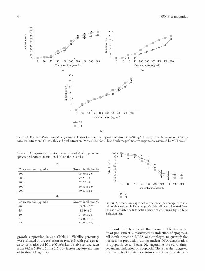
3.1. Effects of Punica granatum L. var. spinosa Extract onProliferation of Prostate Cancer Cell Line (PC3). Peels ofPGS extract at 10 to 600 µg/mL exhibited significant dose-dependent inhibitory effects on the proliferation of PC3.Growth inhibition of peel extract in 24 and 48 h was 61.2 ±2.3% and 67.± 1.75%, respectively (Figure 1(a)), with morethan 75% suppression. The concentrations producing 50%growth inhibition (IC50) of the PGS extract on PC3 wereeffectively suppressed with the IC50 value (250.21 µg/mL)after incubation with the peel extract. However seed extractinduced no significant suppression on the proliferation ofPC3 cells (Figure 1(b)) and the peel extract induced nosignificant suppression on the proliferation of normal L929cells (Figure 1(c)). PC3 cells were compared to elucidate
the cytotoxicity of both peels of PGS extract and Toxol(chemotherapeutic agent, control positive) with more than75% in 600 µg/mL and 90% in 20 µg/mL growth suppressionin 24 h (Table 1).
In 24 and 48 h Dye exclusion assay evaluated viabilityof PC3 cells exposed to peel extract. As the result inFigure 2, the viabilities of cells exposed to peel PGS extractat concentrations of 10 and 600 µg/mL were 96.3± 7.8% and24.1± 2.5%, respectively.
3.2. Effects of Punica granatum L. var. spinosa Extract on CellDeath of Prostate Cancer Cell Line (PC3). As determinedby MTT assay, peel extract at 50, 100, 200, and 300 µg/mLwas chosen for PC3 cell line in cell death detection ELISA.The proportion of dead PC3 cells increased sharply (from32± 8.5%, to 55± 1.9%) upon 24 h incubation with the peelextract at 50–300 µg/mL at 24 h. These results suggested thatthe apoptotic response of PC3 cell lines should be evaluatedat different concentration points.
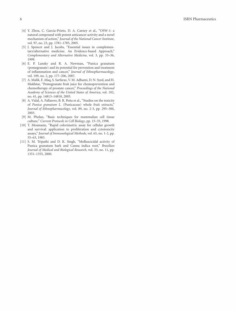
3.3. Punica granatum L. var. spinosa Extract Induced Apoptosisin Prostate Cancer Cell Line (PC3). To test whether or notpeel extract that induced the decrease of cell viability andcytotoxicity contributes to apoptotic death in PC3 cell linesin vitro. Cells were incubated with 250 µg/mL of PGS for24 h and then determined using TUNEL assay. It was foundthat the PC3 cells treated with peel extract (250 µg/mL)for 24 h exhibited apoptotic body formation (Figure 4).PC3 cells treated with peel extract displayed typical mor-phological features of apoptotic cells, with condensed andfragmented nuclei (Figure 4(a)). However, homogenousnuclear chromatin was evident in control cells (Figure 4(b)).The induction of apoptosis by peel extract was confirmed byin situ TUNEL assay. TUNEL assay based on labeling of DNAstrand breaks generated during apoptosis revealed that peelextract induces apoptosis in PC3 cells.
4. Discussion
Apoptosis (programmed cell death) is a physiological mech-anism of cell death. Cancer is one of the major causesof mortality throughout the world. Cancer is a diseasethat is characterized by too little apoptosis. Understandingapoptosis regulation is a main concern in the developmentof chemotherapeutic anticancer drugs on malignant cells[2]. The present study has demonstrated that ethanolicpeel extract of PGS in natural form could significantlysuppress the proliferation of PC3 cells in vitro using theMTT assay. Such antiproliferative activity of peel extractof PGS was characterized by the dose-dependent manner(Figure 1(a)). However seed extract induced no significantsuppression on the proliferation of PC3 cells (Figure 1(b))and the peel extract induced no significant suppressionon the proliferation of normal L929 cells (Figure 1(c)).Toxol at an optimal in vitro concentration was found toselectively induce at least 90% growth suppression on PC3cells but peel extract has more than 75.50% in 600 µg/mLin comparison to 90% inhibition activity Toxol in 20 µg/mL
4 ISRN Pharmaceutics
0
2030
10
405060708090
100In
hib
itio
n (
%)
0 10 20 50 100 200 300 400 500 600
Concentration (µg/mL)
(a)
0
5
10
15
20
25
30
Inh
ibit
ion
(%
)
0 10 20 50 100 200 300 400 500 600
Concentration (µg/mL)
(b)
0
5
10
15
20
25
30
Inh
ibit
ion
(%
)
0 10 20 50 100 200 300 400 500 600
Concentration (µg/mL)
24
48
(c)
Figure 1: Effects of Punica granatum spinosa peel extract with increasing concentrations (10–600 µg/mL with) on proliferation of PC3 cells(a), seed extract on PC3 cells (b), and peel extract on L929 cells (c) for 24 h and 48 h the proliferative response was assessed by MTT assay.
Table 1: Comparisons of cytotoxic activity of Punica granatumspinosa peel extract (a) and Toxol (b) on the PC3 cells.
(a)
Concentration (µg/mL) Growth inhibition %
600 75.50 ± 2.6
500 72.21 ± 8.1
400 70.67 ±7.8
300 66.83 ± 3.9
200 49.67 ± 6.5
(b)
Concentration (µg/mL) Growth inhibition %
20 93.78 ± 5.7
15 82.86 ± 2
10 71.69 ± 2.8
5 63.88 ± 3.2
3.5 51.79 ± 1.5
growth suppression in 24 h (Table 1). Viability percentagewas evaluated by dye exclusion assay at 24 h with peel extractat concentrations of 10 to 600 µg/mL and viable cell decreasesfrom 96.3±7.8% to 24.1±2.5% by increasing dose and timeof treatment (Figure 2).
102030405060708090
100
Via
bilit
y (%
)
0 10 20 50 100 200 300 400 500 600
Concentration (µg/mL)
2448
Figure 2: Results are expressed as the mean percentage of viablecells with 3 wells each. Percentage of viable cells was calculated fromthe ratio of viable cells to total number of cells using trypan blueexclusion test.
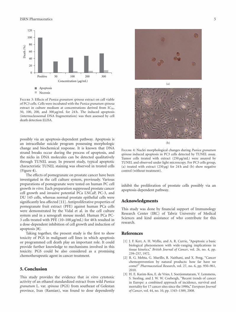
In order to determine whether the antiproliferative activ-ity of peel extract is manifested by induction of apoptosis,cell death detection ELISA was employed to quantify thenucleosome production during nuclear DNA denaturationof apoptotic cells (Figure 3), suggesting dose-and time-dependent induction of apoptosis. These results suggestedthat the extract exerts its cytotoxic effect on prostate cells
ISRN Pharmaceutics 5
0
20
40
60
80
100
120
Dea
th (
%)
Positive 50 100 200 300
Concentration (µg/mL)
Apoptosis
Necrosis
Figure 3: Effects of Punica granatum spinosa extract on cell viableof PC3 cells. Cells were incubated with the Punica granatum spinosaextract in culture medium at concentrations derived from IC50,50, 100, 200, and 300 µg/mL for 24 h. The induced apoptosis(internucleosomal DNA fragmentation) was then assessed by celldeath detection ELISA.
possibly via an apoptosis-dependent pathway. Apoptosis isan intracellular suicide program possessing morphologicchange and biochemical response. It is known that DNAstrand breaks occur during the process of apoptosis, andthe nicks in DNA molecules can be detected qualitativelythrough TUNEL assay. In present study, typical apoptoticcharacteristic TUNEL staining was observed in treated cells(Figure 4).
The effects of pomegranate on prostate cancer have beeninvestigated in the cell culture system, previously. Variouspreparations of pomegranate were tested on human PC cellgrowth in vitro. Each preparation suppressed prostate cancercell growth and invasive potential PCa LNCaP, PC-3, andDU 145 cells, whereas normal prostate epithelial cells weresignificantly less affected [11]. Antiproliferative properties ofpomegranate fruit extract (PFE) against human PCa cellswere demonstrated by the Vidal et al. in the cell culturesystem and in a xenograft mouse model. Human PCa PC-3 cells treated with PFE (10–100 µg/mL) for 48 h resulted ina dose-dependent inhibition of cell growth and induction ofapoptosis [8].
Taking together, the present study is the first to showtoxicity of PGS in malignant cell lines in which apoptosisor programmed cell death play an important role. It couldprovide further knowledge to mechanisms involved in thistoxicity. PGS could be also considered as a promisingchemotherapeutic agent in cancer treatment.
5. Conclusion
This study provides the evidence that in vitro cytotoxicactivity of an ethanol standardized extract from wild Punicagranatum L. var. spinosa (PGS) from southeast of Golestanprovince, Iran (Ramian), was found to dose dependently
(a)
(b)
Figure 4: Nuclei morphological changes during Punica granatumspinosa induced apoptosis in PC3 cells detected by TUNEL assay.Tumor cells treated with extract (250 µg/mL) were assayed byTUNEL and observed under light microscopy. For PC3 cells group,(a) treated with extract (250 µg) for 24 h and (b) show negativecontrol (without treatment).
inhibit the proliferation of prostate cells possibly via anapoptosis-dependent pathway.
Acknowledgments
This study was done by financial support of ImmunologyResearch Center (IRC) of Tabriz University of MedicalSciences and kind assistance of who contribute for thisresearch.
References
[1] J. F. Kerr, A. H. Wyllie, and A. R. Currie, “Apoptosis: a basicbiological phenomenon with wide-ranging implications intissue kinetics,” British Journal of Cancer, vol. 26, no. 4, pp.239–257, 1972.
[2] R. G. Mehta, G. Murillo, R. Naithani, and X. Peng, “Cancerchemoprevention by natural products: how far have wecome?” Pharmaceutical Research, vol. 27, no. 6, pp. 950–961,2010.
[3] H. E. Karim-Kos, E. de Vries, I. Soerjomataram, V. Lemmens,S. Siesling, and J. W. W. Coebergh, “Recent trends of cancerin Europe: a combined approach of incidence, survival andmortality for 17 cancer sites since the 1990s,” European Journalof Cancer, vol. 44, no. 10, pp. 1345–1389, 2008.
6 ISRN Pharmaceutics
[4] Y. Zhou, C. Garcia-Prieto, D. A. Carney et al., “OSW-1: anatural compound with potent anticancer activity and a novelmechanism of action,” Journal of the National Cancer Institute,vol. 97, no. 23, pp. 1781–1785, 2005.
[5] J. Spencer and J. Jacobs, “Essential issues in complemen-tary/alternative medicine. An Evidence-based Approach,”Complementary and Alternative Medicine, vol. 3, pp. 33–36,1999.
[6] E. P. Lansky and R. A. Newman, “Punica granatum(pomegranate) and its potential for prevention and treatmentof inflammation and cancer,” Journal of Ethnopharmacology,vol. 109, no. 2, pp. 177–206, 2007.
[7] A. Malik, F. Afaq, S. Sarfaraz, V. M. Adhami, D. N. Syed, and H.Mukhtar, “Pomegranate fruit juice for chemoprevention andchemotherapy of prostate cancer,” Proceedings of the NationalAcademy of Sciences of the United States of America, vol. 102,no. 41, pp. 14813–14818, 2005.
[8] A. Vidal, A. Fallarero, B. R. Pena et al., “Studies on the toxicityof Punica granatum L. (Punicaceae) whole fruit extracts,”Journal of Ethnopharmacology, vol. 89, no. 2-3, pp. 295–300,2003.
[9] M. Phelan, “Basic techniques for mammalian cell tissueculture,” Current Protocols in Cell Biology, pp. 15–35, 1998.
[10] T. Mosmann, “Rapid colorimetric assay for cellular growthand survival: application to proliferation and cytotoxicityassays,” Journal of Immunological Methods, vol. 65, no. 1-2, pp.55–63, 1983.
[11] S. M. Tripathi and D. K. Singh, “Molluscicidal activity ofPunica granatum bark and Canna indica root,” BrazilianJournal of Medical and Biological Research, vol. 33, no. 11, pp.1351–1355, 2000.

Submit your manuscripts athttp://www.hindawi.com
PainResearch and TreatmentHindawi Publishing Corporationhttp://www.hindawi.com Volume 2014
The Scientific World JournalHindawi Publishing Corporation http://www.hindawi.com Volume 2014
Hindawi Publishing Corporationhttp://www.hindawi.com
Volume 2014
ToxinsJournal of
VaccinesJournal of
Hindawi Publishing Corporation http://www.hindawi.com Volume 2014
Hindawi Publishing Corporationhttp://www.hindawi.com Volume 2014
AntibioticsInternational Journal of
ToxicologyJournal of
Hindawi Publishing Corporationhttp://www.hindawi.com Volume 2014
StrokeResearch and TreatmentHindawi Publishing Corporationhttp://www.hindawi.com Volume 2014
Drug DeliveryJournal of
Hindawi Publishing Corporationhttp://www.hindawi.com Volume 2014
Hindawi Publishing Corporationhttp://www.hindawi.com Volume 2014
Advances in Pharmacological Sciences
Tropical MedicineJournal of
Hindawi Publishing Corporationhttp://www.hindawi.com Volume 2014
Medicinal ChemistryInternational Journal of
Hindawi Publishing Corporationhttp://www.hindawi.com Volume 2014
AddictionJournal of
Hindawi Publishing Corporationhttp://www.hindawi.com Volume 2014
Hindawi Publishing Corporationhttp://www.hindawi.com Volume 2014
BioMed Research International
Emergency Medicine InternationalHindawi Publishing Corporationhttp://www.hindawi.com Volume 2014
Hindawi Publishing Corporationhttp://www.hindawi.com Volume 2014
Autoimmune Diseases
Hindawi Publishing Corporationhttp://www.hindawi.com Volume 2014
Anesthesiology Research and Practice
ScientificaHindawi Publishing Corporationhttp://www.hindawi.com Volume 2014
Journal of
Hindawi Publishing Corporationhttp://www.hindawi.com Volume 2014
Pharmaceutics
Hindawi Publishing Corporationhttp://www.hindawi.com Volume 2014
MEDIATORSINFLAMMATION
of