studies toward isolation and identification...
TRANSCRIPT
An-Najah National University Faculty of Graduate Studies
STUDIES TOWARD ISOLATION AND IDENTIFICATION OF BIOACTIVE
SUBSTANCES FROM MEDICINAL PLANTS
By Derar Issa "Mohammad Hassan" Smadi
Supervisor Dr. Othman Hamed
Submitted in Partial Fulfillment of the Requirements for the Degree of Master of Science in Chemistry, Faculty of Graduate Studies, An- Najah National University, Palestine.
2011


iii
DEDICATION
To my father, my mother, my brothers, my sister,
and to all my friends
iv
ACKNOWLEDGMENT
Praise and thanks to Allah, for helping and directing me to the
right path. Special thanks to my research supervisor Dr. Othman
Hamed for the chance given me to work with his research group. I
am deeply grateful to him for his constant presence, and his
encouragement throughout this research project.
Great thanks to Dr. Nizar Matar for his help during writing the
thesis. Many thanks to Dr. Adham Abu Taha for helping me in
measuring of antibacterial activity of the fractions. My thanks to
the thesis committee members for their willingness to read the
thesis and provide useful suggestions.
Finally, I thank my doctors and all staff in the department of
chemistry at An-Najah University, as well as to Mr. Omair
Nabulsi for helping me in the lab

v
اإلقرار
:أنا الموقع أدناه مقدم الرسالة التي تحت عنوان
Studies toward Isolation and Identification of Bioactive Substances from Medicinal Plants
دراسة عزل وتشخيص مواد نشطة طبياً من اصول نباتات طبية
ستثناء ما تمت اإلشارة اقر بأن ما اشتملت عليه الرسالة إنما هي من إنتاجي الشخصي با
أو أي جزء منها لم يقدم من قبل لنيل أية درجـة علميـة أو , وان الرسالة ككل, إليه حيثما ورد
.بحث علمي أو بحثي لدى أية مؤسسة تعليمية أو بحثية أخرى
Declaration
The work provided in this thesis, unless otherwise referenced, is my
research own work and has not been submitted elsewhere for any other
degree or qualification.
Student’s name: اسم الطالب :
Signature: التوقيع:
Date: التاريخ:

vi
List of Contents No. Contents Page
Dedication iii Acknowledgment iv Declaration v List of Contents vi List of Tables viii List of Figures ix List of Appendices x Abstract xi Chapter One: Introduction 1 1.1 The Significance of Natural Products as Drugs 1 1.2 Sources of Natural products 3 1.2.1 Natural Products from Plants 4 1.2.2 Natural Products From Animals 61.2.3 Natural Products from Microorganisms 7 1.2.4 Natural Products from Marine Organisms 8 1.3 Plant Based Agents Antibacterial Agents 91.3.1 Terpenoids 9 1.3.2 Alkaloids 11 1.4 Aromatic compounds 12 1.4.1 Flavonoids 12 1.4.2 Quinones 13 1.5 Plants Used In Traditional Medicine in Palestine 14 1.5.1 Walnut Tree 14 1.5.2 Plumbago 161.5.3 Salvia L 19 1.6 Aims and Scope of the Work 21 1.7 Chemical Compounds Extracted from Inula viscosa 231.7.1 Sesquiterpenes 23 1.7.2 Terpenes 24 1.7.3 Lactone 24 1.7.4 Azulene 24 Chapter Two: experimental 26 2.1 General Procedure for extractions 26 2.1.1 Requirements of Efficient Extraction 26 2.1.2 Plant Material 272.1.3 Preparation of the Collected plants for Extraction 27
2.2 Evaluation of Concentrate by Thin Layer Chromatography (TLC) 27
2.3 Separation of Extract by Flash Chromatography 28

viiNo Contents Page
2.3.1 Flash Column Chromatography 28 2.4 Evaluation of fractions for Antibacterial Activity 30 2.4.1 Materials 30 2.4.2 Microorganisms used 30 2.4.3 Screening for Antimicrobial Activity 30 2.4.4 Determination of Minimum Inhibitory Method 31 Chapter Three: Results and Discussion 32 3.1 Analysis of the separated fractions 323.1.1 Fraction Three 34 3.1.2 Fraction Four 34 3.2 Analysis the fractions 39 3.2.1 Screening Results 39
3.2.2 Determination of Minimum Inhibitory Concentration (MIC) 40
3.2.3 Determination of Minimal Bactericidal Concentration (MBC) 41
References 43 Appendix 52 ب الملخص

viii
List of Tables No. Tables Page
Table (2.1) A summary of the separated fractions from Inula viscose extract (about 10.0 g) 29
Table (2.2) Screening results (Zone of inhibition in mm) 31 Table (2.3) MIC results of fraction 4 321 Table (2.4) MBC results for S.aureus bacteria 32 Table (3.1) LC/MS Analysis Results of Fraction 2 34Table (3.2) LC/MS Analysis Results of Fraction 4 34
Table (3.3) Summary of the 13C chemical shifts of 3,3’-di-O-methylquercetin (61). 39
Table (3.4) Screening results (Zone of inhibition in mm) 40 Table (3.5) A Summary of MIC Results of Fraction 4 41
Table (3.6) MBC and MIC results of fraction four against S. aureus bacteria 41

ix
List of Figure No. Figure Page
Fig. (1.1) Examples on natural materials with anaesthetic properties 2
Fig. (1.2) Examples on bioactive materials extracted from various plants 3
Fig. (1.3) Natural products with anticancer and antimalarial activities 4
Fig. (1.4) Commercial anticancer with natural origin 5 Fig. (1.5) bioactive material extracted from animals 7
Fig. (1.6) Examples on Bioactive compounds extracted from Bacteria 7
Fig. (1.7) Marine source natural products with bioactivities 8
Fig. (1.8) Terpenoids with antifungal activities extracted from leaves of pinus radita 10
Fig. (1.9) Example on aromatic compounds with antibacterial activity 12
Fig. (1.10) Example on flavonoids with antibacterial activities 13
Fig. (1.11) Example on isoflavonoids with antibacterial activities 13
Fig. (1.12) Structures and name of materials extracted from walnut tree 15
Fig. (1.13) Natural materials available in plumgbago europea L 18
Fig. (1.14) Chemical compounds that are available in Salvia Fruticosa 21
Fig. (3.1) 35
Fig. (3.2) 1H NMR of major component of fraction 4 of I. Viscosa 36
Fig. (3.3) 1H NMR of major component of fraction 4 of I. Viscosa, Showing the coupling constants between major peaks
37
Fig. (3.4) 13C NMR of major component of fraction 3 of I. Viscosa 38

x
List of Appendices No. Appendix Page
Fig a1 1H NMR of fraction 3 of I. Viscosa, showing the presence of a mixture of two components 52
Fig a2 GC/MS of crude of I. Viscosa. 53
Fig a3 LC/MS of fraction 4 of I. Viscosa, showing the presence of a mixture of two components 54
Fig a4 LC/MS of component 1 of fraction 4 after purification 55 Fig a5 MS of component 1 of fraction 4 after purification 56
Fig a6 MS of component 2 of fraction 4 after purification 57
Fig a7 LC/MS of fraction 2 of I. Viscosa, showing the presence of a mixture of five components 58
xiSTUDIES TOWARD ISOLATION AND IDENTIFICATION OF
BIOACTIVE SUBSTANCES FROM MEDICINAL PLANTS By
Derar Issa Smadi Supervisor
Dr. Othman Hamed
Abstract
More than 600 plant species have been used in the Palestinian
traditional medicine to treat various diseases. About fifty of these plants
are used to treat various skin diseases. This work is a continuation of
existing effort to find new medicine from plants grows in Palestine.
Medicinal plant Tayoon was chosen for this work. It was chosen because it
is an important plant in the Palestinian folklore, it has unlimited number of
medical applications. Three main stages were used in separation and
identification of tayoon extracts. In the first stage, tayoon was subjected to
extraction with ethyl acetate. In the second stage, tayoon extracts were
fractionated using flash chromatography, four fractions were separated. In
the third stage, the four separated fractions were evaluated for antibacterial
activities. Results of the antibacterial study showed that only fraction 4 has
activity against bacteria S.aureus. Base on these results fraction 4 was
further fractionated by flash chromatography and two components were
collected. The major component which was identified to have the
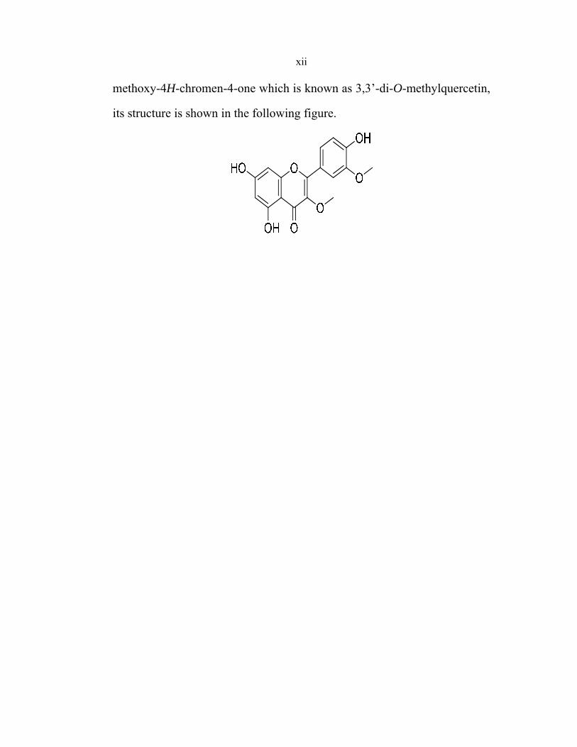
bioactivity was analyzed by various spectroscopic techniques and its
structure was determined. The major compound of fraction four was
identified to be 5,7-dihydroxy-2-(4-hydroxy-3-methoxyphenyl)-3-
xii
methoxy-4H-chromen-4-one which is known as 3,3’-di-O-methylquercetin,
its structure is shown in the following figure.

1
CHAPTER ONE INTRODUCTION
1.1 The Significance of Natural Products as Drugs
Since the old time, natural products have played a very important
role in curing human diseases. The ancient civilizations such as Chinese,
Egyptian, Indians, Greek, and North Africans provide written evidence for
the use of natural products for treatment of various diseases .1
The earliest known written medical prescription is about four
thousand year old, Sumerian clay tablet that shows remedies for various
illnesses .2 For instance, mandrake was prescribed for pain relief, turmeric
possesses blood clotting properties, roots of the endive plant were used for
treatment of gall bladder disorders, and raw garlic was prescribed for
circulatory disorders. These treatments are still being used in several
countries as affordable drugs.
However, it was not until the nineteenth century that scientists
isolated active components from various medicinal plants. Friedrich
Sertürner isolated morphine (1) (Fig 1.1) from Papaver somniferum in
1806. In 1860, a German chemist Carl Koler isolated cocaine (2) (Fig 1.1),
the chemical responsible for certain biological activity. He found that
cocaine could act as a local anaesthetic in eye surgery. As the years
passed, scientists observed that cocaine paralyzed nerve endings
responsible for transmitting pain. As a local anaesthetic, it revolutionized
several surgical and dental procedures and since then natural products have
been extensively screened for their medicinal purposes.

2
Fig. (1.1): Examples on natural materials with anaesthetic properties
More examples on bioactive materials extracted from natural sources
are shown in Fig 1.2. Atropine( 3) obtained from Atropa belladonna,
strychnine (5), a CNS stimulant, identified from a cone snail, Conus magus,
and Taxol® (4)obtained from the bark of the Pacific yew tree are examples
of bioactive materials available in plants.
A recent study conducted by the World Health Organization (WHO)
shows that, about 80% of the world’s population relies on traditional
medicine .3 More than one hundred drugs prescribed in USA today come
from natural sources, more than 90% of which come either directly or
indirectly from plant sources .4 About 50% of the anticancer drugs in the
market come from natural products or natural products derivatives .5 More
than a hundred anticancer drugs have been developed between the years
1981-2006, 25% of which are natural product derivatives, eighteen are
natural product mimics, eleven candidates are derived from a natural
product pharmacophore, and nine are pure natural products .6 Thus natural
products make a very significant contribution to drug discovery.

3
Fig. (1.2): Examples on bioactive materials extracted from various plants
1.2 Sources of Natural Products
Naturally products could be classified into four types based on their
sources.
1. Natural products from plants
2. Natural products from animals
3. Natural products from microorganisms
4. Natural products from marines

4
1.2.1 Natural Products from Plants
The efficiency of medicinal plants in treatment of various diseases is
known and proved thousands of years ago. Farnsworth, N. R et. al 7
showed in a documented article that up to date, 35,000-70,000 plant
extracts have been screened for their medicinal use.
The earliest known records for using plants as medicine are from
Mesopotamia about 2600 B.C., and these are still significant part of
traditional medicine and herbal remedies .8 Important drugs such as
Taxol® (4) (Fig 1.2), camptothecin(7) (Fig 1.3), morphine (Fig 1.1) and
quinine(6) (Fig 1.3) have been isolated from plant sources. The first two
are widely used as anticancer drugs, while the remaining are analgesic and
antimalarial agents, respectively.
Fig. (1.3): Natural products with anticancer and antimalarial activities
Other anticancer agents available in the market today derive their
origin from plants are: podophyllotoxin (11), Etoposide (10),
teniposide(9), Catharanthus roseus, vincristine, (8) and vinblastine (8).

5
O
OO
OHOCH3H3CO
O
O
OHO
OO
Teniposide9
S
Fig. (1.4): Commercial anticancer with natural origin
Podophyllotoxin is one of the early compounds isolated as an
anticancer agent from Podophyllum peltatum. It was initially used
therapeutically as a purgative and in the treatment of venereal warts .9
Later, in 1974, it was shown that it acts as an anticancer agent by binding
irreversibly to tubulin .10 Etoposide (10) and teniposide (9) are modified
analogs of podophyllotoxin .

6
Vincristine (8) and vinblastine (8) extracted from Madagascar
periwinkle, Catharanthu, 11 a member of the Apocynaceae. These natural
products seem to have a diverse medicinal property. They have anticancer
and antihypertensive activities. Both vinblastine and vincristine are now
known to prevent cell division by inhibiting mitosis in the cell cycle. They
irreversibly bind to tubulin, thereby blocking cell multiplication and
eventually causing cell death.12
The anticancer agent Paclitaxel (Taxol®) was extracted from Pacific
yew tree, Taxus brevifolia ,13 it has been used in the treatment of several
types of cancer, but most commonly for ovarian and breast cancers as well
as non-small cell lung tumors .14 It had sales of $750 million in 2002 and
$1.0 billion in 2003 .15
1.2.2 Natural Products from Animals
Animals are also source of biomaterials that can be used as drugs.
For instance, Epibatidine (12) (Fig 1.5), obtained from the skin of an
Ecuadorian poison frog, is ten times more potent than morphine .16 Other
interesting bioactive materials obtained from animals and played a
significant role in designing a multitude of cures for several diseases are
Venoms , toxins, and Teprotide . Teprotide for example, extracted from a
Brazilian viper, has led to the development of cilazapril (14) and captopril
(13) which are effective against hypertension .17
7
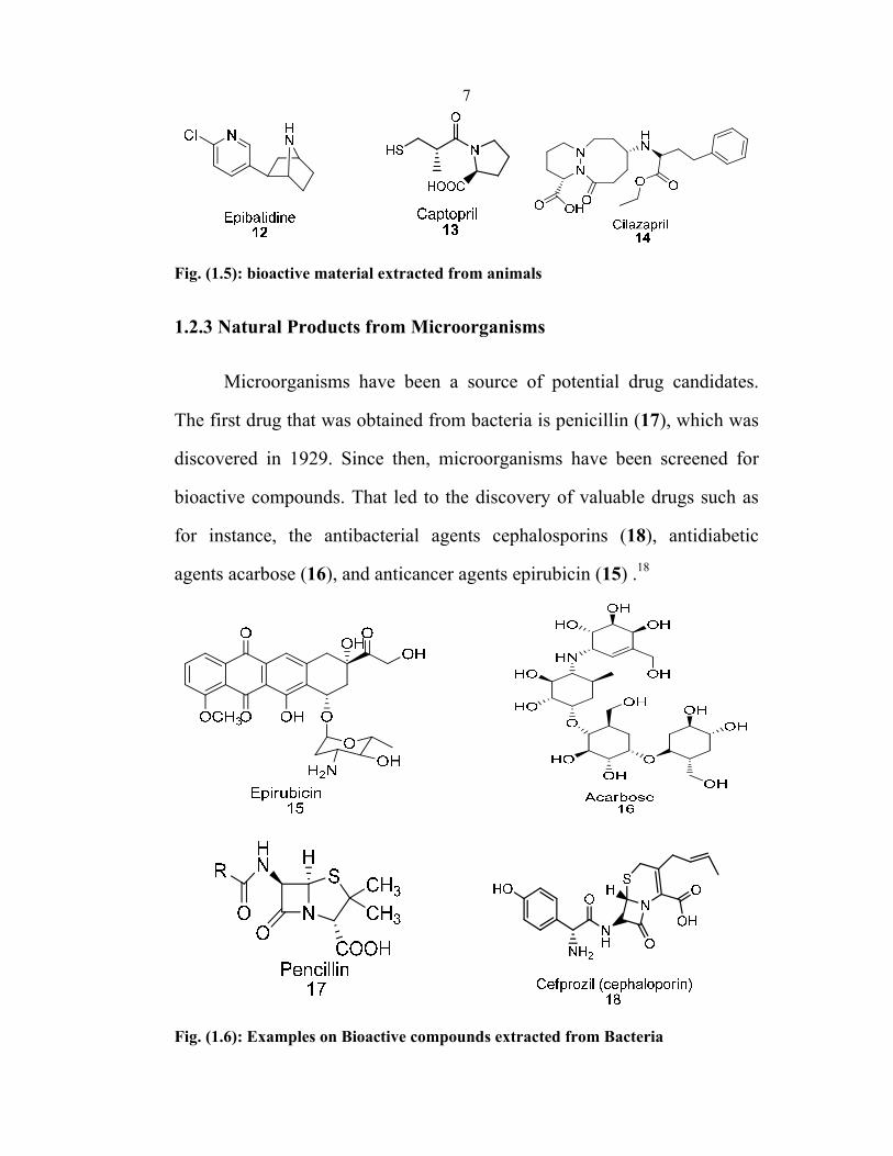
Fig. (1.5): bioactive material extracted from animals
1.2.3 Natural Products from Microorganisms
Microorganisms have been a source of potential drug candidates.
The first drug that was obtained from bacteria is penicillin (17), which was
discovered in 1929. Since then, microorganisms have been screened for
bioactive compounds. That led to the discovery of valuable drugs such as
for instance, the antibacterial agents cephalosporins (18), antidiabetic
agents acarbose (16), and anticancer agents epirubicin (15) .18
Fig. (1.6): Examples on Bioactive compounds extracted from Bacteria

8
1.2.4 Natural Products from Marine Organisms
Marine organisms are also a potential source of drugs. The first
bioctactive compound to be isolated from marine species was
spongouridine (19) in the 1950s .19 Since then several others were isolated
such as spongothymidine (19) from the Carribean sponge Cryptotheca.
These compounds are nucleotides and show great potential as anticancer
and antiviral agents. The discovery of spongouridine led to an extensive
research to identify drug candidates from marine sources. This led to the
discovery of the anticancer agent discodermolide (20), isolated from the
marine sponge, Discodermia dissoluta, which has similar mode of action
to that of paclitaxol® besides possessing a strong antitumor activity. It is
unique in that it exhibits better water solubility as compared to
paclitaxol®.20
OHO
OH
N
HN
O
O
OH
Spongouridine R= HSpongothymidine R= CH3
19
R
Fig. (1.7): Marine source natural products with bioactivities

9
1.3 Plant Based Antibacterial Agents
In spite of the availability of a large number of antibacterial agents,
there is still a great need for more potent antibacterial drugs.
Most of the drugs available is either having toxicity for ecosystem or
bacteria developed some kind of resistance against them.
Therefore, researchers are increasingly turning their attention to folk
Medicine, looking for new leads to develop better drugs against microbial
infections.22 Plants represent a reservoir of effective chemotherapeutants
and can provide valuable sources of drugs which posses antimicrobial
properties. During recent years, large number of plants have been screened
for their antibacterial activity, as a result of that large numbers of plants
have been found to contain ingredients that have some kind of activities
toward bacteria .21 Examples of these are mentioned earlier in this chapter
and will be presented in the next few pages with some details.
1.3.1 Terpenoids
The terpeniods, sometimes called isopreniods, are a large diverse
class of naturally occurring organic chemicals. They are derived from five-
carbon isoprene units assembled and modified in thousands of ways. Some
terpenoids have unusual seven-carbon ring structures .23
Plant terpeniods are used widely for their aromatic qualities; they
play a major role in traditional herbal therapies and are under investigation
for antibacterial, antineoplastic and other pharmaceutical functions.

10
Terpeniods contribute to the scent eucalyptus, the flavors of
cinnamon, cloves, and ginger and the color of yellow flowers.
Monoterpenoids are well known of their antimicrobial activity.
Several terpeniods showed antifungal activity against microsporum
cookie and trichophyton mentagrophyes and fusaruim .24
Examples on these tepenoids are shown in Figure (1.8) which was
isolated from the leaves of pinus radita .25
OHCOOH
(1R,4aS,9S)-9-hydroxy-7-isopropyl-1,4a-dimethyl-1,2,3,4,4a,9,10,10a-octahydrophenanthrene-1-carboxylic acid
22
Fig. (1.8): Terpenoids with antifungal activities extracted from leaves of pinus radita.

11
Another type of biological active terpenoids is saponins, that are a
group of triterpenoids which have been isolated from different plant (e.g.
rapanea, dolichos, camellia, and primula). Saponins show a wide range of
biological activities including antibacterial activity. 26-28
Saponins are considered to be an exception among antifungal
compounds in that their antifungal activity is usually correlated with the
sugar moiety glycosylated to the 3-hydroxyl group of the terpenoids and
thus a polar part of the molecule, whereas most other antifungal costituents
tend to be strongly lipophilic and inactive in glycoside form .29,30
1.3.2 Alkaloids
They are groups of natural chemicals which mostly contain basic
nitrogen atoms. Alkaloids are produced by plants, animals, fungi and
bacteria. Many alkaloids can be purified from crude extracts by acid base
extraction, most alkaloids are toxic to organisms. They often have
pharmacological effects and are used as medications, as recreational drugs,
or in entheogenic rituals. For example, cocaine and morphine are used as
local anesthetic and are also used as stimulant caffeine and nicotine, or the
antimalarial drug quinine. Although alkaloids act on a diversity of
metabolic systems in humans and other animals, they almost uniformly
invoke a bitter taste .31
Extracts from plants containing toxic alkaloids, such as aconitine and
tubocurarine, were used since antiquity for poisoning arrows.32

12
1.4 Aromatic compounds
A large proportion of aromatic substances that are extracted from
plants often show antifungal activity. 33 These include simple and alkylated
phenols, phenolic acids, phenylpropanoids ( 24), coumarins, flavonoids,
isoflavonoids , stillbenoids, quinones and xanthones.
Fig. (1.9): Example on aromatic compounds with antibacterial activity
1.4.1: Flavonoids
Such as those shown in Figure (1.10), and especially isoflavonoids
Figure (1.11) were reported to have high antifungal activity against
richophyton mentagrophytes and T. rubrum .34-37

13
OHHO
OCH3O
OH
(E)-1-(2,4-dihydroxy-6-methoxy-3-(3-methylbut-2-enyl)phenyl)-3-(4-hydroxyphenyl)prop-2-en-1-one
25
Fig. (1.10): Example on flavonoids with antibacterial activities
Fig. (1.11): Example on isoflavonoids with antibacterial activities
1.4.2 Quinones
Some quinones are also known to inhibit mycelial growth of fungi,
examples are two naphthoquinonoid naphthaxirene derivatives and their
glucosides from sesamum angolense pedaliaceae 38 and the benzoquinones
juglone present in a number of plants, e.g pecan carya illinoesis,
junglandaceae 39 and 2, 6- dimethoxybenzoquinone (28) from
crotonlacciferus (euphorbiaceae).

14
The latter constituent displays antifungal activity against
cladosporium cladosporiodes.40
1.5 Plants Used In Traditional Medicine in Palestine
More than 600 plant species have been used in Palestine in
traditional medicine to treat various diseases .41, 42 About fifty species of
these plants are used to treat various skin diseases. The following
paragraphs show a summary about some of these plants, the structure of the
bioactive materials extracted form them and their applications.
1.5.1 Walnut Tree
The scientific name for this plant is Juglans Regial, it belongs to a
family known as Juglandaceae .43 Its name is derived from the Latin glans
jovis, the corn of Jupiter. A very leafy tree with dark green leaves, the tree
grows to a height of about 12-15 m. It has a straight, well-branched trunk
with smooth, grayish-white bark, when young that develops deep,
longitudinal furrows with age and becomes very rough.
The bioactive materials in this plant are available in leaves, bark, and
fruit.

15
In Palestinian tradition, this plant is used to treat bacterial infection
by isolating the juice from the green peels of the fruits and apply it on the
infected skin.
Several bioactive materials have been isolated from walnut tree
among these are inositol, tannin, gallic acid, junglandin, carotene,
pyrogallic acid, monoterponids, sesquiterpenes and juglone. The structures
of these extracted compounds are shown in Fig (1-12).
O
Onaphthalene-1,4-dione
31
Fig. (1.12): Structures and name of materials extracted from walnut tree

16
The bioactive materials extracted from the walnut tree are used as:
antihypoglycaemic, deputative, galactofuge, rubefacient, antiscrophutous
and antidermatosic.
1.5.2 Plumbago
The scientific name for this plant is Plumbago europea L, it belongs
to a family known as plumbaginaceae .44 The Plumbago herb grows to
height of 30-100 cm. It has a herbaceous stem which is ribbed, erect,
branched, and leafy. The leaves are farinose, especially on the lower face,
oblong elliptic obviate or oblanceolate, remotely denticulate. Lower most
leaves are petiolate, whereas middle and upper leaves sessile and
auriculate-clasping. Flowers of this plant are calyx 6-8 mm with corolla
purple to lilac, lobes obovate, obtuse, mucronate. The medicinal parts are
gathered in Summer.
Plumbago is native to South Africa, and is a popular ornamental in
subtropical gardens in Florida and California, Plumbago may be found in
gardens all over the world.
The bioactive materials in these plants are available in leaves, roots
and whole plants.
In Palestinian tradition, green leaves are often macerated and applied
on the infected skin. Also dry leaves are usually moistened and then
macerated before being applied on infected skin parts.

17
Chemical compounds that existing in Plumbago are shown in figure
1.13:

18
Fig. (1.13): Natural materials available in plumgbago europea L

19
1.5.3 Salvia L
The scientific name for this plant is salvia fruticosa and it belongs to
a family known as lamiaceae. Salvia L. is the largest genus of the family
labaite, including over 900 species in the world. Since ancient times,
species of salvia have been used in folk medicine for the treatment of
diabetes and skin diseases such as psoriasis and eczema and are used with
powder alum and other plants to treat mouth fungi.
Salvia due to has a wide range of biological activities such as anti
bacterial activities 45-49antitumor activities 50-57 and antifungal activities 51-56
salvia species also have some useful compounds to preserve raw and
processed food 58and some of them are used as a drink .59
Salvia fruticosa is a very leafy plant which grows to height of about
60-120 cm; the light green gray leaves are used in traditional medicine in
Palestine.

20
O
1,3,3-trimethyl-2-oxabicyclo[2.2.2]octane(1, 8-cineole)
45
(R)-1-methyl-4-(prop-1-en-2-yl)cyclohex-1-ene
(Limonene)46
(1S,4R)-2,2-dimethyl-3-methylenebicyclo[2.2.1]heptane(Camphene)
47
6,6-dimethyl-2-methylenebicyclo[3.1.1]heptane(beta-Pinene)
48
(1S,5S)-2,6,6-trimethylbicyclo[3.1.1]hept-2-ene
(a-Pinene)49
7-methyl-3-methyleneocta-1,6-diene(Myrcene)
50

21
Fig. (1.14): Chemical compounds that are available in Salvia Fruticosa:
1.6 Aims and Scope of the Work
Scarce information on natural bioactive compounds and their
properties in many plants in Palestine, as well as increasing demand for
natural drugs were important motivations to start this study.
The general aims of this study can be placed into four folds:
1. Finding new natural compounds with bioactivities against bacteria in
plants cultivated in Palestine.
2. Assessment of application possibilities of partially purified extracts
containing these compounds.

22
3. Determination of their molecular structures, and
4. Determination of bioactivity.
Previous studies of aromatic and medicinal plants grown in Palestine
have resulted in the discovery of new natural products with antibacterial
activities 60 and the identification of new antioxidants .61 These findings
encouraged to initiate this work.
Inula viscosa (L.) is a perennial weed, native to the Mediterranean
Basin known in the Palestinian folklore as Tayoon. It grows on hills tops,
damp habitats, and roadsides. Extracts of the plant have been widely used
in folk medicine to treat various diseases such as a diuretic, topical anti-
inflammatic, and haemostatic64. Aqueous extracts of I. viscosa were also
shown to exhibit antifungal activity in vitro 65, 66. Cohen et al67 provided
evidence for the antifungal activity in plant of extracts made with organic
solvents, including methanol, ethanol, ethyl acetate, acetone, chloroform,
and n-hexane. Using thin-layer chromatography overlay assays, seven
inhibitory zones against Cladosporium cucumerinum were observed in the
extracts 67 In a recent study,68 it was found that leaf extracts of Inula
viscosa were highly effective in controlling downy mildew of grapevine,
caused by Plasmopara viticola. Other biological activities of Inula viscosa
include antiulcerogenic effects 69, prevent growth of pathogenic fungi 70,
prevent zygote implantation in mammals 71, and have a strong anti oxidant
activity 72.
23
There is also published evidence that Inula viscosa has also
nematicidal/antihelm acologically active compounds 73, 74 including
sesquiterpenes, sesquiterpenes acids 75, azulenes, lactones, flavonoids, and
essential oils 76.
Currently, there is no published data on the cytotoxicity and
genotoxicityof I. viscosa leaf extracts.
Inula viscosa was chosen for this study since it is an important plant
in the Palestinian folklore and it has unlimited number of medical
applications. Below are some examples on commercial medical
applications of inula viscosa:
1- Inula head lice: Inula viscosa extract emulsified with olive oil or water
is used in removing head lice and lice eggs.
2- Inula tea: Inula plant boiled in water is used to tract infection treatment,
reduction of blood pressure, prevention of flu and cold, gum disorder
treatment, and toothache treatment.
1.7 Chemical Compounds Extracted from Inula viscosa
1.7.1 Sesquiterpenes
Sesquiterpenes are a class of terpenes that consist of three isoprene
units 77 and have the molecular formula C15H24. Like monoterpenes,
sesquiterpenes could be acyclic or contain rings, including many unique
combinations.

24
1.7.2 Terpenes
Terpenes are hydrocarbons with the molecular formulas (C5H8)n.
Their building block is the hydrocarbon isoprene 77 and they are classified
according to the number of isoprene units it composing them. Terpenes
are widespread in nature, mainly in plants as constituents of essential oils.
Many terpenes are hydrocarbons, but oxygen-containing terpenes such as
alcohols, aldehydes or ketones (terpenoids) are also found.
1.7.3 Lactones
Lactones are cyclic esters 78 which can be seen as the condensation
product of an alcohol group -OH and a carboxylic acid group -COOH in
the same molecule. It is characterized by a closed ring consisting of two or
more carbon atoms and a single oxygen atom, with a ketone group C=O in
one of the carbons adjacent to the other oxygen.
1.7.4 Azulene
Azulene is an isomer of naphthalene. Whereas naphthalene is
colorless, azulene is dark blue. Its name is derived from the Spanish word
azul, meaning "blue". Two azulenes, vetivazulene (4,8-dimethyl-2-

25
isopropylazulene) 59 and guaiazulene (1,4-dimethyl-7-isopropylazulene)
60. , are found in nature as constituents of pigments in mushrooms, guaiac
wood oil, and some marine invertebrates.

26
CHAPTER TWO EXPERIMENTAL
All chemicals were purchased from Aldrich Chemical Company and
used without any purification. Extracted and purified compounds from
Inula viscosa were characterized by 1H NMR, 13C NMR, GC/MS and
LC/MS. Nuclear Magnetic Resonance Spectra were recorded on Varian
Gemini 2000, 300 MHz instrument, Gas Chromatography mass &
spectrometry were recorded on Perkin Elmer 560D.
All 1H NMR experiments were reported in unit of parts per million
(ppm) downfield from tetramethylsilane.
All 13C NMR spectra were reported in ppm relative to
deuterchloroform (77.0 ppm).
All tests for antibacterial activity were reported in Al-Arabi hospital
lab in Nablus, West Bank, Palestine.
Purification of extracted samples was performed by flash
chromatography on silica gel (100-200) mesh.
2.1 General Procedure for extractions
2.1.1 Requirements of Efficient Extraction
Suitable solvent for extraction of organic compounds from plants
must possess certain properties such as: water insoluble, low boiling point,
medium polarity, and has higher affinity for plants extracts than water,

27
2.1.2 Plant Material
As mentioned earlier in the introduction chapter, the plant chosen for
this study was Inula Viscosa known in Palestinian folklore as tayoon.
Plants were collected from hilly areas around Nablus city far from
agricultural lands. It was collected in spring time, and dried in the shade.
2.1.3 Preparation of the Collected plants for Extraction
The dried material was ground and suspended in ethyl acetate. The
produced suspension was mixed using mechanical mixer for about 48 h.
Another method of extraction was also tried which is soxhlet extraction. In
this method ground plant was extracted with ethyl acetate for about 24 h.
Produced solution was dried over magnesium sulfate and
concentrated under reduced pressure at 50 ᴼC using rotary evaporator.
2.2 Evaluation of Concentrate by Thin Layer Chromatography (TLC)
Thin layer chromatography (TLC) is an extremely useful technique
for chemist in particular. TLC is an inexpensive, simple, rapid technique
used to determining the number of components present in solution and
helps in finding a suitable solvent for separating the components by flash
chromatography as well as for monitoring reactions progress.
Several combinations of solvents of increasing polarity were
evaluated as mobile phase in TLC to determine the number of compounds
in Inula viscose extract. The solvents combinations were: hexane (100 %),

28
ethyl acetate/ n-hexane (3:7), ethyl acetate /n-hexane 1:1), ethyl acetate/n-
hexane (7:3), ethyl acetate (100%), ethyl acetate/methanol (9:1). This
study concludes that all of these solvent combinations are required to
separate Inula viscosa extracts into pure components by flash
chromatography.
2.3 Separation of Extract by Flash Chromatography
2.3.1 Flash Column Chromatography
Flash chromatography is very useful techniques for separating
mixture of organic compounds into pure components.
It was performed by using a column with dimensions of 60 cm in length
and 3.0 cm in diameter packed with silica gel 60 (230-400 mesh) purchased
from Aldrich chemical company
The Inula viscosa extracts were loaded into the column as a solid
mixture with some silica, which was prepared by suspending the mixture of
about 5.0 g silica gel and 20 ml ethyl acetate, then ethyl acetate was
removed under vacuum using rotary evaporator. The separation was started
with pure hexane (low polarity) then the mobile phase polarity was
increased gradually as follows: pure hexane, then hexane containing 20%
ethyl acetate, then ethyl acetate was increased to 40%, then pure ethyl
acetate was used and finally the column was flushed with ethyl acetate
containing 10% methanol.
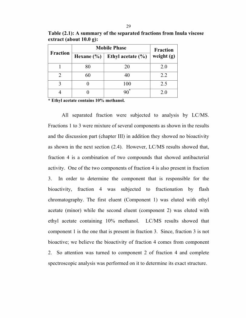
29Table (2.1): A summary of the separated fractions from Inula viscose extract (about 10.0 g):
Fraction Mobile Phase Fraction
weight (g) Hexane (%) Ethyl acetate (%)
1 80 20 2.0 2 60 40 2.2 3 0 100 2.5 4 0 90* 2.0
* Ethyl acetate contains 10% methanol.
All separated fraction were subjected to analysis by LC/MS.
Fractions 1 to 3 were mixture of several components as shown in the results
and the discussion part (chapter III) in addition they showed no bioactivity
as shown in the next section (2.4). However, LC/MS results showed that,
fraction 4 is a combination of two compounds that showed antibacterial
activity. One of the two components of fraction 4 is also present in fraction
3. In order to determine the component that is responsible for the
bioactivity, fraction 4 was subjected to fractionation by flash
chromatography. The first eluent (Component 1) was eluted with ethyl
acetate (minor) while the second eluent (component 2) was eluted with
ethyl acetate containing 10% methanol. LC/MS results showed that
component 1 is the one that is present in fraction 3. Since, fraction 3 is not
bioactive; we believe the bioactivity of fraction 4 comes from component
2. So attention was turned to component 2 of fraction 4 and complete
spectroscopic analysis was performed on it to determine its exact structure.

30
1H NMR of component 2 of fraction 4; compound (61)
CDCl3 δ (ppm): 3.8 ( s, OCH3), 3.83 (s, OCH3), 6.18 ( d, J, 2 Hz, C-
5 H), 6.45 (d, J, 2 Hz C-7 H), 6.93 (d, J, 9 Hz, C-2’ H) , 7.55 (dd, J, 2 and
9 Hz), 7.62 ( d, J= 2Hz) doublet, 9.95 ( bs, C4’-OH), 10.85 (bs, C6-OH)
and 12.65 ( bs, C4 OH).
13C NMR of component 2 of fraction 4; compound (61)
CDCl3 δ (ppm): 56.37, 60.41, 94.53, 99.53, 104.86, 116.32, 121.45,
138.4, 148.12, 150.13, 156.16, 157.03, 161.89, 164.84, 178.58.
2.4 Evaluation of fractions for Antibacterial Activity
2.4.1 Materials
Culture media: Mueller-Hinton, Tryptic Soy Broth (Hylabs ,Israel)
2.4.2 Microorganisms used
Bacteria strains used in the study were clinical isolates of
Staphylococcus aureus, Escherechia coli, Proteus mirabilis, and
Pseudomonas aerginosa, all of them were isolated from patients suffering
from bacterial infections with the relevant bacteria.
2.4.3 Screening for Antimicrobial Activity
Fractions of Inula viscosa extracts were collected by flash
chromatography and were screened for antimicrobial activity by using the
agar well diffusion method reported in the literature by Perez et al.62

31
1. Three colonies of bacteria where transferred to sterile tubes each
containing 5 ml of Tryptic Soy Broth.
2. Turbidity of the bacterial suspensions was adjusted to reach an optical
density equivalent to a 0.5 McFarland standard to give a bacterial
suspension of 10.cfu/ml. (cfu: colony forming unit).
3. Mueller-Hinton agar plates were inoculated by streaking bacterial swabs
over the entire surface of the plates.
4. Plates were allowed to dry at room temperature.
5. Six millimeter wells were pushed into the plates.
6. Fifty microliters of the four fractions were added into duplicate wells.
7. Plates were allowed to stand at room temperature to let the tested
derivative absorbed into the agar, and afterwards, they were incubated at
37 oC for 18 to 24 h.
8. Plates were examined for bacterial growth inhibition and zones of
inhibition were measured in millimeters.
Table (2.2): Screening Results (Zone of Inhibition in mm): Fraction
(10 mg/ml) S.aureus E.coli P.mirabilis P.aerginosa
1 0 0 0 0 2 0 0 0 0 3 0 0 0 0 4 25 0 0 0
2.4.4 Determination of Minimum Inhibitory Concentration Method as
Shown Below63
MIC was determined by broth dilution method as shown below:
1- Two-fold serial dilutions were prepared from fraction 4 in Tryptic Soy
Broth.

32
2- Duplicate tubes of each dilution were inoculated with 5*10 of S.aureus.
3- All tubes were incubated at 37ᴼC for 18 to 24 hours.
4- The highest dilution of the drug that resulted in inhibition of bacterial
growth was considered as the MIC. Table (2.3): MIC results of fraction 4
Conc.(mg/ml) Fraction 4 10 -ve 5 -ve
2.5 -ve 1.25 -ve
0.625 -ve 0.3125 -ve
0.15625 -ve 0.078125 -ve 0.039063 -ve 0.019531 +ve
Positive Control +ve Sterility Control -ve
Bacterial growth (positive: +ve) or (negative: -ve)
Table (2.4): MBC results for S.aureus bacteria: Fraction 4 MIC(mg/ml) MBC (Mg\ml) 4 39 78

33
CHAPTER THREE Results and Discussion
3.1 Analysis of the separated fractions
In this study, a comprehensive analysis was performed on tyoon
extracts. Number of constituents and complete structure determination of
at least one of the components was performed.
Tayoon was collected from areas in Nablus, Palestine, dried in the
shade and grounded. Sample of dried and ground tayoon plant (about 500
g) was extracted with ethyl acetate. The extracts were concentrated in
vacuo to afford about 30 g of extract. Another sample of tyoon was
extracted with ethanol. Again the extracts were concentrated in vacuo to
afford about 15 g of extract. The extracts of the two solvents were
analyzed by HPLC and GC/MS. In the HPLC analysis, the mobile phase
consists of acetic acid, and the results indicate the presence of phenolic
compounds. Analysis by HPLC didn’t give clear picture about the number
of components present in tayoon. Analysis by GC/MS showed the
presence of about 13 compounds as shown in Figure a2 (Appendix).
The ethyl acetate extract (10.0 g) was fractionated by flash
chromatography on silica gel 120 H using hexane–EtOAc solution of
increasing polarity. Four fractions were separated. The first fraction was
eluted with 20% EtOAc in hexane. Analysis of the first fraction by LC/MS
showed the presence of multi components with very close retention time.
The second fraction eluted with 40% EtOAc in hexane. Analysis of the

34
second fraction by LC/MS showed the presence of five components (Figure
a7, page 58, appendix), retention times and molar masses for the five
components are summarized in Table 3.1.
Table (3.1): LC/MS Analysis Results of Fraction 2
Component Retention Time (min)
Molar Mass
1 0.55 392 2 1.27 253 3 1.49 294 4 1.74 409 5 2.40 584
3.1.1 Fraction Three
Analysis of Fraction 3 by LC/MS showed the presence of several
components at low concentrations. No further work was performed on this
fraction except for its biological activity against certain bacteria which was
evaluated (see section below)
3.1.2 Fraction Four
Analysis of the fraction four by LC/MS showed the presence of two
components (Figure 3a, Appendix), retention times and molar masses for
the two components are summarized in Table III.2. Component 2 was the
major product.
Table (3.2): LC/MS Analysis Results of Fraction 4
Component Retention time
Molar Mass
1 1.22 331.9 2 1.29 347.9

35
Fraction 4 was subjected to purification by Flash chromatography;
component with relatively high concentration was collected and analyzed
by 1H NMR and 13C NMR. The results are consistent with the structure
shown in Figure (3.1). The proposed structure was confirmed with that
reported in the literature.79, 80 The compound was identified to be 3,3’-di-
O-methylquercetin (61).
(Fig. 3.1)
The 1H NMR of the major component of fraction four exhibits the
following signals (Figure III.2): two methoxy singlet C-2 and C-3’ at δ 3.8
ppm and 3.83 ppm respectively, doublet C-5 H at 6.18 with coupling
constant of J5,7= 2 Hz, doublet C-7 H at 6.45 with coupling constant of
J5,7=2 Hz, doublet C-2’ at 6.93 with a coupling constant ofJ5,'6'= 9Hz,
doublet of doublet at 7.55 with coupling constants of J6',2'=2 and J6',5'=9 Hz,
doublet 7.62 with a coupling constant of 2Hz, broad singlet C4’-OH at
9.95, broad singlet C6-OH at 10.85, and broad singlet C4 OH at 12.65.
The high desheilding of C4-OH could be attributed to intra H-bonding
between β-hydroxyl group (C4-OH) and the carbonyl group as shown in
Figure II.2.
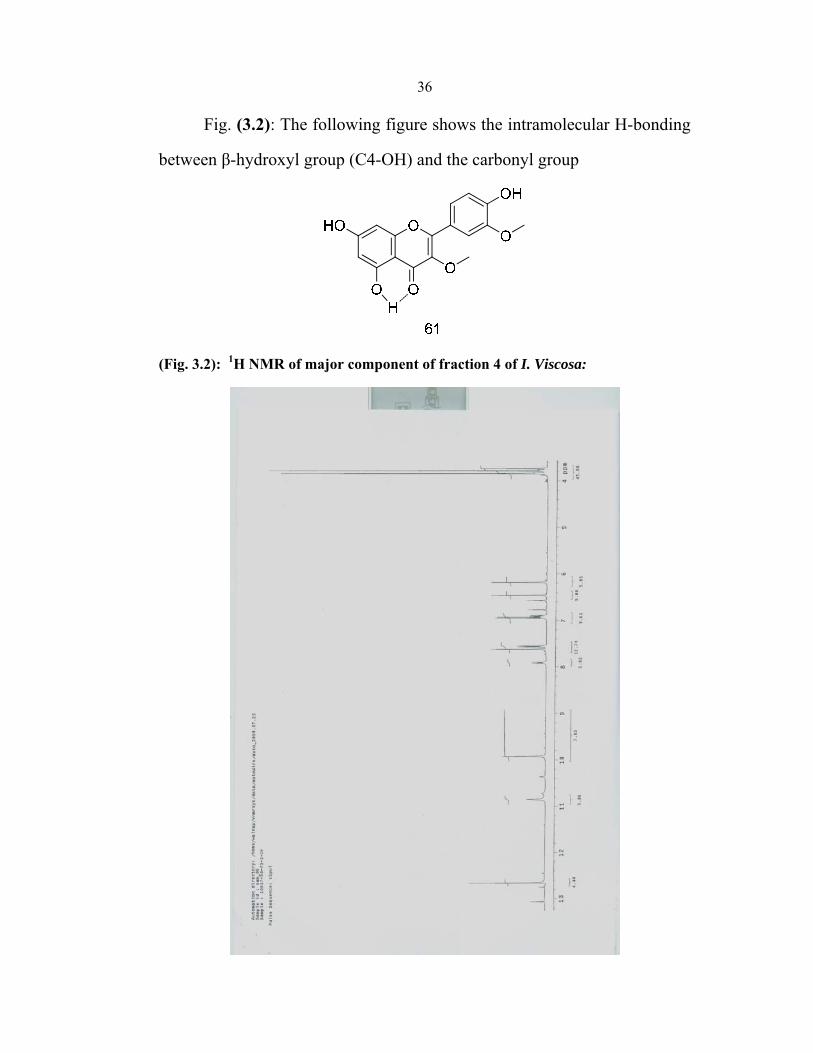
36
Fig. (3.2): The following figure shows the intramolecular H-bonding
between β-hydroxyl group (C4-OH) and the carbonyl group
(Fig. 3.2): 1H NMR of major component of fraction 4 of I. Viscosa:

37
Fig. (3.3): 1H NMR of major component of fraction 4 of I. Viscosa, Showing the coupling constants between major peaks:
Analysis of 3,3’-di-O-methylquercetin (61) by 13C NMR showed the
presence of 15 peaks (figure III.4), they are summarized in table II.3

38
Fig. (3.4): 13C NMR of major component of fraction 3 of I. Viscosa

39Table (3.3): Summary of the 13C chemical shifts of 3,3’-di-O-methylquercetin (61):
Chemical Shift δ (ppm)
56.37 C-3’ OMe 60.41 C-2 OMe 94.53 C-7 99.53 C-9
104.86 C-2’ 116.32 C-5’ 121.45 C-1’ 138.40 C-4’ 148.12 C-3’ 150.13 C-2 156.16 C-1 157.03 C-8 161.89 C-4 164.84 C-6 178.58 C-3
3.2Antibacterial Activity
As mentioned earlier, four fractions were separated from I. Viscosa
using flash chromatography technique. Fractions of components were
evaluated for their antimicrobial activity against four types of bacteria: S.
aureus, E. coli, Proteus mirabilis and Pseudomonas aeruginosa.These
bacterial strains were clinical isolates; all of the strains were isolated from
patients suffering from bacterial infections with the relevant bacteria.
3.2.1 Screening Results
Fractions, separated from Inula Viscosa extracts, were screened for
antimicrobial activity using the well known diffusion method reported in
the literature by Perez et al62, the efficiency of the drug was measured by

40
the zone of inhibition in millimeters of the bacteria cultured on the
Mueller-Hinton agar plate. The results are summarized in Table III.5. As
can be seen in Table 5, all four fractions were inactive (zones of inhibition
were zero), for E. Coli, Proteus mirabilis, and Pseudomonas aeruginosa.
However, fraction four showed antimicrobial activities against S. aureus,
others fractions 1, 2 and 3 showed no activities as shown in table 4, zone of
inhibition for compounds in these fractions were 0 mm. The zone of
inhibition for fraction four was 25 mm.
Table (3.4): Screening Results (Zone of Inhibition in mm): Fraction
(10 mg/ml) S.aureus E.coli P.mirabilis P.aerginosa
1 0 0 0 0 2 0 0 0 0 3 0 0 0 0 4 25 0 0 0
All fractions, except fraction four, were dropped for further
evaluation because they showed zero activities against the four different
types of bacteria studied.
3.2.2 Determination of Minimum Inhibitory Concentration (MIC)
The aim of this test is to determine the minimum concentration of
bioactive material that inhibits the growth of bacteria. Results were
compared to that of triclosan. Both fraction four and triclosan were tested
on clincal isolate S. aureus.63 MIC was determined by broth dilution
method. Two solutions were used to obtain correct results; one is positive
control solution (contains bacteria alone) where bacterial growth always

41
positive, another is sterility control solution (contains curcumin alone)
where bacterial growth always negative.
The results obtained from this test are summarized in table III.5. As
shown in Table 6, MIC of fraction four was about 0.04 mg/mL, which is
almost equal to that of tricolsan (0.025-0.1 mg/mL).81
Table (3.5): A Summary of MIC Results of Fraction 4 Conc.(mg/ml) Fraction 4
10 -ve 5 -ve
2.5 -ve 1.25 -ve
0.625 -ve 0.3125 -ve
0.15625 -ve 0.078125 -ve 0.039063 -ve 0.019531 +ve
Positive Control +ve Sterility Control -ve
3.2.3 Determination of Minimal Bactericidal Concentration (MBC)
The concentration of bioactive material that results in a total
inhibition of bacterial growth is known as Minimal Bacterial Concentration
(MBC). This method was applied on fraction four, the results are
summarized in Table 6.
Table (3.6): MBC and MIC results of fraction four against S. aureus bacteria:
Fraction 4 MIC(mg/ml) MBC (Mg\ml) 4 39 78

42
As shown before, the major component of fraction four was
identified by H1 and 13C NMR to be 3,3’-di-O-methylquercetin (61). As
mentioned earlier in the experimental part, the antibacterial activity of
fraction 4 could be attributed to 3,3’-di-O-methylquercetin (61). Since
fraction 4 contains two components one on them (minor) is also present in
fraction 3 as shown by LC/MS, however, fraction 3 showed no sign
bioactivities which suggested that this minor component is not the active
one, hence, no further evaluation was done to it.
43
References
1- Phillipson, J. D. Phytochemistry and Medicinal Plants.
Phytochemistry, 56, 237−243, 2001.
2- Kong, J. M.; Goh, N. K.; Chia, L. S.; Chia, T. F. Recent advances in
traditional plant drugs and orchids. Acta Pharmacologica Sinica, 24,
7−21, 2003.
3- Traditional medicine strategy launched. (WHO News). 80, 610, 2002.
4- Benowitz, S. As war on cancer hits 25-year mark, scientists see
progress, challenges. Scientist, 10, 1−7, 1996.
5- Newman, D. J.; Cragg, G. M. Natural products as sources of new
drugs over the last 25 years. J. Nat. Prod. 70, 461−477, 2007.
6- Newman, D. J.; Cragg, G. M.; Snader, K. M. Natural products as
sources of new drugs over the period 1981-2002. J. Nat. Prod. 66,
1022−1037, 2003.
7- Farnsworth, N. R.; Akerele, O.; Bingel, A. S.; Soejarto, D. D.; Guo,
Z., Global importance of medicinal plants. In The Conservation of
Medicinal Plants, 1991.
8- Farnsworth, N. R.; Akerele, O.; Bingel, A. S.; Soejarto, D. D.; Guo,
Z., Global importance of medicinal plants. In The Conservation of
Medicinal Plants, 1991.
9- Kaplan, W. Codylamata Acuminata. New Orleans Med. Surg. J. 94,
388, 1942.
10- Wilson, L. B., Jr.; Mizel, SB.; Grisham I. M.; Creswell, KM.,Session
V- Networks and Shared Facilities- Rapporteurs Summary. Fed. Proc.
Fed. Am. Soc. Exp. Biol. 33, 158, 1974.

44
11- Catharanthus Roseus. http://www.nybg.org/bsci/belize/gallery.html as
accessed on 22/07/10.
12- Noble, R. L. The discovery of the vinca alkaloids - chemotherapeutic
agents against cancer. Biochem. Cell Biol. 68, 1344−1351, 1990.
13- Ingram,J.
http://www.daveingram.ca/knowingnature/C1534673850/E200701192
12411/index.html as accessed on 22/07/10.
14- Kinghorn, A. D.; Seo, E. K. Plants as sources of drugs. Agricultural
Materials as Renewable Resources, 647, 179−193, 1996.
15- Oberlies, N. H.; Kroll, D. J. Camptothecin and taxol: Historic
achievements in natural products research. J. Nat. Prod. 67, 129−135,
2004.
16- Spande, T. F.; Garraffo, H. M.; Edwards, M. W.; Yeh, H. J. C.;
Pannell, L.; Daly, J. W. Epibatidine- A novel (chloropyridyl)
Azabicycloheptane with Potent Analgesic Activity from an
Ecuadorian Poison Frog. J. Am. Chem. Soc. 114, 3475−3478, 1992.
17- Blood Pressure. http://www.medicinenet.com/captopril/article.htm.
18- Chin, Y. W.; Balunas, M. J.; Chai, H. B.; Kinghorn, A. D. Drug
discovery from natural sources. Aaps Journal, 8, E239−E253, 2006.
19- Haefner, B. Drugs from the deep: marine natural products as drug
candidates. Drug Discovery Today, 8, 536−544, 2003.
20- Huang, G. S.; Lopez-Barcons, L.; Freeze, B. S.; Smith, A. B.;
Goldberg, G. L.; Horwitz, S. B.; McDaid, H. M. Potentiation of taxol
efficacy and by discodermolide in ovarian carcinoma xenograft-
bearing mice. Clin. Can. Res. 12, 298−304, 2006.

45
21- Davis J. Inactivation of antibiotic and the dissemination of resistance
genes. Science, 264, 375- 382, 1914.
22- Robin EH, Anril W, Alexander M, Loeto M, Keith K (1998)
Nasopharyngeal carriage and antimicrobial resistance in isolates of
Streptococcus pneumoniae and Haemophilus influenzae Type b in
children under 5 years of age in Botswana. International Journal of
Infectious Diseases, 3(1), 18-25, 1998.
23- Harborne J. B. and baxter H.(eds) , Phytochemical Dictionary, Taylor
and Francis, London (1993).
24- Picman A. K. Biochem. Syst. Ecol. 12, 13 (1984).
25- Franich, R. A. , Gadgil. P. D. and Shahin, L. physiol Plant Path. 23,
183 (1983).
26- Marston A. , Gafner F. , Dossagi S. F. and Hostettmann K. ,
Phytochemistry 27, 1325 (1988).
27- Ohtani K. , Mavi S. and Hostettmann K. , Phystochemistry 33,83
(1993).
28- Nagata T., Tsushida T., Hamaya E., Enoki N., Manabe S. and Nishno
C., Agric. Biol. Chem. 49, 1181 (1985).
29- Renee J. G. and Jeffry B. H. Phystochemistry, Vol. 37, #1, pp 19-42
(1994).
30- Takechi M. and Tanaka Y. Phytochemistry 29. 451 (1990).
31- Rhoades, David F (1979). "Evolution of Plant Chemical Defense
against Herbivores". In Rosenthal, Gerald A., and Janzen, Daniel H.
Herbivores: Their Interaction with Secondary Plant Metabolites. New
York: Academic Press. p. 41. ISBN 0-12-597180-X.

46
32- Aniszewski, p.182.
33- Renee J. G. and Jeffry B. H. Phystochemistry, Vol. 37, #1, pp 19-42
(1994).
34- Ingham J. L., Tahra S. and Harborne J. B. Z. Naturforsch 38c, 194
(1983).
35- Tahra S. , Ingham J. L. , Nakahara S. , Mizutani J. and Hrborne J. B.
Phytochemistry 23, 1889 (1984).
36- Geibel M. , Geiger H. and Treutter D. Phytochemistry 29, 1351
(1991).
37- Mizobouchi S. and Sato Y. , Agric. Biol. Chem. 48, 2771 (1884).
38- Pottert O. , Stoecki-Evans H. , Msonthi J. D. and Hostermann K. ,
Helv. Chim. Acta 70, 155 (1987).
39- Henry H. B. , Gary D. C. and James E. O. Instrumental Analysis
(1978).
40- Snyder L. R. and Kirkland J. J. Modern Liquid Chromatography New
York: Wily-Interscience (1974).
41- Palevitch Pros. D. , Yaniv Z., Medicinal Plants of the Hollyland.
42- Ali-Shtayeh M. S. , Medicinal Plants of the West Bank, Department of
Biological Sciences, An-Najah Univ., Nablus, Unpublished work.
43- Brinkman, K.A. (1974) "Juglans L. - Walnut", in: Schopmeyer, C.S.
(ed.), Seeds of woody plants in the United States, Agriculture
Handbook 450, Washington, D.C.: U.S. Department of Agriculture,
Forest Service, p. 454–459, (rev. ed.: 1992), ISBN 0-931146-21-6.

47
44- "Plumbago L.". Germplasm Resources Information Network. United
States Department of Agriculture. 2002-01-02. http://www.ars-
grin.gov/cgi-bin/npgs/html/genus.pl?9608. Retrieved 2010-01-29.
45- Tzakou O, Pitarokili D, Chinou IB. Composition and antimicrobial
activity of the essential oils of Salvia ringens. Planta Med 67: 81-83,
2001.
46- Tepe B, Daferera D, Sokmen A et al. Antimicrobial and antioxidant
activities of the essential oil and various extracts of Salvia tomentosa
Miller (Lamiaceae). Food Chem 90: 333-340, 2005.
47- Dobrynin VN, Kolosov MN, Chernov BK et al. Antimicrobial
substances of Salvia officinalis. Khim Prir Soedin 5: 686-686, 1976.
48- Albayrak S, Aksoy M, Hamzaoglu E. Determination of antimicrobial
and antioxidant activities of Turkish endemic Salvia halophila Hedge,
Turk J Biol 32: 265-270, 2008.
49- Ogutcu H, Sokmen A, Sokmen M et al. Bioactivities of the various
extracts and essential oils of Salvia limbata C.A.Mey. and Salvia
sclarea L., Turk J Biol 32: 181-192, 2008.
50- Kurkcuoglu M, Baser KHC, Duman H. Composition of essential oils
from two varieties of Salvia aucheri Bentham growing in Turkey. J
Essent Oil Res, 14: 241-242, 2002.
51- Honda G, Koezuka Y, Tabata M. Isolation of an antidermatophytic
substance from the root of Salvia miltirrhiza. Chem Pharm Bull 36:
408-411, 1988.
48
52- Soliman KM, Badeea RI. Effect of oil extracted from some medicinal
plants on different mycotoxigenic fungi. Food Chem Toxicol 40:
1669-1675, 2002.
53- Daferera DJ, Ziogas BN, Polission MG. GC-MS analysis of essential
oils from some Greek aromatic plants and their fungitoxicity on
Penicillium digitatum. J Agric Food Chem 48: 2576-2581, 2000.
54- Pitarokili D, Tzakou O, Loukis A et al. Volatile metabolites from
Salvia fruticosa as antifungal agents in soilborne pathogens. J Agric
Food Chem 51: 3294-3301, 2003.
55- Viuda M, Ruiz-Navajas Y, Fernandez-Lopez J et al. Chemical
composition and antifungal activity of the essential oils of Salvia
(Salvia officinalis L.) and rosemary (Rosemarinus officinalis L.).
Alimentaria 4: 101-105, 2006.
56- Gao YG, Song YM, Yang YY et al. Pharmacology of tanshinone. Yao
Hsueh Pao 14: 75-82, 1979.
57- Chen XG, Li Y, Yan CH et al. Cancer chemopreventive activities of
S-3-1, a synthetic derivative of danshione. J Asian Nat Prod Res 3: 63-
75, 2001.
58- Rota C, Carraminana JJ, Burillo J et al. In vitro antimicrobial activity
of essential oils from aromatic plants against selected foodborne
pathogens. J Food Prot 67: 1252-1256, 2004.
59- Lima CF, Andrade PB, Seabra RM et al. The drinking of a Salvia
officinalis infusion improves liver antioxidant status in mice and rats. J
Ethnopharmacol 2: 383-389, 2005.

49
60- Yu, B.-W.; Chen, J.-Y.; Wang, Y.-P.; Cheng, K.-F.; Li, X.-Y.; Qin,
G.-W.; Phytochemistry, 61, 439, 2002.
61- Zygmunt, B.; Namiesnik, J.; J. Chromatogr. Sci. 41, 109, 2003.
62- Perez C, Pauli M, Bazevque P. An Antibiotic Assay by the Agar Well
Diffusion Method. Acta Biologiae et Medicine Experimentalis.
15:113-115, 1990.
63- NCCLS-National Committee for Clinical Laboratory Standards:
Methods for dilution antimicrobial susceptibility tests for bacteria that
grow aerobiacally. Approved Standards M7-A5, Wayne, PA, USA,
2000.
64- A. Shtacher, G.; Kashman, Y. J. Med. Chem. 1970, 13, 1221-1223. b.
Azoulay, P.; Reynier, J. P.; Balansard, G.; Gasquet, M.; Timon-avid,
P. Pinharm. Acta Helv. 61, 345-352, 1986.
65- Maoz, M., and Neeman, I. Antimicrobial effects of aqueous plant
extracts on the fungi Microsporum canis and Trichophyton rubrum
and on three bacterial species. Lett. Appl. Microbiol. 26:61-63, 1998.
66- Qasem, J. R., Al-Abed, A. S., and Abu-Blan, M. A. activity of clammy
inula (Inula viscosa) on Helminthrosporium sativum and Fusarium
oxysporum f. sp. lycopersici. Phytopathol. Mediterr. 34:7-14, 1995.
67- Cohen, Y., Baider, A., Ben-Daniel, B. H., and Ben-Daniel, Y.
Fungicidal preparations from Inula viscosa. Plant Prot. Sci. 38:629-
630, 2002.
68- Wang, W. Q., Ben-Daniel, B. H., and Cohen, Y. Extracts of Inula
viscosa control downy mildew caused by Plasmopara viticola in
grapevines. (Abstr.) Phytoparasitica 32:208, 2004.

50
69- A. Alkofahi and A. H. Atta, “Pharmacological screening of the anti-
ulcerogenic effects of some Jordanian medicinal plants in rats,”
Journal of Ethnopharmacology, vol. 67, no. 3, pp. 341–345, 1999.
70- M. Maoz and I. Neeman, “Effect of Inula viscosa extracton chitin
synthesis in dermatophytes and Candida albicans,”Journal of
Ethnopharmacology, vol. 71, no. 3, pp. 479–482, 2000.
71- N. M. Al-Dissi, A. S. Salhab, and H. A. Al-Hajj, “Effects of Inula
viscosa leaf extracts on abortion and implantation in rats,” Journal of
Ethnopharmacology, vol. 77, no. 1, pp. 117–121, 2001.
72- G. R. Schinella, H. A. Tournier, J. M. Prieto, P. Mordujovich, and J.
L. Rios, “Antioxidant activity of anti-inflammatory plant extracts,”
Life Sciences, vol. 70, no. 9, pp. 1023–1033, 2002.
73- A. Ulubelen, S. O¨ ksu¨z, and N. Go¨ren, “Sesquiterpene acids from
Inula viscosa,” Phytochemistry, vol. 26, no. 4, pp. 1223– 1224, 1987.
74- E. Wollenweber, K. Mayer, and J. N. Roitman, “Exudate flavonoids of
Inula viscosa,” Phytochemistry, vol. 30, no. 7, pp. 2445–2446, 1991.
75- B. Marongiu, A. Piras, F. Pani, S. Porcedda, and M. Ballero,
“Extraction, separation and isolation of essential oils from natural
matrices by supercritical CO2,” Flavour and Fragrance Journal, vol.
18, no. 6, pp. 505–509, 2003.
76- L. Lauro and C. Rolih, “Observations and research on an extract of
Inula viscosa,” Bollettino della Societa Italiana di Biologia
Sperimentale, vol. 66, no. 9, pp. 829–834, 1990.
77- http://en.wikipedia.org/wiki/sesquiterpene.

51
78- March's Advanced Organic Chemisty: Reactions, Mechanisms, and
Structure Michael B. Smith, Jerry March Wiley-Interscience, 5th
edition, 2001, ISBN 0-471-58589-0.
79- Chiappini, I.; Fardella, G.; Menghini, A.; Rossi, C. Planta Med. 44,
159-161, 1982.
80- Grande, M.; Piera, F.; Guenca, A.; Torres, P.; Bellido, I. S. Planta
Med. 51, 414- 419, 1985.
81- Suller, M. T. E.; Russell, A. D. " Triclosan and antibiotic resistance
in Staphylococcus aureus" Journal of Antimicrobial Chemotherapy 46,
(1), Pp. 11-18, 1999.

52
Appendix
(Fig a1): 1H NMR of fraction 3 of I. Viscosa, showing the presence of a mixture of two components

53
(Fig a2): GC/MS of crude of I. Viscosa

54
Figure a3: LC/MS of fraction 4 of I. Viscosa, showing the presence of a
mixture of two components

55
Fig: a4: LC/MS of component 1 of fraction 4 after purification

56
Fig: a5: MS of component 1 of fraction 4 after purification

57
Fig: a6: MS of component 2 of fraction 4 after purification

58
Fig: a7: LC/MS of fraction 2 of I. Viscosa, showing the presence of a
mixture of five components

جامعة النجاح الوطنية
كلية الدراسات العليا
دراسة عزل وتشخيص مواد نشطة طبياً
نباتات طبية أصولمن
إعداد
صمادي "محمد حسن" عيسى ارضر
إشراف
عثمان حامد. د
في الكيمياء بكلية الدراسـات الماجستيراستكماال لمتطلبات درجة األطروحةقدمت هذه
.العليا في جامعة النجاح الوطنية في نابلس، فلسطين
م2011

ب
نباتات طبية أصولدراسة عزل وتشخيص مواد نشطة طبياً من
إعداد
صمادي "حمد حسنم" عيسى ضرار
إشراف
عثمان حامد. د
الملخص
ويسـتخدم نحـو , نوع من النباتات في الطب الشعبي الفلسطيني 600يستخدم أكثر من
هذا العمل اسـتمرارا وعليه جاء .خمسين نوع من هذه النباتات لعالج اإلمراض الجلدية المختلفة
لهـذا هنا د تم اختيار نبات الطيونوق .إليجاد أدوية جديدة تستخرج من نباتات تنمو في فلسطين
ني، اذ ان لـه هذا النبات لما يحمله من أهمية في الطب الشعبي الفلسطيني راختي حيث غرض، ال
.غير محدود من التطبيقات الطبية اعدد
في ف .ثالث مراحل أساسية في فصل وتحديد محتويات الطيوناتباع تم هذه الدراسةفي
وفي المرحلة الثانية تـم ،(ethyl acetate) المرحلة األولى تم نقع الطيون في اسيتات االيثيل
رحلـة مفي الأما .الى اربعة اجزاء flash chromatography ةطريقبفصل محتويات الطيون
ودراسته كمضاد لنـوع مـن في عملية الفصل الجزء الرابع من النبات تخصيصتم فقد الثالثة
) .S.aureus( ع البكتيريا هيأنوا
من أجل تم فصله مرة أخرىبالتاثيرات الطبية المذكورة، فقد الجزء الرابع بسبب تفردو
وتم . ، فرعيا بنسبة ضئيلة ورئيسا بنسبة عاليةمركبين أعطى الحصول على نقاوة مرتفعة حيث
-dihydroxy-2-(4-hydroxy-3-5,7(هـو تركيبـه تشخيص المركب الرئيس حيث كـان
methoxyphenyl)-3-methoxy-4H-chromen-4-one ( شكل المركب كمـا هـو ويظهر
:مبين أدناه