studies on the components of resistance …oar.icrisat.org/945/1/62578.pdfeffect of age of the plant...
TRANSCRIPT
STUDIES ON THE COMPONENTS OF RESISTANCE IN
GROUNDNUT TO LATE LEAF SPOT DISEASE INCITED BY
I ' IIA~OlSA1tIOI~SIS I~I~ItSONA'l'A (l$lCltlZ AND (XJ1t1I') V. AltX.
I ~ Y T. RATNA RAJESH
R.Sc. (Ag.)
THESIS SIJRMI'I'TEL) TO THE ACHAllYA N.G. RANGA AGRICUL'I'URAL UNIVERSITY IN PARTIAL FIJLFIJAMENT OF THE REQIJIREMENTS
FOR THE AWAIl1) OF THE 1)EOItEE MASTER OF SCIENCE
IN THE FACIJLTY OF AGRlCULTUllE
DEPARTMENT OF PLANT PATHOLOGY COLLEGE OF AGRICULTURE
ACHARYA N.G. RANGA AGRICULTURAL UNIVERSITY RAJENDRANAGAR, HYDERABAD - 500 030
Mr. T. Ratna Rajesh has satisfactorily procecuted the course of
research and that the thesis entitled STUDIES ON THE COMPONENTS OF
RESISTANCE IN GROUNDNUT TO LATE LEAF SPOT DISEASE INCITED
BY PHAEOlSARlOPSlS PERSONATA (BERK AND CURT) V. ARX submitted
is the result of original research work and is of sufficiently high standard to
warrant its presentation to the examination. I also certify that the thesis or part
thereof has not been previously submitted by him for a degree of any University.
Date. 16"'Junc, 1999
Place : Hyderabad
(K, CIIANDMSEKIIAKA KAOl Major Advisor

CERTIFICATE
'This is to ccrtify that thc thcsis cntitlcd "STllDlES ON 'I'III:, COMI'ONEN'I'S O F RESIS'I'ANCE 1N C1ZOUNI)NII'I' '1 '0 1,A'I'E; LEAF SPO'I' DISEASE INCITED BY Pllaeoisariopsis persottr~lrt (Rerk and Curt.) V. Arx" submitted in partial fulfillment of the rcquircments h r thc dcgrcc of MASTER 01: SCIISNCIC IN AGIllCULTURE of the Acharya N.C. Rangr Agricultural University, Ilyderabad, is a rccord of the bonafide research work carried out by Mr. 'I'. RA'I'NA RA.IESI1 under our guidance and supcrv~sion. l'hc sul?jccl of the thesis has been approvcd by the Student's Advisory Committee.
No part of the thcsis has becn subniittcd for any other d c g i c ~ or diploma. The published part has bccn fully acknowledged. All asslstancc and help rcccived during thc course of thc investigations havc bccn duly acknowlcdgcd by thc author of thc tllcsis.
*fid-& (Dr. K. ClIANDlZASEKll~~lZA IZAO) Chairinan of the Advisory Colnmitlcc
'I'licsis approvcd by lhc Student Advisory Corninittcc.
Chiiinan . (Dr. K. CIIANDRASEKIIAM RAO) Professor and Hcad Department of Plant l'athology College of Agriculture Itajcndranagar I Iyderabad-500 030 1 \
, ,
Mcmbcr : (Dr. SIJRESII PANDE) .- ._?~JY<D -- - - - . - h -- - 1-43 Scnior Scientist (Pathology) 1,egumcs Pathology Crop I1rotection Division IClZlSAT Asia Cer\trc, Patanchcru Andhra Pradesh-502 324
Mc~nbcr . (Dr. 'r. NAGESWAIZA RAO) Associate Professor Dcpirtment of Genctics and Plant Brecding
'
College of Agriculture I<ajcndranagar I-lydcrabad-500 030

CONTENTS
Chapter No. Title Page No.
I INTRODUCTION I
II REVIEW OF LITERATURE 7~
111 MATERIALS AND METHODS I ;
IV RESULTS I
V DISCUSSION I
VI SUMMARY l
LITERATURE CITED 1 - ' / ~ l
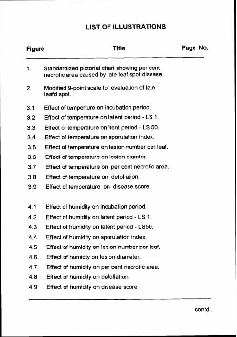
LIST OF ILLUSTRATIONS
Figure Title Page No.
I. Standardized pictorial chart showing per cent necrotic area caused by late leaf spot disease
2. Modified 9-point scale for evaluation of late leafd spot.
Effect of temperture on incubation period.
Effect of temperature on latent period - LS 1.
Effect of temperature on ltent period - LS 50.
Effect of temperature on sporulation index.
Effect of temperature on lesion number per leaf.
Effect of temperature on lesion diamter.
Effect of temperature on per cent necrotic area.
Effect of temperature on defoliation.
Effect of temperature on disease score.
Effect of humidity on incubation period.
Effect of humidity on latent period - LS 1.
Effect of humidity on latent period - LS50. Effect of humidity on sporulation index.
Effect of humidity on lesion number per leaf.
Effect of humidty on lesion diameter.
Effect of humidity on per cent necrotic area.
Effect of humidity on defoliation.
Effect of humidity on disease score
-- - -
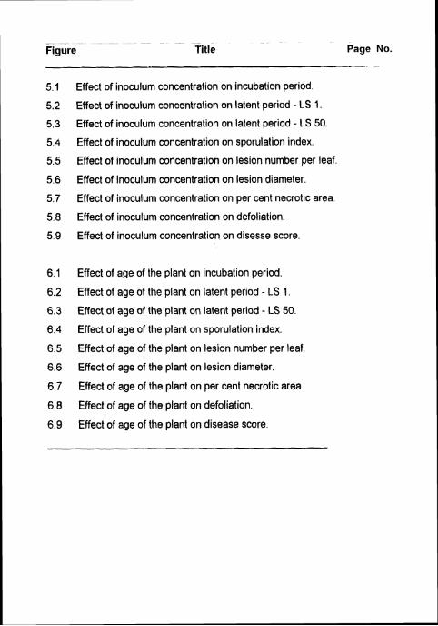
Title Page No.
Effect of inoculum concentration on incubation period.
Effect of inoculum concentration on latent period - LS 1.
Effect of inoculum concentration on latent period - LS 50.
Effect of inoculum concentration on sporulation index.
Effect of inoculum concentration on lesion number per leaf.
Effect of inoculum concentration on lesion diameter.
Effect of inoculum concentration on per cent necrotic area.
Effect of inoculum concentration on defoliation.
Effect of inoculum concentration on disesse score.
Effect of age of the plant on incubation period.
Effect of age of the plant on latent period - LS 1.
Effect of age of the plant on latent period - LS 50.
Effect of age of the plant on sporulation index.
Effect of age of the plant on lesion number per leaf.
Effect of age of the plant on lesion diameter.
Effect of age of the plant on per cent necrotic area.
Effect of age of the plant on defoliation.
Effect of age of the plant on disease score.
LIST OF PLATES
Plate No. Title Page No.
Effect of temperature on the components of resistance to late leaf spot.
Effect of humidity on the components of resistance to late leaf spot in TMV 2.
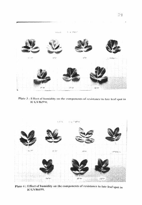
Effect of humidity on the components of resistance to late leaf spot in ICGV 86590.
Effect of humidity on the components of resistance to late leaf spot in ICGV 86690.
Effect of inoculum concentration on components of resistance to late leaf spot in TMV 2.
Effect of inoculum concentration on components of resistance to late leaf spot in ICGV 86590.
Effect of inoculum concentration on components of resistance to late leaf spot in ICGV 86699.
Effect of age of the plant on the components of resistance to late leaf spot in TMV 2.
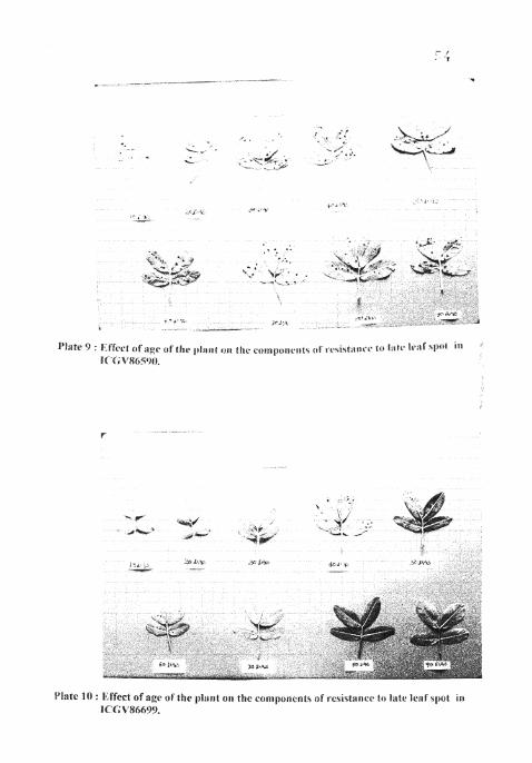
Effect of age of the plant on the components of resistance to late leaf spot in ICGV 86590.
Effect of age of the plant on the components of resistance to late leaf spot in ICGV 86699.
contd.
Plate No. Title Page No.
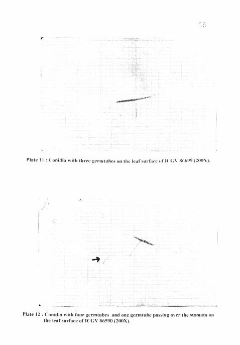
Conidia with germ tubes on the leaf surface of ICGV 86699 (200 x).
Conidia with four germ tubes and a germtube passing over the stomata on the leaf surface of ICGV 86590(200 x)
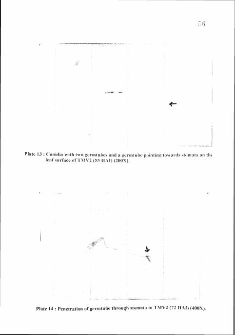
Conidia with two germ tubes and a germtube pointing towards stomata on the leaf surface of TMV 2 (55 HAI) (200 x).
Penetration of termtube through stomata on TMV 2 (72 HAI) (400 x).
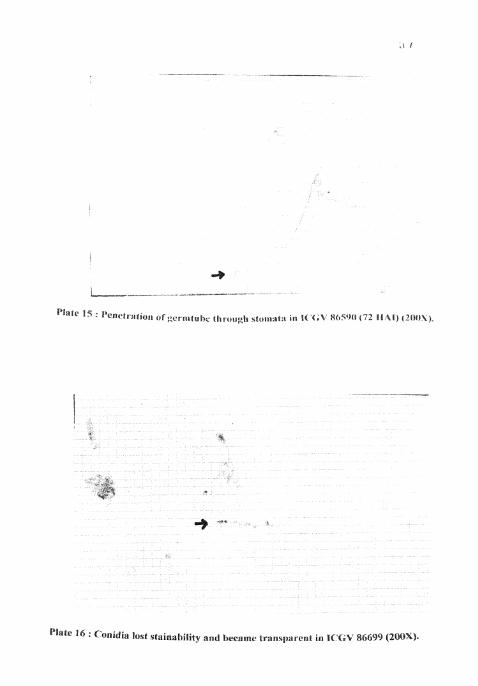
Penetration of gerrritube through stomata on the leaf surface of ICGV 86590 (72 HAI) (200 x).
Conidia lost stainability and became transparent on the leaf surface of ICGV 86699 (200 x).
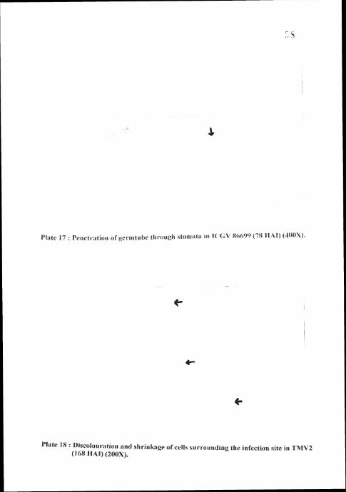
Penetration of germ tube through stomata on the leaf surface of ICGV 86699 (78 HAI) (400 x).
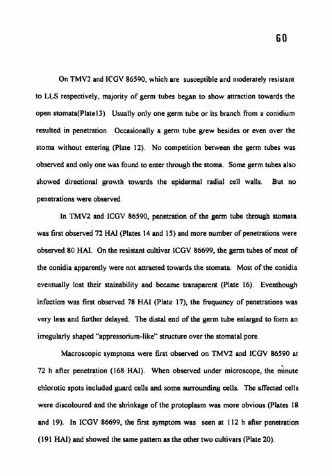
Discoularation and shrinkage of cells surrounding the infection site in TMV2 (168 HAI) (200 x).
Discoularation and shrinkage of cells surrounding the infection site in ICGV 86590 (168 HAI) (200 x).
Discoularation and shrinkage of cells surrounding the infection site in ICGV 86699 (191 HAI) (200 x).

LIST OF TABLES
Table No. Title Page No.
1 . Sporulation index on 1-9 scale (after incubating 24 the leaves for 48-721.1 at 25OC in moist chamber)
2. A 9-point scale for screening of groundnut genotypes :?? for resistance to the late leaf spot disease
3. Effect of temperature on incubation period, latent period and sporulation ndex
4. Effect of temperature on lesion number per leaf, lesion - , diameter, per cent necrotic area and defoliation s
5. Effect of temperature on disease score ? ?
6. Effect of humidity on incubation period, latent period and y, sporulation index
7. Effect of humidity on lesion number per leaf, lesion diameter, per cent necrotic area and defoliation
8. Effcet of humidity on disease score 2 9
9. Effect of inoculum concentration on incubation period, -1 :: latent period and sporulation index
10. Effect of inoculum concentration on lesion number I: !:
per leaf, lesion diameter, per cent necrotic area and defoliation
I I . Effect of inoculum concentration on disease score .i -2
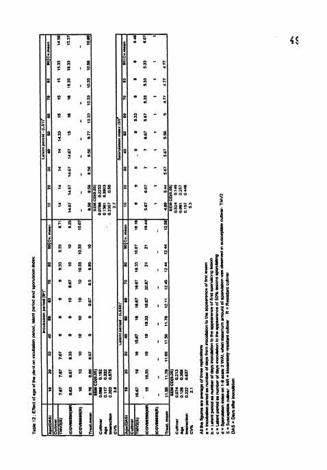
12. Effect of age of the plant on incubation period, latent .? . period and sporulation index
13. Effect of age of the plant on lesion number per leaf, lesion diametre, per cent necrotic area and defoliation
14. Effect of age of the plant on disease score 7- -4
ACKNOWLEDGEMENTS
I express my deep sense of gratitude and sincere thanks to Dr.K.Chandrasekhara Rao, Chairman of the Advisory Committee. Professor and Head, Department of Plant Pathology, College of Agriculture, Rajendranagar, tiyderabad for his encourage~rient, valuable suggesions and guidance throvgtiout the course of this study.
I specially thank Dr. Suresh Pande, Senior Scientist, Legume Pathology, crop protection division (CPD), ICRISAT, Patancheru for his keen interest, constant supervision, constructivecriticism and invaluable suggestions during the course of my work at ICRISAT.
I am highly thankful to Dr.T. Nageswara Rao, Member of the Advisory Committee, Associate Professor, Department of Genetics and Plant Breeding, College of Agriculture, Rajendranagar for his constant help during the course of this study.
I take this opportunity to express my thanks to Mr. J. Narayana Rao, Senior Research Associate, Legume Pathology, LPD, ICRISAT for supervising and helping during the course of this study.
My thanks are also due to all the staff of the Department of Plant Pathology, College of Agriculture, Rajendranagar for their co-operation and help during the course of study.
I also express my sincere thanks to all the staff members of Legume Pathology division, ICRISAT for helping me during the period of my work in their division.
I am thankful to Dr. 8. Divakar, Training and Fellowship programme, ICRISAT for permitting me to work at ICRISAT, Patancheru and also to Mr. V.R. Prabhakar, ICRISAT for his work in statistical analysis.
To my beloved father whose blessings and encouragement are always a source of wisdom and inspiratioin for me and the affectionate and blissful encouragement of my mother is respectfully acknowledged. I extent my sincere thanks to my sister and brother for their constant encouragement throughout my academic career.
It is with pleasure to acknowledge the services of my friends Hazra, Harinarayana, Bheema Raju, Bhukta and all others who stood my side throughout my studies and research work.
I sincerely thank ANGRAU, Hyderabad for providing financial assistance by way of stipend during the course of my study.
Date : t i . t ic; .- \<&b&L LPJ1 - T. RATNA RAJESH
DECLARATION
I, T. RATNA RAJESH, hereby declare that the thesis entilled
"STUDIES ON THE COMPONENTS OF RESISTANCE IN GROUNDNUT
TO LATE LEAF SPOT DISEASE INCITED BY PHAEOlSARlOPSlS
PERSONATA (BERK AND CURT) V. ARX." submitted to Acharya N G.
Ranga Agricultural University for the degree of Master of Science in
Agriculture is a bonafide record of work done by me during the period of
research at International Crops Research Institute for Semi-Arid Tropics
(ICRISAT), Patancheru, A.P., 502 324, India. This thesis has not formed in
whole or in part, the basis for the award of any degree or diploma.
Date : lbm June, 1999 7 'R & Lyh?
T. RATNA RAJESH

Eanie of the author
Title of the thesis
Degree to whicti i t is submitted Faculty
Discipline
Major Advisor
University
Year of submission
Studies oti the co~iil)onet~ts of rrsistn~lrc it1 grollt~d~lut to late leaf spot diseese it~cited by Aueoiscrriop.~isp'nrnt~u~u (Uerk ; ~ n d Curt)V. Arx.
hlhSTER O F SCIENCE
ACRICIILTLIRE
PLAST PATHOl.OC1'
Dr. L Chandrnsekh~ril RHO
Achrpa N.G.Ranga Agricultural l!niversity
ABSTHACI'
Studies on the effect of temperature. humidity. inoculuni concentration and age ol'
the plant on the components of rebistance I~I:., incubation period. latent period fbr
sporulation, number of leions per leaf , lesion diameter, per ccnt necrotic area.
sporulation index, defoliation and disease score to late leaf spot wore undertaken a1
ICRISAT, Patancheru. In all the studies, TMVZ, ICGV 86590 and I('GV 86699 remained
susceptible, moderately resistant and resistant respectively
Over the range of temperatures tested ( I S , 20. 25, 30, 35"('). the severity ol'
disease was the highest at 25 "C i n all the cvs will1 the shoflest incubation period and
latent peroid, the maximum number of lesions per leaf, per cent necro~ic area, defoliation
and disease score At 30 and 3 5 " ~ - no significant amount of disease was observed The
severity of disease progression was the highest between 1 5 and 20°C and the lowest
between 20 and 2 5 " ~ .
Duration of wetness treatment to cause the maximum amount of disease was I6 h
in all the cvs The severity of the disease was the lowest in low humidity treatments (4,8
and I2 h) and the highest in high humidity treatments (16,?0 and 24 h ) with the
maximuni at 16 h treat~nc~it witli the sliol.test ~~icubi~tioti period and latent period . tlic
maximum number of lesions per lent per cent necrotic area. defoliaticln and discasc
score
Inoculuni concentration uas found to tiavc no elt'ect un iricubation period, latent
period for sporulation and sporulation index Tile optiniuni inocuiuni conccrrtra~ion to
cause the highest aniount of disease was 20.000 conidia 1111'' and thcrc was no significant
increase in the amount of disease wit11 further hiylicr corice~itratioll in all the three c ~ s
with the maximum number of lesions per leaf: per ceiit necrotic area, defoliation and
disease score. The amount of disease was ver). niucli restricted in lower concentration
treatments (1000-7500 conidia ml")
Twenty to fiiy day old plants werc found to be most suscepuble to late leaf spot
infection and disease developnlent in all the cvs However in ICGV 86609. no significant
interaction between plant age and Iue leaf spot infection was observed. Ten day old
plants were found to be the moa resistant followcd by 60,70,80 and 90 day old palnts
Defective germination of conidia, delayed penetration and slow iiivasion of host
tissue by the pathogen led to prolonged incubation period in the resistant cultivar, ICGV
86699. The differences between TMV2 and ICGV 86590 were not much for incubation
period

INTRODUCTION

C1IAP'l'EH I
INTRODUCTION
Groundnut (Arucl~is I~jpogucu L ) IS one ot' the world's riiajor food
legume crops It onglnated from South Amer~ca. where the genus Anr~hr\ 15
w~dely d~stnbuted Groundnut 1s an Inlponant 011. food and forage crop
generally d~str~buted ~n trop~cal, subtrop~cal and warm temperate 7ones World's
groundnut production averages to 28 18 n i ~ l l ~ o n t from hdrvebt o f dpprox~mately
21 15 mt l l~on ha and In lnd~a ~t IS 8 2 m l l l~on t from 8 2 m ~ l l ~ o n ha ( Product~on
estimates and crop assessment dlvls~on, USDA. Dec-1997) Ind~a. Ch~na and
USA produce 70 per cent o f the world's groundnut (Poner e f l i l , 1982)
Late leaf spot (LLS) d~sease caused by I'lrueo~sarrol)tis prrrotlttfu (Berk
and Curt) V Arx IS globally w~de spread, and 1s the most Important fol~ar
d~sease o f groundnut L.LS damages the plant by reduc~ng the ava~lable
photosynthetic area, by les~on formallon, and by stlmulat~ng leaflet absc~ss~on
Worldw~de, y~eld losses range from 10 to 50 per cent or even more, but vary
cons~derably from place to place and between the seasons Consewative
est~mates o f y~eld losses caused by the d~sease are ~n the order o f 0 5 tons per
year for l nd~a alone, wh~ch IS about seven per cent o f the total production (T G
Kelley, ICRISAT, Unpublished data, 1992)
Crop protection from the d~seases IS one o f the mandates glven by
human~ty to plant patholog~sts The manayement o f LLS d~sease IS malnly based
on the use o f ava~lable host plant resistance and fung~c~dal spray Explo~tat~on 1
of host plant resistance is the watchword of every agriculturist in developed and
developing countries Advances in screening (Subraholanyanl LV 1 1 1 , 1989) and
breeding for resistance to LLS have resulted in release of a number of promising
lines (Wynne el a/., 1991) However. the degree of res~stance shown by a given
genotype may vary between the locations. possibly owing to the dill'erences in
climate and / or pathogen race So, it is essential to understand the conlponents
of resistance operating in a genotype and the way in which these are affected by
various factors This knowledge will help to develop cultivars with better
resistance to LLS even when the environment favours rapid disease increase
Therefore, the present investigations were carried out to
I Study the effect of temperature and humidity on the components of
resistance to late leaf spot on groundnut.
2. Study the effect of inoculum concentration on the components of resistance
to late leaf spot on groundnut.
3 . Study the effect of age of the groundnut plant on the components of
resistance to late leaf spot on groundnut.
4 Surface studies on the inoculated leaves of d~fferent cultivars for spore
germination and penetration of late leaf spot pathogen.
REVIEW OF LITERATURE

CHAPTER II
REVIEW OF LI'I'ERA'I'IIRE
The literature relevant to the present investigations is reviewed in tllis
chapter under the following sections
1 General
2 Components of resistance
3 Histopathological and histochemical studies
2.1 GENERAL
2.1.1 Distribution
Late leaf spot (LLS) caused by IJh(reoi.\(rrroy~.\,.r p(~r.~orxr/tr (Bcrk and
Curt) V An is the most important disease ofgroundnut worldwide (Jackson and
Bell, 1969; Feakin, 1973, Mc Donald r /cr l , 1985) This disease along with early
leaf spot has also been referred to as Myco.~~~/ iacr t l la leaf spots, ('rrco.~l)oru leaf
spots, peanut cercosporiosis, tikka, viruela, brown spot and black spot (Jackson
and Bell, 1969). LLS is commonly present wherever groundnut is grown
(Feakin, 1973; Mc Donald el a / , 1985) and appears later in the season The
incidence and extent of damage caused by LLS can differ markedly between
locations and seasons In India, LLS is predominant than early leaf spot (Nath
and Kulkami, 1967, Subrahmanyam el a l . , 1980).
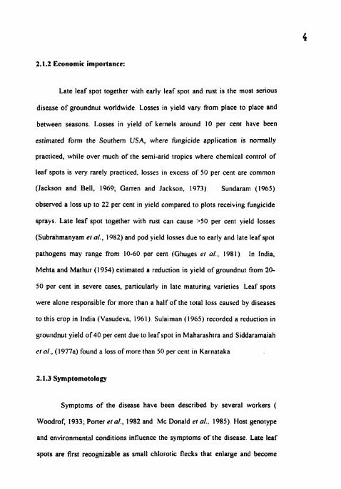
2.1.2 Economic importance:
Late leaf spot together with early leaf spot and rust is the most serious
disease of groundnut worldwide Losses in yield vary from place to place and
between seasons. I.osses in yield of kernels around 10 per cent have been
estimated form the Southern USA, where hngicide application is normally
practiced, while over much of the semi-arid tropics where chemical control of
leaf spots is very rarely practiced. losses in excess of 50 per cent are common
(Jackson and Bell, 1969; Garren and Jackson, 1973) Sundaram (1965)
observed a loss up to 22 per cent in yield compared to plots receiving hngicide
sprays Late leaf spot together with rust can cause >50 per cent yield losses
(Subrahmanyam ex a/., 1982) and pod yield losses due to early and late leaf spot
pathogens may range from 10-60 per cent (Ghuyes er < I / . , 1981) In India,
Mehta and Mathur (1954) estimated a reduction in yield of groundnut from 20-
50 per cent in severe cases, particularly in late maturing varieties Leaf spots
were alone responsible for more than a half of the total loss caused by diseases
to this crop in India (Vasudeva, 1961) Sulairnan (1965) recorded a reduction in
groundnut yield of 40 per cent due to leaf spot in Maharashtra and Siddaramaiah
el a/., (1977a) found a loss of more than 50 per cent in Karnataka
2.1.3 Symptomotology
Symptoms of the disease have been described by several workers (
Woodrof, 1933; Porter er a/., 1982 and Mc Donald el a/., 1985) Host genotype
and environmental conditions influence the symptoms of the disease Late leaf
spots are first recognizable as small chlorotic flecks that enlarge and become
light to dark brown lesions measuring from one to eight mm in d~anreter
Lesions formed by Pharorsar~op.~~.~ pt~rsorraril tend to be smaller, more nearly
circular and darker On the abaxial surface the lesions are black and slightly
rough in appearance The fungus usually sporulates on the abaxial surface and
the conidial tuRs of the pathogen are macroscopically visible as raised clrcles
The yellow halo which is present around early leaf spot is often less
conspicuous or absent in LLS lesions The LLS pathogen also produces lesions
on petioles, stems, stipules and pegs in the later stage of an epidemic These are
oval to elongate and have more distinct margins than the leaflet lesions
2.1.3 The pathogen: Phaeoisariopsi.spe~~onata (Berk and Curt ) V Arx
I'haeorsarropsrs per.sorra/a (Berk and Curt )V Arx Proceedings of the Koninklijke Nederlandse Akademie 86(1), 15-24, 1983 (anamorph),
= ~'ercosporrdrrm~ pcrsorrartmt (Berk & Curt ) Deighton Mycological Papers 112, 71,1967
= ( 'lado.s/~orrrmt per,rotio/o Berk & Curt Grevillea 3, 106, 1875,
= ('erco.s~~ortrprr,~o~ra/a (Berk & Curt ) Ellis & Everhart Journal of Mycology 1, 63, 1885,
= Sep~ogloerrm aruchldr.s Racibolski, Zeitschrifl fuer Pflanze~rkrankheiten und Pflanzenschutz 8.66, 1898,
= i'erco.vpora aruchrdr~ P Hennings tiedwiyia 4 I, 18, 1902,
= Passalora persot~a~a (Berk & Curt ) Khan & Kamal Pakistan Journal of Science 13(4), 188, 1961
~Vfycosphaerella berkelqyi W A Jenkins Journal of Agricultural Research 56, 330, 1938 (teleomarph)
Detcription o r the pathogen
The pertinent morpholoptcal characters of the anamorph arc strollla
dense. pseudo parenchymatous, upto I I p in d~anietcr. con~dlophore\ numerous.
pale to ol~vaceous brown. smooth 1-1 genlculate. 10-100 \ 3 0-6 5 11 In stze.
conld~al scars conspccuous and prom~nently 2-I 11 wtde The conldla are
medlum oltvaceous cyltndr~cal, obclavate usuallv stra~ghr or sllghtlv curved.
wallr usually finely roughened. rounded at the apex, base shonlv tapered with
conspccuous htlum, number of septa vary from 1-9 and 1-4 septa are common
2.2 COMPONENTS OF RESISTANCE
2.2.1 Correlation among the components of reslstance
Reststance to late leaf spot pathogen has been attrtbuted to varlous
morphologtcal and anaromtcal characters of the host plant and to different
chem~cal constituents of leaver and seeds It operates by prolong~ng tncubatton
and latent penods, and by reductng number of lestons per un~t area of leaf
surface, defoltatton, leaf area damage, stze of the les~ons and sporulat~on The
components of reslstance play an Important role ~n tmparttng reslstance to
groundnut genotypes and thus are very useful crlterla ~n select~on of genotypes
for breedtng for LLS reststance In groundnut, however, the assoclatlon among
the components may not be always poclttve Nev~ll (1981) reported that on
some genotypes that defoltate severely, lesions sporulate sparsely whlle on
others they sporulate heavtly Conversely, some genotypes defoliate heav~ly
before 50 per cent of the lesions have began sporulatrng, whlle on others,
les~ons spowlate heavtly before defol~atton The importance of latent perlod for

spomlation has long been recognized (Zadokes.l972) and 11 is the most
important component o f resistance (Nevill, 1980)
Subrahmanyam el ill.. (1982) screened a number o f genotypes in glass
house and found that the parameters iec . lesion diameter, per cent defoliativn
and sporulation gave highly siynificant correlation with field disease scale
Significant differences in lesion diameter on groundnut genotypes in field were
reported (Subrahmanyam tar a/.,1982 and Walls ' I (11.. 1985) S~milarly
significant differences in the amount o f spore production o f ( ln~ov~~orrclrrrm
persot1attrm were reponed amony genotypes GPNC-343 and NC5 and
susceptible check Nc3033 (Walls el a/.. 1985) Differences in necrotic area
produced on groundnut lines by LLS pathogen have also been observed
( Iromue and Knaufl ,1987)
Chiteka et (I/.. (1987) screened 116 genotypes o f which the most
resistant genotypes, UF 81206-1, UF 81206-2 and PI 203396 had both longer
latent period and reduced spomlation Similarly, Jogloy rr a/.. (1987) tested 20
breeding population for resistance to LLS and found that the resistant
population had an increased latent period, decreased number o f lesions, lesion
size, defoliation and reduced spore production
Greatest variability amony genotypes for lesion diameter and latent
period has been recorded by Chiteka el a/., (1980a) These workers (1988 b)
also found that amount o f spomlation, lesion size and latent period were highly
correlated with each other and with per cent necrotic area Based on this they
concluded that these were the most important components o f visual plant
appearance score of which sporulation accounted for most o f the variability in
the score. Shokes and Gorbet (1991) reponed that resistance was due to

decreased sporulation and lenghened latec~t percod in resistant viirieties
(Southern runner and UF81206) as conlpnred to susceptible varier? (1:lomnner)
Recently Watson ur a/.. (1997) observed reduced lesion diacllcter and
longer latent period for sporulation of 50 per cent lesions in paniall\ rescstant
Southern Runner as compared to susceptible cultivar- 1:lonlnner 13111 there were
no differences between spore germination, incubation period and number of
lesions per leaf in the two varieties
2.2.2 Effect of temperature and humidity on the contponests o ~ r e s i s t a ~ ~ c e
It is a well-known fact that tenlperature and humidity play an lmponant
role in the development of diseases L1.S development is highly influenced by
these two weather parameters In the earlier years, Wolf (1914) found no
correlation between temperature and moisture and the prevalence of leaf spot
But in 1938, Jenkins reported in Georgia that cool, humid weather during the
epiphytotic months favoured the spread and development of disease Maublanc
(1925) from Senegal and Kenknight (1941) from the USA. attributed the rapid
spread and severity of leaf spots to heavy rainfall in August-September and in
spring in their respective countries LLS incidence was found relatively more in
damp, warm weather and periods of heavy dew in North Carolina, USA
(Research and Farming, 1943). Miller (1946) opined that the rapid spread of the
leaf spot disease might be correlated with periods of heavy rainfall Sulaiman
and Agashe (1965) studied the influence of climate on the incidence of tikka
disease of groundnut in Maharashtra and Andhra Pradesh and found that the
minimum predisposing factors to disease development were, an average
maximum temperature of 29.3 '~. an average minimum temperature 2 3 " ~ . an
average RH 81 8 per cent and an average rainfall of 240 8mm.
Relative humidity (RH) appears to be a better measure of all the
moisture factors that affect leaf wetness and duration of leaf wetness in the crop
canopy for the development of disease (Jenson and Boyle ,1965) It was also
found that the period required for the maximuni infection decreased with
increasing temperature between 18 and 27 'C which implies that rate of'
infection increases with temperature but with adequate leaf wetness periods
However, the final level of infection would not be affected by temperature
between 20 and 2 7 ' ~ .
Jenson and Boyle (1966) recognized the importance of leaf wetness to
infection and made three important assumptions ( I ) adequate inoculun~ is
always present, (2) free water is necessary for spore germination and the speed
of germination depends on temperature and (3) periods of RI I greater than 95
per cent indicate periods of leaf wetness (this includes wetness from rains) This
implies that under sufficient inoculum level, if favourable temperature persists
along with long periods of leaf wetness (>9S% Rtl) at least for 2-3 days, L1.S
development takes place. Ramakrishna and Appa Rao ( 1968) from Hyderabad ,
India, reported that a 72h period of high RH was ideal for infection and funher
development of leaf spot disease
While evaluating the groundnut genotypes for resistance to leaf spot,
Hassan and Beute (1977) found high environmental variations for lesion count
per leaf They found that plants grown continuously in greenhouses tended to
develop more lesions than did plants grown outside for two weeks before
inoculation. Shew and Beute (1984) reported the requirement of longer mist
periods up to eight days for increase in lesion numbcrs under ,ureenbouse
conditions
Germination of ('c~rco.y)orc7 trrc~t~lrrc/rc~ol~r spores declined as R H was
reduced from 100 through 98% and the perm tubes were longest at 2z°C
(Alderman and Beute. 1986)
Temperature of 16-20'~' \bas found lhvourahle fi)r the germination of
conidia of Cerco.sj~orrdrtml prr.\orrtzrron (Sonlmanya and Reute, 1986) They
also studied the germination of d~fferent isolates of I'lrnrorscrrrops~t j~~~r.$otar~tl
~ t r rbrrro and found that the percentage germination of corlidia decreased alter
48h with increasing temperature between I6 and 3 2 " ~ The nlaximum reduction
was at temperature greater then 2 8 " ~ Contrary to the repons of Jenson and
Boyle (1966), Sommartya and Beute (1986) stated that the germination was the
maximum at 2 0 ' ~ and at this temperature more than 50% of the conidia
germinated
Studies on the effect of duration of leaf wetness on infectiorl at different
temperatures revealed that in majority of genotypes the nlaxlmum number of
lesions were obtained at 2 0 ' ~ but differences between 2 0 and 24°C were not
large (Shew el nl., 1988) They pointed out the importance of long periods of
leaf wetness with intermittent dry periods for the development of I.LS lesions
on inoculated leaves Resistance level of genotypes with high (PI 259747,Nc
Ac17133), moderate (GPNc 343) and low (Nc 3033, Robut 33-1) resistance
decreased with increasing temperature from 20 to 32°C and also by decreasing
high RH shorter than 12hlday.
Lanmou and Blizoua (1989) reponed that on detached leaves at least six
days were required for the establishment of lesions at 2 7 ' ~ and 100 per cent RH
and on potted plants the infection efficiency was the highest when a daily
rythemicity in RH (70-100%) was simulated They concluded that, long period
under humidity saturated conditions, preferably spread out over time was
required for the development of cercos/x~ro leaf spot epidemic
Infection was very rapid at 2 3 ' ~ when the leaf wetness was provided
continuously for five nights (Butler, 1990). These results confirmed earlier
findings of Shew er al., (1988) that infection occurred with intermittent periods
of surface moisture. He therefore concluded that dominant variable affecting
infection at a particular temperature was the total number of hours of leaf
wetness.
In controlled environment experiments, Alderman and Nutter (1994)
found that the minimum of four hours of RH >95 per cent per day was required
for conidial production by ('. perso~~ot~rm and the highest number of conidia
was produced when lesions were subjected to a daily pe:iod of 16h or more at
>95 per cent RH The optimum temperature for spore production was around
20°c
Butler el ul., (1994) found that the temperature response curves for
conidial germination and infection were similar with the optimum close to 2 0 " ~
and the minimum and the maximum :emperature of about 8 and 3 4 " ~
respectively. They found that the number of lesions resulting from a fixed
amount of inoculum was several times greater if the leaves were exposed to
alternate wet and dry periods as compared with continuous wetness
Wadia and Butler (1994) reponed that latent period for ('. persot~ufirm
ranged from 13-38 days in susceptible variety TMV2, between temperatures 12
and 3 3 ' ~ confirming the findings of Nevill (1981) that latent period for
/'. I W ~ ~ J I ~ U ~ U was 14 6 davs at 25°C in ThiV? They establisl~ed cardinal
temperature as T min -loUC, T opt- close to 25°C and T mas-35°C hy relating
the rate of pathogen development (IILP) to temperature These workers (1995)
further observed an increase in mean conidial length and number ot' septa when
the humidity increased from 96 through 100 per cent.
2.2.3 EiTect of inoculum con cent ratio^^ on the components of resistance
Hassan and Beure (1977) screened about I6 cultivars for reststance to
C arachidicob at three inoculum levels (15,000. 10,000 and 5000 corlid~a ml")
and found that there were consistent cultivar differences in number of lesions
Components of resistance such as number of lesions, time to leaflet loss
by (: arachidicola and ('. ~~er.\orrarr~rn were influenced by concentration of
inoculum applied to rhe leaves (Nevill, 1981) Similar results were obtained for
other host-pathogen systems - Solnt~~rnr .sly) to IJhyro~~h~hora rr!/c..sro~r.c (Guzman,
1964) and winter barley cultivar -Vulcan to I<hy~ro.s/~orrrmnr .sccir1~.s (tlabgood,
1972)
Shew and Beure(1984) found increase in lesion number with increasing
spore concentration from 12,500 to 1,00,000 condia ml" in the cultivar Nc3033
2.2.4 ElTect of plant age on the components of resistance
The amount and extent of infection depends on age of plants, rate of
plant growh, method of cultivation and the length of the peanut rotation cycle
(Miller, 1946). Subrahmanyam el a/., (1982) observed significant interaction
between plant age and genotype for all the parameters except sporulation. Fifty
day old plants showed higher per cent defoliation, per cent necrotic area,
infection frequency, and lesion diameter than 30 days old plants o f different
genotypes
Savary and Van Sante~r (1992) while workiny on primary gradients of
LLS in field reported heavy detbllation IN case o f younger plants \rliereas in
older crops accessibility \bas reduced by h i ~ h f o l i a ~ r density and I~i_ul~ leaf area
index Contrary to this, Con~acho de Torres and Suberco (1993) repned that
younger plants were the most resistant to (: ur~~c~i~~clic~olcr independent of' the
cultivar (Red star, Bollvia pintado and Tarapoto)
2.3 HISTOPATHOLOGICAL AND HISTOCHEMICAL S'S1IL)IES
2.3.1 Histopathological studies
Little has been carried out on the mechanisms o f resistance The host
response to L.LS pathogen widely varies from highly resis~ant to highly
susceptible reaction The resistance is influenced by the morphological and
physiological characters o f the host I t is also noted that the number o f lesions,
necrotic area and extent o f sporulation vary greatly from susceptible to resistant
cultivars This may be due to reduced number o f penetrations, reduced stomatal
index, increased thickness o f the cuticletepidermis and/or histochen~ical changes
within the host in response to infection
Jenkins (1938) reported that infection by LLS pathogen was
accomplished through either leaf surfaces The resistance in wild Aruchr.~ sp
appeared to be associated with small stomatal apertures (Decurz and
Upadhyaya, 1961 and Gibbons and Bailey, 1967) Hemingsway (1957)
suggested that the more susceptibility o f sequentially branched early maturing
variety was due to higher proportion o f stomata o f "penetrable size" on the

dorsal leaf surface He also proposed that greater amount o f palisade tissue
might account for slower rate o f pathogen gro\\rli on alterna~ively branched
culitivars Similarly Mazzani er 111.. (1972) found resistance to L1.S ill cultivars
with small stornatal apenures and also observed that leaf spot counts were
higher on cultivars with large light green leaves Contrary to these repons. Cook
(1981) observed no dityerence in conidial germinalion on I ? peanut cultivars
and also observed that variation in stomatal density and s~omatal lenyth were
not related to resistance to infection
Abdou el 01.. (1974) compared post germination behaviour o f
C: yersotrarrrm on the leaf surface o f wild species and cultivated genotypes and
found that germ tubes apparently were not attracted towards the stoniata in
resistant genotypes but were attracted towards stomata in susceptible yenotypes
'They reponed that conidia and germ tubes lost their stainability and became
transparent on resistant genotypes They also noted that resistance aRer
penetration was associated with the formation of barrier in advance around the
infection site in the form o f cell wall swelling and thickening and the deposition
o f pectic substances on the cell wall and in the intercellular spaces
Longer incubation period, reduced sporulation, stomatal exclusion and
absence of directed growlh o f germ tubes towards the stomata are some of the
components o f host resistance (Nevill, 1981 ) Kaui. and Dhillon(1988) reponed
that the epidermal and mesophyll cells were shrunken or collapsed in L.LS
pathogen infection The damage to protoplast was more obvious than to the cell
walls.
Thicker epidermis, smaller stomata and compact palisade in resistant
leaves were accounted for fewer penetration sites o f the pathogen in resistant

cultivars (Basra el ul. 1985) 5fayee and Surya\ra~ishi ( 1005) rsponed that Istc
appearance of symptoms in resistant cultivars (NC Ac 17 133. PI-4051 31.
259747,381622 and 390595) to LLS was due to prolonged iticuhatioti period
because of delayed appearance of niesophyll niass helotv eptder~tiis
Recently Bera cr ~11 . . (1997) reponed that susceptible genotype possessed
wider stomata than resistant i tolerant genotype in luwer surface of the nurnial
leaf They also reponed more distortion of mesophyll tissue ill susceptible
genotype than in resistant 1 tolerant genotype and opined that thick palisade and
spongy tissue in resistant 1 tolerant genotype might have allowed lini~ted and
slow rate of pathogen growth
2.3.2 Histochemical studies
A low level of magnesiuni was either directly or indirectly responsible
for increased susceptibility (Bledsoc el 0 1 , 1946) Yenni (1970) found that
healthy tissues of all cultivars tested had higher maynesiuni content than in
diseased leaves
The histochemical localizations revealed a gradual depletion of
polysaccharides, proteins, ascorbic acid and nucleic acids from the diseased host
tissue at the site of contact with the pathogen and their subsequent accumulation
in the pathogen in the later stages of disease developement (Kaur and Dhillon,
1988).
The level of nitrogen was low and phosphorus and that of potassium was
high in the resistant cultivars than in the susceptible cultivars at all the growh
stages and the elements decreased with the age of the plant in all the cultivars.
Similarly the level of zinc was high and level of iron was low in resistant
cultivars (Jagalan and Sindhan, 1988). High levels of zinc may be responsible

for resistance because deticienc! is thought to restrict RNA synthesis \r.hich III
turn inhibit protein synthesis and lead to pool. ~ r o & ? l l and increased
susceptibility l o various pathogens (Mogle and hlayee. 198 1 )
Sindhan and Parashar (1996) reported that resista~lt cultivars had higher
phenolic contents and lower reduced and non-reducinp sugars, and higher P. K.
Zn & Cu compared to N. hfn. and Fe AAer infection the total phenols increased
in all the cultivars and the carhohydrates decreased

MATERIALS &METHODS

The investigations on the quantification of co~nponents and ~~ieclianis~~is o f
resistance in groundnut to LLS were carried out at International ('rops Rrasercl~ Institu~e
For Semiarid Tropics (ICRISAT). Patancheru, Andhra I'radesh. 502 32.1. India The
materials and methods used in the present investigation are broadlv described under the
following heads
I . General
2 Experimental design
3 . Green house experinients
4. Observations
5 . Histopathological invest~yations
3.1 GENERAL
Three groundnut cultivars (cvs ), TMV 2. I U i V 86590, I<'(iV 86699
representing susceptible, moderately resistant and resistant respectively were selected
to study the effect o f temperature, humidity, spore concentration and the age o f the
plant on the components o f resistance Further, histopathological investigations were
conducted to hrther quantify the components o f resistance in these cultiva;~
3.1.1 Plant material
Groundnut plants o f all the three cvs were grown in 15-cm
diameter plastic pots in a greenhouse. The potting medium consisted o f 60 per cent,
Alfisol, 20 per cent sand and 20 per cent compost. Healthy plants were maintained in
the greenhouse. Four weeks old plants (two1 pot) were used for inoculation
3.1.2 Collection nnd nloltiplica~ion of i ~ ~ u c t ~ l ~ ~ n i
3.1.2.1 The pathogen:
Single lesion isolate o f LLS pathoeen of yrou~idnut available in groundnut
pathology laboratory at IC'R1Sh'l'-Patancheru was used in these i~irestigations
lnoculum of this pathopen was maintained 011 the detached leaves of a susceptible
groundnut genotype, TXIV2 The inoculum was harvested with a cyclone spore
collector and stored at 4°C'
3.1.2.2 Detached leaf techniques for inocu lu t~~ productio~i
3.1.2.3 Inoculation
Mature, undamaged. apparently healthy leaves o f preen Iiouse-grown
groundnut plants were excised through the pulvinus base from the middle portion o f
the main stem . The leaves were thoroughly washed and arranged in plastic trays
(55cm long x 27 5 cm wide and 5 cm deep) with their petioles buried in steam
sterilized ( I 5 Ibs for 30 min) sand There were four leaves per row and six rows per
tray. Trays were covered with clear polyethylenc bays (62 x 18 cm) with the open
ends partially sealed with cellophane tape to maintain high relative liurnid~ty and the
trays were kept in percival incubators at 25°C' and 12h photoperid (4000 lux) After
24 h, the trays were removed from the incubator chambers and the leaves were
sprayed on both surfaces with spore suspension (20,000 conidia rnl") using a plastic
atomizer. The trays were then returned to the incubator (Foster rr 0 1 , 1980)
3.1.2.4 Spore collection
LLS lesions developed and spo~lat ion was observed in two weeks after
inoculation in all the leaves Then the spores were collected from the lesions using a
cyclone spore collector (Flsher Ssienlilic Co . LISA) i l l s111n11 glass v~als ( 7 5 c n ~ h 2 O
crn diameter)
3.1.2.5 lnoculurn preparation
For all the experiments (except for the study of etkct of spore colicrntration) tlw
inoculum was prepared in the following way
The spores were suspended in distilled hater to which a fcw d ~ o p s (10 drops
1000 ml') of Tween-80, (Polyouyethylene sorbiton mono-oleate) wetting agent were
added (Melouk and Banks. 1978). 'The spore suspe~lsion was nirrcd \cell using a
magnetic stirrer (model 213, Fisher Scienlific Co . USA) to make i~iocululn uniform
The spore conczntration was adjusled to 20.000 conidia rill" using a hacmocytome~er
3.2 EXPERIMENTAL DESIGN
All the experiments were carried out under greenhouse conditions Experiments
were conducted in a completely randomized block design with three rcplica~ions.
Each replication contained two healthy plants On each plant the third or fourth fully
expanded leaf from top was tagged prior to inoculation to study the components of
resistance
3.3 CLASS HOUSE EXPERIMENTS
3.3.1 Inoculation
A spore concentration of 20,000 conidia rnl ' was used for all the experiments
except for the study of effect of spore concentration ill which different spore
concentrations were used. Conidial suspension was prepared as already described.
Immediately afler preparation of conidial suspension, the plants were uniformly
inoculated with atomizer, Inoculated plants were kept in dew-chambers to ensure
complete wetting of leaves Next day morning the plants were shified to the
greenhouse. Thus, the plants were kept in dew-chambers in the night (16h for wet
period) and in greenhousc during dav (811 for dly pe~iod) for sis dn\s atid t l~c l i tlie
plants were permanently shined to ylasshouse for the rcsl o f thc espel.inicl>l
3.3.2 Studies on the erect of temperatttre on the cot~ipone~tts o f rcsistnnce
The components o f resistance to L1.S were studied at 15, 20. 7-5. 3 0 and 35°C' Six
dew chambers and six incubators were adjusted to maintain 15.20.?5..;0 atrd 35°C' tbr
this study The plants were kept tn the dew chambers at nlglit for \\el period and
moved to incubator duriny the dav fur dry period Care was laken to keep tlie platits
in the same temperature while shiHing from and 11) dew chanrhers for s ~ s days The11
the plants were permanently shifted to incubators at the respeclive trnrperature lill the
end o f the experiment
3.3.3 Studies on the elTect of humidity on the components o f resistatice
Immediately after inoculation the plants were kept in dew chamber for leaf wetness
for different durations o f 4, 8, 12, 16, 20 and 24 h per day 'The plants were taken oul
o f the dew chambers as per the treatment and shifted to greenhouse everyday The
wet and dry treatments were given for six days and after that. the plants were
permanently shifted to greenhouse till the end o f the experirncnt
3.3.4 Studies on the erect of inoculurn concentration on the components of resistance
To study the effect o f inoculum concentration, the plants were inoculated
with different concentrations of conidial suspension (1,000, 2,500, 5,000, 7,500,
10,000, 15,000, 20,000 and 25,000 condia ml") Immediately afier inoculation, the
plants were kept in dew chamber for wet period during nights and in green house for
dry period during day. The dew chamber treatment was continued for six days and
after that the plants were permanently shifted to greenhouse till the end o f the
experiment.
3.3.5 Studies on the effect of age of the plant on the cornpo~ients olresistance:
To study the effect of the age of the groundnut plant on the components of
LLS resistance, different aye proups of plants ranging from 10 to 90DAS were
maintained by staggered sowing at 10 days interval The plants of all ages were
inoculated at a time with a conidial suspension containing 20,000 conidia nil" Then.
dew chamber treatment was given as in the case of other experin~ents for six days and
after that the plants were permanently shifted to green house for the rest of the
experiment
3.4 OBSERVATIONS:
The components of resistance studied were incubation period (1P). latent period
(LS), lesion count (LC), lesion diameter (LD), necrotic area (NA). defoliation (DEF)
and disease score (DS) Scoring was done at weekly intervals soon after IP was
observed in all the experiments The scoring was continued up to 100 days after
sowing
3.4.1 Incubation period (IP): IP was recorded by counting the number of days from
inoculation to the appearance of first symptoms All the leaves of the plants were
observed daily to record IP
3.4.2 Latent period (LS): Latent period was recorded by counting number of days
from inoculation to appearance of first sporulating lesion (LSI), and 50 per cent of
lesions sporulating (LSSO) everyday with the help of a 20X magnifying lens The
lesions were considered to be spomlatiny tufts of fascicles visible on the lesions
3.5 Surface studies on the i~~ocu l r t ed leates
3.5.1 Plant material
Histopathological studiec \+ere conducted b\ adopting detached leaf technique
The leaves were collected and maintained as descnbed In section I I 2 ?
3.5.2 Inoculation
The detached leaves were inoculated with a conidial suspension coiltaining 50.000
con~dia per ml with the help of an atomirer lmmediately atter inoculation, the travs
containing detached leaves were covered with polythene bags and incubated at 25°C
in an rncubator (Perclval C o , Boone, lowa, USA) Sufficient leaf wetness (RH> 95
%)was maintained at least for 16 h in the trays throughout the experinlent
3.5.3 Collection of leaf samples
Samples were collected everyday at OSOOh and 1600h, starling from four h after
inoculation, till the appearance of first lesion The leaflets were cut into small bits
( IX 1 cm) aRer the mtdrib was incised
3.5.4 Processing of leaf samples
3.5.4.1 F~xation and clearing
The leaf samples were fixed and cleared In a solution containing glacial acetic acid
and absolute ethanol in 2 1 ratio (Johan~on, 1940) The sanlples were left in the
solution for 36h After that, the samples were decanted and stained
3.5.4.2 Staining
M e r clearing, the leaf samples were stained in lactophenol coton blue (0 I%) for
about five mln and then the starned samples were observed under the light microscope
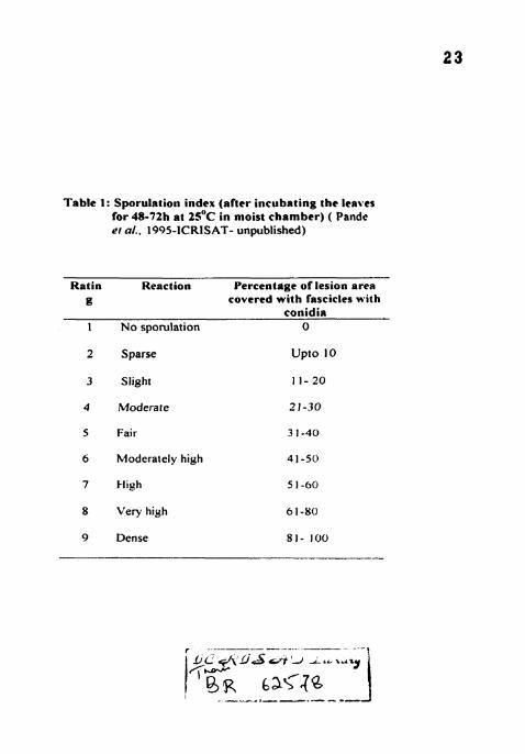
Table 1: Sporulrtion index (after incubating the leaves for 48-72h at 2 5 ' ~ i n moist chamber) ( Pande er a/.. 1995-ICRISAT- unpublished)
Rat in Reaction Percentrrge o f lesion area g covered wi th fascicles with
conidir 1 N o spo~ la t ion 0
2 Sparse Upro 10
3 Slight 11-20
4 Moderate 21-30
5 Fair 3 1-40
6 bloderately high 4 1 -50
8 Very high 6 1-80
9 Dense 81- 100 -
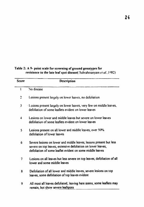
Table 2: A 9- point scale lor screcnir~g olgruiind genotypes for resistai~ce to the late leal s110t discas(( S~~hrahnl;lnya~ii 1'1 (11.. 1082)
Score 1)escription -------
I No disease
? Lcsions present laryely on lower leavcs. no defoliation
3 1.csions present largely on lowcr leaves, very few on nliddle leaves, defoliation of some leaflets evident on lower leavcs
4 Lesions on lower and middle leaves but scvcre on lowcr leaves defoliation of some leaflets evident on lower leaves
5 Lesions present on all lower and middle leaves; over 50% defoliation of lower leaves
6 Severe lesions on lower and middle leaves; lesions present but less severe on top leaves; extensive defoliation on lower leaves, defoliation of some leaflet evident on some middle leaves
7 Lesions on all leaves but less severe on top leaves, defoliation of all lower and some middle leaves
8 Defoliation of all lower and middle leaves, severe lesions on top leaves; some defoliation of top leaves evident
9 All most all leaves defoliated, leaving hare sterns, some leaflets may remain, but show severe leafspols _
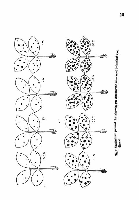
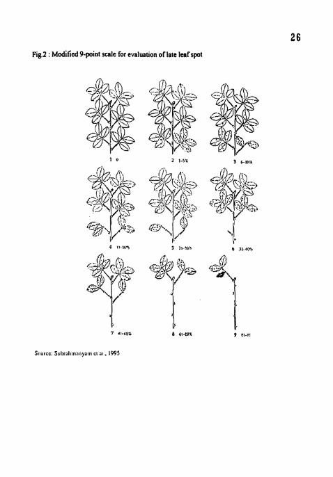
Fig.2 : ~odified Ppoint scale for evaluation of late leaf spot

RESULTS
CHAPTER I V
RESULTS
Investigations were carried out to study the effect of temperature, relative
humidity, inoculum concentration and age of the plant on the components of late
leaf spot. Histopathological investigations were also carried out to further quantify
the components of resistance to LLS. The results obtained from the above
investigations are presented in this chapter.
4.1 Effect of temperature on the components of resistance to LLS
Different temperatures (l5,20,25,30 and 35'C) were selected to study their
effect on the components of resistance. At 35°C there was no disease and plants
eventually died. At 30°C, even though lesions were observed the amount of disease
was insignificant.
4.1.1 Incubation period
The incubation period (IP) was shortest at 25°C and longest at 15°C in all the
cvs (Table 3). Among the cvs, TMV2 showed shortest 1P (8 67 days) at 25°C and
ICGV 86590 showed longest IP (10.67 days) at 20°C. Significant differences were
found between cultivars at all the temperatures However there was no significant
difference between the IP's at 20 and 25°C in all the cultivars No significant
difference was observed between TMV2 and ICGV 86590 at 25°C.
4.1.2 Latent period (LS 1 and LS 50)
No sporulation was observed at all the temperatures in the cv ICGV 86699
Significant differences were seen between the temperatures for both LS 1 (days
from inoculation to the appearance of first sporulating lesion) and LS 50 (50%
lesions sporulating) in TMV2 and ICGV 86590 and also between these two cvs
27
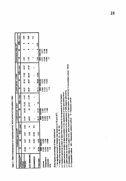
(Table 3). LS 1 and LS 50 were shortes~ at 25°C and longest at 15°C i l l all the
cultivars. TMV2 showed shonest LSI (13.33 days) and LS50 ( 17 33 days) at 25°C
4.1.3 Sporulation Index
Sporulation index (SI) was almost nil in ICGV 86699 at all the temperatures
studied (Table 3). Highest sporulation (9) was recorded 2 weeks after inoculation
(WAI) at 20 and 25°C in TMV2 and at 25°C in ICGV 86590 Significant
differences were observed between 20 and 25°C in ICGV 86590. Significant
differences were found among cultivars at I S'C. At 15"C, SI reached maxirnun~ at
5 WAI in TMV2 and 8 WAI in ICGV 86590. At 20°C, ICGV 86590 showed
maximum S1 (9) at 3 WAI.
4.1.4 Lesion number
The number of lesions per leaf was significantly higher in TMV2 (155 2,
160.8 and 144.2) than in ICGV 86590 (74.3, 70.7 and 66) and ICGV 86699 (74 2,
59.3 and 38.7) at 15.20 and 25°C respectively (Table 4). In susceptible TMV2, the
number of lesions per leaf increased upto 3 WAI at all the temperatures and no
significant differences were found among the temperatures In ICGV 86590 also,
no significant differences were found among the temperatures However lesion
number was increased upto 6 WAI at 1 S°C.
4.1.5 Lesion diameter
Maximum lesion diameter (6.23 mm) was observed in TMV2 5 WAI at
2 5 ' ~ . At 5 WAI significant differences were observed between the temperatures
and also in between the cultivars (Table 4). TMV2 and lCGV 86699 showed
highest (3, 5 and 6.23 mm) and lowest (1, 2 and 3.8 mm) lesion diameters
respectively at 15, 20 and 25°C. Highest lesion diameter was noticed at 2 5 " ~ and
lowest at 1 5 ' ~ in all the cultivars.

4.1.6 Per cent necrotic area
The per cent necrotic area (PNA) per leaf was significantly higher in ThlV2
than all the cultivars at all temperatures (Table 4) The PNA was lowest at 15°C
Eventhough the PNA was maximum in all the cultivars at 20°C. it was on par with
2S°C. ICGV 86699 did not show any significant difference between the
temperatures. Cultivar differences were significant at 15, 20 and 2S'C with highest
PNA (15 83, 35.33 and 31.67) in TMV2 and lowest PNA (2, 6.33 and 7 58) in
ICGV 86699.
4.1.7 Defoliation
At 4 WAI, per cent defoliation was highest (100) at 25°C in TMV2 followed
by ICGV 86590 No defoliation at 1 5 ' ~ and highest defoliation was observed in all
the cultivars. Significant differences were also noticed among the cultivars in all the
temperatures except at 1 5 ' ~ (Table 4)
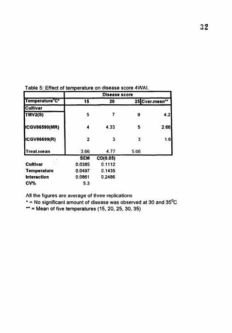
4.1.8 Disease severity
Maximum disease (9) score was observed 6 WA1 in TMV2 at 2 5 " ~
Lowest disease was recorded in ICGV 86699 in all the temperatures. Significant
differences were observed among the cultivars at all the temperatures (Table 5).
The disease score was highest at 25OC and lowest at 15'C in all the three cultivars
(Plate 1). TMV2 and ICGA 86699 showed highest (5, 7 and 9) and lowest (2,3 and
3) disease score respectively at 15, 20 and 25°C.
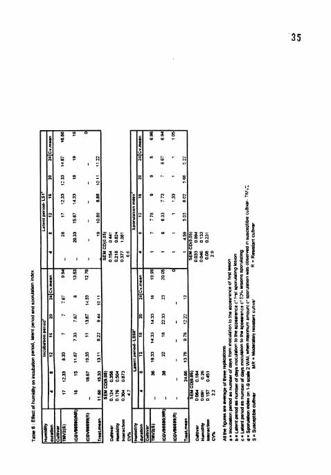
4.2 Effect of humidity on the components of resistance to LLS
4.2.1 Incubation period
The IP was longer at low humidity treatments (4, 8 and 12 h) (Table 6) . but
shortest at 16 h treatment in all the three cvs while the IP's at 20 and 24 h treatments
Cultivar 0.0385 0.1 112 Temperature 0.0497 0.1435 Interaction 0.0861 0.2486 CV% 5.3
Table 5: Effect of temperature on disease score 4WAI.
All the figures are average of three replications * = No significant amount of disease was observed at 30 and 35'~ ** = Mean of five temperatures (15, 20, 25, 30, 35)
TemperatureUC'
Cultivar TMVP(S)
ICGV86590(MR)
ICGV86699(R)
Treat.~t~eati
Disease score 15 20 251~var.rnean**
5 7 9 4.2
4 4.33 5 2.66
2 3 3 1.6
3.66 4.77 5.66 SEM CD(O.05)

i"la¶i- f : I'i'fccf (if r ~ ~ i r r ~ i e r r vxr t t ~ c u.ibirtjliincrtt\ ~ i f ic~.i \ l:t<~ct" 'I, l i l t ~ I t 1 ) ~ ) * i f t ~ t i n grotaintixiirt.
plate 2 : EfTect of haprnittilv orr tire romponcznia c i f rc~,iblasrrc tir IxPr Iesf'qrot i t s I 'MVZ.

were on par with the 16 h treatment in TMV2 and ICGV 86590. IP was shortest (7
days) at 16h in TMV2 among all the treatments. In ICGV 86699, IP was shonest
(1 1 days) at 16 h treatment and no IP was noticed at 4 h. ICGV 86699 showed
lowest IP than TMVZ and ICGV 86590 in all the treatments.
4.2.2 Latent period (LS 1 and LS SO)
No sporulation was observed in ICGV 86699 in any of the treatments while
it was nil in 4h treatment in TMV2 and ICGV 86590. Significant differences were
observed in all the treatments in TMVZ and ICGV 86590 (Table 6). Both LS I and
LS 50 were shonest at 16 h treatment and longest at 8 h treatment in all the
cultivars. In TMV2 no significant difference was found for LS 1 and LS 50
between 16 h and 20 h treatments.
4.2.3 Sporulation index
Sporulation index reached maximum (9) in TMV2 for 16 h and 20 h
treatments at 3 WAI (Table 6). SI was highest (9) in TMV2 and lowest ( I ) in ICGV
in ICGV 86699 in all the treatments. Maximum SI (9) was recorded at 16 h
treatment followed by 20, 24, 12 and 8 h treatments in the same order in TMV2 and
ICGV 86590. At 4 h treatment there was no sporulation in all the three cultivars. In
ICGV 86699, sporulation was observed sparsely (I .33) at 16 h treatment only.
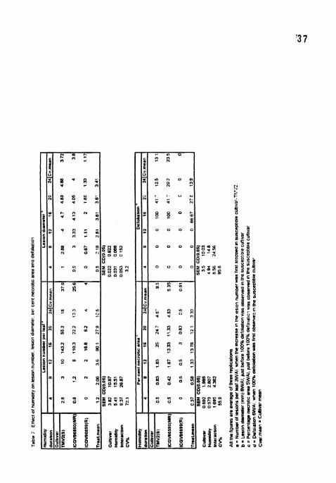
4.2.4 Lesion number
In all the cvs, maximum and minimum number of lesions per leaf were
observed at 16 h and 4 h treatments, respectively 3 WAI (Table 7). Lesion number
was highest (143.2) at 16 h treatment in TMV2 and nil at 4 h treatmnet in ICGV
86699. The number of lesions per leaf was highest in TMV2 and lowest in ICGV
86699, in all the treatments. Significant differences were noticed among cultivars in
lesion number at 16 h treatments

4.2.5 Lesion d ian~e te r
Lesion size significantly varied between the cultivars in all the treatments
Lesion diameter was the highest (4 89 mm) at 20 h treatment in ThtV? and no
lesions were observed at 4 h treatment in lCGV 86699 TMVZ and ICG\' 86699
showed highest and lowest lesion diameter respectively for all the treatnrents
(Tablel). The difference in size of the lesions were not significant in 20 and 24 h
wetness treatments in TMV2 and among 16, 20 and 24 h wetness treatments in
ICGV 86590
4.2.6 Per cent necrotic area
There were no significant differences in PNA in 4, 8, 12 and 24 h wetness
treatments for all the cultivars and also between the cvs 5 WAI (Table 7) The
maximum PNA (25, 13 33 and 3) was recorded in 16 h treatment in TMV2. ICGV
86590 and ICGV 86699 respectively. The PNA was hiyhest (25) in TMV2 at 16 h
and lowest (0) in ICGV 86699 at 4 h treatment. PNA was not recorded in ICGV
86699 in 4 h wetness treatment
4.2.7 Defoliation
At 6 WAI, maximum defoliation (100) was observed in TMV2 and ICGV
86590 at 16 h wetness treatment followed by 20 and 24 h treatments No
defoliation was observed TMV2 and ICGV 86590 at 4. 8 and 12 h wetness
treatments There were no significant differences between TMV2 and ICGV 86590
in all the treatments (Table 7).
4.2.8 Disease severity
Maximum disease score (9) was recorded on TMV2 6 WAI in 16 h wetness
treatment followed by 20, 24, 12, 8 and 4 h wetnes treatments Significant
differences were also observed among cultivars in all the treatments (Plates 2, 3 and
5
? g 5 d
l l l sf. 1 ; g u % u ; e S b g i g $ r;i\ s e g j l i l l , I!$; - s B ; j $ i g 8 g g gqij[
g f x y ! y ; i z - u l~f$i a Z S s;, " k g : $ ! Iji'13 ~ i ; 1 4 =jijsg,
u s 4 8 0 U V O

Table 8 :Elfect of hu~rlidity 011 disease score 6 WAI.
Cultivar 0 1 4 i 04216 Humldlty 0 206 0 596 lnteractlon 0 36 1033 CV% 17 9
Humldily duration Cultivat
TMVZlSl . . ICGVB6590(MR) ICGV86699(R) Treat.mean
All the figures are average of three replications
Disease score 4 8 12 16 20 24ICv.mean
.I 2 2 4 33 9 7 67 6 5 16 1 2 3 9 587 3 3 94 1 2 2 3 2 2 7
1 33 2 3 11 7 5 11 3 SEM CD(0.05)
4). There was no significant difference between I? and 24 h wet~~ess treatments in
ICGV 86590. TMV? and ICGV 86699 showed maximum and ntinimuni disease
score respectively in all the treatments (Table 8)
4.3 Effect of inoculum concentration on the components of resistance to
LLS
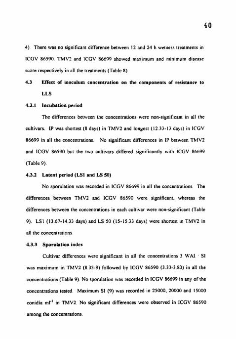
4.3.1 Incubation period
The differences between the concentrations were non-significant in all the
cultivars. IP was shortest (8 days) in TMV2 and longest (12 33-13 days) in ICGV
86699 in all the concentrations. No significant differences in 1P between ThiVZ
and ICGV 86590 bur the two cultivars differed significantly with ICGV 86699
(Table 9).
4.3.2 Latent period (LS1 and LS SO)
No sporulation was recorded in ICGV 86699 in all the concentrations The
differences between TMVZ and lCGV 86590 were significant, whereas the
differences between the concentrations in each cultivar were non-significant (Table
9). LS1 (13.67-14.33 days) and LS 50 (15-15.33 days) were shortest in TMV2 in
all the concentrations.
4.3.3 Sporulation index
Cultivar differences were significant in all the concentrations 3 WAI . S1
was maximum in TMV2 (8 33-9) followed by ICGV 86590 (3.33-3 83) in all the
concentrations (Table 9). No sporulation was recorded in ICGV 86699 in any of the
concentrations tested. Maximum SI (9) was recorded in 25000, 20000 and 15000
conidia ml" in TMV2. No significant differences were observed in ICGV 86590
among the concentrations.

4.3.4 Lesion number
Maximum number of lesions per leaf were observed at in 25000 and 20000
conidia ml'l in all the cultivars with no significant differences between the two
treatments. In TMV2 (7.7-22) and ICGV 86590 (3.3-1 1.2) lesion number was
minimum in 1000 - 7500 conidia ml" concentrations and significant differences
were not noticed both among the treatments and among the cultivars Lesion
number was highest in TMV2 (7.7-125.3) and lowest in ICGV 86699 (0 5-20 7) at
all the concentrations Cultivar differences were significant at 25000 and 20000
conidia mlT' concentrations but no significant differences were observed in 7500.
5000, 2500 and 1000 conidia ml.' concentrations. In 15000 and 10000 conidia n~l"
treatments, no significant differences were observed between TMVZ and ICGV
86590 but both differed significantly with ICGV 86699 (Table 10)
4.3.5 Lesion diameter
Lesion diameter was highest (5.88-7 mm) in TMV2 and lowest (0 67-1.5
mm) in ICGV 86699 in all the concentrations The differences between the
cultivars were significant in all the concentrations. Lesion diameter was maximum
in 25000, 20000, 15000 and 10000 conidia ml" concentrations with no significant
differences among them in all the cultivars. In 7500, 5000. 2500 and 1000 conidia
rnl" treatments, lesion diameter was minimum in all the cultivars with no signilkant
differences among the treatments(Tab1e 10)
4.3.6 Per cent necrotic area
PNA was maximum (3.5-27.5) in TMV2 and minimum (0.5-3) in ICGV
86699 in all the treatments 5 WAI (Table 10) In TMVZ and ICGV 86590, PYA
was maximum in 25000, 20000 and 15000 conidia ml" treatments with no
significant differences among them. In ICGV 86699, no significant differences

( s o 0 ~ 3 ~ Z I S I S W O b W S 0 J 0 0 3 8 1 PEE Zff i 6 % E8 L L C i i S 2.9 L 1 6 9 S Z l U S 1 LL 'SI U . W U o l l
1 9 9 9 ~ 0 0 0 0 Cii Z OS SL 8 0 L 1 9 9 8 S L S Z -1 I L 9 E L 1 6 L L V l L 9 9 1 S L L I(UWh6991119111
LS6L 99.9 W3u . )U I L E L L 96's 3- -mu(
9-0 Z L ~ O 6.9 w - z n w l n a IS0 '0)01 W3S (S0 '0 la1 H3S
I LSE S I C E9E E 9 E 7 6 V Q LC V 8E-V B E 6 9 2 9 %LL 9SW LLZ 9 Z L 9 L - U U . r u l W l
l Z L 1 EBO L 9 0 880 6 0 ZZL S L S l PL 1 1 8 T O 8 1 LZ L Z V L O 1 L Z Z L O 2 1 ( U ) 6 6 S 9 M 0 1 l l

iB!lptc 6 : rn-.ff~:cc of inc)crcIiirn concrn~tratirril O R ibis cor111~un~.~it \ ~ i ' ~ t " i i i # a n ~ ~ 1 0 j ~ t i . E c : I J . ~ ) ~ o iri f4't;F 36590.

were observed among the treatments and significantly lower (0 5-3) P X h was
observed than in TMV2 (3 5-27.5) and ICGV 86590 ( I 5- 17 5) ill all the treatnients
In all the cultivars, PNA gradually decreased with the decreasing incrculuni
concentration.
4.3.7 Defoliation
No defoliation was recorded in ICGV 86699 in all the treatments. T\1V2
and lCGV 86590 also did not show any defoliation in 7500. 5000. 2500 and 1000
conidia ml" treatments (Table 10). Defoliation was maximum and on par in 25000
(100) and 20000 (87 5) conidia ml" treatments followed by 15000 and 10000
conidia ml-I treatments in TMVZ. Similar trend was observed in lCGV 86590 also.
The differences between TMV2 and lCGV 86590 were not significant in all the
treatments
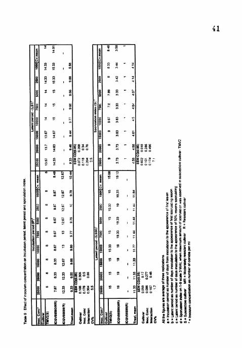
4.3.8 Disease severity
Disease score reached maximum (9) in TMV2 6 WAI in 25000 and 20000
conidia ml'l treatments (Table I I). Disease score was highest and on par in 25000
and 20000 conidia ml" treatments in all the ct~ltivars Lowest disease score (3) was
recorded in 7500, 5000, 2500 and 1000 conidia ml" treatments in TMV2 and ICGV
86590 with no sign~ficant differences among the treatments and between the
cultivars. Significant differences were seen between TMV2 and lCGV 86540 in
25000, 20000, 15000 and 10000 conidia ml". ICGV 86699 showed lowest disease
(2-3) in all the treatments (Plates 5,6 and 7).
4.4 Effect of the age of the plant on the components of resistance to LLS
4.4.1 Incubation period
IP was shortest (7.67-9.33 days) in TMVZ and longest (10-10.33 days) in
ICGV 86699 in all the treatments (Table 12). In TMV2 and lCGV 86590 IP was
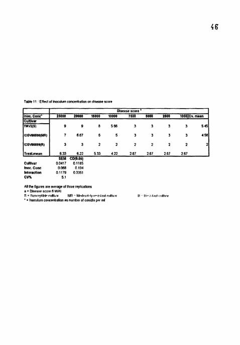
Table I 1 Effect of lnoculum concenlratlon on dlsease score
Inoc. Conc' Cultivar TMW(SI
Cultivar 00417 o11eS Inoc. Conc 0068 0194 Inletacllon 0 1179 0.3351 CVK 5 1
Disease score " 25000 20000 15000 10000 1600 5000 2600 10OO~Cv.mern
9 9 6 566 3 3 3 3 545
ICQVE669O(MRl
ICOVS6699(R)
Treal.mean
AW the ligures are average of lhree replicalions a = Disease score 6 WAI S = SUPI:PIIIIIIIP r ~ ~ l l l v , ~ ~ MI1 - Mn<l#,!,~tr.ly ~P..I..~.III~ rtdll~v.tl I1 I<,~~,I~,I~NIII u~llivat ' = Inoeulu~n coticelitlalion as number of conldia per ml
7 667 6 5 3 3 3 3 458
3 3 2 2 2 2 2 2 2
6.33 622 5.33 422 267 2.67 267 267 SEM CD(B.Obl
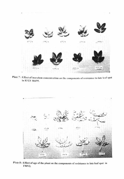
&'l:+te 8 8 @.ffta:ii.ct Of age of the iil:rat rnn ilre cirrnpojlcnn t ~ l . ~.eii\ianc.r: t t ! la/#* i~:ib.\prit in 'l'Pllb2.
shonest and on par in 10, 20, 30, 40 and 50 DAS treatments and lon$cst and on par
in 60, 70, 80 and 90 DAS treatments. Significant differences bet \ \ rr t~ ThlV2 slid
ICGV 86590 were noticed in all the treatments. TMV2 was differing si_unificantly
with ICGV 86699 in all the treatments whereas ICGV 86590 was 011 par with ICGV
86699 in 60, 70, 80 and 90 DAS treatments.
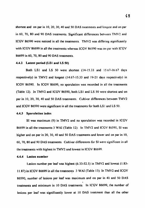
4.4.2 Latent period (LS1 and LS 50)
Both LSI and LS 50 were shonest (14-15.33 and 15 67-16 67 days
respectively) in TMV2 and longest (14.67-15.33 and 19-21 days respectively) in
ICGV 86590. In ICGV 86699, no sporulation was recorded in all the treatments
(Table 12). In TMV2 and ICGV 86590, both LSI and LS 50 were shortest and on
par in 10, 20, 30, 40 and 50 DAS treatments Cultivar differences between TMV2
and ICGV 86590 were significant in all the treatments for both LS I and LS 50.
4.4.3 Sporulation index
SI was maximum (9) in TMV2 and no sporulation was recorded in ICGV
86699 in all the treatments 3 WAI (Table 12). In TMVZ and ICGV 86590, SI was
higher and on par in 20, 30,40 and 50 DAS treatments and lower and on par in 10,
60, 70, 80 and 90 DAS treatments. Cultivar differences for SI were significant in all
the treatments with highest in TMVZ and lowest in ICGV 86699.
4.4.4 Lesion number
Lesion number per leaf was highest (6.33-52.5) in TMVZ and lowest (1.83-
11.87) in ICGV 86699 in all the treatments 3 WAI (Table 13). In TMVZ and ICGV
86590, number of lesions per leaf was masimum and on par in 40 and 50 DAS
treatments and minimum in 10 DAS treatments. In ICGV 86699, the number of
lesions per leaf was significantly lower at 10 DAS treatment than all the other
treatments which are on par. Cultivar differences were significant in all the
treatments except in 10 DAS treatment.
4.4.5 Lesion diameter
Lesion diameter was siynificantly lower (0.5-1.67) in lCGV 86699 in all the
treatments than TMV2 (3-5) and ICGV 86590 (3-4) 4 WAI (Table 13). In all the
cultivars lesion diameter was highest and on par in 20, 30, 4-0 and 50 DAS
treatments and lowest and on par in 10,60, 70,80 and 90 DAS treatments Cultivar
differences were significant in 20, 30, 40 and 50 DAS with mwjmum lesion
diameter (4.67-5) in TMV2.
4.4.6 Per cent necrotic area
PNA was highest (9.33-27.48) in TMV2 and lowest (0.5) in LCGV 86699 in
all the treatments 4 WAI (Table 13). In ICGV 86699, PNA in all the treatments
were on par and significantly lower than TMV2 and ICGV 86590. PNA was
maximum and on par in 20, 30, 40 and 50 DAS treatments in TMV2 and ICGV
86590. Cultivar differences between TMV2 and ICGV 86590 were significant in
all the treatments except in 70,80 and 90 DAS treatments.
4.4.7 Defoliation
No defoliation was recorded in all the treatments in lCGV 86699 and in 60,
70, 80 and 90 DAS treatments in TMV2 and ICGV 86590 at 4 WAI (Table 13).
Significant differences between TMV2 and ICGV 86590 were observed only at 20
DAS treatments. In W 2 , maximum defoliation (40.4 and 33.33) was observed in
20 and 30 DAS treatments and minimum defoliation (8 3) was observed 10 DAS
treatment. In ICGV 86590, no significant differences were observed among 10, 20,
30.40 and 50 DAS treatments (8.3-29.2).

lDV6 0 o o a ce FzL ZP 092 t.s 1 0 0 1 CCL 11.0 ECJ 11 11 FP.IL CL .-EL c r r bors-l
v'i1 r LL M a 190So 99110 WL U Z u w = w UK'O LC010 c%rt Ulsl .4 98910 5659'0 7SZ VZ60 -3 l ssob3 urn 1 9 a o h ~ v l s s
L L Z LL'Z t z s z a r c ~ C E a rc z r c LIZ a s 1 ere1 ~ 1 1 LZ'U =I LC cctz OVDL WI U-VMIL
1sac E c s c LI-I: 95.. s s 1st c (scic s r z ma Ira r w ww szs s rc Ira ccs I IS~MI]

4.4.8 Disease severity
In TMV? and ICGV 86590, disease score was highest and on par in 20, 30,
40 and 50 DAS treatments 4 WAI (Table 14). In ICGV 86699, disease score was
significantly lower (1-2.33) than TMV2 (3-5.67) and ICGV 86590 (3-4.67) in all
the treatments (Plates 8, 9 and 10). Differences between TMV2 and lCGV 86590
were significant in 20, 30, 40, 50 and 60 DAS treatments with highest disease score
in TMV2.
4.5 Surface studio on the inoculated leaves
Leaves from TMV2 and ICGV 86590 and ICGV 86699 which show
differential reactiuu to LLS (susceptible, moderately resistant and resistant
respectively) were inoculated with conidial suspension of LLS pathogen and
incubated at 25°C in incubator. Conidia began to germinate within 6-8 h afler
inoculation irrespective of the cultivar. After eight hours, there were significant
differences in the percentage of germination of conidia between the cultivars At
eight hours after inoculation (HAI), more than 50 per cent conidia germinated in
TMV2 and ICGV 86590. But in ICGV 86699 it took 24 h for 50 per cent wnidial
germination. Conidia usually germinated from the terminal cells, although other
cells occasionally produced germ tubes. At 8 HA1 only a few conidia showed two
germ tubes in TMV2 and ICGV 86590, but by 32 HAI, most of the conidia
produced two or even more germ tubes (Plate I I and 12). The average number of
germ tube s per conidium were comparitively less in ICGV 86699 than in TMV2
and ICGV 85690. The germ tube branching was observed rarely in all the cvs. Germ
tube branching was first sseen in TMV2 and ICGV 86590 at 48 HA1 while in ICGV
86699, it was observed at 72 HAI.
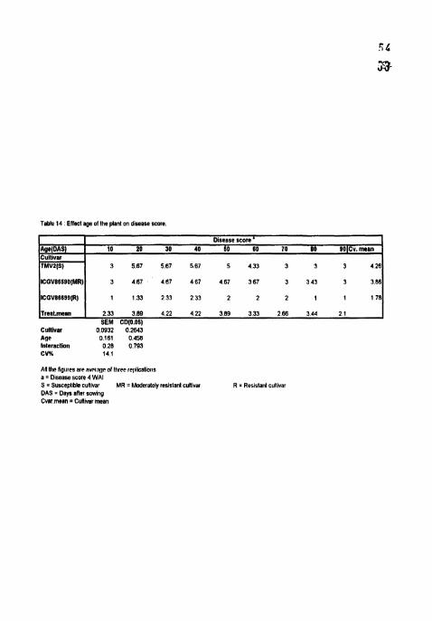
Table 14 Ellect age of Ihe plan1 on diseahe score
Treat.rnean 1 2.33 3.89 4.22 4 22 389 3 33 268 3 44 2 1 I SEM CD(0.06)
Culllvar 0.0932 0.2843 Age 0 I61 0.458 Interaction 0.28 0 793 CVX 14 1
Age(DAS) Cultlvar TMVZ(S)
ICGVS6500(MR)
All llrc liglltes arr RVOI;I~P 01 lllrrc rrplkallons a = O~sease score 4 WAI S = Susceplibls cullivar MR = Moderalely retislant cullivar R = Reslslant cull~var DAS = D a y after sowing Cvar.mean = Cultivar mean
Dlsrase score ' 10 20 30 40 60 80 70 80 SOICv,man
3 5 8 7 567 5 6 7 5 4 3 3 3 3 3 426
3 487 467 467 4 6 7 367 3 343 3 3 86
iJi8Fe : I,.I'fcet cif age rbl'rttc pl:int orb i h p ronitpurii.trt* of rttaiiiart~c~ t t j i:rtr icaf sped ira l d i s b 86609.

On TMV?, and ICGV 86590, which are susceptible and nloderately resistant
to LLS respectively, majority of germ tubes began to show attraction towards the
open stomata(Platel3) Usually only one germ tube or its branch from a conidium
resulted in penetration. Occasionally a germ tube grew besides or even over the
stoma without entering (Plate 12). No competition between the germ tubes was
observed and only one was found to enter through the stoma. Some germ tubes also
showed directional growth towards the epidermal radial cell walls. But no
penetrations were observed.
In TMVZ and ICGV 86590, penetration of the germ tube through stomata
was first observed 72 HA1 (Plates 14 and 15) and more number of penetrations were
observed 80 HAI. On the resistant cultivar ICGV 86699, the germ tubes of most of
the conidia apparently were not attracted towards the stomata. Most of the conidia
eventually lost their stainability and became transparent (Plate L6). Eventhough
infection was first observed 78 HA1 (Plate 17), the Frequency of penetrations was
very less and further delayed. The distal end of the germ tube enlarged to form an
irregularly shaped "appressorium-like" structure over the stornatal pore
Macroscopic symptoms were first observed on TMVZ and ICGV 86590 at
72 h after penetration (168 HAI). When observed under microscope, the m'inute
chlorotic spots included guard cells and some surrounding cells. The afTected cells
were discoloured and the shrinkage of the protoplasm was more obvious (Plates 18
and 19). In ICGV 86699, the first symptom was seen at 112 h after penetration
(191 HAI) and showed the same pattern as the other two cultivars (Plate 20).

CHAPTER V
DlSCUSSlON
In the present investigations, TMVZ, lCGV 86590 and ICGV 86699 remained
susceptible, moderately resistant and resistant in all the experiments Th1V2 is the
most susceptible variety with the shortest incubation period, latent period and uith
the highest sporulation index. lesion count, per cent necrotic area, lesion diameter.
defoliation and disease score over the range of temperatures, leaf wetness periods.
inoculum concentrations and different ages of the plants tested followed by ICGV
86590. lCGV 86699 is the most resistant cultivar with all the components of
resistance highly restricted The susceptibility of TMV2 is due to shortened
incubation period, latent period, increased number of lesions, size of the lesions which
results in increased per cent necrotic area, defoliation, finally leading to increased
disease severity. The resistance in ICGV 86699 is associated with smaller lesions,
longer latent period and reduced sporulation which finally resulted in reduced disease
severity.
In some experiments, the differences between the cultivars found to be non-
significant at certain treatments for incubation period and number of lesions per leaf.
But these two components were not very useful in differentiating the resistance of the
genotypes. Chiteka el a/.. (1988a) reported that incubation period was not a criteria
for isolating resistant genotypes but latent period, amount of sporulation and lesion
size were useful. Number of lesions per leaf is also of limited usehlness because it is
hiyhly influenced by environmental factors (Subrahmanyam el a1 , 1982, Walls el ul.,
1985 and Chiteka el a/., 1988a) But the differences for all the other components of
resistance were highly significant between the cultivars. Genotypic differences were
found to be significant for latent period (Chiteka el a/ . , 1988a), per cent necrotic area
6 1

(Subrahmanyam el a / . 1982, lroume and Knauft, 1987 and Chiteka c1 ol. 1988.1).
lesion diameter (Subrahmanyam e/ a / , 1982, Walls el a/.. 1985 and Chiteka rzr crl ,
1988a), spomlation index and disease score (Nevill, 198 1, Subrahmanyam rr ol.,
1982, Walls el a / , 1985 and Chiteka el a t , 1988a) There was a positive correlation
between per cent defoliation, lesion diameter and spomlation (Subrahrnanyam el ol ,
1982) and between lesion size, latent period and amount of spomlation and all with
per cent necrotic area (Chiteka et a / , 1988b) were reported
Success in efforts to develop high yielding cultivars with better resistance to
late leaf spot depends on identification of resistance that is expressed even when
environment favours disease increase (Shew et a/. 1988) The present investigations
were carried out to determine the temperature, duration of high humidity,
concentration of inoculum and age of the plant that favour infection of groundnut by
Phaeoisarropsrspersonda and to determine the factors favourable for post infection
development of the pathogen to determine if temperature, relative humidity, inoculum
concentration or age of the plant affect the expression of resistance to the LLS
pathogen in peanut The results obtained during the course of investigation are
discussed in this chapter
5.1 EFFECT OF TEhlPERATURE ON THE COMPONENTS OF RESISTANCE
The results indicate that the incubation period of LLS pathogen was the
shortest at 25°C followed by at 20 and 15°C in all the three cultivars But differences
between 20 and 25°C were slight No significant amount of disease was observed at
30 and 35°C and even the plants died subsequently This is because of gradual
decline and sharp inhibition of conidial germination (Sommartya and Beute, 1986)
and complete inhibition of infection and disease development (Shew el a1.,1988) at
higher temperatures (>2g°C) and also because of the continuous growing of the

6 3 inoculated plants at their respective treatment temperatures In the incubators 1111 the
end of the expenment which 1s hrghly unfavourable for the growth of the plant In the
present Investlyatlon, seventy of dlsease was max!murn at 25°C m all the three
cultlvars However, the effect of temperature on PhaeorsarroprsprsotIafa ~nfection
and drsease progression was hlghest between 15 and 20°C and sllght between 20 and
2S°C The nearly complete tnhba~on of leaf spot lnfectlon and development on all the
cultlvars at 30 and 35'C was unexpected, because plants In the field are nearly always
exposed to dally maxlmum temperatures of 30 - 35°C Conldta must sumve
exposures to hrgh temperatures and low humldlty that occur between deposltlon and
favourable lnfectton penods The nunlmum penod of exposure to hgh temperature
that lrreverslbly lnhrblts the post lnfect~on development of leaf spot IS not known, but
lesions on leaves that were transferred From 32°C to lower temperatures at the end of
the expenments (after 28 days exposure to 32°C) drd not resume development w t h n
two weeks of transfer (Shew er a1 1988) In controlled envlronrnent studres,
temperature >3OeC dunng penods of hgh relatrve humldlty had a greater effect on
reduclng spore productron than d ~ d the recommended rate of chlorothalo~ul applred at
7- 10 day Intervals (Labnnos and Nutter Jr , 1993)
Shew el a1 (1988) expected leaf spot seventy to be the highest when
temperatures near 24°C occur dunng long leaf wetness penods as the lnfecttoris
probably occur at nrght or In early momng when leaves are wet and temperatures are
cool Post lnfectlon development probably proceeds rapldly at warm temperatures,
slows dunng the hottest part of the day, and resumes In the wenlng as temperatures
decrease
lncubatlon penod was the shortest at 25°C and the longest at 15°C In all the
cultrvars However, there was no slgn~ficant dtfference between 20 and 25°C
(Fig.3.1). No significant difference was found between T M V ~ and ~ C G V 86590 for
IP at 2 5 " ~ . But there were highly significant differences for latent period which is the
most important component of resistance (Nevill, 1980). Chiteka rr a/. (1988a)
opined that incubation period did not appear to be a useful component for isolating
resistant genotypes. But they found highly significant differences for latent period.
Latent period (LSI and LS 50) was the shortest at 25°C and the longest at
15°C (Figs. 3.2 and 3.3). The longest latent periods occurred at the lowest
temperatures and shortest periods occurred between 209C and 30°C (Wadia and
Butter, 1994). Nevill (1981) observed a latent period of 14.6 days for
Phaeoisariopsispersorlala at 25'C on detached leaves of TMV2. Similar result (13.3
days) was obtained in the present investigations on TMVZ at 2SoC on whole plant.
There was no sporulation in resistant ICGV 86699 at all the temperature.
The number of lesions per leaf at 15°C in all the cultivars was not significantly
differed with that at 20 and 25°C in all the cultivars, but, the severity of disease was
low at 15°C because the lesions were generally small and poorly developed. Lesions
at 13°C were generally small and poorly developed and included many hypersensitive
specks, although the lesion number was large, disease severity was low (Butler el a/.
1994).
In all the cultivars at all the temperatures tested, the increase in number of
lesions continued for 20 or even more days. The increase in number of lesions per leaf
stopped 3 WAI in TMV2 at all the temperatures and at 20 and 25°C in ICGV 86599.
The increase was observed upto 6 WAI at 15°C in ICGV 86590 and at all the
temperatures in lCGV 86699. The number of lesions per leaf were highest in TMVZ
at 3 WAI followed by ICGV 86590 and ICGV 86699 at all the temperatures (Fig
3.5). Lannou and Blizoua Bi (1989) concluded that infection process continues for
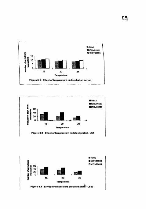
Figure 3.1 . Effect of temperature on incubation perlod - . . --. - -.
---- . - -. .-. . .- 81TMV2 . ICGVR55QO
60 UlCGV86699
z 15 20 25 Temperature
I Flgure 3.2 : Effcct nf lcrnpalnlllrr or1 l a t c ~ ~ t period - L S I
I 15 20 25 Temperature
I ngu. 3.3 : Effect a temperature on iatent p e r J - Ls.

more than six days. Butler CI 01. (1994) observed continuation of infection process as
long as 10 days. Alderman and Beute (1986) observed a continuing increasing i r ~ the
number of lesions for 20 days after inoculation with ('rrco~.v[>or~r ilril~./ridtc~ok~. The
stoppage of increase in number of lesions was earliest at 25°C in all the cultivars
because of the rapid progression of disease. As the disease progresses, the lesion size
will increase and often two or more lesions coalesce to give an appearance of single
lesion. So, the increase in number of lesions apparently stopped and even started
decreasing in TMV2 and ICGV 86590 3 WAI. But in ICGV 86699, the resistance is
associated with appearance of small non-sporulating lesions, hence the increase in
number of lesions was noticed even 6 WAI.
The increase in size of the lesion with disease progression lead to increased
per cent necrotic area. Per cent necrotic area was more at 20 and 25°C in all the
cultivars but it was the highest at 25" as some leaflets were already defoliated.
Defoliation was also highest at 25°C in all the cultivars followed by 20°C
(Fig.3.8). No defoliation was observed at 15OC in all the cultivars. Spomlation index
reached highest respective to the cultivar depending on its resistance behaviour at
25°C followed by 20 and 15'C (Fig 3.4). The disease score on 1-9 scale has given
conformation of highest amount of disease at 25°C and lowest at 15" in all the
cultivars (Fig.3.9).
Therefore it can be concluded that the severity and rapidity of disease
progression was increasing From 15 to 25°C irrespective of the resistance nature of
the host system. The effect of temperature on Pl~oeoisnrio~~.vis personala infection
and disease progression was highest between I5 to 20°C and the lowest between 20
to 25°C. Under controlled environmental conditions, Shew er a/. (1988) found that
infection by LLS pathogen was optimum when exposed to 20°C and at least 12 hrlday
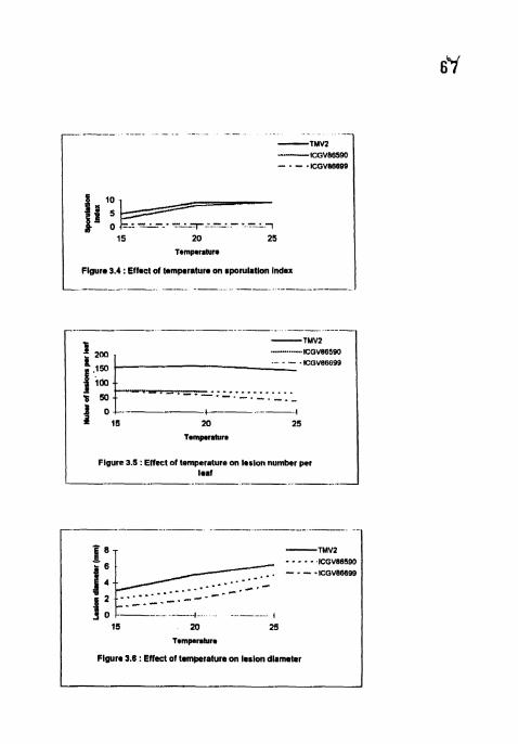
/ Figure 3.4 : Wmet of temperature on sporulation Index
Figure 3.5 : Effect of tempeiature on leslon number per Imaf
15 20 25
Tompcmlura
Figure 3.8 : Effect of temperature on levion dlameter
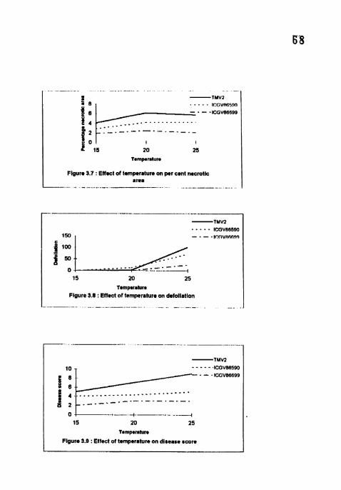
Figure 3.7 : Effect of temperature on per cent necrotic area
-- -....... ... . - - -- . .
15 20 25
T*mp*rmturm Figure 9.6 : Effect of temperature on defoilation
......... 15 20 25
tmmp*ratur*
Flgure 3.0 : Effect of temperature on disease score
duration of >93 per cent RIi for six days Similar results were reported with
temperature optima close to 20°C and minimum and maximum of about eight and
34"C, respectively by Butler c/ 01. (1994). In all the cultivars, there were no
significant differences between 20 and 25°C for incubation period, lesion number, per
cent necrotic area and sporulation index. But the differences were clear with the
lengthened latent period, smaller lesion size, reduced defoliation and disease score at
20°C, compared to 25°C. Eventhough the per cent necrotic area in 20 and 25°C were
on par 4 WAI, defoliation was more in 25°C at that point of time.
5.2 EFFECT OF HUMIDITY OH THE COMPONENTS OF RESIST.4NCE
High daily leaf wetness penoddrelative humidity is very much favourable for
the infection of the LLS pathogen. Incubation period was shortest in 16 h humidity
treatment in all the cultivars (Fig.4.1). Eventhough the incubation period did not
differ much between the high humidity treatments (16, 20 and 24 hr) for TMV2 and
ICGV 86590 and in between the two cultivars, the differences in the other
components of resistance play an important role in differentiating them. Chiteka el a/.
(1 988a) opined that IP was not a usehl component for isolating resistant genotypes
for P. persotlala. There was no difference for spore germination, incubation period
and number of lesions per leaf between Southern runner and Florunner, but Southern
runner has higher LS 50, less sporulation index and lesion diameter (Watson el ul.,
1997). Infection was delayed at low humidity treatments with longest 1P at 4 h
treatment in TMV2 and ICGV 86590. In ICGV 86699, no infection observed at 4 h
treatment. The relative humidity rate appears to be a limiting factor on lesion
development and influential for incubation period (Rapilley, 1983).
Latent period (LSI and LS SO) was also shortest in 16 h treatment followed
by 20, 24, 12 and 8 h treatments in TMV2 and lCGV 86590 (Figs.4.2 and 4.3). In

lCGV 86699, sporulation was greatly restricted at all the huniidity rrratmelits
Alderman and Nutter (1994) reported that in controlled environmental espe~iments. a
minimum of 4 hr relative humidity >95% per day was required for conidial production
and the highest number of conidia were produced when lesions were subjected to
daily periods of 16 or more hours of relative humidity >95% In the present
investigation, though symptoms were seen in 4 h treatment, no sporulation was
recorded even in the most susceptible cultivar TMV2, but sporulation was observed
in 8 h treatment. So, for conidial production, the minimum period of humidity
saturation conditions was probably in between 4-8 h.
Maximum number of lesions per leaf were observed in 16 h humidity
treatment in all the three cultivars (Fig.4.5). The differences in the number of lesions
per leaf among 4, 8, 12, 20 and 24 h treatments were not significant but the severity
of the disease at 20 and 24 h was more with higher lesion size and per cent necrotic
area. The number of lesions resulting from a fixed amount of inoculum was several
times greater when the leaves were exposed to long periods of humidity saturation
alternating with short dry periods (Butler el al. 1994). Lannou and Blizoua Bi (1989)
and Butler el a/. (1994) reported that number of lesions increased if leaves were
exposed to alternate wet and dty periods. Butler el a/. (1994) also found maximum
infection with daily wetness periods of 16 h. The results of the present inveaigation
confirm this as the number of lesions were higher at 16h treatment than other
treatments in all the cultivars. In contrary to this, Shew el a/. (1988) found maximum
lesion number with 24 h of RH per day (i.e., continuous wet period). Butler el a/.
(1994) explained this variation stating that, in high humidity chambers used by Shew
eta/ . (1988), the RH varied between 93 and 99% and although leaves were wetted
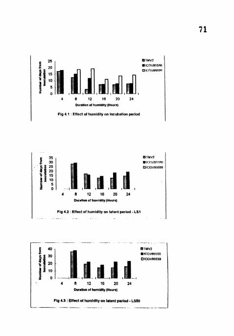
E z5 U l M V l
B 20 ICOVBII~'IO
g 15 OI<.C.V86C'19
t i f = 0
4 8 12 16 20 24
Ourallon of humldity (Hauls)
I Fig 4.r : Eflect of hurnidily on incubalion period I
E 35 IMV?
alf:(;vofi'3nfl
ICGV86609
4 8 12 16 20 24 Ouratton of humldlty (Hours)
I Fig 4.2 ' Effect of humidity on latent period . LS1 I
4 8 12 16 20 24
Duratlon of humldlly (Hours)
L Fig 4.3 Effect of humidly on latent period - LS60 - - - - - - - -- - -- - - - - -- - - -. - I
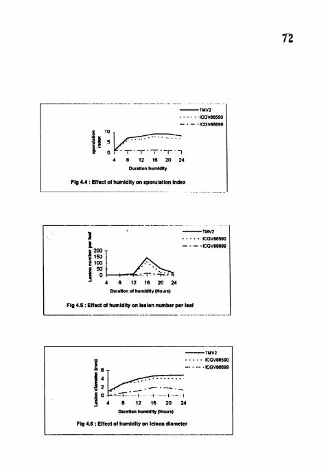
4 8 12 16 20 24
Duratlon humldlty
FIg 4.4 : Effect of humidity on sporulalion index -- -- .. . . - .
- -- - - - --- . .
1 -1MV2 . . . lCGVB6590
ICGV86699
50
"4 8 12 16 20 24 Duratlon of humldlty (Hours)
Fig 4.5 : Effect of humidity on lerlon number per leaf I- _ - -- - --- - . - . . - . -. .
Durallon humldlly (Hours)
I Flg 4.6 : Effect of humldlty on leison diameter
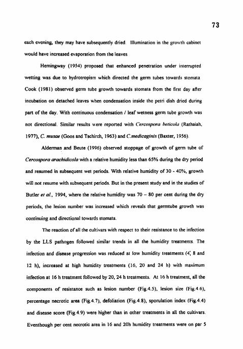
each evening, they may have subsequently dried llluminatio~i ill the griw-rl~ cabilret
would have increased evaporation from the leaves
Hemingway (1954) proposed that enhanced penetration under iliternlpted
wetting was due to hydrotropism which directed the germ tubes towards stomata
Cook (1981) observed serrn tube growth towards stomata from the first day aAer
incubation on detached leaves when condensation inside the petri dish dried during
part of the day. With continuous condensation / leaf wetness germ tube srowth was
not directional. Similar results were reported with C'rrcospom hcricoll~ (Rathaiah,
1977), C. musae (Goos and Tschirch, 1963) and C.nledicagitiis (Baxter, 1956).
Alderman and Beute (1996) observed stoppage of growth of germ tube of
Cercospora arachidicola with a relative humidity less than 65% during the dry period
and resumed in subsequent wet periods. With relative humidity of 30 - 40%, growth
will not resume with subsequent periods. But in the present study and in the studies of
Butler et a]., 1994, where the relative humidity was 70 - 80 per cent during the dry
periods, the lesion number was increased which reveals that germtube growth was
continuing and directional towards stomata.
The reaction of all the cultivars with respect to their resistance to the infection
by the LLS pathogen followed similar trends in all the humidity treatments. The
infection and disease progression was reduced at low humidity treatments (4; 8 and
12 h), increased at high humidity treatments (16, 20 and 24 h) with maximum
infection at 16 h treatment followed by 20, 24 h treatments. At 16 h treatment, all the
components of resistance such as lesion number (Fig.4.5), lesion size (Fiy.4 6).
percentage necrotic area (Fig.4.7), defoliation (Fiy.4.8), sporulation index (Fig.4.4)
and disease score (Fig.4.9) were higher than in other treatments in all the cultivars.
Eventhough per cent necrotic area in 16 and 20h humidity treatments were on par 5
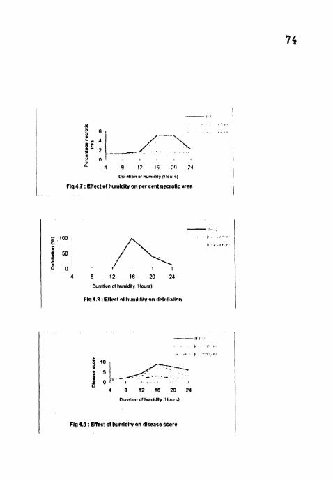
E n ! , , , I
.I 4 1: Ili :f) :'A
Ollliltloll of hurnldlly (Ifolrrs)
Fig 4.7 : Cllect of llutllidity on per cent nectolic area
4 8 12 18 20 24
Durntion of humldlty (Hours)
Flq I R : Ellrrl nl IIIIIIIIIIII~ ntl 11~~ l~~ l l~ l ln t1
I l l ,
I , , , a , , , ,
. - I, . . / ,.,.. 6 t '"c, ; 0
4 8 12 (8 20 24
Dtltntlorn a1 l!tlnlldlty Illotsr)
Fig 4.9 : Effect of li\lmldily on disease score

WAI, percentage of defoliation was more in 16h treatment at that point of time
Therefore, it can be concluded that the severity and rapidity of disease progression
was increasing from 4 to 16 h humidity duration irrespective of the resistance nature
of the host system. The effect of humidity on Phoeoiriop.si.spr.sotrori~ infection and
disease progression was highest between 4 to 16 h and slight between 16 to 24 h with
maximum disease at 16 h in all the cultivars. Eventhough no significant differences
observed for incubation period, sporulation index and lesion diameter between 16 and
20h treatments in TMVZ and ICGV 86590, the severity disease was more at 16h
treatment with higher number of lesions per leaf, pre cent necrotic area and
defoliation. This clearly indicates that intermittent wetness with 16h wet and 8h dry
periods per day f o ~ six days is highly favourable for disease development
5.3 EFFECT OF MOCULUM CONCENTRATION ON THE COMPONENTS OF RESISTANCE
lnoculum concentration has no effect on incubation period, latent period and
sporulation index in all the three cultivars. However, the inoculum concentration has
direct effect on the number of lesions per leaf, per cent necrotic area, lesion diameter,
defoliation and disease score in all the three cultivars. Defoliation and lesion number
were directly related to inoculum level (Nevill, 1981) and lesion number will increase
with increasing spore concentrations (Shew and Beute, 1984). Similar results were
obtained in the present investigation
Incubation periods in all the inoculum levels were on par in all the cultivars
(Fig.S.l). No significant differences were observed between TMV2 and ICGV
86590 for incubation period but both differed significantly with ICGV 86699.
Similarly no significant differences were observed between different concentrations
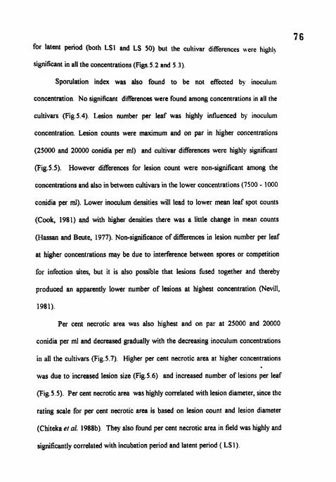
for latent period (both LSI and LS 50) but the cultivar differences were higlll)
significant in all the concentrations (Figs.5.2 and 5.3).
Sporulation index was also found to be not effected by inoculum
concentration. No significant differences were found among concentrations in all the
cultivars (Fig.5.4). Lesion number per leaf was highly influenced by inoculum
concentration. Lesion counts were maximum and on par in higher concentrations
(25000 and 20000 conidia per ml) and cultivar differences were highly significant
(Fig.5.5). However differences for lesion count were non-significant anlong the
concentrations and also in between cultivars in the lower concentrations (7500 - 1000
conidia per ml). Lower inoculum densities will lead to lower mean leaf spot counts
(Cook, 1981) and with higher densities there was a little change in mean counts
(Hassan and Beute, 1977). Non-significance of differences in lesion number per leaf
at higher concentrations may be due to interference between spores or competition
for infection sites, but it is also possible that lesions hsed together and thereby
produced an apparently lower number of lesions at highest concentration (Nevill,
1981).
Per cent necrotic area was also highest and on par at 25000 and 20000
conidia per ml and decreased gradually with the decreasing inoculum concentrations
in all the cultivars (Fig.5.7). Higher per cent necrotic area at higher concentrations
was due to increased lesion size (Fig.5.6) and increased number of lesions per leaf
(Fig.5.5). Per cent necrotic area was highly correlated with lesion diameter, since the
rating scale for per cent necrotic area is based on lesion count and lesion diameter
(Chiteka el al. 1988b). They also found per cent necrotic area in field was highly and
significantly correlated with incubation period and latent period ( LSI)
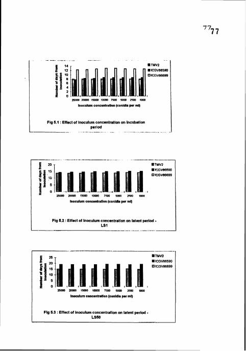
- . m lhXlO 15ca I m m 7500 m 2m lorn
lnoculum concanballen (conldla per ml)
Fig 5.1 :Effect of inoculum concentration on incuballon period
-. . -. -. - - - -- - - - .
lnoculum concanhallan (conidla per ml)
/ Fig 5,) :Effect of lnoculvn concentralion on latent period LS1
-- - -.-. 8TMV2 8 ICGV88590 OICGVBBBQQ
2 m 2 0 0 ~ I H ~ W 1 0 2 ~ 7m m m 1000
lnocuturn cobcentratIan (conldla pmr mi)
Fig 5.3 : Effect of inoculum concentration on latent period - LSSO

I Fig 6.4 : E I M d lnoculurn concentration on wrulallon Index
I Fig 8.5 :Effect of lnoculurn concenlrrtlon on larlon number per leaf - - - - -- - - - -- - - - - - -
Flg 5.8 : Effectt of inoculum concmntralion on lerlon dlameter

In the Present investigation although lesion diameter does not \.ay anlong
lower concentrations (7500 - 1000 conidia per ml), per cent necrotic area was
decreased gradually. This may be due to decrease in lesion number from 7500
through 1000 conidia per ml concentrations. Defoliation was rnaximunr in higher
concentrations (25000 and 20000 conidia per ml) in fMV2 and ICGV 86590
(Fig.5.8). No defoliation was recorded in ICGV 86699 in all the concentrations and
in 7500 - 1000 conidia per ml concentrations in TMV2 and ICGV 86590 hlaximum
disease score 1 plant appearance score (PAS) was recorded in 25000 and 20000
conidia per ml concentrations and lowest and on par in 7500 - 1000 conidia per ml
concentrations in all the cultivars (Fig.5.9).
Except for incubation period, latent period and sporulation index, all the other
components of resistance decreased gradually with the decreasing inoculum
concentrations with highest amount of disease in higher concentrations (25000 and
20000 conidia per ml) and lowest and on par in 7500 - 1000 conidia per ml
concentrations in all the cultivars Hence the optimum concentration to cause
maximum amount of disease was 20000 conidia per ml as there was no further
significant increase in the severity of disease with 25000 conidia per ml concentration.
5.4 EFFECT OF ACE OF THE PLANT ON THE COMPONENTS OF RESISTANCE
In susceptible cultivar - TMV2 and moderately resistant cultivar - ICGV
86590 significant differences were observed in between the different age treatments.
But in lCGV 86699, no significant interaction between plant age and components of
resistance was observed. Irrespective of the resistance reaction of the host system,
disease severity was higher in 20 - 50 day old plants. The disease severity was highly
reduced in 10,80 and 90 day old plants in case of TMV2 and ICGV 86590.
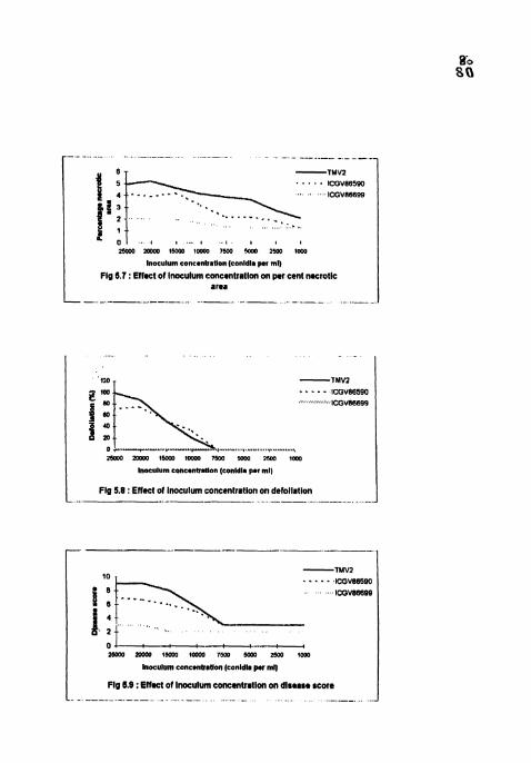
. . . . ICGVB6580 1 / p-:G:866m '.. . . . - _ , , . .
25000 2OWO 1W tom0 7Mo Urn 2Mo ICCO
Inaculum conc*nbatlon leonldla per ml) Fig 6.7 : Effmct of lnoculum concmntratlon on per cent necrotic
area
-1MV2 -..". ICCV86590 . .,..... lCGV86689
?wm rmoo 1m lom0 7m soar Ym la0
Inoculum cons~nbatlon (conldl8 par ml)
Fig 5.8 : Effect of inoculum concentration on defollatlon
0 l. : : : ---. mm ZOWO 1x130 tom0 7x4 H)W lSOO two
ln=ulum concsnbatlon (conldla per ml)
Fig 1.8 : Elfmct of lnoculum concentrmtlon on dlmaso score - - - - - - - - - - - - - - - - -
I FIg 6.t : Effect of age of the plant on Inrubailon period --
I Flg 8.2 : Effect of age of the plant on latent period - LS1
L.. . .. . . . . . . . . . . . - - . - ..
0 TMV2
8 ICGV86590
UICGV86699
10 20 30 40 50 60 70 80 90
Asc of tho plant IDAs)
L Fig 6.3 : Effect of age of lhe plant on UM p n b d - LSEO -" -- - -- - - -. -- ---
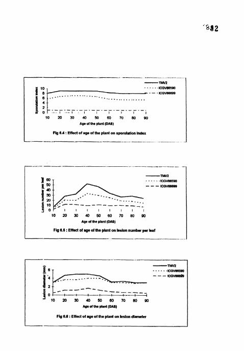
I 10 20 30 40 50 BO 70 80 90
Ape of tne plant IDAS)
I Fig 6.4 : Ewefed of age ofthe plant on rporulalion index
-- - --- - -
-TMY2 . . . . . . ICOV8859O - - - ICOVMIBW
10 20 30 40 M 60 70 80 90 Age ofthe plant IDA81
Fig 9.6 : Ertrct of age of the plant on Iaslon number p r leaf
' 1 0 20 30 40 M 80 70 80 W Age Mthe plant (OW)
Fig 6.4 : Enect of age of the plant on lesion diametw

Incubation period was shortest and on par in 10 - 50 day old plants in TMV2
and ICGV 86590 (Fig.6.1). There was no interaction between incubation period and
plant age in ICGV 86699. Similar results like that of 1P were observed for latent
period (both LSl and LS 50) also in TMVZ and ICGV 86590 (Figs.6.2 and 6.3).
Sporulation of lesions was highly reduced in all the ages in lCGV 86699. Spomlation
index was highest and on par in 20 - 50 day old plants (Fig.6.4) in TMV2 and ICGV
86590. Subrahmanyam er al. (1982) reported that spomlation score was a consistent
component for rating genotypes and they observed no interaction between
spo~lat ion index and plant age (30 and 50 DAS).
Lesion tiumber was significantly high and on par in 40 and 50 day old plants
and lowest in 10 day old plants in TMVZ and ICGV 86590. ICGV 86699 showed no
interaction between plant age and lesion number but lesion number in 10 day old
plants was highly reduced (Fig.6.5). Subrahmanyam er a/. (1982) reported high
infection frequency in 50 day old plants than in 30 day old in TMV2. Lesion count
was not a consistent measure of resistance because of its high environmental
variations (Subrahrnanyam el al,. 1982; Walls el a.,l 1985 and Chiteka el a/., 1988a).
Both lesion diameter (Fig.6.6) and PNA (Fig.6.7) were higher in 20 - 50 days
old plants in all the cultivars. Eventhough lesion number was higher in 40 and 50 day
old plants, lesion diameter and per cent necrotic area were on par with 20 and 30 day
old plants in W 2 and ICGV 86590. This may be due to comparatively smaller
size of leaves in 20 and 30 day old plants. Subrahmanyam el al., (1982) observed
higher per cent necrotic area in 30 day old plants than in 50 day old plants in TMV2
in glasshouse. Shailta (1960) and Cook (1981) found that the youngest (4 week) and
oldest (10 week) leaves of the plants were leest susceptible to infection. However,
Cook (1981) opined that this effect appears to be related to leaf size rather than leaf
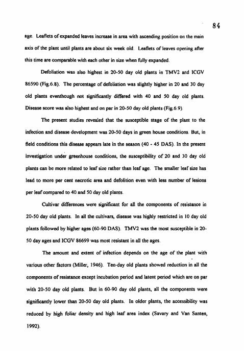
8 4 age. Leaflets of expanded leaves increase in area with ascending position OII the main
axis of the plant until plants are about six week old. Leaflets of leaves opening after
this time are comparable with each other in size when hlly expanded.
Defoliation was also highest in 20-50 day old plants in TMV2 and ICGV
86590 (Fig.6.8). The percentage of defoliation was slightly higher in 20 and 30 day
old plants eventhough not significantly differed with 40 and 50 day old plants.
Disease score was also highest and on par in 20-50 day old plants (Fig.6.9)
The present studies revealed that the susceptible stage of the plant to the
infection and disease development was 20-50 days in green house conditions. But, in
field conditions this disease appears late in the season (40 - 45 DAS). In the present
investigation under greenhouse conditions, the susceptibility of 20 and 30 day old
plants can be more related to leaf size rather than leaf age. The smaller leaf size has
lead to more per cent necrotic area and defolition even with less number of lesions
per leaf compared to 40 and 50 day old plants.
Cultivar differences were significant for all the components of resistance in
20-50 day old plants. In all the cultivars, disease was highly restricted in 10 day old
plants followed by higher ages (60-90 DAS). TMV2 was the most susceptible in 20-
50 day ages and ICGV 86699 was most resistant in all the ages
The amount and extent of infection depends on the age of the plant with
various other factors (Miller, 1946). Ten-day old plants showed reduction in all the
components of resistance except incubation period and latent period which are on par
with 20-50 day old plants. But in 60-90 day old plants, all the components were
significantly lower than 20-50 day old plants. In older plants, the accessibility was
reduced by high foliar density and high leaf area index (Savary and Van Santen,
1992).
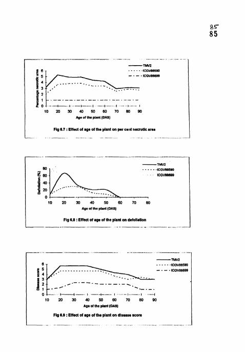
-. .. - ....... .... . ..... ......-.
-m ...... ICGVLlQDO
- . - - ICOV86689
. .....-
.... .- .............................................. --.. -1-., --+-.( ..... .I -- +.- . I
10 20 50 40 50 60 70 80 90 Age of the plant (DASJ
Flg 8.7 : Effect of age of the plant on per cent necrotic ares
10 20 50 40 50 80 70 80 Age ofthc plant (DAS)
Fig 6.8 : Effect of age of the plant on defoliallon
/-------- I.I-.' .. -'."...-. ...... 1 .-I
1 0 2 0 J O 4 0 5 0 6 0 7 0 8 0 9 0 A ~ O of the plant (DM)
Fig 8.S : Effect of age of the plant on disease score
8 6 It was suggested that, in the variation of susceptibility with leaf age and
development stage of a groundnut cultivar, more factors involve than just leaf
wettability. Infection efficiency decreases with increasing development and aye of the
plant as reported in several other host pathogen systems (Schein, 1965; Aust cr 01..
1980 and Savary, 1987). Similar results were reported for latent period ( Parlevliet,
1975 and Savary,1987) and sporulation intensity (Tomerlin et a/., 1983).
Inspite of the diversity among pathosystems, several authors (Popular, 1978;
Zadoks and Schein, 1979 and Vanderplank, 1982) hve suggested generalizations to
account for the variation in the pathological interactions between hngi and ageing
plants. The most general hypothesis for a biotrophic pathogen is, perhaps, that the
younger and healthrer the host tissues, the easier their recognition and use as a
convenient habitat for faster and more intense growth and multiplication. This has led
Zadoks and Schein (1979) to suggest that partial resistance against biotrophic fungi
would generally increase with age and development.
5.5 SURFACE STUDIES ON THE INOCULATED CROUNDNUT LEAVES
The surface studies of the susceptible (TMVZ), moderately resistant (ICGV
86590) and resistant (ICGV 86699) cultivars after inoculation revealed the reaction
of conidia on the leaves. No significant differences observed on the leaves of TMV2
and ICGV 86590. The pathogen reacted more or less similar on both these hosts.
But in ICGV 86699, the difference in the reaction of conidia on the leaves was
differing with the susceptible and moderately resistant cultivars.
The spores of Phaeoisariopsis personata germinated within 6-8 hours after
inoculation (HAI) irrespective of the resistance. In TMV2 and ICGV 86590, more
than 50 per cent of the conidia germinated within 8 HA1 but in ICGV 86699, it took
more than 20 hours. Eventhough the percentage germination of the conidia has no
effect on the resistance, in ICGV 86699 most of the germinated conidia eventually
lost their stainability, became transparent and disintegrated. No significant differences
in germination of conidia were observed on leaves of various peanut cultivars (Cook
1981; Abdou, 1974). The number of germ tubes per conidia did not play any role in
reduced infection of resistant cultivar because generally only one germ tube entered
through the stomata.
The delayed and reduced number of penetrations through stomata and late
appearance of symptoms in the resistant cultivar can be effectively explained by
Mayee and Suryawanshi (1995) that penetration of germ tube through stomata will be
followed by accumulation of intracellular hypha in the cells surrounding the infection
site. The time required for accumulation of such hyphae was longer in resistant
cultivar compared to the susceptible cultivar. This will subsequently lead to delay in
the occlusion of the intercellular spaces, thickening of palisade cells and formation of
conidiophores in the stroma.
The delayed and defective germination and penetration of conidia ill resistant
cultivar lead to reduced infection. However the resistance to LLS pathogen is of
partial type (Nevill, 1981; McDonald, 1985) and is characterized by delayed
incubation period, latent period, reduced spomlation, necrotic leaf area and less
defoliation. In ICGV 86699, all the components of resistance play an important role
in imparting partial resistance. The expression of resistance in lCGV 86699 was
associated with appearance of many small, non-spomlating lesions. These characters
appear to be the consequence of slow invasion of the leaf tissues by the pathogen.
The initial symptoms of LLS infection in susceptible cultivar were coagulation
and shrinkage of protoplasm of the cells surrounding the infection site, disappearance
of chloroplasts, swelling and destruction of nuclei and ultimately the cells became
shrunken and filled with dark brown material. Kaur and Dhillon (1988) noted
shrunken epidermal and mesophyll cells and damage to protoplast in LLS infection.
The reduced number of infections in the resistant cultivar can be explained by the
anatomical differences between the resistant and susceptible cultivars. In the
susceptible cultivar the stomata will be wider than resistant cultivar and the slow rate
of pathogen growth in the resistant tissues is due to the presence of thick palisade and
spongy tissue (Bera et al., 1997). Mayee and Suryawanshi (1995) found thickening of
palisade cells and production of zone of smaller cells around the infection site which
can be related td small size of spot and less production of mycelial mass res;lting in
low sporulation. Basra et at., (1985) observed thicker epidermis, smaller stomata and
compact palisade in resistant leaves which accounted for fewer penetrations in
resistant cultivar. However Cook (1981) reported that variations in stomatal density
and stomatal length were not related to resistance to infection. In the moderately
resistant cultivar (ICGV 86590), the germination of conidia was similar to susceptible
cultivar (TMVZ). There were not much difference observed betweeti these two
cultivars. All the components of resistance were delayed than susceptible cultivar but
never restricted. But both these cultivars significantly differed with the resistant
cultivar (ICGV 86699). The delayed and defective germination of conidia. reduced
number of penetrations, slower rate of invasion of host cells in the resistant cultivar
can be highly correlated with late appearance of symptoms.
The difference in the level of minerals (Sindhan and Parashar, 1996; Jagalan
and Sindhan, 1988; and Mogle and Mayee, 1981), the histochemical changes that
takes place after infection (Kaur and Dhillon, 1988), the anatomical differences (Bera
et al., 1997; Mayee and Suryawanshi, 1995; and Basra el al., 1995) between the
resistant and susceptible cultivars play an important role in imparting panial resistance
along with the defective germination of the conidia, delayed and reduced number of
penetrations/infections and slower rate of pathogen growth in resistant cultivar.
In conclusion, the studies on the effect of temperature, humidity, inoculum
concentration and age of the plant provide the basis for understanding how these
factors affect various components of resistance in the host system The late
appearance of symptoms which impart partial resistance in addition to the other
components of resistance in the resistant cultivar can be related to defective
germination and delayed penetration of the host tissue by the conidia of the pathogen
from the present study. Though it is an established fact that the anatomical characters
and the histochemical changes take place during post infection development in the
resistant cultivar which result in resistance reaction, more concentrated and valiant
effort by the future researchers on the exogenous and endogenous factors which play
an important role in the spore germination and penetration of Phueoisuriopsis
Ixrscvioto on resistant cultivar can add on to the important results emerged out o f tllr 9 0
present study.

SUMMARY
CHAPTER V I
SUMMARY
Studies on the effect of temperature, humidity, inoculum concentration and
age of the plant on the components of resistance to late leaf spot disease of
groundnut were camed out. Histopathological investigations were also carried out
to further quantify the components of resistance. All these studies were conducted
at the International Crops Research Institute for Semi-Arid Tropics (ICRISAT),
Patanchem-502 324, Andhra Pradesh, India The results are summarized below.
In all, the studies, TMV2 and ICGV 86699 were the most susceptible and
resistant cultivars respectively. ICGV 86590 was found to be moderately resistant.
Resistance in ICGV 86699 was expressed by prolonged incubation period, absence
of sporulating lesions (i.e. no latent period), reduced lesion numbw, lesion size,
sporulation index, per cent necrotic area, defoliation and disease score.
Over the range of temperatures studied, 25°C was the most favourable for
infection and disease development in all the three cultivars. However, the effect of
temperature on Phaeoisariopsis persotrala infection and disease development was
the highest between 15 to 20°C and slight between 20 to 25°C. No significant
amount of disease was observed at 30 and 3%
The maximum disease severity was observed in 16 h humidity treatment in
all the three cultivars. The infection and disease development were reduced in low
humidity treatments (4, 8 and 12 h) and higher in high humidity treatments (16,20
and 24 h) with the maximum in 16 h treatment. Althouyh the lesion number did not
vary much between low humidity treatments (9, 8 and 12 h) and 20,24 h treatments,
the differences were clear for all the other components of resistance. 9 1
The optimum concentration of inoculum to cause the highest amount of
disease was 20000 conidia per ml as there was no difference in the severity of
disease with hrther higher concentrations (25000 conidia ml") in all the three
cultivars. Concentration of inoculum has no effect on incubation period, lptent
period and sporulation index Disease severity was reduced in lower concentration
treatments (7500 - 1000 conidia mrl) and no significant differences were observed
among the treatments for all the components of resistance Cultivar differences
were not significant for lesion number in all lower concentration treatments The
higher concentrations (25000 and 20000 conidia ml") significantly differed with
immediate lower concentrations (15000 and 10000 conidia ml'l) for lesion number,
defoliation and disease score
Twenty to fitly day old plants were found to be the most susceptible to LLS
in all the cultivars. However, the interaction between plant age and LLS infection
and disease development was not s i i c a n t in LCGV 86699. Cultivac differences
were also significant for all the components of resistance in 20 - 50 day old plants
Ten day old plants were found to be highly resistant followed by higher ages (60 - 90 DAS) in all the cultivars
Histopathological investigations revealed that defective germination,
delayed penetration, slow invasion of host tissues lead to prolonged!lengthened
incubation period and reduced severity of disease in resistant cultivar - LCGV
86699. The differences between TMV2 (susceptible) and ICGV 86590 (moderately
resistant) were not large.
LITERATURE CITED
LITERATURE CITED
Abdou Y A M, Gregory W C and Cooper W E 1974 Sources and nature of resistance to Cercospora arachidicola and ('ercospriclrr~trr prsotrarttttl in Arachis species. Peanut Science 1 : 6- 1 1 .
Alderman S C and Beute M K 1986 Influence of temperature and moisture on germination and germ tube elongation of Cercr~spora arachidicola. Phytopathology 76 : 715 - 719.
Alderman S C and Nutter F W Jr. 1994 Effect of temperature and relative humidity on development of Cerco.sporiJirtm persor~atrmr on peanut in Georgia. Plant Disease 78 : 690 - 694.
*Aust H J, Bashi E and Rotem 1 1980 Flexibility of plant pathogens in exploiting ecological and biotic conditions in the development of epidemics. In : Palti J and Kranz J (Eds), comparative epidemiology. A tool for better disease management. Pudoc, Wageningen . p. 46 - 56.
Basra R K, Kaur S and Dhillon M 1985 Anatomical and biochemical studies of the resistance and susceptibility of the groundnut varieties to cercospora leaf spot. Annals of Biology 1 : 7-12.
Baxter J W 1956 Cercospora black stern of alfalfa. Phytopathology 46 : 396-400.
Bera G C, Ghosh S K and Das P K 1997 Foliage characters of Cercospora infected leaves in susceptible and resistantttolerant genotypes Annals of Agricultural Research 18 (2) : 188- 193.
Bledsoe R A, Hanis H C and Tisdale W D 1946 1 Leaf spot of peanut associated with magnesium deficiency. Plant Physiology 21 . 237-240.
Butler D R 1990 Weather requirement for infection by late leaf spot in groundnut Proceedings of the Fourth Regional Groundnut Workshop for Southern Africa, ICRISAT, Patanchem, Andhra Pradesh-502 324, India.
Butler D R, Wadia K D R and Jadhav D R 1994 Effect of leaf wetness and temperature on late leaf spot infection of groundnut. Plant Pathology 43 : 112-120.
*Carnacho de Torres F F and Subero M L J 1993 (Institute de Botanica Agricola, Seccion Anatomia Vegetil, Facultad de Agronomia, Universidad Central de Venezuela, Apdo. 4579, Maracay 2101, Venezuele.) Influence of the age of three peanut cultivars on early leaf spot symptoms.. ( Es). Revista de la Facultad de Agronomia, Universidad Central de Venezuele 19(1) : 9-21
9 G Chiteka Z A, Gorbet D W. KnauR D A, Shokes F M. Kucharek T ti and Norden A
J 1987 ( Univ. Florida, Gainsville, FL 3261 1, USA.) Components of resistance to late leaf spot in peanut ( Arch is hylx)gia*ir L.). . (En ) Proceedings. American Peanut Research and Education Society 19 : 25.
Chiteka Z A, Gorbet D N Knaufl D A Shokes F M and Kucharek T A 1988 b 11. Correlations among components and their significance in breeding for resistance. Peanut Science 15 : 76-81
Chiteka Z A, Gorbet D W Knauft D A Shokes F M and Kucharek T A 1988 a Components of resistance to late leaf spot in peanut I . Levels and variability - implications for selection. Peanut Science 15 : 25-30.
Cook M 1981 Susceptibility of peanut leaves to Cercosporidirrnr persoriarrtm. Phytopathology 7 1 : 787-791.
D'cruz R and Upadhyaya B R 1961 Stem and leaf anatomy in Arnchis Indian Oilseeds Journal 5 : 239-244.
Feakin S D (Ed.) 1973 Pest control in groundnuts. PANS Mannual No.2 Centre for Overseas Pest Research, London. pp. 197.
Foster D J, Wynne J C and Buete M K 1980 Evaluation of detached leaf culture for screening peanuts for leafspot resistance. Peanut Science 7 : 98-100.
*Garren K H and Jackson C R 1973 Peanut disease. ln Peanuts - culture and uses. APREA Stone Printing Co., Roanoke, VA. pp.429-494.
Ghuges S S, Mayee C D and Godbole G N 1981 Assessment of losses in peanut due to rust and tikka leafspots. Indian Phytopathology 34 : 179-182.
Gibbons R W and Bailey B E 1967 Resistance to Cercospora arnchrdrcola in some species of Arachis. Rhodesia, Zambia, Malawi Journal of Agriculture 5 : 57- 59.
Goos R D and Tschirch M 1963 Green house studes on the cercospora leaf spot of banana. Transactions ofBritish Mycological Society 46 : 321 - 330.
Guzman N J 1964 Nature of partial resistance of certain clones of three Solattttm species to Phyrophrhora itfeslatrs. Phytopatholoyy 54 : 1398-1404:
Habgood R M 1972 Resistance to Rhy,?cospori~rm srcalis in the winter barley cultivar-Vulcan. Annals of applied biology 72 : 265-271.
Hassan R M and Beute M K 1977 Evaluation of resistance to cercospora leafspot in peanut germplasm potentially useful in a breeding programme. Peanut Science 4 : 78-83.
*Hemingway J S 1954 Cercospora leaf spots of groundnuts in Tanganyika. E. Afr. Agric. J. 19 : 263-267.
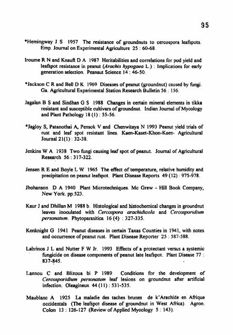
*Hemingway J S 1957 The resistance of groundnuts to cercospora leafspots Emp. Journal on Experimental Agriculture 25 : 60-68.
lroume R N and Knaufi D A 1987 Heritabilities and correlations for pod yield and leafspot resistance in peanut (Arachis hypgaea L.) : Implications for early generation selection. Peanaut Science 14 : 46-50.
*Jackson C R and Bell D K 1969 Diseases of peanut (groundnut) caused by fungi. Ga. Agricultural Experimental Station Research Bulletin 56 : 156.
Jagalan B S and Sindhan G S 1988 Changes in certain mineral elements in tikka resistant and susceptible cultivars of groundnut, Indian Journal of Mycology and Plant Pathology 18 (1) : 55-56.
*Jaglay S, Patanothai A, Pensok V and Chenwitaya N 1993 Peanut yield trials of rust and leaf spot resistant lines. Kaen-Kaset-Khon-Kaen- Agricultural Journal 21(1) : 32-38.
Jenkins W A 1938 Two fungi causing leaf spot of peanut. Journal of Agricultural Research 56 : 31 7-322.
Jensen R E and Boyle L W 1965 The effect of temperature, relative humidity and precipitation on peanut leafspot. Plant Disease Reports 49 (12) : 975-978.
Jhohanson D A 1940 Plant Microtechniques. Mc Grew - Hill Book Company, New York. pp.523.
Kaur J and Dhillan M 1988 b. Histological and histochemical changes in groundnut leaves inoculated with Cercosmra arachidicola and Cercosporidirrm personatrrm. Phytoparasitica 16<4) : 327-335.
Kenknight G 1941 Peanut diseases in certain Taxas Counties in 1941, with notes and occurrence of peanut rust. Plant Disease Reporter 25 . 587-588.
Labrinos J L and Nutter F W Jr. 1993 Effects of a protectant versus a systemic ' fungicide on disease components of peanut late leafspot. Plant Disease 77 .
837-845.
Lannou C and Blizoua bi P 1989 Conditions for the development of Cercosporidium personaium leaf lesions on groundnut after artificial infection. Oleagineux 44 (1 1) : 53 1-535.
Maublanc A 1925 La maladie des taches brunes de k'kachide en Afrique occidentals (The leafspot disease of groundnut in West Africa). Agron. Colon 13 : 126-1 27 (Review of Applied Mycology 5 143).

Mayee C D and Suryawanshi A P 1995 Structural defence ~necl~anisnls in groundnut to late leaf spot pathogen, Indian Phytopatholog!. 48 (2) . 160- 165.
'Mazzani B, Allievi J and Bravo P 1972 Relacio'n entre la incidencia de nlanchas foliares por Cercvspvra spp. Y algunos caracter'istics varietales del rnani. Agron. Trop. (Mara cay) 22 : 119-1 32.
McDonald ,D Subrahmanyam Gibbons R W and Smith D H 1985 Early and late leafspots of groundnut. Information Bulletin No.2 1, ICRISAT, Paranchem, A.P. 502324, India, pp 19.
Mehta P R and Mathur S C 1954 Efficacy of various hngicides in controlling cercospora leafspots of groundnut (Arachis hypogaea L.) . Agric. Anim. Husb. Unar Pradesh 5 : 11-12 (Review of Applied Mycology 34 : 509).
Melouk H A and Banks D J 1978 A method of screening peanut genotypes for resistance to cercospora leaf spot. Peanut Science 5 : 112-1 14
*Miller L I , 1946 Peanut leafspot control. Virginia Agricultural Experimental Station Technical Bulletin 104.
Mogle T R and Mayee C D 1981 Alterations in the nutrient levels in susceptible, moderately resistant and resistant pearlmillet lines following downy mildew infection. Indian Journal of Mycology and Plant Pathology 1 1 : 189- 194.
Nath V R and Kulkarni L G 1967 Effect of different dates of sowing of groundnut on the development and intensity of leafspot disease by Cercospora spp. Indian Journal of Agricultural Sciences 37 : 362-368
Nevill D J 1980 Studies of resistance to foliar pathogens. In Gibbons R W ed. Proceeding of the international workshop on groundnut. 13-17 October 1980 Patanchem, India : ICRISAT, 199-202.
Nevill D J 1981 Components of resistance of C'rrcospvra aruchidicoku and Cercosporiditrmpersot~arrtrn in groundnuts. Annals of Applied Biology 99 : 77-86.
Nevill D J 1981 Components of resistance to Cerc'o.y>ora aruchidicola and Cercosporidirrm persottaturn in peanut genotypes in field plots. Plant Disease 68 : 395-397.
Parlevliet J E 1975 Partial resistance of barley to leafrust Arcciniu hordri. I. Effect of cultivar and development staye on latent period. Euphytica 26 : 249-255.
Popular C 1978 Changes in host susceptibility with time. In : Horsfall J G and Cowling E B (Eds), Plant disease. An advanced treatise, Vol.111. Academic Press, New York. p. 239-262.
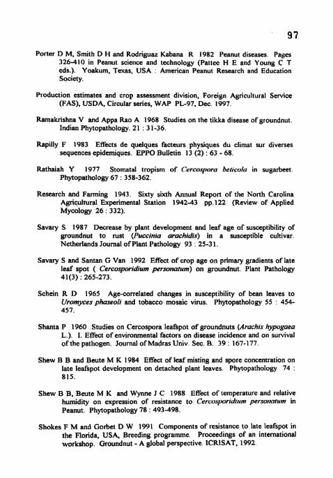
Porter D M, Smith D H and Rodriguaz Kabana R 1982 Peanut diseases. Pages 326-410 in Peanut science and technology (Pattee H E and Young C T eds.). Yoakum, Texas, USA : American Peanut Research and Education Society.
Production estimates and crop assessment division, Foreign Agricultural Service (FAS), USDA, Circular series, WAP PL-97, Dec. 1997.
Ramakrishna V and Appa Rao A 1968 Studies on the tikka disease of groundnut. Indian Phytopathology. 21 : 3 1-36.
Rapilly F 1983 Effects de quelques facteurs physiques du climat sur diverses sequences epidemiques. EPPO Bulletin 13 (2) : 63 - 68.
Rathaiah Y 1977 Stomatal tropism of ('ercospora beticola in sugarbeet. Phytopathology 67 : 358-362.
Research and Farming 1943. Sixty sixth Annual Report of the North Carolina Agricultural Experimental Station 1942-43 pp.122 (Review of Applied Mycology 26 : 332).
Savary S 1987 Decrease by plant development and leaf age of susceptibility of groundnut to rust (Puccinia arachidis) in a susceptible cultivar. Netherlands Journal of Plant Pathology 93 : 25-3 1.
Savary S and Santan G Van 1992 Effect of crop age on primary gradients of late leaf spot ( Cercosporidium perso~~attim) on groundnut. Plant Pathology 41(3) : 265-273.
Schein R D 1965 Age-correlated changes in susceptibility of bean leaves to Uromyces phaseoli and tobacco mosaic virus. Phytopathology 55 : 454- 457.
Shanta P 1960 Studies on Cercospora leafspot of groundnuts (Arachis hypogaea L.). I. Effect of environmental factors on disease incidence and on survival of the pathogen. Journal of Madras Univ. Sec. B. 39 : 167-177.
Shew B B and Beute M K 1984 Effect of leaf misting and spore concentration on late leafspot development on detached plant leaves. Phytopathology 74 : 815.
Shew B B, Beute M K and Wynne J C 1988 Effect of temperature and relative humidity on expression of resistance to CercosporiJiltm personlatum in Peanut. Phytopathology 78 : 493-498.
Shokes F M and Gorbet D W 1991 Components of resistance to late leafspot in the Florida, USA, Breeding programme. Proceedings of an international workshop. Groundnut - A global perspective. ICRISAT, 1992.
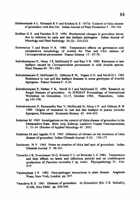
Siddaramaiah A L Hiremath R V and Krishna K S 1977a Cointrol of tikka disease of groundnut with Der-Ter. lndian Journal of Plant Protection 5 : 193-194.
Sindhan G S and Pareshar R D 1996 Biochemical changes in groundnut leaves due to infection by early and late leafspot pathogens. lndian Journal of Mycology and Plant Pathology 26 (2) : 2 10-2 12.
Sommanya T and Beute N K 1986 Temperature effects on germination and comparative morphology of conidia for Thai and USA isolates of Cercosporidicrm persoIiatirm. Peanut Science 13 : 67-70.
Subrahmanyam P, Moss J P, McDonald D and Rao V R 1985 Resistance to late leafspot caused by Cercosporidiirm perso~iarirm in wild Arachis species. Plant Disease 69 : 95 1-954.
Subrahmanyam P, McDonald D, Gibbons R W, Nigam S N and Nevill D J 1982 Resistance to lust and late leafspot diseases in some genotypes of Arachis hypogaea. Peanut Science 9 : 6- 10.
Subrahmanyam P, Mehan V K, Nevill D J and McDonald D 1980 Research on fbngal diseases of groundnut. In ICRISAT Proceedings of International Workshop on Groundnut, 13-17, October 1980, Patancheru, India. pp. 193-198.
Subrahmanyam P, Ramanadha Rao V, McDonald D, Moss J P and Gibbons R W 1989 Origins of resistance to rust and late leafspot in peanut (Arachis hypogaea, Fabaceae). Economic Botany 43 : 444-455.
Sulaiman M 1965 Investigations on the control of tikka disease of groundnut in the Maharashtra State. Beitr. trop. Subtrop. Landwrit Tropen Veterinarmedizin 2 : 51-54 (Review of Applied Mycology 45 : 305).
Sulaiman M and Agashe N G 1965 Influence of climate on the incidence of tikka disease of groundnut. Indian Oilseeds Journal 9 (3) : 176-1 77.
Sundaram N V 1965 Notes on creation of tikka leaf spot of groundnut. lndian Oilseeds Journal 9 : 98-101.
Tomerlin J R, Eversmeyer M G, Kramer C L nd Browder L E 1983. Temperature and host effects on latent and infectious periods and on urediniospore production of Pttccinia recotidira f sp. tririci. Phytopathology 73 : 414- 419.
Vanderplank J E 1982 Host-pathogen interactions in plant disease. Acsdemic Press. New York, London. pp. 207.
Vasudeva R S 1961 Diseases of groundnut. In Groundnut (Ed. C.R. Seshadri), ICAR, New Delhi. pp. 229-240.

Wadia K D R and Butler D R 1994 Relationship between temperature and latent periods of rust and leafspot diseases of groundnut. Plant Pathology 43 : 121-129.
Wadia K D R and Butler D R 1995 Effect of humidity on conidial morpliology of Phaeoisarir~j~sispersot~ar~~, International Arachis Newsletter I5 : 33-34
Walls S B, Wynne J C and Beute M K 1985 Resistance to late leafspot. Peanut Science 12 : 17-22.
Watson G R, Kucharek T A and Shokes F M 1997 Components of resistance in Southern Runner Peanut to Late Leafspot. Phytopathology 87 : S 116.
Woodroof N C 1933 Two leafspots of the peanut (Arachis Ivpogma L.) Phytopathology 23 : 627-640.
*Wolf F A 1914 Leaf spot and some fruit rots of peanuts. Albania Agricultural Research Station Bulletin 180 : 127-150
Wynne J C, Beute M K and Xigam S N 1991 Breeding for disease in peanut (Arachis hypogaea L ). Annual Review of Phytopathology 29 . 279-303.
Yenni Y R 1970 Physiological changes associated with tikka disease of groundnut and field control of the disease. Mysore Journal of Agricultural Sciences 6 . 522-523.
Zadoks J C 1972 Methodology of epidemiological research. Annual Review of Phytopathology 10 : 253-76.
Zadoks T C and Schein R D 1979 Epidemiology and plant disease management. Oxford University Press, New York, Oxford. pp.427.
Original not seen. The thesis is presented according to the guidelines framed by Acharya N.G Ranga Agricultural University.