studies on glycosphingolipids of eggs of the brine shrimp ... · chs, were characterized as novel...
TRANSCRIPT

1
Biochemical Studies on Sphingolipids of Artemia franciscana (II): Novel Neutral Glycosphingolipids
Hisao Kojima1, 2, Takemasa Shimizu2, Mutsumi Sugita2, Saki Itonori2, Norihisa Fujita3, Masahiro Ito1*
1Department of Bioinformatics, Institute of Science and Engineering, Ritsumeikan University, 1-1-1 Nojihigashi,
Kusatsu, Shiga 525-8577, Japan
2Department of Chemistry, Faculty of Liberal Arts and Education, Shiga University, 2-5-1 Hiratsu, Otsu, Shiga
520-0862, Japan
3Laboratory of Pharmcoinformatics, Faculty of Pharmacy, Ritsumeikan University, Nojihigashi, Kusatsu, Shiga
525-8577, Japan
*Correspondence to:
Masahiro Ito, Department of Bioinformatics, Institute of Science and Engineering, Ritsumeikan University, 1-1-1
Nojihigashi, Kusatsu, Shiga 525-8577, JAPAN
Phone: +81 77 561 5301 Fax: +81 77 561 5301
E-mail: [email protected]
Abbreviated title for a running footline: Neutral glycosphingolipids of Artemia franciscana
Abbreviations
GC, gas-liquid chromatography; GC-MS, combined gas-liquid chromatography-mass spectrometry; MALDI-TOF
MS, matrix-assisted laser desorption/ionization time-of-flight mass spectrometry; 1H-NMR, proton nuclear
magnetic resonance; CMS, ceramide monosaccharide; CDS, ceramide disaccharide; CTS, ceramide trisaccharide;
CTeS, ceramide tetrasaccharide; CPS, ceramide pentasaccharide; CHS, ceramide hexasaccharide; GSL,
glycosphingolipid; MacCer, mactosyl ceramide; At3Cer, arthrotriaosylceramide; At4Cer, arthrotetraosylceramide;
by guest, on January 25, 2019w
ww
.jlr.orgD
ownloaded from
2
At5Cer, arthropentaosylceramide; At6Cer, arthrohexaosylceramide; At7Cer, arthroheptaosylceramide; HPTLC,
high performance thin-layer chromatography
by guest, on January 25, 2019w
ww
.jlr.orgD
ownloaded from

3
Abstract
Neutral glycosphingolipids containing one to six sugars in their oligosaccharide chains have been isolated from
cysts of the brine shrimp Artemia franciscana. The structures of these glycolipids were identified by
methylation analysis, partial acid hydrolysis, gas-liquid chromatography, combined gas-liquid
chromatography-mass spectrometry, matrix-assisted laser desorption/ionization time-of-flight mass spectrometry,
and proton nuclear magnetic resonance spectroscopy to be Glcβ1-Cer (CMS), Manβ1-4Glcβ1-Cer (CDS),
Fucα1-3Manβ1-4Glcβ1-Cer (nAtCTS), GlcNAcβ1-3Manβ1-4Glcβ1-Cer (AtCTS),
GlcNAcα1-2Fucα1-3Manβ1-4Glcβ1-Cer (nAtCTeS), GalNAcβ1-4GlcNAcβ1-3Manβ1-4Glcβ1-Cer (AtCTeS),
GalNAcβ1-4(Fucα1-3)GlcNAcβ1-3Manβ1-4Glcβ1-Cer (CPS), and
GalNAcβ1-4(GlcNAcα1-2Fucα1-3)GlcNAcβ1-3Manβ1-4Glcβ1-Cer (CHS). Two glycosphingolipids, CPS and
CHS, were characterized as novel structures. Since Artemia contains a certain series of glycosphingolipids
(-Fucα3Manβ4GlcβCer), which differ from the core sugar sequences reported thus far, we tentatively designated
the glycosphingolipids characterized as non-arthro-series ones. Furthermore, CHS exhibited a hybrid structure
of arthro-series and non-arthro-series sugar chain.
[Short Conclusion]
Two novel glycosphingolipids were characterized from the brine shrimp Artemia franciscana; one was composed
of arthrotetraose and a branching fucose attached to N-acetylglucosamine residue, and the other was composed of
CPS with an additional N-acetylglucosamine residue attached to the branching fucose.
Supplementary key words (up to ten key phrases)
sphingolipid, sphingosine, long chain base, oligosaccharide, fucomannolipid, terminal alpha-N-acetylglucosamine
residue, structure characterization, conventional destructive analysis, Branchiopoda, dormant cyst
by guest, on January 25, 2019w
ww
.jlr.orgD
ownloaded from

4
Introduction
Glycosphingolipid (GSL) is composed of sugar chain and ceramide, the latter of which consists of a
fatty acid and a sphingoid. GSLs, which are expressed on the outer side of the lipid bilayer in animal
cells, form patches with sphingomyelin (microdomains) and play a foundational role in intercellular
adhesion, cellular recognition, differentiation/growth, and immune response (1-3). However, a
comprehensive understanding of GSL function has not yet been attained, because of the structural
complexity in the sugar chain of GSLs even in vertebrates and invertebrates (4, 5). We have
analyzed the GSL structures in lower animals on the assumption that their complexity plays an
important role in maintaining the biological activity of multicellular organisms.
In invertebrates, structural analyses of GSLs have been performed in several phyla and are
well known in Arthropoda, Mollusca, and Nematoda. The immune response by Galα-Cer from a
marine sponge (3) and the induction of cytokine secretion by zwitterionic GSLs from a parasitic
nematode (6) are interesting in terms of GSL function, and investigation of invertebrate GSL
structures could therefore be a productive research area. In Arthropoda, structural analyses of
GSLs have begun for flies (Diptera Insecta) (7, 8), and a characteristic arthro-series sugar chain
(GlcNAcβ3Manβ4GlcCer; At3Cer) has been identified. In Drosophila, mactosyl ceramide
(MacCer) and arthrotriaosylceramide (At3Cer) are biosynthesized by catalytic
β4-mannosyltransferase (egghead, egh) and β3-N-acetylglucosaminyltransferase (brainiac, brn),
respectively (9-12). It was reported that the mutant of brn was shown to be a lethal phenotype at
pupal stage (9): therefore, the arthro-series trisaccharide appears to be essential for development in
insects.
In this report, we analyzed GSL structures in cysts of the brine shrimp Artemia franciscana,
a crustacean arthropod. This species is a kind of plankton inhabiting saline environments such as
by guest, on January 25, 2019w
ww
.jlr.orgD
ownloaded from

5
the Great Salt Lake in the U.S.A. Analytical reports on this species have mainly related to
nutritional analyses in aquaculture using the species as instant live food (13). There have only been
two reports regarding sphingolipid characterization: one on a ceramide tetrasaccharide (CTeS) in
newly hatched nauplii (14), and the other on sphingomyelin in diapausing eggs (15). Here we
present novel non-arthro-series and arthro-series GSLs, as well as hybrid structure GSL composed of
core arthro-series sugar chains with a branching non-arthro-series disaccharide in the brine shrimp.
Materials and Methods
Isolation of Neutral Glycosphingolipids
Great Salt Lake brine shrimp cysts (1.8kg) purchased from A & A Marine LLC (Salt Lake City, Utah,
U.S.A) were ground to powder by using automatic mortars. Lipids were extracted once with 7.2 l
of chloroform-methanol (2:1, v/v), once with 5.3 l of chloroform-methanol (2:1, v/v) and once with
4.4 l of chloroform-methanol (1:1, v/v). The combined chloroform-methanol extracts were
concentrated by a rotary evaporator and subjected to mild alkaline hydrolysis with 0.5 M KOH in
methanol. The hydrolyzate was acidified (pH 1) with several drops of conc. HCl, kept in an ice
bath for 1 h, and dialyzed against tap-water for 2 days. The inner fluid was concentrated to near
dryness in vacuo at 40 °C, and precipitated by addition of cold acetone (yield: 8.6 g, alkaline-stable
product). The alkaline-stable product was dissolved in chloroform-methanol-water (30:60:8, v/v/v)
and applied to a column (φ3.5×48 cm) packed with QAE-Sephadex A-25, OH- form (GE Healthcare
Co.) equilibrated with the same solvent. The column was successively eluted with the same solvent
(5 column volumes) and pure methanol (1 volume) as neutral solvents, and with 0.45 M ammonium
acetate in methanol (5 volumes) as a polar solvent. The separation of neutral and acidic glycolipids
by guest, on January 25, 2019w
ww
.jlr.orgD
ownloaded from

6
was monitored by thin-layer chromatography (TLC) as described below. The eluates obtained from
this column using the neutral solvents were pooled and evaporated to dryness (yield: 610 mg, crude
neutral GSL fraction). The crude neutral GSL fraction was acetylated and then fractionated on a
column (φ2.0×57 cm) packed with Florisil, 60 ~ 100 mesh (Nacalai Tesque, Inc.) by slightly
modifying the method of Saito and Hakomori (16). The column was successively eluted with 3
column volumes of n-hexane-dichloroethane (1:4, v/v), 3 volumes of pure dichloroethane, 3 volumes
of dichloroethane-acetone (1:1, v/v), 6 volumes of dichloroethane-methanol (9:1, v/v), 3 volumes of
dichloroethane-methanol (3:1, v/v), 3 volumes of dichloroethane-methanol-water (2:8:1, v/v/v), 6
volumes of chloroform-methanol-water (6:4:1, v/v/v), and 3 volumes of chloroform-methanol-water
(2:8:1, v/v/v). In the course of this fractionation, different species of neutral GSLs were eluted with
the various mixtures of dichloroethane-acetone and dichloroethane-methanol solvents. The
solutions of acetylated GSLs were each evaporated to dryness, deacetylated with 0.5 M KOH in
methanol at 37°C for 6 h, and dialyzed against tap-water for 2 days. The inner fluids were
concentrated to near dryness in vacuo at 40°C. The concentrated lipid fractions were tested by
TLC as described below and combined (yield: 102 mg, neutral GSL fraction).
Isolation of Neutral Glycosphingolipid
For isolation of each neutral GSL, the neutral GSL fraction (97mg) was applied to an Iatrobeads
(6RS-8060, Mitsubishi Kagaku Iatron Inc., Tokyo) column (φ1.0×111cm). The neutral GSLs were
eluted with two linear gradient elution systems of chloroform-methanol-water with compositions of
[80:20:1 (v/v/v) 255 ml ∼ 50:50:5 (v/v/v) 325 ml] and [50:50:5 (v/v/v) 252 ml ∼ 20:80:10 (v/v/v)
330ml], respectively. Fractions of 3 ml were collected in each tube and aliquots from every three
tubes were tested by HPTLC.
by guest, on January 25, 2019w
ww
.jlr.orgD
ownloaded from

7
Solvent System for Thin-Layer Chromatography (TLC)
The following solvent system was used; chloroform-methanol-water (60:40:10, v/v/v). In
QAE-Sephadex and Florisil column chromatography, the eluates on TLC plates of silica gel 60
(Merck KGaA) were visualized by spraying with orcinol-sulfuric acid reagent (17) followed by
heating at 110°C and by spraying Dittmer-Lester reagent (18) to detect sugar and phosphate groups,
respectively. In Iatrobeads column chromatography, GSLs on HPTLC plates of silica gel 60
(Merck KGaA) were visualized by spraying with orcinol-sulfuric acid reagent followed by heating at
110°C.
Analysis of Fatty Acid and Sugar Components
For determination of the composition of fatty acids and sugars, 0.1 ~ 0.2 mg of each GSL was
methanolyzed with 1 M anhydrous methanolic HCl at 100°C for 3h. The produced fatty acid
methyl esters were extracted with n-hexane and analyzed by GC and GC-MS. The remaining
methanolic phase was neutralized by adding silver carbonate, and evaporated to dryness after
removal of silver chloride. The residue containing methyl glycosides was N-acetylated by adding
10µl of pyridine and 50µl of anhydrous acetate in 0.5 ml of methanol for 30 min (19), evaporated
under a nitrogen stream, and dried in a vacuum desiccator with a water aspirator. The N-acetylated
residue was trimethylsilylated (20), and subjected to GC.
Methylation Analysis
by guest, on January 25, 2019w
ww
.jlr.orgD
ownloaded from

8
About 0.2 mg of each GSL was permethylated according to the method of Ciucanu and Kerek (21).
In brief, each GSL was dried in a screw-capped glass test tube and dissolved in 0.2 ml of
dimethylsulfoxide while sonicating for 5 min. Immediately after about 20 mg of fine powdered
NaOH and 0.2 ml of methyl iodide were added, the test tube was capped tightly and vigorously
stirred for 2 min. The methylated GSL was extracted with chloroform, washed with water six times,
dried under a nitrogen stream, and hydrolyzed with 0.3 ml of acetic acid-HCl-water (16:1:3, v/v/v)
using a microwave oven (22, 23). The acetolyzate was dried under a nitrogen stream using a few
drops of toluene, dried in a desiccator with a water aspirator for 1 h, and then reduced by adding 0.25
ml of 0.01M NaOH followed by 0.25 ml of 0.01 M NaOH solution containing 2% sodium
borohydride (final concentration: 1% sodium borohydride, 0.01 M NaOH) at room temperature
overnight. The reduction was stopped by adding a few drops of acetic acid. The reduced and
partially methylated alditols were dried under a nitrogen stream while adding ~ 1 ml of methanol a
few times, and then dried in a desiccator with a water aspirator for 1 h. The residue was acetylated
by adding 0.25 ml of pyridine followed by 0.25 ml of acetic anhydride, and then incubated in boiling
water for 12 min. The partially methylated alditol acetates were extracted with chloroform, washed
with water six times, and then subjected to GC and GC-MS.
Analysis of Partial Acid Hydrolysis
About 4 mg of CHS was hydrolyzed with 1 ml of 0.1 M HCl in boiling water for 60 min. The
hydrolyzate was extracted into a lower phase by adding 5 ml of chloroform-methanol (2:1, v/v),
evaporated to dryness under a nitrogen stream, and dissolved in 2 ml of chloroform-methanol (2:1,
v/v). The GSL fragments produced were separated on a silica gel 60 TLC plate, developed in the
solvent mixture of chloroform-methanol-water (60:40:10, v/v/v) for 20 min, and dried. After slight
by guest, on January 25, 2019w
ww
.jlr.orgD
ownloaded from
9
exposure to iodine vapor, the spots corresponding to mono-, di-, tri- and tetraglycosyl ceramide were
scraped off, extracted with chloroform-methanol-water (2:1:0.1, v/v/v) and subjected to sugar
composition analysis.
Analysis of Sphingoids
Sphingoid composition was determined by the method of Gaver and Sweeley (24). In brief, about
0.2 ∼ 0.3 mg of each GSL was measured into a screw-capped glass test tube and dried under a
nitrogen stream. After adding 0.2 ml of aqueous HCl-methanol reagent (methanolic reagent
containing 8.6% conc. HCl and 9.4% water), and the mixture was hydrolyzed at 70°C in the oven for
18 h. The hydrolyzate was washed with n-hexane to remove fatty acids. The residual methanolic
phase was dried under a nitrogen stream, alkalized with 0.6 ml of methanol -1 M NaOH solution
(4:3, v/v), extracted with 0.72 ml of chloroform, and washed twice with 0.4 ml of methanol-water
(1:1, v/v). The residual chloroform solution was evaporated under a nitrogen stream, dried in a
vacuum desiccator with a water aspirator, trimethylsilylated, and subjected to GC and GC-MS.
Gas-liquid Chromatography (GC) and Gas Chromatograph-Mass Spectrometry (GC-MS)
Compositional analyses of methylation, fatty acids, sugars and sphingoids were carried out using a
Shimadzu GC-18A gas chromatograph with a Shimadzu HiCap-CBP 5 capillary column (0.22 mm x
25 m). The temperature increase was programmed at 2°C/min from 140 to 230°C for sugar
component analysis, 4°C/min from 140 to 230°C for the methylation study, 4°C/min from 170 to
230°C for fatty acid analysis, and 2°C/min from 210 to 230°C for sphingoid analysis. Electron
impact ionization mass spectra were obtained using a Shimadzu GCMS-QP5050 gas
by guest, on January 25, 2019w
ww
.jlr.orgD
ownloaded from

10
chromatograph-mass spectrometer with the same capillary column under the following conditions;
interface temperature of 250°C, an injection port temperature of 240°C, a helium gas pressure of
100kPa, and an ionizing voltage of 70eV. The oven temperatures for GC-MS analyses were 80 (2
min) to 180 (20°C/min) to 240°C (4°C/min) for the methylation study, 80 (2min) to 170 (20°C/min)
to 240°C (4°C/min) for fatty acid analysis, and 80 (2min) to 220 (20°C/min) to 280°C (6°C/min) for
sphingoid analysis, respectively.
Matrix-Assisted Laser Desorption Ionization Time-of-flight Mass Spectrometry (MALDI-TOF
MS)
MALDI-TOF MS analysis was performed using an Applied Biosystems/Voyager-DE STR
Biospectrometer with a nitrogen laser (337 nm) operating in the reflector positive-ion mode at an
acceleration voltage of 20 kV. The matrix used was α-cyano-4-hydroxycinnamic acid (Proteomics
Grade, Wako Chemical Co.). External mass calibration was provided by the [M+Na]+ ions of
angiotensin I (1296.96 mass units; Sigma Chemical Co.) and bradykinin fragment I-V (573.31 mass
units; Sigma Chemical Co.).
Proton-Nuclear Magnetic Resonance Spectroscopy (1H-NMR spectroscopy)
NMR spectra were obtained using a JEOL-ECS400 400MHz NMR spectrometer at an operating
temperature of 60°C. The purified GSL was dissolved in 0.5 ml of d6-dimethylsulfoxide containing
2% D2O. The chemical shift was referenced to the solvent signals (δH 2.49 ppm) in
d6-dimethylsulfoxide as the internal standard.
by guest, on January 25, 2019w
ww
.jlr.orgD
ownloaded from

11
Results
Purified Neutral Glycosphingolipids
Crude sphingolipids (8.6 g) were obtained from chloroform-methanol extracts (285 g) of brine
shrimp (Artemia franciscana) cysts (1.8 kg) by alkaline- and acid-treatment, and chromatographed
on a QAE-Sephadex column using a solvent mixture of chloroform-methanol-water (30:60:8, v/v/v).
In the TLC analysis of the resultant fraction (610 mg), GSLs (102 mg) and sphingomyelin (359 mg)
were detected with orcinol-sulfuric acid and Dittmer-Lester reagent, respectively. Zwitterionic
GSL could not be detected. GSLs were separated using Florisil and silica gel column
chromatography into 26 fractions. GSLs with one to six sugar residues were developed by TLC
(Fig. 1). The yields of the purified GSLs obtained from 1.8 kg of brine shrimp cysts were 2.8
(CMS), 2.7 (CDS), 5.1 (nAtCTS), 2.1 (AtCTS), 5.1 (nAtCTeS), 6.9 (AtCTeS), 1.9 (CPS), and 5.3
mg (CHS). GSLs with more than six sugar residues are currently under investigation.
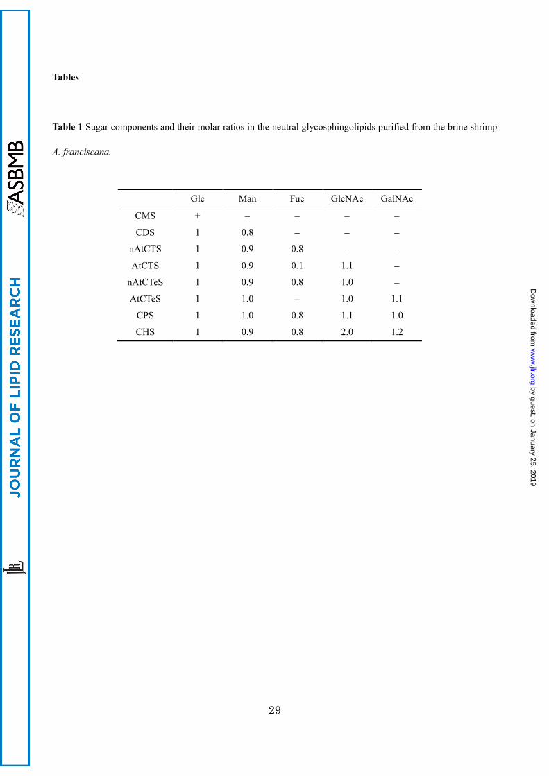
Sugar Composition
Each GSL was methanolyzed and the methylglycosides were converted to N-acetyl-O-trimethylsilyl
derivatives for GC analysis. Their gas chromatograms defined the sugar composition of CMS as
glucose; CDS as glucose and mannose (molar ratio, 1:1); nAtCTS as glucose, mannose and fucose
(1:1:1); AtCTS as glucose, mannose and N-acetylglucosamine (1:1:1); nAtCTeS as glucose,
mannose, fucose and N-acetylglucosamine (1:1:1:1); AtCTeS as glucose, mannose,
N-acetylglucosamine and N-acetylgalactosamine (1:1:1:1); CPS as glucose, mannose,
N-acetylglucosamine, N-acetylgalactosamine and fucose (1:1:1:1:1); and CHS as glucose, mannose,
by guest, on January 25, 2019w
ww
.jlr.orgD
ownloaded from

12
N-acetylglucosamine, N-acetylgalactosamine and fucose (1:1:2:1:1) (see Table 1). Sugar
compositional analysis of AtCTS indicated 0.1 mol of fucose corresponding to the impurities in
nAtCTeS which could not be separated by the Iatrobeads column chromatography.
Methylation Analysis
The partially methylated alditol acetates derived from the obtained GSLs were separated by GC as
shown in Fig. 2. The identification was confirmed by GC-MS analysis according to the data of
Jansson et al. (25) and Tai et al. (26). The methylation analysis revealed
1,5-di-O-acetyl-2,3,4,6-tetra-O-methylglucitol (1Glc) from CMS;
1,4,5-tri-O-acetyl-2,3,6-tri-O-methylglucitol (1,4Glc) and
1,5-di-O-acetyl-2,3,4,6-tetra-O-methylmannitol (1Man) from CDS; 1,4Glc,
1,3,5-tri-O-acetyl-2,4,6-tri-O-methylmannitol (1,3Man), and
1,5-di-O-acetyl-2,3,4-tri-O-methylfucitol (1Fuc) from nAtCTS; 1,4Glc, 1,3Man, and
1,5-di-O-acetyl-3,4,6-tri-O-methyl-N-acetylglucosaminitol (1GlcNAc) from AtCTS; 1,4Glc, 1,3Man,
1,2,5-tri-O-acetyl-3,4-di-O-methylfucitol (1,2Fuc), and 1GlcNAc from nAtCTeS; 1,4Glc, 1,3Man,
1,4,5-tri-O-acetyl-3,6-di-O-methyl-N-acetylglucosaminitol (1,4GlcNAc), and
1,5-di-O-acetyl-3,4,6-tri-O-methyl-N-acetylgalactosaminitol (1GalNAc) from AtCTeS; 1,4Glc,
1,3Man, 1,3,4,5-tetra-O-acetyl-6-O-methyl-N-acetylglucosaminitol (1,3,4GlcNAc), 1GalNAc, and
1Fuc from CPS; and 1,4Glc, 1,3Man, 1,3,4GlcNAc, 1GalNAc, 1,2Fuc, and 1GlcNAc from CHS.
Although 1,4Glc and 1,3Man are not separated by GC analysis using the Shimadzu HiCap-CBP 5
capillary column, their identities were determined from their electron impact ionization (EI) mass
spectra. Their identities were subsequently confirmed by repeating the analysis using a column of
DB-210 capillary column (0.25mm × 30 m, Agilent Technology) that provided separation and
by guest, on January 25, 2019w
ww
.jlr.orgD
ownloaded from

13
characteristic retention times for both 1,4Glc and 1,3Man (data not shown).
Position of Glycosidic Substitution on the Branching Saccharide by Partial Acid Hydrolysis
The methylation analysis described above for CHS indicated the presence of a saccharide branch on
the N-acetylglucosamine residue. To determine the position of glycosidic substitution on
N-acetylglucosamine to N-acetylgalactosamine residue, CHS was hydrolyzed with 0.1 M HCl at
100°C for 60 min, and the resulting products were separated with preparative TLC. From the
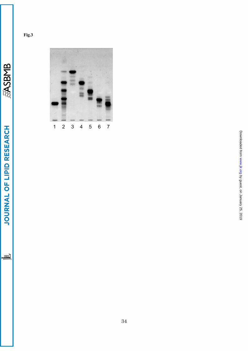
hydrolysis, four products corresponding to GSLs with one to four sugar residues (CHS-M, -D, -T
and -Q) were obtained (Fig. 3), methanolyzed, trimethylsilylated, and subjected to GC. The gas
chromatogram revealed the sugar composition of CHS-M as Glc, CHS-D as Glc and Man (molar
ratio, 1:1); CHS-T as Glc, Man and GlcNAc (1:1:1); CHS-Q as Glc, Man and GlcNAc and GalNAc
(1:1:1:1). This result indicates the presence of the sugar chain, GalNAc-GlcNAc-Man-Glc, in the
CHS structure as a core sequence. Therefore we concluded that the branching Fuc residue attaches
to the 3-position on GlcNAc residue in the core AtCTeS, GalNAc1-4GlcNAc1-3Man1-4Glc.
Aliphatic Components
The compositions of fatty acid and sphingoid in the obtained GSLs are given in Table 2. The fatty
acids were mainly normal saturated acids ranging in length from C16 to C24, of which C22 acid was
the most predominant. In some GSLs, odd-numbered saturated acids ranging in length from C19 to
C23 were also detected in low amounts. The monoenoic acids of C22 and C24 were common in all
by guest, on January 25, 2019w
ww
.jlr.orgD
ownloaded from

14
GSLs. CMS contained 2-hydroxy acids as minor components. The sphingoids of the GSLs were
composed of hexadeca- and heptadeca-4-sphingenines. In each case, the amount of the shorter base
accounted for more than 57.5% of the total amount.
MALDI-TOF MS Analysis
The putative structures of the eight purified GSLs were confirmed by the positive-ion reflector mode
of MALDI-TOF MS analysis, as shown in Fig. 4 and summarized in supplemental table. Their
mass spectra have two to four sodium adducted ion species, [M+Na]+ ion species, the mass
difference of which ranged from −319 to +463 ppm against the values calculated from the proposed
structures, throughout the whole measurement period: that is, for CMS, [M+Na]+ ions at m/z 722.78,
738.77, 778.86, and 792.87 with one mole each of glucose, fatty acid (assigned as 18:0, h18:0 or
22:0), and sphingoid (assigned as d16:1 or d17:1); for CDS, [M+Na]+ ions at m/z 884.62, 898.64,
940.69, and 954.70 with one mole each of glucose, mannose, fatty acid (assigned as 18:0 or 22:0),
and sphingoid (assigned as above); for nAtCTS, [M+Na]+ ions at m/z 1030.67, 1044.68, 1086.73,
and 1100.74 with one mole each of glucose, mannose, fucose, fatty acid (assigned as above), and
sphingoid (assigned as above); for AtCTS, [M+Na]+ ions at m/z 1143.69 and 1157.69 with one mole
each of glucose, mannose, N-acetylglucosamine, fatty acid (assigned as 22:0), and sphingoid
(assigned as above); for nAtCTeS, [M+Na]+ ions at m/z 1233.88, 1247.89, 1289.95, and 1303.96
with one mole each of glucose, mannose, fucose, N-acetylglucosamine, fatty acid (assigned as 18:0
or 22:0), and sphingoid (assigned as above); for AtCTeS, [M+Na]+ ions at m/z 1290.78, 1304.80,
1346.84, and 1360.85 with one mole each of glucose, mannose, N-acetylglucosamine,
N-acetylgalactosamine, fatty acid (assigned as above), and sphingoid (assigned as above); for CPS,
[M+Na]+ ion at m/z 1493.18 and 1507.16 with one mole each of glucose, mannose,
by guest, on January 25, 2019w
ww
.jlr.orgD
ownloaded from

15
N-acetylglucosamine, N-acetylgalactosamine, fucose, fatty acid (assigned as 22:0), and sphingoid
(assigned as above); and for CHS, [M+Na]+ ion at m/z 1695.54 and 1709.55 with one mole each of
glucose, mannose, N-acetylgalactosamine, fucose, two moles of N-acetylglucosamine, and one mole
each of fatty acid (assigned as 22:0) and sphingoid (assigned as above), respectively.
Anomeric Configurations of the Sugar Residues
Anomeric configurations of the sugar residue in GSLs were determined by a 1H-NMR spectrometer
(Fig. 5). Configurations were assigned by reference to the data of MacCer and At3Cer (11), that of
At5Cer (27), that of Fuc (28), and that of α-GlcNAc (29, 30) in terms of chemical shifts and coupling
constants, these assignments of which are listed in Table 3. The anomeric assignments of α-Fuc
and α-GlcNAc in nAtCTeS were determined by downfield shift of glycosyl-substituted fucose
compared to the data of nAtCTS, which was consistent with the data of Xu et al (14). In the
anomeric region of the spectrum for each GSL, the following anomeric proton resonances were
observed: at 4.09 ppm (J1,2=7.8Hz) for β-Glc (Fig. 5A, CMS); at 4.15 ppm (J1,2=7.8Hz) for β-Glc
and at 4.50 ppm (J1,2= ~1Hz) for β-Man (Fig. 5B, CDS); at 4.15 ppm (J1,2=7.8Hz) for β-Glc, at 4.52
ppm (J1,2= ~1Hz) for β-Man and at 4.79 ppm (J1,2=3.7Hz) for α-Fuc (Fig. 5C, nAtCTS); at 4.16 ppm
(J1,2=7.8Hz) for β-Glc, at 4.53 ppm (J1,2= ~1Hz) for β-Man, at 4.53 ppm (J1,2=7.3Hz) for β-GlcNAc
(Fig. 5D, AtCTS); at 4.15 ppm (J1,2=7.8Hz) for β-Glc, at 4.52 ppm (J1,2= ~1Hz) for β-Man, at 4.95
ppm (J1,2=3.7Hz) for α-Fuc, and at 4.80 ppm (J1,2=3.7Hz) for α-GlcNAc (Fig. 5E, nAtCTeS); at 4.15
ppm (J1,2=7.8Hz) for β-Glc, at 4.52 ppm (J1,2= ~1Hz) for β-Man, at 4.55 ppm (J1,2=8.2Hz) for
β-GlcNAc and at 4.35 ppm (J1,2=8.7Hz) for β-GalNAc (Fig. 5F, AtCTeS); at 4.16 ppm (J1,2=7.8Hz)
for β-Glc, at 4.52 ppm (J1,2= ~1Hz) for β-Man, at 4.62 ppm (J1,2=6.4Hz) for β-GlcNAc, at 4.29 ppm
(J1,2=8.7Hz) for β-GalNAc, and at 4.83 ppm (J1,2=3.7Hz) for α-Fuc (Fig. 5G, CPS); at 4.15 ppm
by guest, on January 25, 2019w
ww
.jlr.orgD
ownloaded from

16
(J1,2=7.8Hz) for β-Glc, at 4.52 ppm (J1,2= ~1Hz) for β-Man, at 4.64 ppm (J1,2=7.3Hz) for β-GlcNAc,
at 4.31 ppm (J1,2=8.2Hz) for β-GalNAc, and at 5.01 ppm (J1,2=2.7Hz) for overlapping α-Fuc and
α-GlcNAc (Fig. 5H, CHS).
Discussion
This study shows the structures of cerebroside, mannolipid, and fucomannolipids as neutral GSLs in
the cysts of the brine shrimp Artemia franciscana. There are some differences in the sphingolipids
between the brine shrimp and insect culture cell/flies; the differences are that the dominant
sphingoids are mainly hexadeca-4-sphingenine/heptadeca-4-sphingenine and
tetradeca-4-sphingenine/hexadeca-4-sphingenine, respectively, and that the dominant fatty acids are
behenic acid and arachidic acid/stearic acid/behenic acid, respectively(7, 31). Compared with the
dominant sphingoids of the CTeS (BSG-I & II) in newly hatched nauplii of the brine shrimp (14),
those of nAtCTeS are similar, while the dominant fatty acids are quite different between the nauplii
and the cysts. In the CTeS isolated from the hatched shrimp, the dominant fatty acid was
2-hydroxy behenic acid, followed by behenic acid, as inferred from the density of BSG-I and BSG-II
on the TLC plate (14). On the other hand, in the CTeS isolated from the cysts, behenic acid was
largely dominant and 2-hydroxy behenic acid was below detection limits. There are many sibling
Artemia species, which cannot mate with each other despite being indistinguishably similar in
morphology, among Artemia species: A. franciscana, A. monica, A. parthenogenetica, A. persimilis,
A. salina, A. sinica, A. tibetiana, A. tunisiana, and A. urmiana. It would be interesting to know
whether the difference due to sibling species or stages is due to the difference in the fatty acid
composition of CTeS in two stages. Apart from CTeS, it appears that the hatched brine shrimp
contains GSLs, such as CMS, CDS, CTS, CPS, and CHS, as indicated by a previous report on the
by guest, on January 25, 2019w
ww
.jlr.orgD
ownloaded from

17
CTeS in the hatched shrimp by Horibata et al. (32). The dominant GSLs are CTS and CTeS: of
these, CTS appears to be Fucα3Manβ4GlcβCer since CTeS is a non-arthro-series GSL
(GlcNAcα2Fucα3Manβ4GlcβCer). As for CPS and CHS, the cysts apparently contain a higher
amount than the hatched shrimp; this could be due to the fact that Horibata et al. used only the
solvent of chloroform/methanol 2:1 (v/v) as the solvent in the lipid extraction and recovered only the
lower-phase extract after a Folch partition in the purification of lipids. Compared with the GSLs in
the hatched shrimp, those in the cysts differ considerably regarding the amount of CDS. Although
only a small difference exists in the simple GSLs between the cysts and the hatched shrimp, further
studies are required in the future to determine whether there are further differences in the complex
GSLs between the two developmental stages and the other stages. In addition, it is also interesting
to note that identical dominant ceramide components of sphingomyelin and GSLs have been
detected in the cysts of the brine shrimp Artemia franciscana (15). It seems likely that in the case
of neutral GSLs as well as the sphingomyelin ceramide, biosynthesis in this species utilizes a
common ceramide pool.
It was assumed that the arthro-series GSL would be detected in the brine shrimp as a major
GSL because this species belongs to the phylum Arthropoda. Structural analyses of neutral GSLs
with some sugar residues have been performed in several Arthropodae; the green-bottle fly Lucilia
caesar (7) and the blowfly Calliphora vicina (8), the fruit fly Drosophila melanogaster (33), the
insect culture cell line High Five Cell derived from the cabbage looper Trichoplusia ni (31), the
moth Manduca sexta (34) and the millipede Parafontaria laminata armigera (28)—all of which
have been verified to contain At3Cer. From the brine shrimp, arthro-series GSL (At3Cer) was
characterized as a minor CTS, but on the other hand, and surprisingly, a non-arthro-series GSL
(nAtCTS) was characterized as a major CTS. However, non-arthro-series GSLs account for
approximately 10% of the total 102 mg of neutral GSLs. Both CTSes are elongated from MacCer
by guest, on January 25, 2019w
ww
.jlr.orgD
ownloaded from

18
as the common substrate by the two enzymes involved, β3-N-acetylglucosaminyltransferase and
α3-fucosyltransferase (see Scheme 1). And both CTSes independently elongate a saccharide to
synthesize each series of CTeS. Interestingly, not only nAtCTeS but also At4Cer were abundant as
CTeS in the brine shrimp Artemia franciscana, in spite of the fact that there is no report which has
detected At4Cer to be abundant, except for reports of flies' GSLs (5, 7, 8, 33). It has been shown in
Arthropoda that the non-arthro-series sugar chain was dominant in a cultured insect cell line, namely
High-Five cells (31). The report discussed the followings; 1) the existence of an alternative
non-arthro CTS (Galβ3Manβ4Glcβ1Cer) to At3Cer in High Five GSLs implies that there is potential
for the generation of multiple pathways, with a possibility for core switching as observed in
mammalian cells, 2) the fact that the dominant CTS of the cell is itself a substrate for further
glycosylation supports the premise that the potential for sequence variability is inherent in the nature
of the machinery for glycosylation, and 3) although the potential existence of a
Galβ3Manβ4Glcβ1Cer core pathway is not sufficient to rescue the brn mutation, the observation that
brn is essential for development does not rule out the existence of one or more alternative
glycosylation pathways operating during the life cycle of a normal fly. It was also indicated that
more complex GSLs such as At4Cer and At5Cer were necessary for normal development in
Drosophila (27). In the current report on Artemia, we show that AtCTeS was abundant in amount
(6.9 mg) while AtCTS was minor (2.1 mg), and we propose that the likely source of GSL with more
than five sugar residues is the core arthro-series sugar chain, elongated by transfer of the sugars
occurring at the terminus of the non-arthro-series, as a branch (see Scheme 1). In Artemia,
although both arthro-series and non-arthro-series GSLs were detected as both CTS and CTeS, the
only CPS detected was derived from AtCTeS. Thus CPS is composed of only the core arthro-series
sugar chain plus a branching fucose, while CHS is characterized by the additional presence of an
N-acetylglucosamine residue on this branching fucose. CHS is therefore a hybrid structure, as it
by guest, on January 25, 2019w
ww
.jlr.orgD
ownloaded from

19
were, composed of the core arthro-series sugar chain, plus the sugars from the non-reducing end of
the non-arthro-series chain as a branch. We speculate that Artemia contains two pools (arthro and
non-arthro) of CTS and CTeS, each pool serving individual biological functions, and that the
organism may divert either form of CTS and CTeS as needed for the biosynthesis of more complex
GSLs, such as a sequence variability.
The observations reported herein that a fucosylated LacdiNAc trisaccharide
(GalNAcβ1-4[Fucα1-3]GlcNAcβ) determinant has been found in CPS and internally in CHS is
interesting in several respects. This trisaccharide is a Lewis X analog previously reported in the
parasite Schistosoma mansoni (35, 36), albeit on different aglycone structures. It has also been
found as a carbohydrate terminus on a human immunosuppressive glycoprotein, glycodelin (37).
An enzyme α1,3-fucosyltransferase, which is capable of producing fucosylated LacdiNAc
trisaccharide from LacdiNAc, has been cloned from the nematode Caenorhabditis elegans as
CEFT-1 (38) and from the honeybee Apis mellifera (39). Although the presence of this enzyme has
not been studied, it might be found in the brine shrimp. As far as we know, there is no report about
whether terminal αGlcNAc, terminal αFuc or the disaccharide of GlcNAcα2Fucα3 in GSL are
involved in the brine shrimp development. Since CPS and CHS contain fucosylated LacdiNAc
structure, a LeX analogue, it is possible that these novel GSLs interact with each other and aggregate
to form patches of microdomain in which developmental signals are transduced according to
GSL-GSL interaction, such as on Lex-Lex, Ley-H, and Gb4-Gg3 (40).
The structure of CHS consisted of core arthro-series GSLs with a branching
non-arthro-series disaccharide (GlcNAcα2Fucα-). In the structures of nAtCTeS and CHS,
“α-anomeric” GlcNAc was attached at the non-reducing end. This type of sugar chain in GSL
structure is reported in only a few cases; GlcNAcα4GalαCer from the marine sponges Axinella
damicornis (29), GlcNAcα1-HPO3-6GalCer from the liver fluke Fasciola hepatica (30), and CTeS
by guest, on January 25, 2019w
ww
.jlr.orgD
ownloaded from

20
(BSG-I & II) from newly hatched nauplii in the brine shrimp (14). Mucin-type O-glycan with
terminal αGlcNAc is also reported in only two cases; gastric mucins from pig
[Fucα2Galβ4GlcNAcβ6(GlcNAcα4Galβ3)GalNAc-ol and
GlcNAcα4Galβ4GlcNAcβ6(GlcNAcα4Galβ3)GalNAc-ol] (41) and a human gastric mucin
[GlcNAcα4Galβ4GlcNAcβ6(GlcNAcα4Galβ3)GalNAcαSer/Thr] (42). Interestingly, it was
reported that these mucins arrested proliferation of the spiral-shaped gram-negative bacterium
Helicobacter pylori which causes stomach upsets and peptic ulcers (43). It will be interesting to
examine whether nAtCTeS and CHS from the brine shrimp arrest the growth of H. pylori as the
mucins do, and to investigate the presence of further unique sugar chains in more complex GSLs.
This report on Artemia shows the existence of a sugar chain with rare terminal αGlcNAc.
To the best of our knowledge, although the sugar addition appears to be catalyzed by
α1,2-N-acetylglucosaminyltransferase (α2GnT), it has not been reported among animals to date.
Its homologous transferase, α1,4-N-acetylglucosaminyltransferase (α4GnT) has been characterized
in human (42). By using the data of nucleic or amino acid sequences of α4GnT, homology
searches were performed via BLAST searches (blastn, tblastn, and tblastx) with the filter of
“invertebrate” on the DDBJ web server (http://blast.ddbj.nig.ac.jp/top-e.html). As a result, only
two homologous sequences were found: α1,4-N-acetylgalactosaminyltransferase (GalNAcT) from
the fruit fly Drosophila melanogaster and lactosylceramide 4-α-galactosyltransferase putative
mRNA from the salmon lice Lepeophtheirus salmonis. Further homology searches were performed
according to the method mentioned above by using the data of nucleic or amino acid sequences of
the fly GalNAcT, and again only two records from the same organisms as mentioned above were
found. Thus, α2GnT appears rare. This warrants further study in organisms in which sugar chain
with the terminal αGlcNAc is detected.
by guest, on January 25, 2019w
ww
.jlr.orgD
ownloaded from

21
Acknowledgements
The authors thank Dr. John T. Dulaney who kindly read this manuscript while it was in preparation and for
providing helpful comments. This work was supported in part by the Grand-in Aid for Scientific Research from
the Japanese Ministry for Education, Science, Culture and Sports under the Grand No. 10010417, and a
scholarship from the society for the advancement of science and technology at Ritsumeikan University.
by guest, on January 25, 2019w
ww
.jlr.orgD
ownloaded from

22
References
1. Hakomori, S. 2000. Cell adhesion/recognition and signal transduction through glycosphingolipid
microdomain. Glycoconj. J. 17: 143-151.
2. Yu R.K, Nakatani Y., and Yanagisawa M. 2009. The role of glycosphingolipid metabolism in the developing
brain. J. Lipid Res. 50: S440-445.
3. Kawano, T., Cui, J., Koezuka, Y., Toura, I., Kaneko, Y., Motoki, K., Ueno, H., Nakagawa, R., Sato, H.,
Kondo, E., Koseki, H., Taniguchi, M. 1997. CD1d-restricted and TCR-mediated activation of Vα14 NKT
Cells by Glycosylceramides. Science 278: 1626-1629.
4. Yu, R.K., Yanagisawa, M., and Ariga, T. 2007. Glycosphingolipid structures. in Comprehensive Glycosci. 1
Introduction Glycosci. Synthesis Carbohydr. (ed) Kamerling, J.P., Elsevier
5. Itonori, S., and Sugita, M. 2007. Glycophylogenetic aspects of lower animals. in Comprehensive Glycosci. 3
Biochem. Glycoconj. Glycans Carbohydr.-mediated Interactions (ed) Kamerling, J.P., Elsevier
6. Lochnit, G., Dennis, R.D., Ulmer, A.J., and Geyer, R. 1998. Structural elucidation and monokine-inducing
activity of two biologically active zwitterionic glycosphingolipids derived from the porcine parasitic
nematode Ascaris suum. J. Biol. Chem. 273: 466–474.
7. Sugita, M., Nishida, M., and Hori, T. 1982. Studies on glycosphingolipids of larvae of the green-bottle fly,
Lucilia caesar. I. Isolation and characterization of glycosphingolipids having novel sugar sequences. J.
Biochem. 92: 327-334.
8. Dennis, R., Geyer, R., Egge, H., Peter-Katalinic, J., Li, S., Stirm, S., and Wiegandt, H. 1985.
Glycosphingolipids in insects; chemical structures of ceramide tetra-, penta-, hexa-, and heptasaccharide
from Calliphora vicina pupae (Insecta: Diptera). J. Biol. Chem. 260: 5370-5375.
9. Müller, R., Altmann, F., Zhou, D., and Hennet, T. 2002. The Drosophila melanogaster brainiac protein is a
glycolipid-specific β1,3N-acetylglucosaminyltransferase. J. Biol. Chem. 277: 32417–32420.
10. Wandall, H.H., Pedersen, J.W., Park, C., Levery, S.B., Pizette, S., Cohen, S.M., Schwientek, T., and Clausen,
H. 2003. Drosophila egghead encodes a β1,4-mannosyltransferase predicted to form the immediate precursor
by guest, on January 25, 2019w
ww
.jlr.orgD
ownloaded from

23
glycosphingolipid substrate for brainiac. J. Biol. Chem. 278:1411-1414.
11. Schwientek, T., Keck, B., Levery S.B., Jensen, M.A., Pedersen, J.W., Wandall, H.H., Stroud, M., Cohen,
S.M., Amado, M., and Clausen, H. 2002. The drosophila gene brainiac encodes a glycosyltransferase
putatively involved in glycosphingolipid synthesis. J. Biol. Chem. 277: 32421-32429.
12. Wandall, H.H., Pizette, S., Pedersen, J.W., Eichert, H., Levery, S.B., Mandel, U., Cohen, S.M., and Clausen,
H. 2005. Egghead and brainiac are essential for glycosphingolipid biosynthesis in vivo. J. Biol. Chem. 280:
4858–4863.
13. Dhont, J., and Sorgeloos, P. 2002. Artemia and aquaculture. in Artemia: Basic and Applied Biology (Biology
of Aquatic Organisms) (eds) Abatzopoulos, T.J., Beardmore, J.A., Clegg, J.S., and Sorgeloos, P. , Kluwer
Academic Publishers
14. Xu, X., Horibata, Y., Inagaki, M., Hama, Y., Sakaguchi, K., Goda, H.M., Okino, N., and Ito, M. 2009. A
novel fucosyl glycosphingolipid of brine shrimp that is highly sensitive to endoglycoceramidase. Glycobiol.
19: 1446–1451.
15. Kojima, H., Inoue, T., Sugita, M., Itonori, S., Ito, M. 2010. Biochemical Studies on Sphingolipid of Artemia
franciscana (I) Isolation and Characterization of Sphingomyelin. Lipids 45:635-643.
16. Saito, T., and Hakomori, S. 1971. Quantitative isolation of total glycosphingolipids from animal cells. J.
Lipid Res. 12: 257-259.
17. Svennerholm, L. 1956. The quantitative estimation of cerebrosides in nervous tissue. J. Neurochem. 1: 42-53.
18. Dittmer, J.C., and Lester, R.L. 1964. A simple, specific spray for the detection of phospholipids of thin-layer
chromatograms. J. Lipid Res. 5: 126-127.
19. Kozulić, B., Ries, B., and Mildner, P. 1979. N-acetylation of amino sugar methyl glycosides for gas-liquid
chromatographic analysis. Anal. Biochem. 94: 36-39.
20. Kishimoto, Y., and Hoshi, M. 1972. Isolation, purification, and assay of fatty acids and steroids from the
nervous system. in Methods Neurochem. (ed) Fried, R., Marcel Dekker
21. Ciucanu, I., and Kerek, F. 1984. A simple and rapid method for the permethylation of carbohydrates.
by guest, on January 25, 2019w
ww
.jlr.orgD
ownloaded from

24
Carbohydr. Res. 131: 209-217.
22. Itonori, S., Takahashi, M., and Sugita, M. 2000. Application of microwave-mediated reaction to lipid analysis.
Proc. J. Oil Chem. Soc. / Am. Oil Chem. Soc. World Congress, Kyoto , 214
23. Itonori, S., Takahashi, M., Aoki, K., and Sugita, M. 2000. Microwave-mediated acetolysis of
glycosphingolipids. Mem. Faculty Ed. Shiga Univ. 50: 17-24 (in Japanese).
24. Gaver, R.C., and Sweeley, C.C. 1965. Methods for methanolysis of sphingolipids and direct determination of
long-chain bases by gas chromatography. J. Am. Oil Chem. Soc. 42: 294-298.
25. Jansson, P.E., Kenne, L., Liedgren, H., Lindberg, B., and Lönngren, J. 1976. A practical guide to the
methylation analysis of carbohydrates. Chem. Commun. Stockholm Univ. 8: 1-75.
26. Tai, T., Yamashita, K. and Kobata, A. 1975. Synthesis and mass fragmentographic analysis of partially
O-methylated 2-N-methylglucosamines. J. Biochem. 78: 679-86.
27. Chen, Y., Pedersen, J.W., Wandall, H.H., Levery, S.B., Pizette, S., Clausen, H., and Cohen, S.M. 2007.
Glycosphingolipids with extended sugar chain have specialized functions in development and behavior of
Drosophila. Develop. Biol.306: 736-749.
28. Sugita, M., Hayata, C., Yoshida, T., Suzuki, M., Suzuki, A., Takeda, T., Hori, T., and Nakatani, F. 1994. A
novel fucosylated glycosphingolipid from the millipede Parafontaria laminata armigera. Biochim. Biophys.
Acta. 1215:163-169.
29. Costantino, V., D'Esposito, M., Fattorusso, E., Mangoni, A., Basilico, N., Parapini, S., and Taramelli, D. 2005.
Damicoside from Axinella damicornis: The influence of a glycosylated galactose 4-OH group on the
immunostimulatory activity of α-galactoglycosphingolipids. J. Med. Chem. 48: 7411-7417.
30. Wuhrer, M., Grimm, C., Zähringer, U., Dennis, R.D., Berkefeld C.M., Idris, M.A., and Geyer, R. 2003. A
novel GlcNAcα1-HPO3-6Gal(1-1)ceramide antigen and alkylated inositol-phosphoglycerolipids expressed
by the liver fluke Fasciola hepatica. Glycobiol. 13: 129-137.
31. Fuller, M.D., Schwientek, T., Wandall, H.H., Pedersen, J.W., Clausen, H., and Levery, S.B. 2005. Structure
elucidation of neutral, di-, tri-, and tetraglycosylceramides from High Five cells: identification of a novel
by guest, on January 25, 2019w
ww
.jlr.orgD
ownloaded from

25
(non-arthro-series) glycosphingolipid pathway. Glycobiol. 15: 1286–1301.
32. Horibata, Y., Sakaguchi, K., Okino, N., Iida, H., Inagaki, M., Fujisawa, T., Hama, Y., and Ito, M. 2004.
Unique catabolic pathway of glycosphingolipids in a hydrozoan, Hydra magnipapillata, involving
endoglycoceramidase. J. Biol. Chem. 279: 33379-33389.
33. Seppo, A., Moreland, M., Schweingruber, H., and Tiemeyer, M. 2000. Zwitterionic and acidic
glycosphingolipids of the Drosophila melanogaster embryo. Eur. J. Biochem. 267: 3549-3558.
34. Abeytunga, D.T.U., Oland, L., Somogyi, A., Polt R. 2008. Structural studies on the neutral
glycosphingolipids of Manduca sexta. Bioorg. Chem. 36: 70–76.
35. Khoo, K.-H., Chatterjee, D., Caulfield, J.P., Morris, H.R., and Dell, A. 1997. Structural mapping of the
glycans from the egg glycoproteins of Schistosoma mansoni and Schistosoma japonicum: identification of
novel core structures and terminal sequences. Glycobiol. 7: 663 – 677.
36. Nyame, A.K., Yoshino, T. P., and Cummings, R.D. 2002. Differential expression of LacdiNAc, fucosylated
LacdiNAc, and Lewis X glycan antigens in intramolluscan stages of Schistosoma mansoni. J. Parasitol. 88:
890-897.
37. Dell, A., Morris, H.R., Easton, R.L., Panico, M., Patankar, M., Oehninger, S., Koistinen, R., Koistinen, H.,
Seppala, M., and Clark, G.F. 1995. Structural Analysis of the oligosaccharides derived from Glycodelin, a
human glycoprotein with potent immunosuppressive and contraceptive activities J. Biol. Chem. 270:
24116-24126.
38. DeBose-Boyd, R.A., Nyame, A.K.,and Cummings, R.D. 1998. Molecular cloning and characterization of an
α1,3 fucosyltransferase, CEFT-1, from Caenorhabditis elegans. Glycobiol. 8: 905-917.
39. Rendić, D., Klaudiny, J., Stemmer, U., Schmidt, J., Paschinger, K., and Wilson, I.B.H. 2007. Towards
abolition of immunogenic structures in insect cells: characterization of a honey-bee (Apis mellifera)
multi-gene family reveals both an allergy-related core α1,3-fucosyltransferase and the first insect
Lewis-histo-blood-group-related antigen-synthesizing enzyme. Biochem. J. 402: 105-115.
40. Hakomori, S. 2008. Structure and function of glycosphingolipids and sphingolipids: Recollections and future
by guest, on January 25, 2019w
ww
.jlr.orgD
ownloaded from

26
trends. Biochim. Biophys. Acta. 1780: 335.
41. Ishihara, K., Kurihara, M., Goso, Y., Urata, T., Ota, H., Katsuyama, T., and Hotta, K. 1996. Peripheral
α-linked N-acetylglucosamine on the carbohydrate moiety of mucin derived from mammalian gastric gland
mucous cells: epitope recognized by a newly characterized monoclonal antibody. Biochem. J. 318: 409-416.
42. Nakayama, J., Yen, J.C., Misra, A.K., Ito, S., Katsuyama, T., and Fukuda, M. 1999. Expression cloning of a
human α1,4-N-acetylglucosaminyltransferase that forms GlcNAcα1→4Galβ→R, a glycan specifically
expressed in the gastric gland mucous cell-type mucin. Proc. Natl. Aca. Sci. USA. 96: 8991-8996.
43. Kawakubo, M., Ito, Y., Okimura, Y., Kobayashi, M., Sakura, K., Kasama, S., Fukuda, N. M., Fukuda, M.,
Katsuyama, T., Nakayama, J. 2004. Natural antibiotic function of a human gastric mucin against
Helicobacter pylori infection. Science 305: 1003-1006.
by guest, on January 25, 2019w
ww
.jlr.orgD
ownloaded from
27
Figure Legends
Fig.1 Thin-layer chromatogram of neutral glycosphingolipids isolated from the brine shrimp A. franciscana. Lane
1, Total neutral glycosphingolipid fraction obtained by QAE-Sephadex A-25 and Florisil column
chromatographies; lanes 2 to 9, isolated CMS, CDS, nAtCTS, AtCTS, nAtCTeS, AtCTeS, CPS, and CHS,
respectively. The HPTLC plate was developed in chloroform-methanol-water (60:40:10, v/v/v). The spots
were visualized by orcinol-sulfuric acid reagent.
Fig.2 Gas chromatograms of partially methylated alditol acetates derived from the isolated GSLs.
A, CMS; B, CDS; C, nAtCTS; D, AtCTS; E, nAtCTeS; F, AtCTeS; G, CPS; H, CHS; a, 1Glc; b, 1Man; c, 1,4Glc;
d, 1Fuc; e, 1,4Glc and 1,3Man; f, 1GlcNAc; g, 1,2Fuc; h, 1GalNAc; i, 1,4GlcNAc; j, 1,3,4GlcNAc
Fig.3 Thin-layer chromatogram of hydrolyzates of CHS with HCl.
1, intact CHS; 2, hydrolyzates before separation; 3, product with mono-saccharide residue (CHS-M); 4, product
with di-saccharide residue (CHS-D); 5, product with tri-saccharide residue (CHS-T); 6, product with
tetra-saccharide residue (CHS-Q); 7, remaining CHS. The plate was developed with chloroform-methanol-water
(60:40:10, v/v/v). Spots were visualized with orcinol-sulfuric acid reagent.
Fig.4 Positive-ion reflector mode MALDI-TOF MS spectra of the isolated GSLs. (A) CMS; a, [M+Na]+ ion at m/z
722.78; b, [M+Na]+ ion at m/z 738.77; c, [M+Na]+ ion at m/z 778.86; d, [M+Na]+ ion at m/z 792.87; (B) CDS; a,
[M+Na]+ ion at m/z 884.62; b, [M+Na]+ ion at m/z 898.64; c, [M+Na]+ ion at m/z 940.69; d, [M+Na]+ ion at m/z
954.70; (C) nAtCTS; a, [M+Na]+ ion at m/z 1030.67; b, [M+Na]+ ion at m/z 1044.68; c, [M+Na]+ ion at m/z
1086.73; d, [M+Na]+ ion at m/z 1100.74; (D) AtCTS; a, [M+Na]+ ion at m/z 1143.69; b, [M+Na]+ ion at m/z
1157.69; (E) nAtCTeS; a, [M+Na]+ ion at m/z 1233.88; b, [M+Na]+ ion at m/z 1247.89; c, [M+Na]+ ion at m/z
1289.95; d, [M+Na]+ ion at m/z 1303.96; (F) AtCTeS; a, [M+Na]+ ion at m/z 1290.78; b, [M+Na]+ ion at m/z
by guest, on January 25, 2019w
ww
.jlr.orgD
ownloaded from

28
1304.80; c, [M+Na]+ ion at m/z 1346.84; d, [M+Na]+ ion at m/z 1360.85; (G) CPS; a, [M+Na]+ ion at m/z 1493.18;
b, [M+Na]+ ion at m/z 1507.16; (H) CHS; a, [M+Na]+ ion at m/z 1695.54; b, [M+Na]+ ion at m/z 1709.55.
Fig.5 Anomeric proton regions of the 1H-NMR spectra of the isolated GSLs. (A) CMS;(B) CDS; (C) nAtCTS; (D)
AtCTS; (E) nAtCTeS; (F) AtCTeS; (G) CPS; (H) CHS; I, Glcβ; II, Manβ; III, Fucα; IV, GlcNAcβ; V, GlcNAcα;
VI, GalNAcβ; *, Fuc5H
by guest, on January 25, 2019w
ww
.jlr.orgD
ownloaded from
29
Tables
Table 1 Sugar components and their molar ratios in the neutral glycosphingolipids purified from the brine shrimp
A. franciscana.
Glc Man Fuc GlcNAc GalNAc
CMS + − − − −
CDS 1 0.8 − − −
nAtCTS 1 0.9 0.8 − −
AtCTS 1 0.9 0.1 1.1 −
nAtCTeS 1 0.9 0.8 1.0 −
AtCTeS 1 1.0 − 1.0 1.1
CPS 1 1.0 0.8 1.1 1.0
CHS 1 0.9 0.8 2.0 1.2
by guest, on January 25, 2019w
ww
.jlr.orgD
ownloaded from

30
Table 2 Ceramide composition of the neutral glycosphingolipids purified from A. franciscana.
a2h, 2-hydroxy. bd, dihydroxy sphingoid. tr., trace. -, not detected.
FA (%)a CMS CDS nAtCTS ArCTS nAtCTeS AtCTeS CPS CHS 16:0 1.4 1.3 tr. tr. tr. tr. 1.8 tr. 18:0 14.5 8.7 5.4 2.4 7.9 5.4 3.8 4.9 19:0 tr. - tr. - tr. - - - 20:0 1.9 2.6 1.6 1.5 2.0 1.6 1.5 1.7 21:0 tr. 1.2 tr. 1.0 1.0 tr. tr. tr. 22:1 1.4 2.7 8.3 3.5 5.9 3.0 3.0 2.7 22:0 66.4 83.5 77.4 86.4 80.7 87.4 87.2 88.0 23:0 tr. tr. tr. 1.3 tr. tr. 1.3 tr. 24:1 1.7 tr. 6.0 1.6 2.5 1.2 tr. 1.1 24:0 1.1 tr. 1.3 2.3 tr. 1.4 1.4 1.6 2h16:0 1.0 - - - - - - - 2h18:0 9.2 - - - - - - - 2h20:0 tr. - - - - - - - 2h22:0 1.4 - - - - - - -
LCB (%)b
d16:1 75.8 64.2 57.5 65.3 61.2 60.6 58.7 64.0 d17:1 24.2 35.8 42.5 34.7 38.8 39.4 41.3 36.0
by guest, on January 25, 2019w
ww
.jlr.orgD
ownloaded from

31
Table 3 Chemical shifts and J1,2 coupling constants of the protons of the isolated glycosphingolipids in the
anomeric regions.
CMS Glc1 I Chemical shifts (ppm) 4.09 Coupling constants (Hz) 7.8 CDS Man1- 4Glc1 II I Chemical shifts (ppm) 4.50 4.15 Coupling constants (Hz) ~1 7.8 nAtCTS Fuc1- 3Man1- 4Glc1 III II I Chemical shifts (ppm) 4.79 4.52 4.15 Coupling constants (Hz) 3.7 ~1 7.8 AtCTS GlcNAc1- 3Man1- 4Glc1 IV II I Chemical shifts (ppm) 4.53 4.53 4.16 Coupling constants (Hz) 7.3 ~1 7.8 nAtCTeS GlcNAc1- 2Fuc1- 3Man1- 4Glc1 V III II I Chemical shifts (ppm) 4.80 4.95 4.52 4.15 Coupling constants (Hz) 3.7 3.7 ~1 7.8 AtCTeS GalNAc1- 4GlcNAc1- 3Man1- 4Glc1 VI IV II I Chemical shifts (ppm) 4.35 4.55 4.52 4.15 Coupling constants (Hz) 8.7 8.2 ~1 7.8 CPS GalNAc1-4 (Fuc1-3) GlcNAc1- 3Man1- 4Glc1 VI III IV II I Chemical shifts (ppm) 4.29 4.83 4.62 4.52 4.16 Coupling constants (Hz) 8.7 3.7 6.4 ~1 7.8 CHS GalNAc1-4 (GlcNAc1- 2Fuc1-3) GlcNAc1- 3Man1- 4Glc1 VI V III IV II I Chemical shifts (ppm) 4.31 5.01 5.01 4.64 4.52 4.15 Coupling constants (Hz) 8.2 2.7 2.7 7.3 ~1 7.8
by guest, on January 25, 2019w
ww
.jlr.orgD
ownloaded from

32
Fig.1
CMS
CDSnAtCTS
AtCTSnAtCTeS
AtCTeSCPSCHS
CMS
CDSnAtCTS
AtCTSnAtCTeS
AtCTeSCPSCHS
by guest, on January 25, 2019w
ww
.jlr.orgD
ownloaded from

34
Fig.3
1 2 3 4 5 6 7
by guest, on January 25, 2019w
ww
.jlr.orgD
ownloaded from


37
GlcNAcβ3Manβ4Glcβ1Cer
【AtCTeS (At4Cer)】GalNAcβ4GlcNAcβ3Manβ4Glcβ1Cer
【CPS】
【CHS】
Glcβ1Cer【CMS】
Fucα3Manβ4Glcβ1Cer【nAtCTS】
GlcNAcα2Fucα3Manβ4Glcβ1Cer【nAtCTeS】
【AtCTS (At3Cer)】
3GalNAcβ4GlcNAcβ3Manβ4Glcβ1Cer
Fucα1
Manβ4Glcβ1Cer【MacCer】
3GalNAcβ4GlcNAcβ3Manβ4Glcβ1Cer
GlcNAcα2Fucα1
At5CerAt6CerAt7Cer
Flie
s
GlcNAc-Man-Glc-CerGlcNAc-FucGlcNAc-GalNAcGlcNAc-Fuc
GalNAc
GlcNAcβ3Manβ4Glcβ1Cer
【AtCTeS (At4Cer)】GalNAcβ4GlcNAcβ3Manβ4Glcβ1Cer
【CPS】
【CHS】
Glcβ1Cer【CMS】 Glcβ1Cer【CMS】
Fucα3Manβ4Glcβ1Cer【nAtCTS】
GlcNAcα2Fucα3Manβ4Glcβ1Cer【nAtCTeS】
【AtCTS (At3Cer)】
3GalNAcβ4GlcNAcβ3Manβ4Glcβ1Cer
Fucα1
Manβ4Glcβ1Cer【MacCer】 Manβ4Glcβ1Cer【MacCer】
3GalNAcβ4GlcNAcβ3Manβ4Glcβ1Cer
GlcNAcα2Fucα1
At5CerAt6CerAt7Cer
Flie
s
GlcNAc-Man-Glc-CerGlcNAc-FucGlcNAc-GalNAcGlcNAc-Fuc
GalNAcGlcNAc-Man-Glc-CerGlcNAc-Fuc
GlcNAc-GalNAcGlcNAc-FucGalNAc
Scheme 1 Dominant pathways for glycosphingolipid glycosylation in A. franciscana.
by guest, on January 25, 2019w
ww
.jlr.orgD
ownloaded from