studies on antimicrobial activity, in vitro, of physalis ... · mem inst oswaldo cruz, rio de...
TRANSCRIPT

779779779779779Mem Inst Oswaldo Cruz, Rio de Janeiro, Vol. 100(7): 779-782, November 2005
Studies on antimicrobial activity, in vitro, of Physalis angulata L.(Solanaceae) fraction and physalin B bringing out the importance
of assay determinationMelissa TG Silva/*/+, Sonia M Simas**, Terezinha GFM Batista**, Paola Cardarelli***,
Therezinha CB Tomassini*
Programa de Pós Graduação em Vigilância Sanitária *Laboratório de Química de Produtos Naturais, Far-Manguinhos**Laboratório de Controle Microbiológico de Antibióticos ***Laboratório de Biologia Molecular-INCQS-Fiocruz, Av. Brasil
4365, 21040-900 Rio de Janeiro, RJ, Brasil
Complex physalin metabolites present in the capsules of the fruit of Physalis angulata L. have been isolated andsubmitted to a series of assays of antimicrobial activity against Pseudomonas aeruginosa ATCC 27853, Staphylo-coccus aureus ATCC 29213, S. aureus ATCC 25923, S. aureus ATCC 6538P, Neisseria gonorrhoeae ATCC 49226,Escherichia coli ATCC 8739; E. coli ATCC 25922, Candida albicans ATCC 10231 applying different methodologiessuch as: bioautography, dilution broth, dilution agar, and agar diffusion techniques. A mixture of physalins (pool)containing physalins B, D, F, G inhibit S. aureus ATCC 29213, S. aureus ATCC 25923, S. aureus ATCC 6538P, andN. gonorrhoeae ATCC 49226 at a concentration of 200 mg/µl, using agar dilution assays. The mixture was inactiveagainst P. aeruginosa ATCC27853, E. coli ATCC 8739; E. coli ATCC 25922, C. albicans ATCC 10231 when applyingbioautography assays. Physalin B (200 µg/ml) by the agar diffusion assay inhibited S. aureus ATCC 6538P by ±85%; and may be considered responsible for the antimicrobial activity.
Key words: Physalis angulata L. - antimicrobial methods - physalins
Physalis is an important genus of the Solanaceae fam-ily. Most of the species are herbaceous annuals or peren-nials, native to tropical North and South America. Somespecies have edible fruits and the tea of their roots isconsidered in popular medicine. The medical uses ofPhysalis are numerous: a wide variety of species are usedfor asthma, urinary problems, rheumatism, and tumors.Their anti-inflammatory and anti-spasmodic properties arealso known.
Ray and Gupta (1994) and Tomassini et al. (2000) re-port some data on therapeutic applications and describethe pharmacological activity of the Physalis species asanti-parasitic, anti-viral, and anti-neoplasic.
Trypanocidal activity was described by Freiburhauset al. (1996) for P. angulata L. dichloromethane and etherextracts. Acetone, ethyl acetate, ethanolic extracts fromleaves, stems, and roots of the specimen were assayedagainst Biomphalaria tenagophila yielding positive re-sults for molluscicidal activity (Dos Santos 2003).
Antimicrobial activity was described by Ongulana andRamstad (1975) for methanol/water extracts of P. angulataagainst Bacillus subtilis. Cáceres et al. (1995) reportedthe results of the antimicrobial tests with the ethanolicextract leaves of Neisseria gonorrhoeae in agreement withthe reported popular use in Guatemala. Aqueous andethanolic extracts of P. angulata inhibited the growth of
Staphylococcus aureus and Escherichia coli (Sancheset al. 1997, Silva et al. 1999).
Pietro et al. (2000) described the tuberculostatic activ-ity of P. angulata chloroform extracts from epigeal partsagainst Mycobacterium tuberculosis, M. avium, M.kansaii, M. malmoense, and M. intracellulare.
Januário et al. (2003) reported activity against M. tu-berculosis for physalins B and D.
Physalin F isolated from P. angulata ethanolic extract(Chiang et al. 1993) shows a potent antineoplasic activity.Soares et al. (2003) described the immunosuppressiveactivity of physalins B, F and G extracted from the stemsof P. angulata L.
The continuous development of antibiotic resistanceof pathogenic microorganisms and particularly of Strep-tococcus pneumoniae to penicillin (PRSP), Staphylococ-cus aureus to methicillin (MRSA), and Enterococcus tovancomycin (VRE) is a major health concern worldwidewith economic, social and political implications.
The screening of plant materials and their isolatedsubstances for new antimicrobial compounds representan important potential source for new effective medicines.
Several techniques are routinely available to test anti-microbial properties, among which, the most popular arethe following:
Agar diffusion technique - Known as the Kirby-Bauermethod, this assay was standardized by Bauer et al. in1966. It is the test, which is most widely used in clinicalpractice and is recommended by Clinical and LaboratoryStandards Institute (CLSI 2001).
The method measures microbial growth inhibition atthe surface of an inoculate medium around a paper diskimpregnated with the antimicrobial substance at a stan-dard concentration. The result may show, or not, the pres-
Financial support: Far-Manguinhos-Fiocruz+Corresponding author. E-mail: [email protected],[email protected] 16 June 2005Accepted 10 November 2005

780780780780780 Antimicrobial studies on P. angulata L • Melissa TG Silva et al.
ence of an inhibition zone around the paper disk, the di-ameter of this zone being a good indicator of the antibi-otic activity.
Bioautography - This assay is a variation of the agardiffusion methodology. The sample to be analyzed is trans-ferred from the chromatographic adsorbent to the inocu-lated agar. The spots containing the substances are visu-alized using microbial indicators (tetrazolium salts) as agrowth detector of dehydrogenase activity (Kline & Golab1965, Rahalison et al. 1994, Nostro 2000).
Dilution tests - These can be applied in solid (agardilution) or liquid (micro and macrodilution broth method)media. The results obtained allow a quantitative estimateof antimicrobial activity. Several dilutions of the antimi-crobial substance are incorporated to the liquid or solidmedia to determine the minimal inhibitory concentration(MIC). MIC is the smallest concentration of the antimi-crobial agent, which is capable of inhibiting the growth ofthe microorganism in vitro. These data are important todetermine the optimal dosage as well as the correct ad-ministration route of the antimicrobial substance intherapy.
The present study deals with the antimicrobial activ-ity of Physalin B and enriched physalin fractions (mixtureof B, D, F, and G physalins) against pathogenic gram posi-tive and gram negative microorganisms in a search for anew antibiotic, as well as to find the best conditions forantimicrobial determination.
MATERIALS AND METHODS
Plant collection - P. angulata L. was collected inBelém, Pará, North of Brazil and a voucher (RFA# 23907/8) was deposited at the Botanical Department of theUniversidade Federal do Rio de Janeiro.
Plant extraction, fractioning, and identification -Dried and powdered capsules (1000 g) from the fruits of P.angulata L. were extracted exhaustively with hot ethanol(Soxhlet) to yield, after evaporation under reduced pres-sure, 101 g of crude material. This ethanolic extract wastreated with lead acetate under the experimental condi-tions described by Tomassini et al. (1999) to yield 9.7 g ofa pool of physalins from which pure physalin B (58 mg)was purified by chromatography on silica gel with elutionby hexane ethyl acetate 30:70. Physalin B was identifiedby spectroscopic analyses and by comparison with lit-erature data. The pool of physalins and physalin B wereobtained from the P. angulata ethanolic capsule extract in9.6 and 1.2% yield respectively.
Microorganisms - The microorganisms used in theantimicrobial assay were C. albicans ATCC 10231, E. coliATCC 8739, E. coli ATCC 25922, N. gonorrhoeae ATCC49226, P. aeruginosa ATCC 27853, S. aureus ATCC 29213,S. aureus ATCC 25923, S. aureus ATCC 6538P. These mi-croorganisms were obtained from the Instituto Nacionalde Controle da Qualidade e Saúde (INCQS) Fiocruz, Riode Janeiro, Brazil.
Antimicrobial tests were performed by bioautography,broth dilution, agar dilution, and agar diffusion proce-dures.
Bioautography - Tests were performed with 100 µg/mlof physalin pool methanolic solutions at: 1000 µg/ml, 2500µg/ml, 5000 µg/ml respectively. Each aliquot (100 µl) wasapplied over a chromatosheet of Silica gel 60 F254 fol-lowed by elution with a system of ethyl acetate/hexane(7:3) and air-drying. The chromatogram was placed on aPetri dish and inoculated agar was distributed over thechromatogram inside the Petri dish with 40 ml of an appro-priate culture medium such as Iso-sensitest agar inocu-lated with 0.5% from a standardized suspension for S.aureus and P. aeruginosa, 0.7% for E. coli, and Sabouraudagar 1% for a standardized suspension of C. albicans.The standardized suspensions of microorganisms wereprepared by using a spectrophotometer at 25% T – 580nm (FDA 1991). For N. gonorrhoeae the chromatogramwas deposited on 40 ml of solid GC agar enriched with 1%Isovitalex sowed with a culture of N. gonorrhoeae inocu-lum adjusted to the 0.5 McFarland turbidity standards(Bauer 1966, CLSI 1993). All plates were incubated at 35°C(29°C for C.albicans) for 18 h, with 5-8% CO2 for N.gonorrhoeae and in an ambient air incubator for the oth-ers. The inhibition zones were visualized with 2.5%methylthiazolyltetrazolium (MTT) solution to detect thedehydrogenase activity. This test was performed in du-plicate.
Dilution methods - Both dilutions methods, i.e,microdilution broth and agar dilution, were carried out byfollowing the procedure outlined by CLSI (1993, 2001)using physalin pool concentrations of 1600 µg/ml, 800µg/ml, 400 µg/ml, 200 µg/ml, 100 µg/ml, 50 µg/ml, 25 µg/ml,12.5 µg/ml, 6.3 µg/ml, and 3.1 µg/ml.
Microdilution trays were incubated at 35°C for 18 h inan ambient air incubator. Controls were prepared: one with-out physalins and the other without the microorganismused in the assay. This method was applied only for theaerobic microorganisms.
The agar dilution technique was used for aerobic mi-croorganisms and fastidious microorganisms. Controlswith methanol/water 100, 50, 25, and 12% were used for allmicroorganisms. The plates were incubated at 35°C for 18h in an ambient air incubator for aerobic pathogens at35°C for 18 h, with 5-8% CO2 (for fastidious microorgan-ism N. gonorrhoeae). All tests were performed six times.
Agar diffusion - Tests were carried out according tothe Farmacopéia Brasileira (1988) and USP XXIII Pharma-copoeia (1995) procedures using disks impregnated withthe following amounts applied in methanol of the physalinpool: 1600 µg, 800 µg, 400 µg, 200 µg, 100 µg, and 50 µg.Based on the linearity range determination and values ofMIC obtained from the physalins pool, the physalin Bconcentrations assayed were 400 µg, 200 µg, and 100 µg.The plates were incubated at 35°C for 18 h in an ambientair incubator. The zone diameters were measured with theFisher Lilly apparatus. The tests were performed ninetimes.
RESULTS AND DISCUSSION
The bioautography screening method was the firstmethod applied to detect the microorganism sensitivityto physalins. The bioautography involved a solvent that
781781781781781Mem Inst Oswaldo Cruz, Rio de Janeiro, Vol. 100(7), November 2005
completely dissolved the physalin pool and pure physalinB. Bioautography as described, with fully dissolvedphysalins was reproducible at all concentrations used.
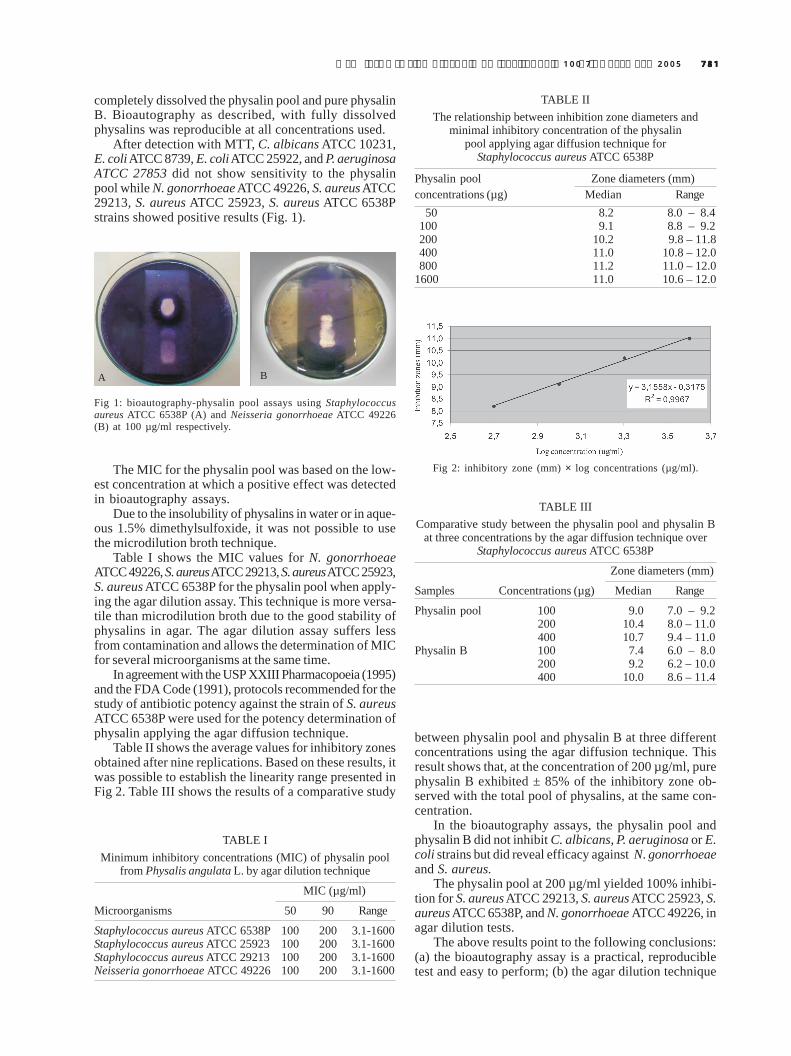
After detection with MTT, C. albicans ATCC 10231,E. coli ATCC 8739, E. coli ATCC 25922, and P. aeruginosaATCC 27853 did not show sensitivity to the physalinpool while N. gonorrhoeae ATCC 49226, S. aureus ATCC29213, S. aureus ATCC 25923, S. aureus ATCC 6538Pstrains showed positive results (Fig. 1).
between physalin pool and physalin B at three differentconcentrations using the agar diffusion technique. Thisresult shows that, at the concentration of 200 µg/ml, purephysalin B exhibited ± 85% of the inhibitory zone ob-served with the total pool of physalins, at the same con-centration.
In the bioautography assays, the physalin pool andphysalin B did not inhibit C. albicans, P. aeruginosa or E.coli strains but did reveal efficacy against N. gonorrhoeaeand S. aureus.
The physalin pool at 200 µg/ml yielded 100% inhibi-tion for S. aureus ATCC 29213, S. aureus ATCC 25923, S.aureus ATCC 6538P, and N. gonorrhoeae ATCC 49226, inagar dilution tests.
The above results point to the following conclusions:(a) the bioautography assay is a practical, reproducibletest and easy to perform; (b) the agar dilution technique
A B
Fig 1: bioautography-physalin pool assays using Staphylococcusaureus ATCC 6538P (A) and Neisseria gonorrhoeae ATCC 49226(B) at 100 µg/ml respectively.
The MIC for the physalin pool was based on the low-est concentration at which a positive effect was detectedin bioautography assays.
Due to the insolubility of physalins in water or in aque-ous 1.5% dimethylsulfoxide, it was not possible to usethe microdilution broth technique.
Table I shows the MIC values for N. gonorrhoeaeATCC 49226, S. aureus ATCC 29213, S. aureus ATCC 25923,S. aureus ATCC 6538P for the physalin pool when apply-ing the agar dilution assay. This technique is more versa-tile than microdilution broth due to the good stability ofphysalins in agar. The agar dilution assay suffers lessfrom contamination and allows the determination of MICfor several microorganisms at the same time.
In agreement with the USP XXIII Pharmacopoeia (1995)and the FDA Code (1991), protocols recommended for thestudy of antibiotic potency against the strain of S. aureusATCC 6538P were used for the potency determination ofphysalin applying the agar diffusion technique.
Table II shows the average values for inhibitory zonesobtained after nine replications. Based on these results, itwas possible to establish the linearity range presented inFig 2. Table III shows the results of a comparative study
TABLE IMinimum inhibitory concentrations (MIC) of physalin pool
from Physalis angulata L. by agar dilution technique
MIC (µg/ml)
Microorganisms 50 90 Range
Staphylococcus aureus ATCC 6538P 100 200 3.1-1600Staphylococcus aureus ATCC 25923 100 200 3.1-1600Staphylococcus aureus ATCC 29213 100 200 3.1-1600Neisseria gonorrhoeae ATCC 49226 100 200 3.1-1600
TABLE IIThe relationship between inhibition zone diameters and
minimal inhibitory concentration of the physalinpool applying agar diffusion technique for
Staphylococcus aureus ATCC 6538P
Physalin pool Zone diameters (mm)concentrations (µg) Median Range
50 8.2 8.0 – 8.4100 9.1 8.8 – 9.2200 10.2 9.8 – 11.8400 11.0 10.8 – 12.0800 11.2 11.0 – 12.0
1600 11.0 10.6 – 12.0
TABLE IIIComparative study between the physalin pool and physalin B
at three concentrations by the agar diffusion technique overStaphylococcus aureus ATCC 6538P
Zone diameters (mm)
Samples Concentrations (µg) Median Range
Physalin pool 100 9.0 7.0 – 9.2200 10.4 8.0 – 11.0400 10.7 9.4 – 11.0
Physalin B 100 7.4 6.0 – 8.0200 9.2 6.2 – 10.0400 10.0 8.6 – 11.4
Fig 2: inhibitory zone (mm) × log concentrations (µg/ml).

782782782782782 Antimicrobial studies on P. angulata L • Melissa TG Silva et al.
is more versatile than the broth dilution assay and doesnot present the problems encountered with this latter as-say with the sample solution, contamination and MICdetermination; (c) the agar diffusion technique can be usedonly for pure substances because when it is applied tomixtures containing constituents, which exhibit differentdiffusion factors, and, thus the results may be unreliable;(d) the methods related here-in permit the choice of thebest tool for determination of the antimicrobial constitu-ents from plant material.
ACKNOWLEDGEMENTS
To the National Institute of Health and Control for techni-cal support.
REFERENCES
Bauer AW, Kirby MM, Sherris JC, Turck M 1966. Antibioticsusceptibility testing by a standardized single disk method.Am J Clinl Pathol 45: 493-496.
Cáceres A, Menéndez H, Méndez E, Cohobón E, Samayoa BE,Jauregui E, Peralta E, Carrillo G 1995. Antigonorrhoeal ac-tivity of plants used in Guatemala for the treatment ofsexually transmitted diseases. J Ethnopharmacol 48: 85-88.
Chiang HC, Jaw SM, Chen CF, Kan WS 1993. Antitumor agent,physalin F from Physalis angulata L. Anticancer Res 12:837-844.
CLSI-Clinical and Laboratory Standards Institute 1993. Meth-ods for dilution antimicrobial susceptibility tests for bacte-rial that grow aerobically, 3rd ed., Approved Standard (M7-A3), Wayne, PA.
CLSI-Clinical and Laboratory Standards Institute 2001. Per-formance standards for antimicrobial susceptibility testing11th ed., Information supplement (M2-A7 e M7-A5),Wayne, PA.
Dos Santos JAA, Ribeiro IM, Drummond D, Silva MTG,Morais ZB, Tomassini TCB 2003. Mollusccidal activity ofPhysalis angulata l. extracts and fractions on Biomphalariatenagophila (d’ Orbigny, 1835) under laboratory conditions.Mem Inst Oswaldo Cruz 98: 425-428.
Farmacopéia Brasileira 1988. Ensaio Microbiológico de Anti-bióticos, 4a ed., São Paulo, v.5.2.1.7.
FDA-Food and Drug Administration 1991. Code of FederalRegulation: Food and Drugs - CFR: 21 parts, 300-499,Washington DC: Office of the Federal Register NationalArchives and Records Administration as a Special Editionof the Federal Register, p. 328-345.
Freiburhaus F, Kaminstry R, Nkunya NHH, Brun R 1996. Evalu-ation of African medicinal plants for their “in vitro” try-panocidal activity. J Ethnopharmacol 55: 1-11.
Januário AH, Rodrigues Filho E, Pietro RCLR, Kashima DN,Sato DN, França SC 2002. Antimycobacterial physalinsfrom Physalis angulata L. (Solanaceae). Phytother Res 16:445-448.
Kline RM, Golab T 1965. A simple technique in developingthin-layer bioautography. J Chromatogr 18: 409-411.
Nostro A 2000. Extraction methods and bioautography for evalu-ation of medicinal plant antimicrobial activity. Lett ApplMicrobiol 30: 379-384.
Ogunlana EO, Ramstad E 1975. Investigations into the antibac-terial activities of local plants of Nigeria. Planta Med 27:354-360.
Pietro RCLR, Kashima S, Sato DN, Januário AH, França SC2000. In vitro antimycobacterial activities of Physalisangulata L. Phytomedicine 7: 335-338.
Rahalison L, Hamburguer M, Monod M, Hostettmann K 1994.Antifungal tests phytochemical investigations: comparisonof bioautographic methods using phytopathogenic and hu-man pathogenic fungi. Planta Med 60: 41-44.
Ray AB, Gupta M 1994. Withasteroids, a growing group ofnaturally occurring steroidal lactones. In W Herz, GW Kirby,RE Moore, W Steglich, CH Tamm (eds), Progress in theChemistry of Organic Natural Products, SpringerVerlag,Austria, p. 2-13 and 56-58.
Sanches EG, Silva MTG, Ribeiro IM, Tomassini TCB 1997.Evaluation of the antibacterial activity of Physalis angulataL. extracts. Boll Chim Farm 2: 136.
Silva MTG, Marques GH, Ribeiro IM, Sanches EG, TomassiniTCB 1999. Evaluation of the bacteria inhibitory profilebetween native Belém do Pará and Rio de Janeiro cultivatedP.angulata L. species against S.aureus, E.coli , K.pneumoniae. Boll Chim Farm 2: 138.
Soares MBP, Bellintani MC, Ribeiro I M, Tomassini TCB, DosSantos RR 2003. Inhibition of macrophage activation andlipopolysaccharide-induced death by seco-steroids purifiedfrom Physalis angulata L. Europ J Pharmacol 459: 107-112.
Tomassini TCB, Barbi NS, Ribeiro IM, Xavier, DCD, inven-tors; Fiocruz, Far-Manguinhos assignee 1999. Processo deisolamento de fisalinas a partir de plantas e composiçõesfarmacêuticas com atividade anti-protozoa contendofisalinas. Brazilian patent BR 9904635, July 23, Int. Cl 09/417779.
Tomassini TCB, Barbi NS, Ribeiro IM, Xavier DCD 2000.Gênero Physalis – Uma revisão sobre vitaesteróides. Quí-mica Nova 23: 47-57.
USP-United States Pharmacopoeia 1995. Biological tests andassays. antibiotics-microbial assays, 23rd ed., United StatesPharmacopoeia Convention, INC. 1690-1696, TwinbrookParkway, Rockville, MD.