studies of the life cycle of diplocarpon rosae wolf on roses and
TRANSCRIPT
Institut für Pflanzenkrankheiten
der
Rheinischen Friedrich-Wilhelms-Universität Bonn
Studies of the life cycle of Diplocarpon rosae Wolf on roses and the
effectiveness of fungicides on pathogenesis
Inaugural-Dissertation
zur
Erlangung des Grades
Doktor der Agrarwissenschaften
(Dr. agr.)
der
Hohen Landwirtschaftlichen Fakultät
der
Rheinischen Friedrich-Wilhelms-Universität
zu Bonn
vorgelegt am 29.12.2004
von
Emma Wanjiru Gachomo
aus
Nyeri, Kenya

Referent: Prof. Dr. H.-W. Dehne
Korreferent. Prof. Dr. W. Schumacher
Tag der Mündlichen Prüfung: 28.04.05
Gedruckt bei: Cuvillier Verlag, Goettingen
Erscheinungsjahr: 2005
II

Dedicated to
Rachel Mūthoni, Gachomo Karwega and Bryan Gachomo
III

Abstract The blackspot disease of roses caused by Diplocarpon rosae Wolf teleomorph (anamorph
Marssonina rosae) is a widespread and important disease on outdoor grown roses. The
development of D. rosae in rose leaves is not clearly understood and no detailed well-
documented photographs of the fungus development in the host are available. The control of
this pathogen heavily relies on fungicides. The objective of this study was to provide a
detailed growth pattern the D. rosae inside the rose leaves and to test the effectiveness of
fungicides, the strobilurins and azoles, on the various fungal structures formed by this fungus.
The aggressiveness of various Kenyan and Germany isolates was investigated. A German
isolate was used to study the life cycle of D. rosae. The life cycle of the fungus was studied
using different staining techniques for the light microscopy and the conventional preparation
for electron microscopy: A conidium germinated to form a germ tube, from which an
appressorium was sometimes formed. A brown ring that was presumably melanized was
formed at the point of penetration of the host cuticle. Upon penetration an infection vesicle
was formed in the subcuticular region. Primary hyphae spread from the infection vesicle into
the subcuticular and intercellular regions in the host. The subcuticular, intercellular and
intramural hyphae formed haustoria of varying shapes in the epidermal cells. The intercellular
hyphae also formed haustoria in palisade mesophyll cells. Intracellular hyphae were formed
just before the formation of the reproduction structures. Brown heavily ornamented structures
formed in the overwintering leaves, opened in spring of the following year to release small
thin-walled structures. No ascospores were formed in the subepidermal apothecia-like
structures. The strobilurins completely inhibited germination of the conidia of D. rosae but
the azoles did not. The azoles were able to destroy the fungal structures in the host even when
they were applied after the fungus was fully established in the host but the effectiveness of the
strobilurins was limited. Protective and curative tests of the effectiveness of the fungicides
were carried out with 100 ppm of the active ingredients.
The study showed that D. rosae is a hemibiotroph: The biotrophic phase is marked by the
formation haustoria and the necrotrophic phase by the formation of the intracellular hyphae.
The weather conditions in Germany may not be suitable for the development of the
ascospores in the subepidermal apothecia. The strobilurins were only effective when applied
protectively. The azoles were effective when applied pre and post inoculation.
IV

Zusammenfassung Der Sternrußtau (Schwarzfleckigkeit), Hauptfruchtform Diplocarpon rosae Wolf, (Neben-
fruchtform Marssonina rosae), ist eine häufige und weit verbreitete Krankheit an
Freilandrosen. Bisher war weder die Entwicklung des Pilzes im Blattgewebe geklärt, noch
waren in der Literatur fotografische Aufnahmen zur endophytischen Pilzentwicklung zu
finden. Die Bekämpfung des Pilzes wird überwiegend mit Fungiziden durchgeführt. Die
Untersuchungen vorliegender Arbeit umfassen sowohl mikroskopische Studien zur exakten
Ausbreitung des Erregers im Rosenblatt als auch die Wirkung unterschiedlicher Fungizide,
insbesondere Strobilurine und Azole, auf pilzliche Strukturen.
Zunächst wurde die Aggressivität verschiedener Isolate aus Kenia und Deutschland getestet.
An einem deutschen Isolat konnte der Entwicklungszyklus von D. rosae mit verschiedenen
Färbetechniken lichtmikroskopisch sowie nach konventioneller Präparation elektronenmikro-
skopisch studiert werden: Nach der Konidienkeimung entsteht eine Keimhyphe, von der aus
meist ein Appressorium gebildet wird. Auf der Wirtskutikula zeigt sich dann an der Stelle der
späteren Penetration ein brauner Ring, der vermutlich melanisiert ist. Nach der Penetration
bildet sich in der subkutikulären Region ein Infektionsvesikel. Primäre Hyphen durchdringen
von diesem Infektionsvesikel ausgehend subkutikuläre und interzelluläre Wirtsgewebezonen.
Subkutikuläre, interzelluläre und intramurale Hyphen bilden in den epidermalen Zellen
Haustorien unterschiedlicher Formen. Interzelluläre Hyphen produzieren ebenfalls Haustorien
im Palisadenparenchym. Intrazelluläre Hyphen entstehen erst kurz vor der Bildung der
Reproduktionsorgane. Braune, stark ornamentierte Strukturen werden vom Pilz auf den
überwinternden Blättern gebildet. Diese öffnen sich im Frühjahr des Folgejahres und
entlassen dünnwandige Organe. In diesen subepidermalen Apothezien-ähnlichen Strukturen
entstehen aber keine Ascosporen. Strobilurine hemmten die Konidienkeimung vollständig,
während Azole einen weniger starken Einfluss auf die Keimung hatten. Dagegen zerstörten
die Azole alle Pilzstrukturen im Inneren des Gewebes, auch wenn sie sich bereits gut etabliert
hatten. Diese Reaktion konnte bei Einsatz der Strobilurine nicht so deutlich beobachtet
werden. Alle Fungizidtests zur protektiven und kurativen Wirkung wurden mit jeweils 100
ppm reinem Wirkstoff durchgeführt.
Die Untersuchungen zeigten, dass D. rosae eine hemibiotrophe Lebensweise besitzt: die
biotrophe Phase besteht aus der Haustorienbildung und die nekrotrophe Phase aus der Bildung
interzellulärer Hyphen. Die deutschen Witterungsbedingungen eignen sich offenbar nicht für
eine subepidermale Apothezienbildung. Strobilurine zeigten nur nach protektiver Applikation
eine Wirkung. Azole wirkten sowohl bei prä- als auch bei post-inokulativer Behandlung.
V

Table of contents………………………………………………................…….Page
1 Introduction…………………………………………………………………….……1
2 Materials and Methods…………………………………………………………….10
2.1 Research organisms.………………………………………………….…….…….10
2.1.1 Pathogen……………………………………………………………………...10
2.1.2 Plants………………………………………………………………….……...10
2.2 Plant cultivation…………………………………………………………….…....10
2.3 Pathogen isolation and inoculum harvesting…………………….…..……..……11
2.4 Inoculation of plants……………………………………………….……….…....11
2.5 Disease assessment…………………………………………………..…………..12
2.6 Maintenance of the pathogen and multiplication of the inoculum…..…………..14
2.7 Evaluation of the fungal growth in vitro………………………………………...14
2.7.1 Preparation of growth media………………………………………….……..14
2.7.2 Incubation on growth media…………………………………………………15
2.8 Microscopical investigations……………………………………………….……15
2.8.1 Light microscopy…………………………………………………………….16
2.8.2 Transmission electron microscopy…………………………….……….……16
2.8.3 Specimen preparation techniques…………………………………………....16
2.8.3.1 Fresh specimen………………………………………………….……….16
2.8.3.2 Whole specimen………………………………………....………………17
2.8.3.2.1 Fixation with chloral hydrate……….……………………...………..17
2.8.3.3 Sectioned specimen…………...….……………………………………...17
2.8.3.3.1 Embedding in ERL-resin…………………………………………….18
2.8.3.3.2 Sectioning…………………………………………………….……...19
2.8.3.3.3 Contrasting…………………………………………………………..20
2.8.4 Staining techniques…………………………………………………….…….21
VI

2.4.8.1 Blancophor……………………………………………………………….21
2.8.4.2 Diethanol (Uvitex 2b)……………………………………………………21
2.8.4.3 Fuchsin acid……………………………………………………………...22
2.8.4.4 Aniline blue……………………………………………………………....23
2.8.4.5 Toluidine blue……………………………………………………………23
2.9 Estimation of reactive oxygen species (ROS) production in leaf tissue………....24
2.9.1 Superoxide anion (O2-)...................................................................…….…..24
2.9.2 Hydrogen peroxide (H2O2)..........................................................…………..25
2.10 Fungicide tests…………………………………………………….…………....25
2.10.1 In vitro tests……………………………………………………..……..…...25
2.10.2 In planta tests………………………………………………………….…...26
2.10.2 1 Protective activity tests……………………………………….………...26
2.10.2.2 Curative activity tests…………………………………………….….....27
2.11 Statistical analysis and software programmes…………………………..……...29
3 Results……………………………………………………………………………....30
3.1 Symptomatology of the blackspot disease caused by
Diplocarpon rosae on roses………………………………………..…….……...30
3.1.1 Symptoms on inoculated plants……………………………….….……….....30
3.1.2 Disease scoring………………………………………………………....…....32
3.2 Characterization of D. rosae isolates……………………………………….…....32
3.2.1 Germination rate………………………………….…………………….…....32
3.2.2 Process of symptom development………………………………….………..33
3.2.2.1 Effect of different temperatures………………………………….……....35
3.2.3 Assessment of the aggressiveness in vivo: accumulation of
reactive oxygen species…………………………………………………….37
3.2.3 1 Superoxide anion (O2-) in the leaf tissue………………………………...37
VII

3.2.3 2 Hydrogen peroxide (H2O2) in the leaf tissue………………………………38
3.3 Microscopical studies of the infection process…………………….……………...39
3.3.1 Growth on the host leaf surface…………………………………………….....39
3.3.1.1 Germination of conidia…………………………………………….……...39
3.3.1.2 Appressorium Formation………………………………………………....40
3.3.2 Penetration of the host cuticle and growth in the host tissue…………………41
3.3.3 Subcuticular growth………………………………………………..………....42
3.3.4 Invasion of the host tissue………………………………………….………....44
3.3.4.1 Intercellular hyphae……………………………………………………....44
3.3.4.2 Intramural mycelia……………………………………………….……….45
3.3.4.3 Haustorium…………………………………………………….………....46
3.3.4.4 Intracellular hyphae……………………………………………………....49
3.3.5 Fructification…………………………………………………………………50
3.3.6 Growth through the lower side of leaf……………………………….……….52
3.3.7 Overwintering………………………………………………………….……..53
3.3.7.1 On fallen leaves…………………………………………………….…….53
3.3.7.2 On the canes……………………………………………………..……….54
3.4 Investigations of the effect of nutrient source
on the fungal development……………………………………………………….55
3.4.1 Development of D. rosae on artificial growth media………………………..55
3.4.2 Fungus development in detached leaves and intact plants……….….……….69
3.5 Investigations into the effectiveness of fungicides on the
development of D. rosae………………………………………………..………..70
3.5.1 Effect on symptom development……………………………………….……70
3.5.2 Effect on pre-infection structures…………………………………..………...76
3.5.2.1 In vitro analysis………………………………………………….……….76
VIII

3.5.2.2 In vivo analysis…………………………………………………..………76
3.5.3 Effectiveness on the different fungal structures
after curative applications……………………………………………………78
3.5.3.1 Effectiveness of application on the 3rd dpi……………………………….78
3.5.3.2 Effectiveness of application on the 7th dpi……………………………….88
3.5.3.3 Effectiveness of application at the 10th dpi………………………………92
4 Discussion…………………………………………………………………………..96
5 References…………………………………………………………………………127
IX

Abbreviations
dpi days post inoculation
PDA potato dextrose agar
MEA malt extract agar
BMA biomalt agar
WA water agar
NBT nitroblue tetrazolium
ERL Epoxy resin low (Spurr’s resin)
a.i. active ingredient
ppm parts per million
G1 Germany isolate 1
K1 Kenya isolate 1
TEM transmission electron microscope
EHM extrahaustorial membrane
EHX extrahaustorial matrix
SD standard deviation
X
1 Introduction
The rose is a widely studied plant and much has been written about it. Without delving
much in to the history suffice it to say that the wild roses are believed to have their
origins in Europe, America, the orient and China, and in the Middle East (Beales 1997,
Joyaux 2003). Rose cultivation dates way back to around 3000 B.C. in the ancient
Chinese gardens (Beales 1997). Three important organizations are concerned with the
formulation of classes and the nomenclature of roses: The American Rose society
through their publications “Modern Roses”, The World Federation of Roses Societies,
and the British Association Representing Breeders (Baeles 1997). Beales (1997) has also
given what he calls a simplified classification of roses based on their flower
characteristics. The details of rose classification and the associated controversies are not
the subject here, but it is important to mention that roses belong to the family Rosaceae,
and Genus Rosa.
The rose has established itself as the most popular garden plant in the world as well as the
most important commercial cut flower grown under glass (Horst 1983). Cut flowers
contributed to 2,787 million of trade turnover in Europe in the year 2000 (Eurostat 2000).
A fifth of the cut flowers were from none European countries and roses made 47 % of the
imports from none European countries, which was worth 282 million Euros in the year
2000 (Eurostat 2000). According to the year 2000 statistical analysis the main destination
for all cut flower imports into Europe was Netherlands but the highest consumer of cut
flowers in Europe was Germany. The highest ranked exporters of cut flowers into the
Europen Union (EU) were Kenya (25 %), Colombia (17 %) and Israel (16 %) to mention
but a few (Eurostat 2000).
1

Despite the popularity of the rose, its cultivation is associated with various diseases. The
most important rose diseases caused by fungal pathogens are: powdery mildew caused by
Sphaerotheca pannosa, the blackspot disease by Diplocarpon rosae, and rust by
Phragmidium species. The most important bacterial disease is the bacterial wilt caused by
Agrobacterium tumefaciens.
The blackspot of roses is a foliar disease recognized by black spots on the upper side of
the leaf. The symptoms are brown to black circular spots with an irregular margin on the
upper surface of the leaf, which are followed by yellowing and premature defoliation.
The disease is caused by a fungal pathogen with the perfect stage Diplocarpon rosae wolf
(Wolf 1912) and the imperfect stage that is called Marssonina rosae (Lib.) Lind (Baker
1948). Other synonyms to the imperfect stage are Asteroma rosae, Actinonema rosae,
and Marsonina rosae (Baker 1948, Horst 1983) among many others.
In temperate regions the first infections in spring are caused by the conidia or ascospores
found in the overwintered plant material. The fungus overwinters on dormant stems,
thorns, fallen leaves, and buds (Lyle 1943, Cook 1981). The conidia are spread by wind,
rain splash and probably by animal vectors including insects and arachnids (Palmer et al
1978). Records of the apothecia containing eight ascospores are rare and the importance
of the genetic variation generated by sexual recombination is not clear (Walker 1995).
The formation of apothecia has been reported in North America (Wolf 1912, Aronescu
1934), Great Britain (Knight and Wheeler 1977, Cook 1981) and in Russia (Dudin 1972).
2
The disease was first described in Europe in Sweden in 1815, then in Belgium in 1827
and later in other European countries (Baker 1948). The first report in North American
were in 1831, South America in 1880, Australia 1892, Africa in 1920, and in China in
1910 as summarized by Baker (1948). Today the disease has a worldwide distribution,
even occurring on oceanic islands like the Philippines and Hawaii (Horst 1983, Drewes-
Alvarez 2003).
Blackspot of roses is an important, devastating and widespread disease. In out door
growing of roses black spot disease is generally present, often as a major problem, and
frequently in epidemic proportions (Horst 1983). It is a minor problem in greenhouse
roses because humidity is regulated very carefully but is the most important disease of
out door roses (Horst 1995). The rose plant is used in gardens and landscaping for its
aesthetic value, but the blackspot infections make the roses unsightly due to the black
spots on the leaves, yellowing and premature defoliation. The pathogen causes
defoliation and weakening of the plants (Drewes-Alvarez 2003). The premature
defoliation leads to reduced vigour (Smith et al 1988) and even death in very susceptible
varieties (Black et al 1994). The damage caused by the D. rosae is greater than the leaf
spots because of the added effect of the premature defoliation. Disease damage cannot be
assessed only in terms of size of lesion but always includes the defoliation aspect.
Symptom expression in the blackspot disease has been reported to differ with pathogenic
races, varieties, and environmental conditions. Rose cultivars differ in their susceptibility
to D. rosae and pathogenic races of this pathogen have been reported by several authors
(Debener et al 1998, Malek et al 1996, Bolton and Svejda 1979, Jenkins 1955, Knight
3

and Wheeler 1978b). In a study using a set of differentials of rose cultivars, Malek et al
(1996) identified at least five physiological races of D. rosae.
The widely cultivated tea hybrids and floribunda roses are susceptible to the blackspot
pathogen (Reddy et al 1992, Walker et al 1995). Some of the wild species of roses have
resistance but they lack the desired aesthetic traits (Reddy et al 1992). High resistance to
blackspot has been found in genotypes of R. banksiae Ait, R. carolina L., R. laevigata
Michx., R. multiflora Thunb. Ex Murray, R. rugosa Thunb., R. roxburghii Trait., R.
virginiana Herrm. and R. wichurana Crèp (Drewes-Alvarez 2003). The resistance
mechanism in roses is thought to operate through mechanical resistance of the cuticle
(Castledine et al 1981), diffusible substances (Walker et al 1995, Knight and Wheeler
1978) or to be a post penetration defence response (Wiggers et al 1997). Debener (2003)
reported that in a recent screening of tetraploids and diploids carrying resistance from R.
multiflora with single conidial isolates the presence of two dominant genes Rdr1 and
Rdr2 was revealed, but this does not in any way rule out horizontal resistance.
The resistance found in the wild roses needs to be transferred into the modern cultivars.
Attempts to transfer resistance from the wild species to the modern roses by introgressive
breeding led to a resistant F1 hybrids but the resistant was lost in the first backcross
maybe due to lack of pairing partner at meiosis (Walker et al 1995). The integration of
resistance genes by crosses with wild species is complicated due to varying ploidy levels
in rose species and possible linkage of resistance genes with undesirable traits (Dohm et
al 1996). In addition to that, breeding of rose cultivars that are resistant to blackspot is
very time consuming because of the generative reproduction and complex genetic
4

constitution of roses (Malek et al 1996). Research for the development of resistance
against the disease are still under way (Debener et al 1996, Debener 1996), but the
control of black spot still requires intensive use of systemic fungicides (Walker et al
1995, Reddy et al 1992).
Once established on plants, blackspot is difficult to control despite a combination of
practices that include sanitation measures and fungicide applications (Behe et al 1993).
Good cultural practices include: Removing disease leaves from the ground and pruning
canes with infected leaves to reduce the overwintering potential of the pathogen; avoiding
dense planting to allow good air circulation through the leaf canopy (Horst 1983);
avoiding overhead irrigation since it favours infection; avoiding excessive watering
during dark and humid weather; avoiding keeping the rose leaves wet for long hours, as
this provides the water needed for the conidia to germinate.
In general, a good understanding of a pathogen’s life cycle is a prerequisite to its
effective control. Despite several research contributions to the understanding of the
development of D. rosae in roses, there are still discrepancies in the observations made
on its life cycle. The existing histological studies of D. rosae (Aronescu 1934, Frick 1944
and Palmer et al 1978) were limited by the scope of the then available techniques and
equipment. Aronescu (1934) and Frick (1944) made drawings of their microscopic
observations, while Palmer et al (1978) commented on the inability to make better
photographs due to poor sectioning apparatus. There are no detailed ultra structure
studies of the infection process of D. rosae on roses. Therefore, there is need to have a
detailed and more accurate documentation of the infection process of D. rosae using
5

modern technology and there is need to shed more light into the discrepancies in the life
cycle of this pathogen.
Given that the management of D. rosae heavily relies on chemical control, there is need
to keep on testing effectiveness of new fungicides active ingredients in controlling this
pathogen. Due to the increase in the general environmental protection awareness world
wide, the need to replace the fungicides in the market with more environmentally friendly
ones has never been more timely. This can be achieved through testing of new more
environmental friendly fungicidal active ingredients with the hope of offering better
disease management.
The conidia of the blackspot pathogen germinate to form germ tubes on the host surface
but further development of the fungus takes place below the host cuticle and even within
the host cells. The control of this type of pathogen requires the use of contact as well as
systemic fungicides. Couch (1995) distinguishes fungicides by their topical or physical
mode of action: contact fungicides stay outside of plants, and penetrants penetrate the
plant in some manner. Localized penetrants diffuse into leaf surfaces. Acropetal
penetrants are transported by xylem towards the leaf tips after they penetrate (upward
movement) but do not move in the phloem towards the root tips. Systemic penetrants are
fungicides that are transported by both xylem and phloem. Penetrant fungicides in
general offer the possibility of prolonging disease control, as the fungicide protects plant
surfaces, but they also may inhibit pathogens in the early stages of infection inside the
plant.
6

One group of fungicides that have been widely used for the control of many leaf spot
pathogens including D. rosae are the demethylation inhibitors or DMI fungicides. This is
a homogenous group of fungicides showing a common mode of action within the fungal
sterol biosynthesis pathway i.e. the inhibition of demethylation at position 14 of
lanosterol or 24-methylene dihydrolanosterol, which are precursors of sterols in fungi
(Kuck et al 1996). Azoles such as imidazole and triazoles derivatives are the most
important DMI fungicides. DMIs have a broad spectrum of fungicidal activity with most
fungi from the Ascomycetes, Basidiomycetes and Fungi Imperfecti. As reported by Kuck
et al (1996), a multitude of leaf spot pathogens are effectively controlled by several
DMIs. According to the report of Newmann and Jacob (1996), all DMIs are able to
penetrate the plant cuticle and /or seed coat to some extent and they are then translocated
in either the apoplast (transport in coherent network of free space, cell walls and non-
living cells) or symplast (transport in the coherent network of protoplasts connected by
plasmodesmata). DMIs have minimal symplastic/basipetal movement i.e. movement in
the living part of the plant which is the phloem and cytoplasm, but their translocation is
predominantly apoplastic and protection is confined to such cases where sufficient
quantities of active ingredients have been applied to the basal parts of the leaf or shoot
(Kuck et al 1996). The DMIs of interest in this study are two triazoles, namely
myclobutanil and tebuconazole.
A new group of fungicides with much promise in the control of a wide range of fungal
pathogens are the strobilurins. Strobilurin fungicides were developed following the
discovery of naturally occurring β-methoxyacrylates, the simplest being strobilurin A and
oudemansin A produced naturally in certain species of mushroom fungi, including
7

Strobilurus tenacellus and Oudesmansiella mucida (Ypema and Gold 1999). These
fungicides are now more properly referred to as QoI. Industrial chemists improved these
natural fungicides by making chemical modifications that resulted in compounds that
were less subject to break down on the leaf surface by sunlight. More strobilurins
continue to be released in the market. Some of the fungicides active ingredients found in
this group include trifloxystrobin, pyraclostrobin, kresoxim-methyl and azoxystrobin
(Bartlett et al 2001).
As reported by Sauter et al (1995), all QoI fungicides share a common biochemical mode
of action: they all interfere with energy production in the fungal cell. They block the
electron transfer at the site of quinol oxidation (the Qo site) in the cytochrome bc1
complex, thus preventing ATP formation. They are site-specific fungicides.
Ebeling (2003) reporting on one of these active ingredients, namely trifloxystrobin, said
that it has a very favourable profile concerning toxicology, residue behaviour,
environmental fate and ecotoxicology. It has low acute toxicity, no likelihood of causing
acute hazards, no genotoxic or carcinogenic potential and also no signs of neurotoxicity.
The active ingredient rapidly degrades in the environment and no risk to the consumer is
expected by the way of residue intake through food.
With important exceptions, the QoI control an unusually wide array of fungal diseases,
including diseases caused by water molds, downy mildews, powdery mildews, leaf
spotting and blight fungi, fruit rotters, and rusts. They are used on a wide variety of crops,
including cereals, field crops, fruits, tree nuts, vegetables, turf grasses and ornamentals
(Stark-Urnau et al 1997, Gold and Leinhos 1995). However, the effectiveness of these
8

new fungicide active ingredients on the fungal structures formed by D. rosae has not
been studied in detail.
This study was undertaken with the main aim of carrying out a detailed study of the life
cycle of Diplocarpon rosae in roses leaves in order to clarify the discrepancies found in
literature. The objectives also included an investigation into the state and the plant parts
in which the fungus overwinters in the weather conditions in Germany. The study
targeted at assessing the aggressiveness of D. rosae isolates from Kenya and Germany by
visual observation of the isolates that led to the earlier development of disease symptoms,
and through biochemical assays of relative accumulation of reactive oxygen species
(ROS) in leaves of plants inoculated with different isolates. The objectives encompassed
a disease control aspect in which the effectiveness of the novel fungicide active
ingredients, the strobilurins, on the fungal structures within the host tissues at different
developmental stages was compared with the effectiveness of the fungicide active
ingredients already in the market.
9

2 Materials and methods
2.1 Research organisms
2.1.1 Pathogen
The pathogen used was Diplocarpon rosae Wolf, which is the teleomorph state and the
anamorph is called Marssonina rosae (Lib.) Lind. Two isolates from Germany collected
in 2001 and 2002 from Duesseldorf and Bonn respectively, and 20 isolates from Kenya
collected in 2001 from Kiambu, Nairobi, Naivasha, Nyeri and Thika districts were used.
2.1.2 Plants
The following cultivars of roses were used in this work: a floribunda rose cv. Frensham
of parentage [Floribunda seedling X crimson Glory] obtained from Belle Epoque
Rozenwekerij breeders, Netherlands; two hybrids teas cv. Claudia and cv. Rosita that
were obtained from Koster breeders in Grefrath, Germany.
2.2 Plant cultivation
Rose cv. Frensham bushes were cut back to 3-5 cm from the budding point and the roots
wetted before potting. The pots were at least 17 cm in diameter and 25 cm deep as
recommended by the breeders. The initial planting soil was composed of “Klasmann
special mixture”: field soil: compost soil in the ratio of 2:2:1 and 5 g “plantosan” long-
term fertilizer (20:10:15). The hybrids teas were planted in pots of 10.5 cm and in the
same soil mixture but only 2 g of the “plantosan” fertilizer per plant was added. The
potted plants were maintained in the green house with a minimum temperature of 20 oC ±
5 and 16 h light all the year round. Heaters that came on when the temperature fell below
21 °C and air circulation that automatically started by the opening of the windows when
10

the temperature was above 25 °C maintained the greenhouse temperature. The day length
of 16 h was maintained by using Philips sodium vapour lamps SGR 140 with a lighting
strength of 300 µE/m2. The plants were fertilized with 2.5-3 g/l “Flory 2 spezial”
(16:9:22) at the rate of 200 ml per plant every 2 weeks for the cv. Frensham and 20 ml
per plant for the two hybrid tea cultivars.
2.3 Pathogen isolation and inoculum harvesting
Diseased leaves with fully developed black spot symptoms that had been sampled from
different locations were incubated in moist chambers at room temperature (20 oC ± 5) for
3 days. Leaves from each location were incubated separately. The leaves were then
washed with sterile water and the concentration of conidia in the suspension adjusted to 1
x 105 conidia/ml. Inoculum from each location was considered as an isolate from that
particular region.
2.4 Inoculation of plants
Inoculations were carried out on both intact plants growing in pots and detached leaves.
Intact plants: The intact plant inoculations were carried out on plants with a minimum of
six fully opened leaves. Potted plants were placed overnight in an incubation chamber
with almost 100 % relative humidity and a temperature of 20 °C. The plants were then
sprayed with a conidial suspension (1 x10⁵ conidia/ml) and placed back in the incubation
chamber for another 2 d. The plants were then taken out of the incubation chamber and
put back in the green house. The untreated control plants were sprayed with sterile
distilled water.
11

Detached leaves: For the detached leaf technique, the youngest fully opened
(penultimate) leaves, -which is usually about the fifth youngest leaf and not from a
flowering stem -were used. Leaflets were detached from the plants and washed in running
tap water for 10 min and then surface sterilized in 3 % sodium hypochloride for 3 min.
The leaflets were then rinsed in three changes of sterile distilled water and placed on
sterile moist filter papers in 90 mm petri dishes with the adaxial side up as described by
Palmer et al (1966). Alternatively 1cm diameter leaf discs were cut out from the sterilized
leaflets on either side of the midrib using a sterile core borer and placed on the filter
papers. The leaflets or the discs were inoculated with 5 µl droplets of the conidial
suspension (5 x 10⁴ conidia/ml) and incubated at 20 °C. The use of discs was avoided
because they dried up earlier than the intact leaflets and they were also more often
contaminated.
A lower inoculum concentration was used on the detached leaves so that the germinating
conidia at infection site could be well spread out from each other in a manner that
observations of each germinating conidia could be made without obstruction from the
others. In the control the leaflets were inoculated with 5 µl droplets of sterile distilled
water.
2.5 Disease assessment
The conidial suspension of the different isolates was used to inoculate two hybrid tea
roses/cultivar/isolate. The hybrid teas were used in disease assessment with the aim of
isolate characterization because of their ability to produce new leaves within a short time.
The growth pattern of the different isolates on the plants was compared with respect to
12

germination rate, and disease severity. The germination percentage for each isolate was
calculated and a disease scale of 0 - 100 % was used to determine the disease severity.
The disease scale was divided into five classes of disease severity as follows:
0 % - no symptoms,
1-20 % - spots less than 5 mm, no yellowing and no defoliation,
20- 40 % - spots less than 5 mm, yellowing and/or defoliation,
40-60 % - spots 5-10 mm, no yellowing and no defoliation,
60-80 % - spots of 5-10 mm, yellowing and/or defoliation,
80-100 % - spots of a diameter greater than 10 mm, yellowing and/or defoliation
The results were used to select isolates showing significant differences in germination
rate and disease severity, which were then used as representative isolates in subsequent
experiments.
The same disease scale was used to compare the growth rate of the representative isolates
on both intact plants and detached leaves, and at different incubation temperatures, 10 °C,
15 °C, 20 °C and 25 °C. The number of days required from inoculation to symptom
expression on the detached leaves and intact plants were recorded. On the detached
leaves ten droplets of inoculum were put on each leaflet and the number of black spots
that developed per leaflet were recorded.
2.6 Maintenance of the pathogen and multiplication of the inoculum
The diseased leaves with fully developed black spot symptoms were incubated in a moist
chamber for 3 d and then stored at –20 oC for long-term storage. To make a conidial
suspension the leaves were allowed to defrost, then washed in sterile distilled water and
13
the conidia concentration adjusted to 1 x 10⁵ conidia/ml before being used to inoculate
the healthy plants. To multiply the inoculum detached leaves were inoculated and leaves
with fully developed symptoms were stored at –20 °C.
2.7 Evaluation of the fungal growth in vitro
Three different types of artificial growth media namely, Potato dextrose agar (PDA),
Biomalt agar (BMA) and Malt extract agar (MEA) were used in their full and half
strengths. In addition to these was 1 % water agar (WA) also used.
2.7.1 Preparation of growth media
Following are the contents of the different growth media:
Water agar: 10 g agar, 1000 ml H2O.
PDA: 39 g industrially prepared PDA in 1000 ml H2O or pulp from 200 g potato cubes,
15 g dextrose and 20 g agar, in 1000 ml H2O. Half strength PDA was composed of 19.5 g
industrially manufactured PDA and 7.5 g agar, in 1000 ml H2O, or pulp from 100 g
potato cubes, 7.5 g dextrose, 20 g agar, in 1000 ml H2O.
Biomalt agar: 20 g biomalt and 20 g agar in 1000 ml H2O. Half strength biomalt agar was
composed of 10 g biomalt and 20 g agar in1000 ml H2O.
Malt extract agar: 30 g malt extract, 3 g peptone from soja bean meal and 15 g agar in
1000 ml H2O. Half malt extract agar was composed of 15 g malt extract, 1.5 g peptone
from soja bean meal and 15 g agar, in 1000 ml H2O.
To a litre of each of the autoclaved and cooled (below 80 °C) artificial media were 50 mg
of each of the following antibiotics added: streptomycin sulphate, penicillin G. and
14

chlortetracycline hydrochloride. 10-15 ml of artificial media were poured into each petri
dish.
2.7.2 Incubation on growth media
Four points at the bottom of each of the petri dishes were marked with a permanent
marker and the growth medium above these marked points was seeded with 10 µl of a
conidial suspension containing 1x 10⁴ conidia/ml. The seeded petri dishes were then
incubated at 20 °C. Germination of the conidia and further development until a new crop
of conidia was formed were closely followed. The evaluations were carried out by cutting
out a block of inoculated artificial media and mounting it on a microscopic slide in 10 µl
of diethanol. The block was then observed under UV-light in the fluorescence mode as
described in section 2.8.3.2.
2.8 Microscopic investigations
To investigate the growth pattern of D. rosae on Rosa species, different microscopic and
histochemical techniques were used. Microscopic evaluations were done using a light
microscope as well as a transmission electron microscope.
2.8.1 Light Microscopy
For all light microscopy, a Leitz DMR photomicroscope from Leica was used. The Leitz-
microscope was equipped with Nomarski-interference-contrast and with UV-excitation
for epifluorescence. The filter combinations that were used are given in table 1. Images of
the observed specimens were photographed with a fitted digital camera and could be
observed on a connected screen. The images were saved using the program “Discus”
(Technisches Büro Hilgers, Koenigswinter, Germany).
15

Table 1: Filter combinations for the incident light fluorescence microscope
exciter filter(nm) chromatic beam splitter (nm) barrier filter (nm)
BP 340-380 FT 400 LP 430
BP 355-425 FT 455 LP 460
2.8.2 Transmission electron microscopy
A Zeiss EM 109 transmission electron microscope was used for all electron microscopy
evaluations.
2.8.3 Specimen preparation techniques
2.8.3.1 Fresh specimen
Samples taken at 4-h intervals for the first 72 h post inoculation (hpi) were used in the
observation of post-germination and pre-penetration fungal structures found on the
surface of the leaf and on artificial growth media. Leaf pieces of 1 cm2 were cut out from
either side of the midrib and mounted on a microscopic glass slide in 10 µ l of 0.05 %
diethanol with the adaxial side to the glass slide. The specimens were evaluated without
discolouration of leaf tissue or fixation of fungal tissue. 1 cm2 blocks of artificial growth
media were also treated the same way.
2.8.3.2 Whole specimen
Leaflet samples from plants or detached leaves (inoculated as described in section 2.4)
were taken at 4-hour intervals for the first 72 hours post inoculation (hpi). After the 3rd
day post inoculation (dpi) samples were collected every day. These samples were fixed in
16

chloral hydrate fixing solution (section 2.8.2.2.1) and stained (section 2.8.3) before the
light microscopic observations. 1cm2 pieces were cut out from either side of the midrib
before mounting on the microscopic slide. The same method was also used to prepare
fallen leaves, overwintered leaves and stem barks for microscopic observations.
2.8.3.2.1 Fixation with chloral hydrate
In order to observe and describe the development of the fungal structures inside the leaf
tissue the chlorophyll was first removed and the samples then stained with the desired
staining solutions. The fungal structures were fixed on the leaf surface and the leaflets
discoloured in saturated chloral hydrate (250 g/100 ml H2O) at 60 oC for at least 3 d
(older tissue required longer). After the leaflets were fully cleared off chlorophyll, they
were used in the light microscopic evaluations.
2.8.3.3 Sectioned specimen
To observe the fungal structures inside the host cells using the transmission electron
microscope, vertical sections of the leaf tissue were necessary. They were prepared in the
method below.
2.8.3.3.1 Embedding in ERL-resin
Samples collected from the inoculated leaflets at intervals of 4 h from 48 hpi to 72 hpi,
and then every day up to the symptom appearance were cut into small pieces of 2 x 4
mm2. These pieces were immediately placed in a fixing solution of 8 % formaldehyde: 8
% glutaraldehyde: 0.2 M cacodylic acid sodium salt trihydrate buffer (1:1:2 v/v/v) and
0.005 g calcium chloride (Karnovsky 1965) for a minimum of 2 h at room temperature.
The eppendorf tubes containing the cut pieces in the fixing solution were placed in an air
17

tight glass jar connected to a water stream pump to enable the fixing solution to quickly
penetrate and fix the structures in the leaf sections and also to remove excess air from the
leaf pieces. Cutting the samples into small pieces enabled the fixing solutions to quickly
penetrate into the cells before any cellular alterations had occurred, therefore the fixation
preserved the cell contents in as much of their original state as possible. The samples
were then rinsed about ten times in cacodylic acid sodium salt trihydrate buffer (pH
7.35), and then further fixed in 2 % Osmium tetraoxide (OsO4, Next Chimica, S. Africa)
fixing solution of 0.4 % KCr2O7: 3.4 % NaCl: 2 % OSO4 (1:1:2 v/v/v) according to
Dalton (1955), and modified according to Wohlfahrt-Bottermann (1957) for 1-2 h.
Osmium tetraoxide increases the electron absorption and leads to a better photo-contrast.
The chromium in the fixing solution helps in maintaining the tissue structure and in
fixation of the lipids (Plattner 1981).
The samples were then rinsed up to eight times in cacodylic acid sodium salt trihydrate
buffer (pH 7.35) for 20 min per rinse followed by dehydration in increasing
concentrations of ethanol/aqua bi-distilled water as follows:
20 min in 15 %,
20min in 30 %,
20min in 50 % 2 changes
20 min in 70 % 2 changes
20 min in 90 % 2 changes
60 min in 96 % 4 changes
60min in 100 % 2 changes
Finally the samples were rinsed in 2 changes of propylene oxide of 10 min each. This
was followed by infiltration of the samples in increasing concentrations of ERL-resin.
18

The contents of ERL-resin (Spurr, 1969) are as follows:
10.0 g ERL-4206
4.0 g D.E.R. 736
0.4 g S-1 (Agar Aids)
The samples were put in different ratios of ERL-resin: propylene oxide (v/v) in the
following order:
16 h in 1:3
8 h in 1:1
16 h in 3:1
16 h in 100 % ERL-resin
The samples were then polymerised in 100 % ERL-resin in flat embedding trays (Agar-
Aids) at 70 oC for 8-12 h.
2.8.3.3.2 Sectioning
Semi-thin sections
Semi thin sections of 500 nm thickness were cut using a 45o glass knife and thereby being
directly suspended in distilled water. The semi-thin sections were later suspended in
distilled water on a glass slide, dried on an electric plate at 70 oC and stained in 0.05 %
toluidine blue (w/v) in 0.01 M phosphate buffer (pH 7.4). The sections were rinsed in tap
water to removed excess stain, followed by a rinse in demineralised water and in xylene
for 5 min each. The sections were mounted and sealed in entellan rapid mounting media
(Merck) and allowed to dry over night in a fume chamber before being viewed under the
bright field of a compound light microscope.
19

Ultra-thin sections
When the observations under the light microscope confirmed the presence of the desired
structures in the semi-thin sections, ultra-thin sections were cut out of the same block end
as the semi-thin sections. The ultra-thin sections (70-75 nm thickness) were cut with a
Reichert-Jung Ultramicrotome Ultracut E of 70-72 nm thickness using a diamond knife
(Sitte 1982, 1985) and placed on copper or nickel grids.
2.8.3.3.3 Contrasting
The ultra-thin sections were then contrasted. The enhancement of the contrast before
observations under the electron microscope was achieved through the use of electron
dense material in form of heavy metal salts. The contrasting was done according to Geyer
(1973). The grids were laid out in drops of saturated 2 % uranyl acetate for 8 min and
then rinsed twice in aqua bi-distilled water, then put in drops of lead acetate solution
(Reynolds 1963) for 2 min and rinsed in 2 changes of aqua bi-distilled water. The uranyl
acetate and lead acetate were centrifuged before use. The lead acetate solution was
prepared as follows:
-1.33 g Pb(NO3)2
-1.76 g Na3(C6H5O7) x 2 H2O in 30 ml aqua bi-distilled water
-thoroughly mixed and stored in darkness for 30 min
-the pH was adjusted to a value of 12 using 8 ml 1 N NaOH
-topped to 50 ml with aqua bi-distilled water.
20

The grids were allowed to air dry before being stored in a grid box at room temperature.
The contrasting was carried out in a darkened petri dish lined with parafilm. The grids
were then observed with a Zeiss EM 109 transmission electron microscope.
2.8.4 Staining techniques
2.8.4.1 Blancophor
The conidia germination and formation of pre-penetration structures were confirmed by
staining the specimen with fluorescence stain blancophor. The same stain was used to
stain fungal structures on artificial growth media. Blancophor binds to polysaccharides
with β-glycoside bonds. This stain does not penetrate the plant cuticle, it binds the cell
wall of the pathogen on the plant surface so that conidia, germ tubes and appressoria
clearly fluorescent under UV-light.
The fresh specimen of inoculated leaflet samples or small blocks of artificial medium
were mounted in 10 µl of 0.1 % blancophor solution in 10 % ethanol/H2Obd (v/v) before
being observed. The specimen were covered with a cover slip and observed with the
BP340-380/FT 400/ LP 430 filter combination at the Leitz-microscope with the incident
lights fluorescence mode.
2.8.4.2 Diethanol (Uvitex 2B)
To determine the germination rate and describe the pre-penetration fungal structures
found on the leaf surface, fresh leaf specimen as well as artificial media blocks were
stained in 10 µl of 0.05 % diethanol (w/v). Diethanol binds to polysaccharides with β-
glycosidic bonds. The stain does not penetrate the plant cuticle; therefore it stains the cell
21

wall of the pathogen on the plant surface, so that the conidia, germ tubes and
appressorium on the plant surface fluorescence under UV-light.
The fresh specimen of inoculated leaflet samples or small blocks of artificial medium on
which the fungus were growing were mounted in 10 µl of 0.05 % diethanol (w/v) before
being observed. The specimen was covered with a cover slip and observed with the
BP340-380/FT 400/ LP 430 filter combination at the Leitz-microscope with the incident
light fluorescence mode.
2.8.4.3 Fuchsin acid
The development of the pathogen in the plant tissue as well as the plant defence reaction
to the pathogen invasion at different intervals after inoculation were described after
observing leaflets fixed and discoloured with chloral hydrate and then stained with
fuchsin acid. Fuchsin acid is a stain for light as well as fluorescence microscopy. It binds
to carbohydrates. It stains the cell contents of the pathogen and damaged plant mesophyll
cells. Fuchsin acid stains carbohydrates and fungal structures pink.
Samples that had been fixed and discoloured with chloral hydrate were stained for 12 h in
0.01 % fuchsin acid (Merck) in lacto phenol. The samples that were very darkly stained
were left in chloral hydrate solution for sometime until they obtained the desired stain
shade. Small pieces of 1 cm2 were cut from the leaflets and mounted in lactophenol. The
microscopic slide was then sealed with nail varnish. The leaf pieces were observed under
the interference contrast.
22

2.8.4.4 Aniline blue
The fluorochrome aniline binds to various glucans of both plant and fungal origin
(Nicholas et al 1994, Van Sengbusch et al 1983), and to plant polysaccharides (Smith and
McCully, 1978). Its strong association with β-1,3-glucans such as callose is due to the
loose packing of this polymer, thus providing greater accessibility of the fluorochrome to
them.
A modification of the preparation method described by Hood and Shew (1996) was used.
The leaflets that had been fixed and discoloured in saturated chloral hydrate were let to
stand for 1-2 h in 1 M KOH. The leaflets were then rinsed twice in water and once in
0.067 M K2HPO4 solution at pH 9.0 (buffer). The pH adjustment is normally not needed.
The leaflets were let to stay overnight in the buffer solution and were then stained for 10-
20 min in 0.05 % (w/v) aniline blue (Serva) in 0.067 M K2HPO4. They were then
mounted in the buffer or in 50 % glycerol in buffer at pH 9.0 with the abaxial surface to
the glass. Stain solutions were stored in brown-glass bottles at room temperature for not
more than several months.
2.8.4.5 Toluidine blue
Toluidine blue is a metachromatic stain that stains phenols, papillae, polysaccharides as
well as other macromolecules, giving different colour tones. Since almost all plant and
pathogen structures take up the stain, the metachromatic effects like the typical green –
blue staining of phenols containing structures could only be differentiated on sectioned
preparations.
23

Semi-thin sections prepared as already described were stained with 0.1 % (w/v) toluidine
blue in 0.01 M phosphate buffer (pH 7) for 1 min, then rinsed once in tap water and twice
in H2Odemin. Due to the metachromatic properties of this stain the plant cells and fungal
structures stained violet and could easily be differentiated under the light microscope.
The sections were then mounted and sealed in Entellan (Sigma). The Microscopic slides
were allowed to dry over night in a fume chamber before microscopic evaluations.
2.9 Estimation of reactive oxygen species (ROS) production in leaf tissue
The production of reactive oxygen species such as superoxide anion (O2-) and hydrogen
peroxide (H2O2) in leaf tissue was estimated.
2.9.1 Superoxide anion (O2
-)
The quantity of the O2- in the inoculated leaflets was determined using the nitroblue
tetrazolium (NBT) reduction test described by Doke (1983a). Fourteen leaf discs were
taken from the infected plants at 4 h intervals from the 24th hpi to 48th hpi. The discs were
placed in a test tube containing 3 ml of a reaction solution. The reaction solution was
composed of 10 mM potassium phosphate buffer (pH 7.8), 0.05 % (w/v) NBT and 10
mM NaN3. The discs were incubated in the suspension for 1 h at room temperature (20
oC ± 5) and the reaction was stopped by heating the test tubes containing the discs in a
water bath at 85 °C for 15 min and cooling immediately in ice. The absorbance of the
solution was measured at 580 nm. The NBT reduction by each set of discs was expressed
as the mean absorbance of each of the three replicates taken at each interval. The means
were compared statistically by Duncan’s multiple range test.
24

2.9.2 Hydrogen peroxide (H2O2)
H2O2 production was assayed as described by von Tiedemann (1997). Ten fresh leaf discs
were kept in the dark for 2 h in a solution containing 50 ml phosphate buffer (pH 7), 0.05
% (v/v) guiacol and 12.3 units/ml peroxidase (Sigma). After 2 h reaction time the
absorbance of the reagent mixture was measured at 450 nm. Results were expressed as
mean absorbance x 1000 without further conversions (von Tiedemann, 1997).
2.10 Fungicide tests
Four fungicides active ingredients (a.i.) with different modes of action were used for the
fungicide tests (Table 2).
Table 2: The active ingredients of fungicides used in the study and their respective concentrations, common names and manufacturers Active ingredient Quantity Common name Manufacturer
Trifloxystrobin 500 g/kg Flint Bayer CropScience
Pyraclostrobin 250 g/l Cabrio BASF
Myclobutanil 60 g/kg Biosynthase Rohm and Haas
Tebuconazole 250 g/l Folicur Bayer CropScience
2.10.1 In vitro tests
Water agar (1 %) was prepared (see preparation of artificial growth media), autoclaved
and then allowed to cool to 70 oC before a given amount of the active ingredients (a.i.) of
the desired fungicides (Table 2) were added. The end concentration of the a.i. required
was 10 ppm per litre of water agar. To maintain the end concentration of the a.i., water
25

agar was autoclaved with 50 ml water less. The amount of fungicide required to give 10
ppm in the end water agar solution was dissolved in 50 ml water and filtered into the
autoclaved water agar using bacterial filters. The water agar was thoroughly mixed before
being poured out into petri dishes. The water agar was inoculated with 10 ml conidial
suspension containing 1 x 10⁴ conidia/ml and the petri dishes were incubated in a moist
chamber at 20 oC for 48 h. At 48 hpi a piece of water agar was cut out, mounted on a
microscope slide in 0.05 % (w/v) diethanol and observed under the fluorescence light as
already described. Germination rate was determined by observing the number of
germinated conidia.
2.10.2 In planta tests
The protective and curative activities of the four fungicide active ingredients,
trifloxystrobin, pyraclostrobin, myclobutanil, and tebuconazole were investigated on the
test plants inoculated with the fungus D. rosae. The aim was to find out the effects of the
active ingredients on disease development and on the pathogenesis related fungal
structures. The concentration of the inoculum used was 1 x 10⁵ conidia/ml.
2.10.2.1 Protective activity tests
Suspensions of 100 ppm a.i. of each of the four fungicides were prepared. To test the
protective activity of the a.i., plants were first sprayed with 100 ppm suspensions of the
fungicides. Leaves were detached from these fungicide treated plants after a determined
period of time, placed on a moist filter papers in 90 mm petri dishes and inoculated with
the conidial suspension. The petri dishes were then incubated at 20 °C. Some leaves from
the fungicide treated plants were inoculated on the same day that the plants were treated
26

with the fungicides and others were inoculated a week after. Samples were collected from
the 3rd day after inoculation to determine the effect of the fungicides on the conidia
germination rate. In the untreated control, the leaflets were collected from plants sprayed
with sterile water instead of the fungicide.
A time plan of the fungicide application, inoculation and sample collection intervals is
schematically illustrated in figure 1.
Inoculation a
a
b Inoculation after 1 week
Inoculation same day
Sample 3dpi
One week
Sample 3 dpi
Application
b
Figure 1: A schematic representation of the inoculation, fungicide application times and the respective sample collection intervals in experiments set up to determine the protective activity of the fungicide active ingredients. 2.10.2.2 Curative activity test
For the curative tests the plants were first inoculated and then treated with the fungicide
after a predetermined time. Three experiments were set up; fungicides were applied 3 dpi
27

in one experiment, 7 dpi in another and 10 dpi in yet another set up. In the experimental
set up where the plants were treated with the fungicides 3 dpi, the samples were collected
from the plants on the 7th and 10th dpi. Where the plants were treated with the fungicides
7 dpi, the samples were collected on the 10th dpi. In the set up where the plants were
treated with the fungicides 10 dpi, the samples were collected 4 d after fungicide
application i.e. 14 dpi. Untreated control samples for each experimental set up were
collected at similar intervals from plants that had been inoculated with the same inoculum
but sprayed with water instead of the fungicides. All the plants were observed for
symptom development. The disease severity was assessed according to the disease scale
given before. The disease scoring was done at 20 dpi to give the surviving inoculum time
to reproduce.
A schematic representation of the inoculation, fungicide application and sample
collection intervals is given in Figure 2.
28

Inoculation S2, S1
S1 S1
10 dpi3 dpi 7 dpi
Application
S1 – 1st sample collectionS2 – 2nd sample collection
Figure 2: A schematic representation of the inoculation times, fungicide application times and the respective sample collection intervals in experiments to determine the curative activity of fungicide active ingredients. 2.11 Statistical analysis and software programmes.
Unless otherwise stated all statistical analysis were done using the MSTAT program. The
mean comparison of more than two means was done using the Duncan’s multiple range
test and that of only two means using the LSD test. The photographs quality was
improved using the computer program “Corel photo paint 9”. The negatives of the
electron microscope images were scanned using a 7400c hp scanner.
29

3 Results
3.1 Symptomatology of the black spot disease caused by Diplocarpon rosae on roses
3.1.1 Symptoms on inoculated plants
The first symptoms on the inoculated plants appeared as small brown spots from the 7th to
the 10th dpi. Several very small brown spots were formed near each other to compose the
fleck that one saw with the naked eye i.e. the spots observed at the beginning of symptom
development were made up of several tiny brown points growing near each other (Figure
3b). On these spots the fungus grew under the cuticle to form what appeared like a
network of railway tracks. On the surface of the leaves, acervuli, small blister-like
structures that pushed the cuticle slightly upwards were formed (Figure 3c). The spots
enlarged as more subcuticular mycelia and more acervuli were formed. The spots turned
from brown to black with time and those next to each other merged to form bigger flecks.
The spots increased in size to a maximum of 15 mm but where two or more spot merged
the spot was bigger. Yellowing of the leaf tissues near the black spot occurred from the
14th dpi (Figure 3a). The leaf surface gradually turned yellow except for the black spot
and the area around the spot that remained green forming “green islands” (Figure 3b). A
closer examination of the black spots under a binocular microscope showed a mass of
white conidia oozing out of the blister-like structures (Figure 3d). Infected leaves easily
detached from the plant with the slight touch of a finger or slight wind movements.
Where the diseased leaves did not turn yellow, they could still be more easily detached
from the plants than the uninfected ones.
30

c d
ba
Figure 3: Photographs of rose leaves infected with Diplocarpon rosae showing, (a) theblackspot symptoms on leaves, note the complete yellowing of some of the leaves, (b) aclose up of one of the black spots, the subcuticular hyphae appear like a network of roadsor railway tracks, blister–like structures (acervuli) can be observed on the spots, the areaaround the black spot remains green but the rest of the leaf turns yellow to give the“green island” phenomen, (c) a close up of a sporulating spot showing the dome shapedunopened acervuli that have pushed the cuticle upwards, (d) a close up of a sporulatingspot where a mass of white conidia oozes out of the acervuli.
31

3.1.2 Disease scoring
The disease scoring was done on the 20th dpi on the plants inoculated with the different
isolates from Kenya and Germany. Although the symptom development process for the
different isolates showed some slight variations, the end symptoms on the 20th dpi were
the same. The variations that were observed in the symptom development in plants
infected with the different isolates are discussed below.
According to the disease scale all the isolates from Kenya had a 100 % disease severity
and those of Germany had also a 100 % disease severity. This was characterized by leaf
spots on the upper side of the leaf with an average diameter greater than 10 mm. The
diseased leaves turned yellow and fell from the plant prematurely. Since no noticeable
differences were recognizable among the isolates from different regions, a representative
isolate was chosen from each country. The isolate from Duesseldorf (G1) was chosen to
represent the German isolates and an isolate from Thika (K1) was chosen to represent the
Kenyan isolates.
3.2 Characterization of D. rosae isolates.
3.2.1 Germination rate
At 20 °C the isolate G1 had an average germination rate of 78.3 %. Out of all the
germinated conidia 14.07 % formed more than one germ tube per conidium, and 7.36 %
formed germ tubes from both cells of the conidia (Figure 4). The isolate K1 had an
average germination rate of 57.9 %, and 20.83 % of the germinated conidia formed more
than one germ tube per conidium. An average of 7.86 % of the germinated conidia
germinated from both cells (MSTAT, Duncan’s multiple Range tests). In general the very
32

long germ tube did penetrate the host but seem to just grow aimlessly over the host
surface.
0
10
20
30
40
50
60
70
80
90
Germination More than one GT GT from both cells
Num
ber o
f con
idia
(%)
G1 K1
Figure 4: The germination rate (%) of conidia of Diplocarpon rosae isolates G1 and K1 on rose leaves: The percent of conidia that formed more than one germ tube (GT) and those that formed germ tubes from both cells of a conidium, (the data represent the mean values ± SD of 3 replicates n = 50, P<0.05, Duncan’s multiple range test). 3.2.2 Process of symptom development
Disease symptom expression on intact plants inoculated with the representative isolates
K1 and G1 were first observed 7 and 9 dpi respectively (Figure 5a, d). The symptoms
formed on infected leaves were similar for both of the isolates and the only difference
was that they appeared 2 d earlier for the isolate K1 (Figure 5b, e). On plants inoculated
with K1, yellowing started on the 11th dpi (Figure 5c) but on those inoculated with G1 it
started on the 14th dpi (Figure 5f). On all the infected plants the diseased leaves turned
yellow and defoliation followed soon after. By the time of disease assessment (20 dpi) no
noticeable differences were visible between the plants inoculated with isolate K1 and G1
(Figure 5c, f).
33

d e f
cba
Figure 5: Photographs of rose leaves infected with Diplocarpon rosae isolates G1 and K1showing the stages in disease symptom development: (a) a rose leaf inoculated with isolateK1 photographed 10 dpi, (b) a close up of (a), (c) the rose leaves inoculated with isolate K1photographed 20 dpi, (d) a rose leaf inoculated with isolate G1 photographed 10 dpi, (e) aclose up of (d), (f) rose leaves inoculated with isolate G1 photographed 20 dpi.
34

3.2.2.1 Effect of different temperatures
Detached leaves inoculated with the isolates G1 and K1 were incubated at different
temperatures in order to monitor the effect of temperature on disease symptom
development. At 25 °C the first symptoms appeared on the 4th dpi on leaves inoculated
with either of the isolates (Table 3). These symptoms were small dark spots and an
acervulus was visible when the spot was viewed under a hand lens. On the 5th dpi the
spots were approximately average 1-2 mm and the number of acervuli per spot had also
increased. On the 7th dpi a mass of white conidia could be observed on the spots on
leaves inoculated with either of the isolates and the leaves had started to yellow (Table 3).
On leaves inoculated with either of the isolates, the black spots with an average diameter
of 10 mm by the 9th dpi. The leaves had also turned yellow except for the areas with the
spots (Table 3).
At 20 °C the first disease symptoms were observed on the 5th dpi on leaflets inoculated
with either of the isolates. 16 out of the 20 (80 %) leaflets inoculated with the isolate K1
had more than 5 spots per leaflet, while on those inoculated with the isolate G1 all the 20
leaflets had less than 5 spots per leaflet. On leaves inoculated with either of the isolates,
all the spots had an average diameter of less than 1 mm. On the 7th dpi, all the leaflets
inoculated with isolate K1 had more than 5 black spots with acervuli per leaflet and on
the 9th dpi the average diameter of the spots was about 10 mm and yellowing had started.
On the leaflets inoculated with isolated G1, 12 out of the 20 (60 %) leaflets had more
than 5 spots per leaflet but no acervuli were observed on the 7th dpi. On the 9th dpi the
spots were 7-8mm in diameter with some acervuli and only slight yellowing was
35

observed. By the 11th dpi many of the spots grew into each other making the spots
suddenly larger (Table 3).
At 15 °C faint appearances of the symptoms were observed 7 dpi when the petri dishes
containing the leaflets were held against the light. On the 9th dpi 10 leaflets per isolate
had at least one black spot each. None of the leaflets had 5 or more spots per leaflet even
after 20 dpi. No disease symptoms were observed on detached leaves inoculated with
either of the isolates and incubated at 10 oC even after 20 dpi. This information is
summarized in Table 3.
Table 3: The development of symptoms on detached rose leaves inoculated with Diplocarpon rosae isolates G1 and K1, incubated at different temperatures (n=20).
10 oC 15 °C 20 °C 25 °C dpi observed character isolate G1 isolate K1 isolate G1 isolate K1 isolate
G1 isolateK1
isolate G1
isolate K1
5 average lesion diameter
no symptoms
no symptoms
no symptoms
no symptoms
1 mm 1 mm 1-2 mm 1-2 mm
7 acervuli formation
no symptoms
no symptoms
no symptoms
no symptoms
none formed formed formed
average lesion diameter
no symptoms
no symptoms
1 mm 1 mm 7 mm 10 mm 10mm 10 mm 9
yellowing of the leaflet
no symptoms
no symptoms
none none yellowed yellowed yellowed yellowed
20 diseased leaf surface (%)
no symptoms
no symptoms
< 1 <1 60 % 60 % 60 % 60 %
36

3.2.3 Assessment of the aggressiveness in vivo: accumulation of reactive oxygen
species.
Further test were carried out to confirm the differences in the aggressiveness of the two
isolates. In plants under stress, the amount of reactive oxygen species (ROS) is higher
than in plants undergoing no stress. In the following experiment the ROS, superoxide
anion (O2-) and hydrogen peroxide (H2O2), in plants inoculated with both isolates was
estimated.
3.2.3 1 Superoxide anion (O2
-) in the leaf tissue
The production of the superoxide anion in leaves inoculated with the isolate K1 increased
steadily from 24 hpi to 48 hpi. The amount of superoxide anion produced in leaves
inoculated with isolate K1 at 48 hpi was double that produced at 24 hpi in leaves
inoculated with the same isolate. The production of superoxide anions in leaves
inoculated with the isolate G1 increased from 24 hpi and peaked 36 hpi before gradually
reducing with increasing length of time after inoculation for the given time span. The
amount of superoxide anions produced in leaves inoculated with K1 at 48 hpi was more
than double that produced in leaves inoculated with the isolate G1. The concentration of
the superoxide anions correlated directly with the reduction of nitroblue tetrazolium
(NBT), which was measured as the relative optical density of the solution in which the
leaf discs from plants inoculated with the different isolates had been placed. The mean
reduction of NBT reduction measured at 580 nm was plotted against the number of hour
post inoculation as show in Figure 6.
37

0
0.5
1
1.5
2
2.5
3
24 28 32 36 40 44 48
Hours post inoculation
K1 G1
O2- C
onte
nt (O
D58
0/mg
Fw)
Figure 6: Reactive oxygen species (ROS) content in rose leaves of plants inoculated with Diplocarpon rosae isolates G1 and K1: The accumulation of the superoxide anions (O2
-) with time (hpi) is expressed as the optical density at 580 nm/mg fresh weight, (the data represent the mean value ± SD of 3 replicates, n = 14, P < 0.05, Duncan’s multiple range test). 3.2.3 2 Hydrogen peroxide (H2O2) in the leaf tissue
The hydrogen peroxide content in leaves inoculated with both isolates increased with
time post inoculation (Figure 7). The H2O2 content in leaves inoculated with isolates K1
was higher than in those inoculated with isolate G1. The H2O2 content in leaves
inoculated with isolate K1 reached its peak at 40 hpi and then remained relative constant
until 48 hpi, while in leaves inoculated with isolate G1 the H2O2 production reached its
peak at 36 hpi and remained constant until 44 hpi after which an increase was recorded.
38

0
0.05
0.1
0.15
0.2
0.25
0.3
0.35
0.4
0.45
24 28 32 36 40 44 48
Hours Post Inocualtion
H2O
2 co
nten
t (O
D 4
50nm
/mg
Fw)
K1 G1
Figure 7: Reactive oxygen species (ROS) content in rose leaves of plants inoculated with Diplocarpon rosae isolates G1 and K1: The accumulation of hydrogen peroxide (H2O2) with time (hpi) is expressed as optical density at 450 nm/mg fresh weight, (the data represent the mean value ± SD of 3 replicates, n = 14, P < 0.05, Duncan’s multiple range test). 3.3 Microscopical studies of the infection process
The purpose of this section of the study was to make detailed observations of the growth
cycle of the fungus. A detailed study of the growth pattern of the fungus carried out with
only one isolate, G1, is described in this section. All further investigations were carried
out with only this one isolate.
3.3.1 Growth on the host leaf surface
3.3.1.1 Germination of conidia
The inoculum was composed of two-celled conidia. The average size of the conidium
was 20.48 µm (± 1.2) by 5.87 µm (± 0.51). The two-celled conidia germinated to form
one or several germ tube(s) (Figure 8a and b), from the 9th hour post inoculation
39

The germ tube was sometimes separated from the conidium by a septum (Figure 8a). The
length of the germ tube varied from very short to very long ones growing over several
cells. Where the germ tube was very short it was seen an extension of the conidium cell
wall (Figure 8e and 9b) or it was found directly beneath the conidium (Figure. 8c and 9a).
Such germ tubes were difficult to observe under a light microscope. Germination
occurred at any place on the conidia surface.
3.3.1.2 Appressorium formation
The distal end of the germ tube became enlarged and rounded off to form an appresorium
(Figure 8a) or the germ tube penetrated the cuticle directly (Figure 8b and 9b), i.e. an
appressorium was not always formed. Where an appressorium was formed it was
sometimes separated from the germ tube by a septum. The appressorium did not have an
appressorial core. On the surface of the appressorium that was in contact with the cuticle,
a penetration peg with a very small diameter was formed. On Figure 8f one can see the
penetration pore through which the penetration peg grows. The fungus penetrated the
host cuticle using this penetration peg. The wall of the appressorium in contact with the
leaf surface and that of the penetration pore were observed to be thicker than the conidial
cell wall. The walls of the conidium and the appressorium stained with fuchsin acid (0.05
%), but where penetration had occurred the conidium did not stain with fuchsin acid
(Figure 9c). Brown rings were observed around the penetration pore (Figure 8c and d).
The thickness of the cuticle at the penetration site was cleanly severed (Figure 9f). Only
conidia germination, germ tube growth and appressorium formation took place on the
surface of the leaf, further fungal growth after penetration of the cuticle occurred between
the upper and lower leaf cuticles. No hyphae were found on the leaf surface.
40

3.3.2 Penetration of the host cuticle and growth in the host tissue
Upon penetration the penetration peg gave way to an infection vesicle (Figure 9b-d). The
infection vesicle was found in the subcuticular space and it partly encroached on the
periclinal wall of the epidermal cell directly below. At the point of penetration the
epidermal cell wall below reacted by forming a papilla on the inner side of the cell wall.
At the site of penetration the cuticle was cleanly severed with no inward deformation and
host cell wall around the penetration peg was different from the rest of the host cell wall
(Figure 9f). The periclinal wall of the underlying epidermal cell was dissolved ahead of
the fungal structure but the plasma membrane appeared intact. The effects of the
penetration peg on the periclinal cell wall were both lateral and vertical as can be
observed on Figure 9f. Electron opaque layers were observed between the appressorium
and the cuticle, in the upper layers of the host cell wall below the point of penetration,
and in the lower host cell wall layers just above the plasma membrane of the epidermal
cell (Figure 9f). The host cell wall below the point of penetration increased in diameter
and changed in its staining properties (Figure 9e). Where the fungus was growing the
wall stained lighter with toluidine blue than the surrounding wall. Hyphae grew from the
infection vesicle. The hyphae grew in the subcuticular space and some few branches in
the intercellular region between the epidermal cells and rarely in the periclinal wall of the
epidermal cells. The hyphae were observed to have septa arising at short intervals from
each other and their cells appeared to be almost rectangular in shape under the light
microscope.
41

3.3.3 Subcuticular growth
Most of the primary hyphae grew in the subcuticular space and in the upper layers of the
periclinal wall of the epidermal cell, while some grew in the intercellular spaces between
the epidermal cells. The subcuticular hyphae multiplied and rapidly spread in a radial
pattern from the point of penetration towards the periphery of the leaf. These subcuticular
hyphae initially spread laterally without invading the epidermal cells below them. The
periclinal cell wall in or near where the mycelia grew was observed to be wider and
stained light violet unlike that in the uninfected parts of the plant where the cell wall
stained dark violet when stained with toluidine blue (Figure 9e). The cuticle above each
strand of primary subcuticular hyphae was pushed slightly upwards. The subcuticular
stromata were one cell thick except where an acervulus was formed and here the
subcuticular stromata were two to five cells thick. At such points the cuticle was pushed
upwards to form a dome shaped structure.
The subcuticular primary hyphae grew in close association with each other in pairs, or
groups of three or more parallel hyphae that were tightly pressed together (Figure 10a).
Although the hyphal strands grew in a radial pattern away from the point of penetration,
they met at several points to form what appeared to be a network of hyphae under the
cuticle. Where two fascicles met, they combined to form a bigger fascicle i.e. one fascicle
changed its direction of growth by separating its constituting hyphae. These hyphae
separated to grow in two opposite directions but parallel and in close association with the
other fascicle had they joined (Figure 10c). On very rare occasions did one fascicle cross
another by dipping under it temporarily, and then coming back to the old level after the
passage was completed (Figure 10c). A new hyphal branch arose by forking of the parent
42

cell at the distal end of the growing hypha to produce two new hyphae, which continued
to grow tightly pressed together side by side, while new fascicles arose by hyphae that
were originally growing side by side separating to grow at an acute angle to each other
and to the main fascicle (Figure 10a). Each hypha in a fascicle branched independently.
As the primary subcuticular hyphae grew away from the point of penetration, they gave
rise to new intercellular hyphae at intervals along their length. These intercellular hyphae
grew into the lower parts of the host tissue. The wall of the subcuticular hyphae was
initially smooth along the whole length when observed under a compound light
microscope, but as the disease progressed, the wall formed folds or was convoluted at
several points along the outer walls of the hyphae. At these points the diameter of the
hyphae was increased, and from the resulting larger cell of the subcuticular hyphae a
narrow hypha was formed. The narrow hypha (Figure 10f) penetrated the periclinal wall
of the epidermal cell. This narrow hypha either penetrated into the epidermal cell directly
below it to form a haustorium (Figure 11b) or grew as a hypha in the periclinal cell wall
(Figure 10f). A narrow hypha formed from a normal sized subcuticular hypha enlarged
just outside the epidermal cell wall and then penetrated the cell wall (Figure 10d) to form
a haustorium on its inner side. Therefore the subcuticular mycelia also directly invaded
the epidermal cells to form haustoria. Many haustoria arose from a point on the hyphae of
one fascicle and were formed in a single epidermal cell (Figure 11a). The haustoria
formation was at intervals along the length of the subcuticular mycelia. The haustoria
were formed from sections of the fascicle growing over the periclinal wall of an
epidermal cell and never at points where the subcuticular mycelia grew over the anticlinal
cell wall.
43

3.3.4 Invasion of the host tissue
3.3.4.1 Intercellular hyphae
At the point of penetration some of the primary hyphae that arose from the infection
vesicle grew into the middle lamella between the epidermal cells (Figure 11c and d).
These intercellular hyphae were septate. The septa were observed to have woronin
bodies. The intercellular hyphae grew both downwards towards the lower parts of the leaf
tissue and also laterally between the epidermal cells, such that an epidermal cell was
almost enclosed by intercellular hyphae. Growth of the intercellular hyphae was observed
to be associated with partial or complete dissolution of the surrounding anticlinal walls as
they progressively grew towards the lower parts of the leaf tissue (Figure 11c).
Occasionally a branch from the intercellular hyphae invaded the adjacent epidermal cell
wall to form a haustorium (Figure 11e, f and 12a) or continued to grow as a hypha on the
inner side of the cell wall but exterior to the plasma membrane (Figure 13a). The
intercellular hyphae and those growing on the inner side of the epidermal cell wall grew
progressively towards to the palisade mesophyll cells. Upon contact with the palisade cell
wall the intercellular hypha invaded the palisade mesophyll cell wall to form a
haustorium (Figure 13a and b).
At the point of invasion of the mesophyll cell the diameter of the host cell wall around
the penetration point was less than that of the surrounding wall (Figure 12f). The hypha
growing on the inner side of the epidermal cell wall penetrated the double cell wall
(epidermal and the palisade cell wall) to form a haustorium on the inner side of the
palisade cell wall (Figure 13a). At the point of cell invasion the penetrating hypha
44

drastically reduced in diameter and the host cell wall surrounding the penetrating hypha
was very thin. Upon penetration the section of the penetration hypha that grew on the
inner side of the wall formed the neck of the haustorium. The host palisade cell reacted to
this invasion by forming a thick callose (Figure 12b and f) through which the penetrating
hypha grew before expanding to the haustorium body at the distal end.
3.3.4.2 Intramural mycelia
In the advanced disease state where palisade mesophyll layer had been invaded and the
subcuticular mycelia had spread to some extent away from the point of penetration,
branches of intramural hyphae were actively formed from the subcuticular hyphae in the
periclinal walls of epidermal cells (Figure 13c). The cell of the subcuticular hypha from
which the intramural hypha was formed enlarged before invading the periclinal wall to
form the intramural hypha (Figure 10d). These intramural hyphae were formed at
intervals along the length of the subcuticular hyphae. Initially the intramural hyphae were
observed in the walls of a few epidermal cells but two days after they had started forming
they were observed to be in almost all periclinal walls of the of the cells in the area
covered by the subcuticular mycelia. The first intramural hypha was formed in the walls
of epidermal cells near the penetration point. The intramural hyphae were formed at
variable angles to the subcuticular hyphae.
The growth of each intramural hypha was limited to the region between the anticlinal
walls of a single cell i.e. each intramural hypha extended only over a single cell. Upon
coming into contact with the anticlinal wall, it formed haustoria in each of the adjacent
epidermal cells (Figure 13e). Rarely were haustoria subtended into the underlying
45

epidermal cell from the intramural hyphae as it grew in the periclinal wall before coming
into contact with the anticlinal wall (Figure 13d). The terminal cell of the intramural
hypha increased in size just outside the host cell wall before penetrating it (Figure 13f).
As already mentioned the intramural hypha was limited to each cell and on very rarely
did it grow through the double anticlinal cell wall to the periclinal wall of the
neighbouring cell. Where this happened the diameter of the intramural hypha reduced
drastically as it grew through the double walled anticlinal cell wall and then expanded to
original size on reaching the periclinal wall of the neighbouring cell.
3.3.4.3 Haustorium
aHaustorium
by the fung
haustoria w
formed very
Subcuticula
the next ba
haustoria at
single fasci
epidermal c
be found in
In an adva
intramural m
HY
-like structures were formed in the epidermal cells and palisade cells invadedus, these structures are referred to as haustoria in this study. The initial
ere formed in the epidermal cells from the intercellular mycelia. These were
early in the disease development i.e. in the first 24 hours post inoculation.
r hyphae that grew progressively away from the point of penetration formed
tch of haustoria in the epidermal cells. The subcuticular mycelia formed
intervals along their length into the epidermal cells below them. Hyphae of a
cle in most cases formed haustoria at about the same time into a single
ell below them. Therefore many haustoria of different shapes and sizes could
a single epidermal cell (Figure12a).
nced disease state, haustoria were formed in the epidermal cells from the
ycelia growing in the periclinal wall of the epidermal cell. This batch of
46

haustoria from the intramural hyphae was formed just before the beginning of the
reproduction process.
The subcuticular runner hyphae progressively colonised new areas in the plant tissues,
therefore the whole pattern of haustoria formation kept repeating itself. In a newly
colonised area the haustoria from the intercellular hyphae were formed first followed by
those from the subcuticular hyphae and then by those from intramural hyphae. In an
infection site, the disease was at different levels of advancement in the various cells.
Haustoria were being formed in host cells by hyphae growing in different parts of the
host tissue.
The hyphal cell from which the penetrating hypha was formed enlarged just before host
cell invasion. This enlarged cell was delimited from the rest of the hypha by a septum
with woronin bodies and was closely appressed to the host cell wall. A penetrating hypha
was formed from the portion of the hypha in contact with the host cell. The penetration
hypha was used to invade the host cell. The tip of the penetrating hypha had a very small
diameter compared to the rest of the hypha. The host cell wall directly below the tip of
the penetrating hypha was cleanly severed, with no inward wall deformation. The plant
cell reacted to the invasion by forming a thick callose on the inner side of the cell wall
directly below the point of invasion. The penetrating hypha grew through this callose and
later expanded into the haustorium body on appearing on the other end of it (Figure 12b
and f). The callose was in form of a hemispherical papilla beneath the penetration points
where the penetrating hypha had not fully penetrated the host cell wall or in form of
collar surrounding the neck of the penetrating hypha when penetration was complete. The
47

callose fluorescent brighter than the other structures in leaves stained with aniline blue
and was easily distinguishable from the fungal mycelia. The penetrating hypha became
the haustorium neck, such that the haustorium appeared to be subtended into the cell by a
narrow and long neck.
Therefore a haustorium was formed when the hyphal cell in contact with the host cell
wall enlarged outside of the host cell wall before penetrating it. The portion of the hyphal
cell in contact with the cell wall formed a penetrating hypha whose diameter was
drastically reduced in comparison to that of the normal intercellular hypha. This
penetrating hypha penetrated the host cell wall and pushed the host cell plasma
membrane away from the cell wall. The penetrating hypha expanded to a haustorium
body on the inner side of the host cell wall but exterior to the host cell plasma membrane.
The haustorium was enclosed by part of the host plasma membrane, which convoluted
around it. The part of the host plasma membrane surrounding the haustorium body was
called the extrahaustorial membrane (EHM) (Figure 12c). Between the haustorium body
and the EHM was an extrahaustorial matrix (EHX). The EHM was very close to the
collar ensheathing the neck of the haustorium and in the neck region no EHX was
observed (Figure 12b). A section of the haustorium body was sometimes covered by a
very thin layer of the EHX and had the EHM lying very close to the haustorium cell wall,
but the EHX around the distal end of the haustorium was substantial. The haustoria were
therefore formed on the inner side of the cell wall but exterior to the plasma membrane.
The haustoria were also observed to lie close to the host nucleaus of the epidermal cells.
48

The haustoria were of different shapes and sizes depending on their developmental stage.
Within the epidermal cell walls the shapes varied from pyriform for the very young ones
through ventricose to oval for the older ones. Some of the haustoria had the shape of two
or three ventricose sections joined to each other by their distal ends (Figure 11f). The
ventricose sections were separated by sections of very narrow diameter. Haustoria formed
in the palisade mesophyll cells were clavate shaped (Figure 12f). Some of the haustoria
were very long (> 30 µm). The very long haustorium had an appearance of an
intracellular hypha, but the two could be differentiated from each other in that the
intracellular hyphae had no neck regions and no collar material. The young haustoria
were small in size but later enlarged to occupy the whole length of the host cell. Where
further growth of the haustorium was hindered by the host cell wall the haustorium grew
in a curved manner away from the host cell wall.
3.3.4.4 Intracellular hyphae
During the advanced state of the disease development, hyphae with a small diameter were
formed from the intercellular mycelia and they grew through the cell lumen. These
intracellular hyphae were distinguished from the other hyphae in that they had a much
smaller diameter and they were intracellular. They were distinguished from the haustoria
in that they did not have a neck region like the haustoria. The initial intracellular hyphae
formed in a cell were enclosed in a callose sheath (Figure 14c) but those formed later
were surrounded by an electron opaque layer (Figure 14d). They were also not enclosed
in the extrahaustorial membrane. These intracellular hyphae run from one end of a cell to
the other and several intracellular hyphae were found in a single cell (Figure 14b). The
intracellular hyphae also grew from one cell to another (Figure 14d). At the point where
49

they penetrate the neighbouring cell the tip of the penetrating hypha was greatly reduced
in diameter and the host cell wall was locally degraded. No haustoria mother cell was
formed. Longitudinal sections showed that in these advanced disease state the host cells
lost their integrity as they became more heavily invaded by intracellular hyphae. When an
epidermal cell was fully occupied by intracellular hyphae the normal cell contents were
no longer visible. The cell had been reduced to a mere outline with the cell wall
beginning to slowly disintegrate or be digested away (Figure 16f).
Intracellular hyphae were mainly found in the epidermal cells and very few were found in
the palisade cells. In the older infections more intracellular subepidermal hyphae were
formed. In fallen leaves the number of intracellular hyphae per host cell increased (Figure
16d).
3.3.5 Fructification
In the advanced disease state after the formation of the intracellular hyphae had begun
finger-like projections were formed at intervals along the length of the subcuticular
hyphae (Figure 14e). These projections had shorter cells than the hyphae, grew in a radial
pattern from a point on the hyphae, and branched dichotomously to give a fan-shaped
appearance. This fan shaped structure became the base of an acervulus. The tip of each of
these finger-shaped projections curved upwards and became conidiogeneous cutting off a
two- celled or a single celled conidium (Figure 15c and f). The two-celled conidium was
separated from the conidiophore by a septum. The conidium ontogeny was observed to be
thalloblastic and the septum separating the conidium from the conidiophore was formed
before that separating the two cells of the conidium. The narrower and more or less
50

pointed cell of a conidium was attached to the conidiophore, while the cell with a more
rounded end pointed upwards (Figure 15c). The conidiophore was very short and only a
single conidium was cut off from each conidiophore. In the cytoplasm of each the cells of
the two-celled conidia, were what appeared like vacuoles (Figure 15c and f), each cell
had at least one of these vacuole-like structures, but in some cells two were found. In the
same acervulus containing the two-celled conidia were found single celled microconidia
(Figure 15c and f). The microconidia were clavate shaped, were borne singly on
conidiophores in the same acervulus as the two-celled conidia. The microconidia were
shorter and narrower compared to the two-celled conidia and were borne on a separate
conidiophore from those bearing the two-celled conidia. The microconidia were
distinguishable from other round cells that were also found in every acervulus. These
round cells were not borne on conidiophores and their formation preceded conidia
formation.
The roof of the acervuli was the plant cuticle. Upon formation of the conidia in the
acervuli was the cuticle pushed upwards to form a dome shaped structure that was more
or less symmetrical (Figure 15a). The acervuli had an appearance of small dark points
when viewed with the naked eye or dark blisters when viewed with a hand lens. Under
the acervulus roof were found the conidia and conidiophores, the acervulus base
composed of the finger like projecting mycelia and part of the subcuticular hyphae from
which the acervulus base stroma was formed. Soon after conidia formation the acervuli
stromata become dark brown (Figure 15d). This browning was responsible for the further
darkening of the infected spot on the leaf to give the typical black spot symptoms. This
51

brown pigmentation could not be removed with saturated chloral hydrate solution
regardless of how long the samples are left to stand in this clearing solution.
The browning of the mesophyll cells upon invasion by the fungus was responsible for the
initial browning at the infection site. It was not however established if only the invaded
cells turned brown. When the roof of the acervuli tore open, and the mass of mature
conidia oozed out (Figure 15e and 16a). The acervuli roof had opening slits running
down it whole middle length but some were observed to open at other points (Figure 15e
and 16a).
3.3.6 Growth through the lower side of leaf
On leaves collected from the institute garden where the infection occurred under natural
conditions, germinated conidia were observed on the lower surface of the leaves. TEM
micrographs of these leaves revealed that the fungus had penetrated the lower cuticle of
the leaf and into the epidermal cells. In the epidermal cell a thick callose was formed
around these hyphae such that they did not manage to grow beyond the lower epidermal
cells (Figure 17d).
On the other hand, on leaves of garden plants that had been trapped by thorns into an
almost vertical position, were several brown spots observed on the lower side. When
these leaves were fixed and discoloured with chloral hydrate, stained in aniline blue and
then observed under the light microscope, it was established that D. rosae was
responsible for the black spots on the lower side of the leaf. The fungal conidia on the
lower side of the leaf had germinated and the germ tube had penetrated the cuticle and
caused infection on the lower side of the leaf. The parallel subcuticular hyphal strands
52

similar to those normally observed on the upper side of the leaf were seen to form
underneath the lower cuticle (Figure 17a). Haustoria were formed in the epidermal cells
and in the guard cells (Figure 17b). Subcuticular acervuli were also formed on the lower
side and they released their conidia when the lower cuticle was burst open (Figure 17c).
3.3.7 Overwintering
The state in which state it overwinters under the weather conditions in Germany is
unknown. Likewise the plant parts in which the fungus overwinters is unknown.
3.3.7.1 On fallen leaves
Infected rose leaves fall relatively easily with a slight touch of a finger or gentle air
movement from wind. Some diseased leaves remained attached to the rose plants in the
garden during the winter. From the leaves collected in September 2003 and stored outside
on a tray of soil, a sample picked in October had many clearly visible subcuticular
mycelia that stained well with aniline. Anastomosis of the subcuticular hypha was
observed. Haustoria could also be made out in the lifeless epidermal cells. Subcuticular
acervuli containing conidia were also present in the overwintering leaves. The
subcuticular hyphal walls were brown in colour and were thicker than the walls the
normal hyphae i.e. they were heavily melanized (Figure 17e). Dark brown round to oval
structures, with heavily ornamented outer surface, were formed from the dark thick-
walled subcuticular mycelial and the fungal mycelia growing in the epidermal cells.
These structures seemed empty in October and had very thick walls which were difficult
to crush (Figure 18a).
53

Observation of the leaf samples collected at the end of December 2003 from those leaves
that had been stored outside in September, showed that the heavily melanized
subcuticular hyphae had cut off two celled conidia-like structures (Figure 17f). When
compared to the normal conidia these two-celled conidia like structure were shorter
(average length 9.69 µm), double walled, and heavily melanized. In these leaves the
round brown-black coloured heavily ornamented structures were also observed, but by
this time a small opening could be seen at one end of these structures. Small oval shaped
cells could be seen inside these round heavily ornamented structures (Figure 18d).
Semi-thin sections made from sample picked in April 2004 showed that the plant tissue
had heavily degenerated, and in the epidermal cells were found many of the round
heavily ornamented structures. These structures were filled with small cells (Figure 18b).
In a few of the leaf sections made from the leaves incubated on soil trays from September
2003 to April 2004, subepidermal apothecia-like structures were observed but the internal
structures were not mature (Figure 18c).
3.3.7.2 On the canes
Canes picked on the 20th of February 2004 from the garden belonging to the Institute for
Plant Disease at the University of Bonn were found to have conidia of Diplocarpon rosae
on their bark. On the bark of the canes picked in March 2004 were observed germinated
conidia, subcuticular hyphae (Figure 18e), haustoria in the cells and opened acervuli from
which conidia oozed (Figure 18f). The rose plants in the garden started to produce new
leaves in mid February 2004 and those young leaves found on the plants in March were
still symptom free. The first disease symptoms in the year appear at the beginning of
54

May. Any diseased plant material found on the plants March and February was what had
over wintered on the plants from the previous year autumn infections.
3.4 Investigations of the effect of nutrient source on the fungal development.
3.4.1 Development of D. rosae on artificial growth media
D. rosae conidia germinated from the 9th hour on all the artificial growth media used.
Water Agar: The fungal colonies were not visible to the naked eye even after 3 weeks of
growth. Very few hyphae were formed and conidia formation started on the 8th day of
growth (Figure 19a).
Potato dextrose agar: Growth on potato dextrose agar (PDA) and ½ PDA was very
similar. The mass of fungal mycelia could be seen as a speck with the naked eye on from
the 14th day of growth. The top and edge of the fungal colony was made up of white
mycelium but the bottom of the colony and the reverse side were yellowish orange in
colour. When viewed under the binocular microscope the lower mycelia at the centre of
the colony were orange. The fungus formed extensive hyphal network on these growth
media growth and the first conidia were identified on the 10th day of growth. However,
after 3 weeks of growth the ratio of conidia to hyphae was still very low i.e. the fungus
had formed very extensive mass of hyphae but very few conidia (Figure 19d).
Malt extract agar: The growth pattern of the fungus on malt extract agar and ½ MEA
was very similar. The fungal mycelia could be seen with the naked eye from the 14th day
of growth as small white points scattered on the surface of the growth medium. The top
and the bottom of the fungal colony were white. A closer look with the binocular
microscope revealed that some of the colonies had a beige wet appearing mat at the top.
55

The beige mat was a mass of conidia. There was little hyphal network and the conidia
were formed from the mycelia at the centre of the colony. The conidia held together in
loosely packed mat. Conidia formation started on the 8th day of growth on the fungal
colonies with very short hyphae. Few conidia and hyphae were formed (Figure 19b).
Biomalt: The fungus showed a very similar growth patterns on biomalt and ½ biomalt
agar. The fungal colony could be seen with the naked eye as whitish points scattered on
the growth media from the 14th day of growth, but when the colonies grew together the
top mycelia was white and the reverse side appeared yellowish orange. The old mycelia
and the dead conidia appeared orange in colour. The fungus formed extensive hyphal
network and equally many conidia. Conidia formation started on the 7th day on the short
hyphae. An abundant mass of conidia was formed and the ratio of conidia to hyphae was
relatively high. Conidia were formed even on two adjacent hyphal cells (Figure 19c).
After 4 weeks of growth all of the colonies were observed to be pale brown in colour on
all the growth media. The inoculum obtained on these artificial growth media could be
rapidly multiplied on the artificial growth media but it had an infection rate of 10 % (± 5)
on rose leaves.
56

20 µma
S
GT
A
C
20 µm
GT C
b
20 µmc
BR
10 µme 10 µm
PP
f
AP
10 µmd
Figure 8: Light microscope photographs of Diplocarpon rosae infecting rose leavesshowing, (a) a germinating conidium (C), a germ tube (GT), a septum (S) and anappressorium (A), (b) a conidium (C) and a short germ tube (GT) penetrating the hostwithout forming an appressorium, (c) a brown ring (BR) formed at the point ofpenetration, (d) a brown ring (arrow) formed at the point of penetration where anextremely short germ tube is formed, (e) a very short germ tube formed directly belowthe conidium note the reinforced walls of the appressorium (arrow), (f) a penetrationsite showing print of an appressorium (AP) and a penetration pore (PP).
57

1 µm
10 µmd
PH
V
10 µme
PS
10 µmb
V W
C
10 µma
10 µmc
PP
V
C
f
Figure 9: Light microscope photographs and an electron microscope micrograph (f) ofDiplocarpon rosae infecting rose leaves showing, (a) a penetration point (arrow) justbelow the conidium, (b) a conidium (C) and a direct penetration of the host without awell defined appressorium, the walls of the short germ tube and penetration pore (W)are reinforced and stain with fuchsin acid, a vesicle is (V) formed upon penetration, (c) apenetration site below a conidium, with a penetration pore (PP), infection vesicle (V),(d) an infection vesicle (V) and primary hyphae (PH) formed from it, (e) a penetrationsite with the penetrating hypha (PS) in the periclinal cell wall, note the change in thehost cell wall thickness and its staining properties, (f) a penetration site with cuticledissolution (arrow) and cell wall disintegration around the penetration hypha.
58

20 µm 20 µm
20 µm 20 µm
20 µm 20 µm f
c
e
d
H
ba
Figure 10: Light microscope photographs of Diplocarpon rosae in rose leaves showing ,(a) subcuticular hyphae growing in a fascicle of parallel hyphae, which branchindependently at acute angles, (b) enlarged cells formed just before penetration of theepidermal cells below the subcuticular hyphae in (a), (c) two fascicles crossing each otherby one of them dipping temporarily below the other (arrow), (d) a hypha (H) formed bythe subcuticular hyphae into the periclinal wall and enlarges just before penetrating theepidermal cell wall, (e) two fascicles crossing each other by one separating its constitutinghyphae to join the other and the two form one big fascicle, (f) a subcuticular hyphainvading the periclinal cell wall, the terminal cell expands just outside the cell wall.
59

I.6 µm
10 µm 10 µm
10 µm
10 µm20 µm
e
H
IH
c
IH
fHB
dIH
H
b
HB
HN
a
Figure 11: Light microscope photographs and an electron microscope micrograph (h) ofDiplocarpon rosae in rose leaves showing, (a) haustoria formed from a fascicle of subcuticularhyphae in the epidermal cell below, (b) a haustorium formed by the subcuticular hyphae in theepidermal cells below it, haustorium body (HB), haustorium neck (HN) cover with a callosecollar, (c) an intercellular hypha (IH) growing between two epidermal cell, note the particialdissolution of the cell wall (arrows), (d) haustoria (H) formed in an epidermal and a mesophyllcell from the intercellular hyphae (IH), (e) intercellular hyphae, (IH), that have formed haustoria,(H), in epidermal cells, (f) a haustorium formed in an epidermal cell from the intercellularhyphae, note the shape of the haustorium body (HB).
60

IC
c
EHM
0.45 µmEHX
H
1.3 µm
C
b
1.2 µm
d
EHMH
0.65 µm f
C
IC
H
1.2 µm
IC
e
20 µm a
Figure 12: A light microscope photographs (a) and electron microscope micrographs (b, c,d, e, f,) of Diplocarpon rosae growing inside roses leaves showing, (a) haustoria of differentshapes and sizes formed in epidermal cells by hyphae growing in different parts of the host,(b) the neck of a haustorium enclosed by a callose material (C) and the extrahaustorialmembrane (EHM) lies close to the callose plug with no EHX being formed in this part ofthe haustorium, (c) a haustorium body (H) surrounded by an extrahaustorial matrix (EHX)that is enclosed in EHM, (d) EHM lies close to the proximal end of the haustorium (H), (e)an intracellullar hyphae growing through an epidermal cell, (f) a haustorium formed in amesophyll cell by the intracellular hyphae in (e), note the haustorium neck in enclosed by acallose plug (C).
61

H
20 µm
e
20 µm
H
d
0.45µm
b
IH
0.45µm
a
HY
H
1.8 µm
IM
f
20 µm
c
Figure 13: Light microscope photographs (c, d, e) and electron microscope micrographs (a, b, f) ofDiplocarpon rosae growing inside roses leaves showing, (a) a hypha (HY) growing inside theepidermal cell wall but exterior to the plasma membrane invades the underlying mesophyll cell, thelower sections of the bodies of two haustoria (H) in the same cell can be seen, (b) an intercellularhypha, (IH), invades a mesophyll, (c) intramural hyphae (arrow) formed from the subcuticular hyphae,(d) intramural hyphae form haustoria in the underlying epidermal cells, (e) intramural hyphae formhaustoria in an epidermal cell upon coming in contact with the anticlinal wall, (f) an intramural hypha,(IM), swells before forming a haustorium, (H), in the epidermal cell below, cell wall dissolution isobserved at the point of invasion.
a
62

0.45 µmc
20 µma
20 µmb
20 µme20 µm
f
0.45 µmd
Figure 14: Light microscope photographs (a, b, e, f) and electron microscope micrographs (c, d)of Diplocarpon rosae in rose leaves showing, (a) browning of some mesophyll cells uponinvasion of the mesophyll layer by the fungus, (b) intracellular hyphae in an epidermal cell, (c)one of the initial intracellular hyphae in the epidermal cells enclosed in a callose plug, (d) anintracellular hypha in an epidermal cell penetrates the double cell wall to invade the underlyingmesophyll cell. Note the electron opaque layer around the intracellular hyphae and the cleanlysevered host cell wall below the penetrating hypha, (e) fan-shaped finger-like projections growingfrom the subcuticular hyphae to form the base of the acervuli, (f) the acervuli base stroma formeda close-knight mat on both sides of the subcuticular hyphae.
63

e 20 µm
20 µma
20 µmb
20 µmc 20 µmd
0.45 µmf
Figure 15: Light microscope photographs and an electron microscope micrograph (f) ofDiplocarpon rosae in rose leaves showing, (a) a developing acervulus with the cuticleabove it slightly pushed upwards, (b) conidia in an acevulus, (c) a mature acervulus withboth double and single celled conidia, round structures can be seen in each of theconidium cells, (d) browning of an acervulus, (e) a mature acervulus that has openedfrom the top, (f) mature conidia with a septum dividing the conidium cells, single celledconidia arising from a different conidiophore
64

20µme
20 µma
20 µmb
20 µmc
20 µmd
5.0 µmf
Figure 16: Light microscope photographs and an electron microscope micrograph (f)of Diplocarpon rosae in rose leaves showing, (a) a mature acervulus that has openedfrom the top, (b) two empty acervuli that opened at different points (arrows), (c) anempty acervulus with the base stroma continuing to increase in size, (d) sub-epidermalintracellular mycelia formed at old infection sites, (e) intracellular hyphae in anepidermal cell, (f) intracellular hyphae in an epidermal cell with slight dissolution ofthe cell wall (arrow).
65

1.2 µmd
20 µme
20 µmf
100 µm
S
Ac
c
20 µma
50 µm
H
b
Figure 17: Light microscope photographs and an electron microscope micrograph (d) ofDiplocarpon rosae in rose leaves showing, (a) subcuticular mycelia growing around astomata on the lower leaf surface, (b) haustoria (H) in stomatal guard cells, (c) an openacervulus (Ac) near stoma (S) on the lower side of a leaf, (d) callose plug enclosing fungalhyphe in the lower epidermal cell, (e) browned subcuticular mycelia on leaves collected inSeptember 2003 and kept on a tray in the garden until December 2003, (f) conidia–likestructures cut from the browned mycelia on the overwintering leaves kept in the gardenfrom September till December 2003.
66

20 µma 20 µmb
FT
20 µmc 20 µmd
20 µme 20 µmf
Ac
C
Figure 18: Light microscope photographs of Diplocarpon rosae infecting rose leaves showing,(a) a heavily ornamented structure found in overwintered leaves kept in garden from September2003 until April 2004, (b) fungal structures, FT, observed in rose leaves that were collected inSeptember 2003 and let to stand on a soil tray until April 2004, (c) a sub-epidermal structureformed in leaves that had overwintered in the garden from September 2003 to March 2004, (d)the heavily ormanented structure with an opening at one end (arrow), (e) parallel hyphae formedunder the cuticle of the rose stem, (f) the cuticle (C) peels back to exposed an acervulus (Ac)formed on the stem.
67

20 µmc
20 µma
20 µmd
20 µmb
Figure 19: Light microscope photographs of Diplocarpon rosae growing on artificialmedia: (a) 10 days old fungal growth on water agar, (b) 10 days old fungal growth onmalt extract agar (c) 10 days old fungal growth on biomalt agar (d) 10 days old fungalgrowth on potato dextrose agar.
68

3.4.2 Fungus development in detached leaves and intact plants
The detached leaf technique has been recommended in the study of the blackspot disease
because similar symptoms are formed on both the detached leaves and the whole plants.
But the differences likely to occur in disease development when the two techniques of
study are used have not been looked into. On detached leaves the first disease symptoms
were observed between the 4th to the 5th dpi and on the intact plants the symptoms first
appeared between the 7th and the 10th day. Microscopic observations revealed that
subcuticular hyphae, intercellular hyphae, intracellular hyphae, haustoria were formed in
infected intact plants and as well as on the detached leaves. In the detached leaves the
first intracellular hyphae were observed on the 4th dpi and the first acervuli formed from
the 5th dpi. On the intact plants the intracellular hyphae were formed from the 7th dpi. The
base of the acervulus was formed from the 7th dpi but the first acervulus appeared on
average from the 10th dpi (Table 4).
Table 4: The development of Diplocarpon rosae in the leaves of intact rose plants and in detached rose leaves.
days post inoculation (dpi) to the formation of
plant part infected symptom intracellular hyphae acervuli base acervuli
detached leaves 3 4 4 5
intact plants 7 7 8 10
69

3.5 Investigations into the effectiveness of fungicides on the development of D. rosae
3.5.1 Effect on symptom development
The effectiveness of four fungicides’ active ingredients, trifloxystrobin, pyraclostrobin,
myclobutanil and tebuconazole, on disease symptom development was investigated. The
disease symptom development if any, on detached leaves treated with the fungicides
before inoculation and on plants treated with fungicides at different times post
inoculation was compared with the untreated control plants. The disease severity based
on the size of the black spots, yellowing or lack of it, and premature defoliation were
used to assess the degree of disease severity. The disease symptom development was
assessed on the 20th dpi.
Untreated
The untreated control plants were taken to represent 100 % of disease severity and they
were used as a reference point in the assessment of the effect of the fungicide active
ingredients on disease symptom development. These untreated control plants showed
symptoms of black spots with a diameter of more than 10 mm. The leaf area around the
black spots remained green but the rest of the leaf surface turned yellow, leading the
“green island formation”. There was premature defoliation.
Trifloxystrobin
On detached leaves treated with trifloxystrobin on the same day they were inoculated
with the conidial suspension and on those treated one week before inoculation, no disease
symptoms developed. On plants treated with the fungicide at different intervals post
inoculation, the observed symptoms were similar to those observed on the untreated
70

control plants. The black spots observed on the leaves had an average diameter of more
than 10 mm, yellowing of the leaves was observed, formation of “green islands” and
premature defoliation were also observed. The symptoms observed on the 20th dpi on
plants treated with the fungicides on the 3rd, 7th and 10th dpi were not noticeably different
from each other (Figure 20 and 24).
0
20
40
60
80
100
120
Untreated Protective 3 dpi 7 dpi 10 dpi
Dis
ease
seve
rity
(%
Time of fungicide application
Figure 20: The effectiveness of 100 ppm trifloxystrobin on symptom development when applied both protectively and curatively on rose leaves inoculated with Diplocarpon rosae, (the data represent the mean value of 3 replicates, n = 4, P<0.05, Duncan’s multiple range test). Pyraclostrobin
On the detached leaves treated with pyraclostrobin on the same day they were inoculated
with the conidial suspension and one week before inoculation, no disease symptoms
developed. However, on plants treated with the fungicide at different times post
inoculation, there were differences in the degree of disease severity observed. On plants
treated with pyraclostrobin 3 dpi the black spots formed were between 5 and 10 mm in
71

diameter, there was no yellowing, no green island formation and no premature defoliation
observed. The disease severity was rated at an average of 36.7 %. On plants treated with
pyraclostrobin 7 dpi black spots had with an average diameter of 10 mm, there was no
yellowing, but there was premature defoliation and the average disease severity was rated
at 70 %. On plants treated with fungicides 10 dpi, the black spots were more than 10 mm
in diameter and there was yellowing, “green island” formation and premature defoliation.
The average disease severity was rated at 83.3 % (Figure 21 and 24).
0
20
40
60
80
100
120
Untreated Protective 3 dpi 7 dpi 10 dpi
Dis
ease
seve
rity
(%)
Time of fungicide application
Figure 21: The effectiveness of 100 ppm pyraclostrobin on symptom development when applied both protectively and curatively on rose leaves inoculated with Diplocarpon rosae, (the data represent the mean ± SD of 3 replicates, n = 4, P< 0.05, Duncan’s multiple range test). Myclobutanil
On detached leaves treated protectively with myclobutanil no disease symptoms
developed. On plants treated with myclobutanil at different intervals post inoculation
were the disease symptoms different. On plants treated with the fungicide 3 dpi no
72

disease symptoms developed but some small yellow spots were observed on the leaves.
On plants treated with myclobutanil 7 dpi, the spots formed were less then 5 mm in
diameter and with very small halos formed around them. There was no yellowing and no
premature defoliation observed. The disease severity was rated at an average of 6.7 %.
On plants treated with the fungicide 10 dpi, the black spots formed had an average
diameter of 5 mm and they were accompanied by “green island” formation, yellowing
and premature defoliation. The disease severity was rated at an average of 30 % (Figure
22 and 24). The applications of myclobutanil left heavy chemical residues on the leaves.
0
20
40
60
80
100
120
Untreated Proctective 3 dpi 7 dpi 10 dpi
Dis
ease
seve
rity
(%)
Time of fungicide applicationFigure 22: The effectiveness of 100 ppm myclobutanil on symptom development when applied both protectively and curatively on rose leaves inoculated with Diplocarpon rosae, (the data represent the mean ± SD of 3 replicates, n = 4 and P < 0.05, Duncan’s multiple range test).
73

Tebuconazole
On detached leaves treated protectively with tebuconazole no disease symptoms
developed. On plants treated with tebuconazole 3 dpi, no disease symptom developed but
yellow specks were observed on the leaves. On those plants treated with tebuconazole 7
dpi flecks similar to those observed in hypersensitive reactions developed but no typical
black spot symptoms developed; no defoliation and the typical yellowing was also
absent. The disease severity on plants treated on the 3rd and 7th dpi was rate at 0 %. On
plants treated with the fungicide 10 dpi the typical black spot symptoms developed and
the spots had an average diameter of 5 mm, with no yellowing, no ‘green island’
formation and no premature defoliation. The disease severity was rated at an average of
10 % (Figure 23 and 24).
0
20
40
60
80
100
120
Untreated Proctective 3 dpi 7 dpi 10 dpi
Dis
ease
seve
rity
(%
Time of fungicide applicationFigure 23: The effectiveness of 100 ppm tebuconazole on symptom development when applied both protectively and curatively on rose leaves inoculated with Diplocarpon rosae, (the data represent the mean ± SD of 3 replicates, n = 4 and P < 0.05, Duncan’s multiple range).
74

Untreated Trifloxystrobin Pyraclostrobin Myclobutanil Tebuconazole
a
b
c
Figure 24: Photographs of rose leaves taken from plants inoculated with Diplocarponrosae and treated with different fungicides at different intervals post inoculation. (a),(b), and (c) mark rows of leaves taken from plants treated with 100 ppm of fungicidesactive ingredients 3, 7 and 10 dpi respectively. Trifloxystrobin, Pyraclostrobin,Myclobutanil and Tebuconazole mark columns of leaves taken from plants treatedwith the fungicide active ingredients, trifloxystrobin, pyraclostrobin, myclobutanil,tebuconazole, while untreated marks the for untreated control plants. All photographswere taken on the 20th dpi.
75

3.5.2 Effect on pre-infection structures
The effect of the fungicide active ingredients (trifloxystrobin, pyraclostrobin,
myclobutanil, and tebuconazole) on the germination and the formation of subsequent pre-
infection structures on artificial growth media and on the detached leaves were
investigated.
3.5.2.1 In vitro analysis.
Observations made on different water agar blocks stained with diethanol revealed that the
conidial did not germinate on water agar containing 10 ppm of the active ingredient of
any of the fungicides tested. The germination on the untreated control was 82.1 %.
3.5.2.2 In vivo analysis
i) Analysis on leaves treated with fungicides on the same day they were inoculated
The conidia germination percentage on leaves treated with fungicides on the same day
that they were inoculated was determined on the 3rd dpi. The germination rate was 14.7
%, 10.7 %, 0.0 %, 0.0 %, and 80 % for tebuconazole, myclobutanil, pyraclostrobin,
trifloxystrobin and untreated control respectively. The conidia did not germinate on
leaflets treated with trifloxystrobin and pyraclostrobin. The percentage germination on
the untreated leaflets was significantly different from that on the leaflets treated with
fungicides. The germination percentage on the leaflets treated with trifloxystrobin and
pyraclostrobin was not significantly different but was significantly different from that on
the leaflets treated with myclobutanil and tebuconazole (p< 0.05) (Figure 25).
76

ii) Analysis on leaves treated one week before inoculation
The percent germination rate on the 3rd dpi was 9.34 %, 6.7 %, 0.0 %, 0 %, and 80 % for
tebuconazole, myclobutanil, pyraclostrobin, trifloxystrobin and the untreated control
respectively (Figure 25). The germination percentage on the untreated leaflets was
significantly different from that on fungicides treated leaflets, but the percentage
germination on the leaflets treated with the fungicides was not significantly different from
each other (P< 0.05).
0
10
20
30
40
50
60
70
80
90
Untreated Trifloxystrobin Pyraclostrobin Myclobutanil Tebuconazole
Ger
min
atio
n (%
) .
same day one week
Time of fungicide applicationFigure 25: The germination rate (%) of Diplocarpon rosae conidia on rose leaves treated with 100 ppm either of the fungicide active ingredients: trifloxystrobin, pyraclostrobin, myclobutanil, tebuconazole, one week before and on the same day they were inoculated, (the data represent the mean ± SD of 3 replicates, n = 4, P< 0.05, Duncan’s multiple range test).
77

3.5.3 Effectiveness on the different fungal structures after curative applications
3.5.3.1 Effectiveness of application on the 3rd dpi
The fungal structures that were already formed in the host at this point of fungicide
application were the subcuticular mycelia, intercellular hyphae and haustoria. Germinated
conidia and their germ tubes were present on the leaf surfaces.
(i) Samples collected 7 dpi
Untreated
The subcuticular hyphae were observed and haustoria could be clearly identified on the
inner side of the wall of the epidermal cells. Intercellular hyphae from which haustoria
were formed could be easily identified. The cytoplasm of some of the epidermal cells at
the infection site appeared crystalline. The acervulus base had started to form at few
points along the length of the subcuticular hyphae. Intracellular hyphae were also
observed in the some epidermal cells.
Trifloxystrobin
Subcuticular runner hyphae were reduced to mere fragments that were scattered in the
subcuticular region. Haustoria were observed in some epidermal cell but no intercellular
hyphae to which such haustoria are normally attached were observed. The germ tubes of
several conidia joined together and a haustorium-like structure was formed directly from
this common germ tube. The contents of most of the fungal structures were crystalline
appearance. No intracellular hyphae were observed (Figures 26 and 27).
78

Pyraclostrobin
The subcuticular mycelia had been destroyed and only fragments of them were observed.
Haustoria were observed in the epidermal cells but no intercellular hyphae were
observed. Some of the haustoria had a crystalline appearance and fluorescent very
brightly. Germinated conidia bore only stubs of the germ tubes that had been destroyed.
Some conidia germinated directly to form a bulbous haustorium-like structure upon
germination. No intracellular hyphae were observed (Figures 26 and 27).
Myclobutanil
Subcuticular hyphae were destroyed and only fragment of them were observed. These
mycelia fragments did not fluorescent evenly i.e. parts of a single hyphal fragment in
close proximity with each other fluorescent with different intensities to give alternating
bands of brightly fluorescence parts and non-fluorescence parts. Geminated conidia bore
only stubs where the germ tubes were previously attached. No intracellular hyphae were
observed. The applications of myclobutanil left heavy chemical residues on the leaf
surfaces (Figures 26 and 27).
Tebuconazole
Small fragments of the subcuticular mycelia could be seen scattered at an infection site.
These fragments were torn and shrivelled up, and sometimes lacked a cell wall, such that
the cell contents spilt out. There were distortion fungal structures to a point where one
could no longer identify them. Germinated conidia with torn up stubs of the germ tubes
were observed. Few haustoria identified in the epidermal cells but had no attaching
79

intercellular hyphae. Intercellular hyphae were absent from all infection sites observed
(Figures 26 and 27).
0
5
10
15
20
25
Untreated Trifloxystrobin Pyraclostrobin Myclobutanil Tebuconazole
Infe
ctio
n si
tes w
ith h
aust
oria
Fungicides active ingredientsFigure 26: The number of infection sites on rose leaves that had been treated with different fungicides ingredients: trifloxystrobin, pyraclostrobin, myclobutanil, tebuconazole, on the 3rd dpi, in which haustoria were observed on the 7th dpi, (the data represent the mean ± SD of 3 replicates, n = 20, P< 0.05, Duncan’s multiple range test).
80

0
5
10
15
20
25
Untreated Trifloxystrobin Pyraclostrobin Myclobutanil Tebuconazole
Infe
ctio
n si
te w
ith su
bcut
icul
ar h
ypha
Fungicides active ingredients
Figure 27: The number of infection sites on rose leaves that had been treated with different fungicides ingredients: trifloxystrobin, pyraclostrobin, myclobutanil, tebuconazole, on the 3rd dpi, in which subcuticular hyphae were observed on the 7th dpi, (the data represent the mean ± SD of 3 replicates, n = 20, P < 0.05, Duncan’s multiple range test). (ii) Samples collected 10 dpi
Untreated
There was a network of subcuticular runner hyphae, haustoria were visible in the
epidermal cells, and at least one acervulus per infection site had been formed. Intramural
and intracellular hyphae were also observed. Some epidermal cells were almost fully
enclosed by intercellular fungal hyphae. The cellular contents of the invaded epidermal
cells fluorescent brightly. Haustoria were observed in epidermal cells. Each haustorium
had a long thin neck that attached it to the intercellular hyphae.
Trifloxystrobin
In some infection sites only fragments of the subcuticular mycelia were observed but in
others the fungus seemed to be growing in the normal way or just like in the untreated
control plants. The subcuticular hyphae in some parts did not fluorescent. The contents of
81
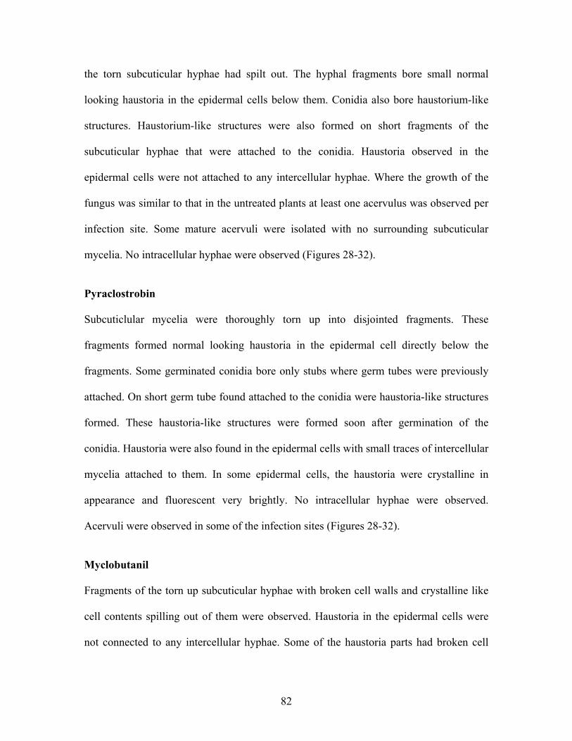
the torn subcuticular hyphae had spilt out. The hyphal fragments bore small normal
looking haustoria in the epidermal cells below them. Conidia also bore haustorium-like
structures. Haustorium-like structures were also formed on short fragments of the
subcuticular hyphae that were attached to the conidia. Haustoria observed in the
epidermal cells were not attached to any intercellular hyphae. Where the growth of the
fungus was similar to that in the untreated plants at least one acervulus was observed per
infection site. Some mature acervuli were isolated with no surrounding subcuticular
mycelia. No intracellular hyphae were observed (Figures 28-32).
Pyraclostrobin
Subcuticlular mycelia were thoroughly torn up into disjointed fragments. These
fragments formed normal looking haustoria in the epidermal cell directly below the
fragments. Some germinated conidia bore only stubs where germ tubes were previously
attached. On short germ tube found attached to the conidia were haustoria-like structures
formed. These haustoria-like structures were formed soon after germination of the
conidia. Haustoria were also found in the epidermal cells with small traces of intercellular
mycelia attached to them. In some epidermal cells, the haustoria were crystalline in
appearance and fluorescent very brightly. No intracellular hyphae were observed.
Acervuli were observed in some of the infection sites (Figures 28-32).
Myclobutanil
Fragments of the torn up subcuticular hyphae with broken cell walls and crystalline like
cell contents spilling out of them were observed. Haustoria in the epidermal cells were
not connected to any intercellular hyphae. Some of the haustoria parts had broken cell
82

walls such their contents spilt out or only disjointed parts of a haustorium were observed.
The contents of the haustoria appeared crystalline and fluorescent brighter than the
surrounding. No intercellular or intracellular hyphae were observed. No acervulus or
acervulus base were observed. The applications of myclobutanil left heavy chemical
residues (Figures 28-32).
Tebuconazole
The subcuticular mycelia were almost completely destroyed except for a few small
fragments that were found scattered at the infection site. These mycelia fragment were
also deformed to the extent that it was difficult to recognise them, therefore no
photographs of the torn subcuticular hyphae were made. Some of the germinated conidia
bore deformed structures that were difficult to identify. The haustoria found were fewer
than in infection sites on untreated leaves. Haustoria-like structures were formed directly
from the conidia. A very short hypha sometimes connected the conidium to a haustorium.
The wall of this short hypha was destroyed and its contents spilt out. No intercellular or
intracellular hyphae were observed. No acervuli were observed (Figures 28-32).
83

0
5
10
15
20
25
Untreated Trifloxystobin Pyraclostrobin Myclobutanil Tebuconazole
Infe
ctio
n si
tes w
ith h
aust
oria
Fungicides active ingredientsFigure 28: The number of infection sites on rose leaves that had been treated with different fungicides ingredients: trifloxystrobin, pyraclostrobin, myclobutanil, tebuconazole, on the 3rd dpi, in which haustoria were observed on the 10th dpi, (the data represent the mean ± SD of 3 replicates, n = 20, P < 0.05, Duncan’s multiple range test).
0
5
10
15
20
25
Untreated Trifloxystrobin Pyraclostrobin Myclobutanil Tebuconazole
Infe
ctio
n si
tes w
ith su
bcut
icul
ar h
ypha
e
Fungicides active ingredients
Figure 29: The number of infection sites on rose leaves that had been treated with different fungicides ingredients: trifloxystrobin pyraclostrobin, myclobutanil, tebuconazole, on the 3rd dpi in which subcuticular hyphae were observed on the 10th dpi, (the data represent the mean ± SD of 3 replicates, n = 20, P < 0.05, Duncan’s multiple range test).
84

0
5
10
15
20
25
Untreated Trifloxystrobin Pyraclostrobin Myclobutanil Tebuconazole
Infe
ctio
n si
tes w
ith a
cerv
uli
Fungicides active ingredients Figure 30: The number of infection sites on rose leaves that had been treated with different fungicides ingredients: trifloxystrobin, pyraclostrobin, myclobutanil, tebuconazole, on the 3rd dpi, in which acervuli were observed on the 10th dpi, (the data represent the mean ± SD of 3 replicates, n = 20, P < 0.05, Duncan’s multiple range test).
85

10 µm
10 µm
20 µm
20 µm b
dc
a
Figure 31: Light microscope photographs of Diplocarpon rosae in rose leavesshowing subcuticular hyphae in leaves of: (a) an untreated control plant, (b) a planttreated with 100 ppm trifloxystrobin, (c) a plant treated with 100 ppmpyraclostrobin, (d) a plant treated with 100 ppm myclobutanil.
86

IH
IH
10 µm b
H
20 µma
IH
10 µmc
10 µm d
10 µme
10 µm f
Figure 32: Light microscope photographs of Diplocarpon rosae in rose leavesshowing haustoria in leaves from: (a) and (b) untreated control plants, note theintercellular hyphae (IH), (c) a plant treated with 100 ppm trifloxystrobin, (d) aplant treated with 100 ppm pyraclostrobin, (e) a plant treated with 100 ppmmyclobutanil, note the absence of intercellular hyphae, (d) a plant treated with100 ppm tebuconazole, note the absence of intercellular hyphae
87

3.5.3.2 Effectiveness of application on the 7th dpi
The fungal structures already present in the host at this point of fungicide application
were subcuticular, intercellular, intramural and a few intracellular hyphae. Haustoria had
also been formed and the base of the acervuli had started to form at several points along
the subcuticular hyphae.
(i) Samples collected 10 dpi
Untreated
There was a network of subcuticular runner hyphae under the cuticle. Haustoria were
observed in the epidermal cells, and at least one acervulus was found per infection site.
The intramural hyphae in the epidermal periclinal walls and intracellular hyphae in the
epidermal cells were observed. Some epidermal cells were almost fully enclosed by
intercellular hyphae. The epidermal cells below the runner hyphae had a crystalline-like
cytoplasm that fluorescent and in which haustoria were found. The haustoria had a long
thin neck that attached to the intercellular hyphae.
Trifloxystrobin
Some of the subcuticular hyphae network was still in place but it did not fluorescent,
while in other places the subcuticular hyphae were torn into fragments that were scattered
at the infection site. On other infection sites the fungus grew in a similar manner as in the
untreated plants. Haustoria were found at some infection sites but no intercellular hyphae
were observed.
88

The haustoria formed were fewer compared to those in the untreated leaves. Fewer
acervuli were formed compared to those in the untreated leaves. Some of the acervuli
observed were not connected to any subcuticular hyphae (Figures 33-35).
Pyraclostrobin
Fragments of the subcuticular hyphae were seen scattered at an infection site and where
the network of the subcuticular hyphae was still in place, the hyphae did not fluorescence.
No intercellular or intracellular hyphae were observed at any infection site. Few haustoria
were observed in the epidermal cells but they were not connected to any intercellular
hyphae. Few acervuli were formed but no subcuticular mycelia were connected to them
(Figures 33-35).
Myclobutanil
No subcuticular hyphae were observed except for a few small fragments that were
scattered in the subcuticular region. There were few haustoria in some epidermal cells.
These haustoria were not connected to any intercellular hyphae. No intracellular and
intercellular hyphae were visible. In some epidermal cells the cytoplasm appeared
crystalline. No acervuli were formed. The applications of myclobutanil left heavy
chemical residues (Figures 33-35).
Tebuconazole
No intact subcuticular hyphae were observed but a few short fragments were occasionally
found. These hyphal fragments however had a discontinuous or broken cell wall.
Haustoria were observed in some epidermal cells. The haustoria were not connected to
89

any visible fungal mycelia. No intracellular, intramural or intercellular hyphae were
observed at any infection site. No acervuli were formed (Figures 33-35).
0
5
10
15
20
25
Untreated Trifloxystrobin Pyraclostrobin Myclobutanil Tebuconazole
Infe
ctio
n si
tes w
ith h
aust
oria
Fungicides active ingredientsFigure 33: The number of infection sites on rose leaves that had been treated with different fungicides ingredients; trifloxystrobin, pyraclostrobin, myclobutanil, tebuconazole, on the 7th dpi, in which haustoria were observed on the 10th dpi, (the data represent the mean ± SD of 3 replicates, n = 20, P< 0.05, Duncan’s multiple range test).
90

0
5
10
15
20
25
Untreated Trifloxystrobin Pyraclostrobin Myclobutanil Tebuconazole
Infe
ctio
n si
tes w
ith su
bcut
icul
ar h
ypha
e
Fungicides active ingredients
Figure 34: The number of infection sites on rose leaves that had been treated with different fungicides ingredients; trifloxystrobin, pyraclostrobin, myclobutanil, tebuconazole, on the 7th dpi, in which subcuticular hyphae were observed on the 10th dpi, (the data represent the mean ± SD of 3 replicates. n = 20, P< 0.05, Duncan’s multiple range test).
0
5
10
15
20
25
Untreated Trifloxystrobin Pyraclostrobin Myclobutanil Tebuconazole
Infe
ctio
n si
te w
ith a
cerv
uli
Fungicides active ingredientsFigure 35: The number of infection sites on rose leaves that had been treated with different fungicides ingredients; trifloxystrobin, pyraclostrobin, myclobutanil, tebuconazole, on the 7th dpi, in which acervuli were observed on the 10th dpi, (the data represent the mean ± SD of 3 replicates, n = 20, P< 0.05, Duncan’s multiple range test).
91

3.5.3.3 Effectiveness of application at the 10th dpi
The fungal structures that were already formed in the host at this time of fungicide
application were subcuticular, intercellular, intramural, and intracellular hyphae,
haustoria, and several fully developed acervuli containing conidia. The disease symptoms
had started to appear at the infection sites.
(i) Samples collected 14 dpi
Untreated
There was a massive subcuticular network. Intercellular, intracellular and intramural
hyphae were observed. Haustoria formed in the epidermal cells from the intercellular,
intramural and subcuticular hyphae were observed. The haustoria were connected to the
subtending hyphae. Infection sites had grown into each other and therefore the size of an
individual infection sites was difficult to determine. Several acervuli were observed near
each other at an infection site.
Trifloxystrobin
Subcuticular hyphal network was observed. The intercellular, intracellular and intramural
hyphae were also observed. Some of the epidermal cells were almost fully enclosed by
the intercellular hyphae. Haustoria formed by the intercellular, intramural and
subcuticular hyphae were observed in the epidermal cells. In some infection sites the
acervuli appeared similar to those in the untreated but in others the acervuli contents
(base, conidiophores and conidia) had been reduced to a mass of small indistinguishable
fluorescence pieces. In some of the acervuli, the conidia were deformed i.e. the cells of
the conidium were separated from each other and seemed to have lost their turgidity. In
92

some other infection sites the destruction was minimal and one could observe a few
conidia in the mass of fluorescence pieces (Figure 36).
Pyraclostrobin
Subcuticular hyphal network was still in place, intercellular, intramural and intracellular
hyphae were also observed but they were fewer than in the untreated. Haustoria formed
from intercellular, intramural and subcuticular hyphae were observed in the epidermal
cells. Acervuli with normal looking conidia are found at each infection site but the
acervuli formed were fewer than those in the untreated control. The viability of these
normal looking conidia was not investigated (Figure 36).
Myclobutanil
Only fragments of subcuticular hyphae were observed. There was increased branching of
the subcuticular hyphae in that a single fascicle had more hyphae than in the untreated
control. There were no intercellular and intracellular hyphae. Very few intramural hyphae
were observed. There was extensive destruction the acervuli; the bases of the acervuli
were reduced to a mass of indistinguishable fluorescence pieces. The conidia were torn
up into pieces or deformed; the 2 cells of the conidia had been separated from each other.
Individual conidium cells seemed to have burst open (Figure 36). The application of
myclobutanil left heavy chemical residues.
Tebuconazole
Fragments of the subcuticular hyphae were present at each of the infection sites. A single
fascicle had more hyphae than in the untreated leaves i.e. there was increased branching
on each hyphae strand compared to the untreated. Fewer haustoria were observed in the
93

epidermal cells compared to the untreated leaves. At each infection site there were
reduced intercellular and intramural hyphae compared to the untreated leaves, but there
were no intracellular hyphae. The acervulus base and most of the conidia were torn up,
although a few of the conidia appeared normal (Figure 36).
94

20 µm
10 µm 10 µm
20 µm 20 µm
20 µm
e
c
f
d
ba
Figure 36: Light microscope photographs of Diplocarpon rosae in rose leavesshowing conidia in an acervulus in a rose leaf from: (a) an untreated controlplant, (b a plant treated with 100 ppm trifloxystrobin, (c) a plant treated with100 ppm pyraclostrobin, (d) a plant treated with 100 ppm myclobutanil, (e) aplant treated with 100 ppm tebuconazole, (f) a plant treated with 100 ppmmyclobutanil.
95

4 Discussion
The understanding of a host-pathogen interaction is an important prerequisite for the
control of plant diseases. Only when one knows how the pathogen interacts with the host
can one chose the management strategies to employ. Different isolates of a pathogen may
interact differently with the host and respond differently to the control methods in use.
Therefore the understanding of a pathogen’s life cycle and how the different isolates
interact with the host is paramount to disease control. This in turn will determine which
methods of control are more appropriate.
The invasion of plants by pathogens is perceived as biotic stress in the plant cells. Under
stress conditions there is excess production of reactive oxygen species (ROS), which
leads to disruption of the electron transport systems in the plant (Kovtun et al 2000, Arora
et al 2002). Measuring the relative accumulation of such molecules can be a good
indicator of the stress in the plant cells. Reactive oxygen species (ROS) is a collective
term used to for oxygen radical and the non-radical hydrogen peroxide (von Tiedemann
1997). The production of ROS is termed as oxidative burst and it is a rapid and transient
response of plant cells to elicitors or plant pathogens in the early phase of interactions
(Doke 1982, Baker et al 1995, Mehdy et al 1996). Elicitors include plant propagules or
products such as Phytophthora infestans hyphae (Doke 1983), chemicals such salicylic
acid (Milosevic and Slusarenko 1996) or mechanical stress (Legendre et al 1993).
Localized pressure and enzymatic degradation of the plant cell wall elicits generation of
H2O2 (Gus-Mayer et al 1998, Mellersh 2001). Many fungal pathogen gain access to the
plant tissues by penetrating the plant cell walls through various mode of penetration
which include use of mechanical force or use of cell wall degrading. The plant cell wall is
96

the first line of defence and plants have evolved means to perceive and respond
defensively to the physical or chemical events associated with such penetration. The
destructive effects of a pathogen are also perceived as stress in the plant cells and the
greater the damage caused to the plant cells the greater the stress and amount ROS
produced. The reactive oxygen species are able to interconvert into each other through
several reactions and H2O2 has the longest half-life and provides a good estimate of the
relative ROS in the plant cells (Elstner 1987, Baker et al 1995). Measuring the relative
accumulation of ROS can be a good indicator of the aggressiveness of different isolate.
In this study the Kenyan isolate K1 caused more stress to the plant cells within studied
time span compared to the German isolate G1. K1 can therefore be said to be more
aggressive. This was also confirmed by the fact that the K1 lead to symptom expression
earlier than G1. The differences observed in the isolates used in this study were not
enough to group them into races but were concluded to be differences in aggressiveness.
G1 however had a higher germination rate than the K1. Further investigations using plant
differentials similar to those used by Debener (1996) need to be done to check if the
disease expressions caused by these isolates on other rose cultivars are different. It
necessary to standardise the differentials used worldwide in order to obtain comparable
data. In addition, carrying out molecular studies will shed more light into the genetic
diversity of the fungal isolates.
At 20 oC isolate K1 lead to symptom expression earlier than G1 but at other incubation
temperatures the effect of the temperature was higher than the differences between the
isolates. The development of disease symptoms was much slower at 15 oC than at 20 and
97

25 oC, and no disease symptoms developed at 10 oC. This indicates D. rosae was able to
germinate and penetrate the host cuticle at 15 oC, but it developed very slowly compared
to the development at 20 and 25 oC. Studies by Palmer et al. (1978), and Horst (1983) led
to the conclusion that the conidia do not germinate if they have not been wetted for a
minimum of 6 hours at relative humidities below 90 % and for at least 5 min even at 100
% RH. The percent infections increase with length of leaf wetness. Horst (1983) had also
reported that the pathogen tolerates a wide range of temperatures from 15-27 oC, mainly
through an inverse relation of humidity and moisture. With this in mind, one can then
presume that the pathogen has evolved a mechanism whereby it can germinate at low
temperatures when free water is more readily available than at high temperatures. The
pathogen then remains in the host until the temperatures are favourable for mycelial
development. The optimum temperature for conidium germination is 18 oC and it is lower
than the optimal temperatures for mycelia and disease development, which are 21 oC and
24 oC respectively (Horst 1983). This compares very well with Peronospora sparsa, the
causal agent of downy mildew of roses. P. sparsa infects roses even at 5 oC or lower
depending on the isolate, but the optimum temperature for infection and disease
development are 15 to 20 oC and 20 to 25 oC respectively (Aegerter et al 2003, Breese et
al 1994). The infection and colonization of many hosts by their respective pathogens
depends on the temperature, length of leaf wetness and relative humidity among many
other factors.
The colour and size of conidia of D. rosae observed in this study were within the range
observed by other researchers (Ali et al 2000, Wenefida and Spencer 1993). Germination
started within eight to nine hours after inoculation and within 12 hpi appressoria
98

formations had started and in some cases the penetration had taken place. Commonly
only one of the cells of the conidia germinated but even up to three germ tubes per
conidium were observed About 7 % of the germinated conidia formed germ tubes from
both cells of the conidium. The germination pattern observed in this study was very
similar to that described by Aronescu (1934), Palmer et al (1978), and Wiggers et al
(1997). Formation of more than one germ tube per conidium has also been observed in
other fungal pathogens like Colletotrichum species, where the germ tubes also have
variable lengths (Smith et al 1999). As observed in this study and also recorded by
Aronescu (1934), the very long germ tubes of D. rosae fail to form appressoria and
probably never succeed in penetrating. These long germ tubes may have depleted their
energy reserves and were therefore not able to penetrate the host cuticle. Niks (1990)
reported that increase in germ tube number and length decreased the amount of energy
available for Puccinia hordei sporelings to reach the stomatal entry points during
infection of barley. He believed that the sporelings were exhausted due to depletion of
endogenous energy reserves before completion of the infection. Thines et al (2000)
demonstrated that appressorium mediated host penetration by Magnaporthe grisea
involves many metabolic reactions and requires much energy.
In this study, well-defined appressoria did not always form at the ends of the germ tube
but many of the germ tubes were simply swollen at their tips, a fact that had also been
reported by Aronescu (1934), Palmer et al (1978) and Wiggers et al (1997). As opposed
to other fungi like Colletotrichum species where the appressoria are well defined and
delimited from the germ tube by a septum (Emmett and Parbery 1975, O’Connell et al
1985, Wharton et al 2001), the appressorium formed by D. rosae were extensively
99

variable, some were just swellings on the germ tube, while others were well defined
rounded and delimited swellings at the end of a germ tube, but sessile structures were not
uncommon. On some germ tubes no changes in their morphology were observed prior to
penetration or the changes may have been too subtle to be noticed. Therefore, in this
study the term appressorium was the used to define a structure formed on the germ tube
for purposes of adhering firmly to the host to achieve penetration.
The reason(s) why much variability with respect to appressorium formation was observed
is not known, and it could have been due to host factors as well as fungal factors or a
combination of the two. In many plant-fungal interactions where well-differentiated
appressoria are formed, factors such as chemical signals (for example potassium and
calcium ions, simple sugars), pH gradient and temperature shifts have been said to induce
appressorium formation (Hoch and Staples 1991). Host related factors such as
hydrophobicity (Lee and Dan 1993), hardness (Xiao et al 1994), components of host
surface such as wax (Podila et al 1993) and topographical properties, like the correct
dimensions of a ridge formed by the stomatal lip of the guard cell (Hoch and Staples
1991) are recognized as major signals. Tucker and Talbot (2001) recently reviewed the
factors that influence pre-penetration stages in various fungal pathogens. Principally
physical and chemical factors of host origin govern germ tube growth and the formation
of appressorium but the fungi respond to other environmental factors such as light and
temperature (Emmett and Palberry 1975).
Nair and Ellingboe (1965) argued that spore (or conidium) populations of the same
species might be composed of different genotypes that require different environmental
100

conditions for germination. It is therefore possible that the conidia in a population will
not be homogenous in the conditions required for appressorium formation (Emment and
Parbery 1975). The variation could also be attributed to observation of premature
structures or incompletely formed structured due to the environment being unsuitable.
Given that the population used in this study was not a single conidium population, this
argument could also be acceptable.
None of the earlier reports on D. rosae mentioned the presence of the brown rings,
presumed to be melanin rings, formed at the point of penetration. Melanization of the
appressoria has been well studied in fungi that form fully melanized appressoria such as
Magnaporthe and Colletotrichum species. Howard et al (1991) indicated that fungi with
melanized appressoria such as Magnaporthe species might be able to penetrate the host
cuticle and cell wall mainly by means of turgor pressure. Melanization of the appressoria
has been found to be essential for penetration of the host by M. grisea (Mendgen et al
1996), and treatment of its conidia with chemicals that inhibit melanization or generation
of mutants by targeted disruption of genes involved in the melanin biosynthesis resulted
in formation of non-pigmented appressoria and the mutants did not penetrate the host
surface (Woloshuk and Sisler 1982, Chumley and Valent 1990, Howard and Ferrari 1989,
Kubo and Furusawa 1991, Kimura et al 2001). Turgor pressure of melanin deficient
mutants is only 30-70 % of that measured in fully melanized appressoria (Dean 1997).
Melanization has been estimated to reduce porosity of appressorial cells of M. grisea to
less than 1 nm from approximately 2 nm and be sufficient to block efflux of cytosolic
solutes responsible for generating appressorial pressure (Howard et al 1991). Although
101

much is known about the role of melanin in completely melanized appressoria, the role of
melanin rings is yet to be elucidated.
In addition to their formation by D. rosae in its interaction with rose leaves, melanin
rings have been reported at the host pathogen interface of Venturia inaequalis and apple
leaves (Steiner and Oerke 2001). In V. inaequalis–apple interaction, the rings are thought
to play a role in the adhesion of the pathogen to the cuticle. Pre-inoculation applications
of melanin biosynthesis inhibitors to the apple leaves largely inhibited melanization of
the rings and reduced infection by 50-80 %. This indicated that melanization of the rings
was important for the penetration of apple leaves by V. inaequalis. The melanin rings are
assumed to play a similar role in the penetration of rose leaves by D. rosae, and
preliminary studies (data not shown) are pointing in this direction.
In D. rosae the penetration pore wall was reinforced suggesting that turgor pressure may
also play a role in the penetration of intact host walls. This reinforcement of the wall may
ensure that the pressure generated by turgor and by the cytoskeleton was exerted over a
restricted area of the pore by the growing penetration hypha. The turgor pressure
generated in appressoria of different pathogens varies e.g. in M. grisea its 20-80 bars
(Howard et al 1991) and in Uromyces appendiculatus 3-5 bar (Terhune et al 1993). M.
grisea forms fully melanized appressoria, while the appressoria of U. appendiculatus
have no melanin. Terhune et al (1993) reported that with the 3-5 bars turgor pressure the
U. appendiculatus appressorium base can distort polystyrene ridges and the emerging
penetration hypha can curl the stomata lip inward. This shows that even in non-melanized
appressoria an enormous pressure is generated, therefore it would be logical to presume
102

that turgor pressure build-ups in appressoria of D. rosae. Although the force generated by
the cytoskleton may only be of importance if the turgor pressure is very low (Mendgen et
al 1996), the cytoskeleton plays a role in stabilizing the tip of the penetration hypha.
The importance of turgor pressure in penetration of rose leaves by D. rosae is not known
and other mechanisms of penetration other than mechanical force seem to be also
involved. The host cell wall at the point of penetration was observed to be locally
degraded with no evidence of inward deformation or material being dislodged, a factor
that Pring et al (1995) in their study of the Colletotrichum species associated with
enzymatic action. The localized changes in staining properties of the host cell wall
adjacent to the penetration peg implicated enzyme action as also reported by Wharton et
al (2001) in their study on Colletotrichum sublineolum.
In fungi that establish an intimate relationship with their host, such as those causing rust
diseases, the differentiation of appressoria and subsequent penetration is accompanied by
a complex pattern of hydrolytic enzyme secretion (Dean 1997). As reported by Fernando
et al. (2001), Bucheli et al (1990) and Sweigard et al (1992a and b), cell wall degrading
enzymes are also associated with penetration of host by fungal pathogen that form well
differentiated melanized appressoria such Colletrotrichum and Magnaporthe. Pryce-
Jones et al (1999) demonstrated that non-melanized appressoria of Erysiphe graminis f.
sp hordei generate a turgor of 2-4 Mpa. Extracellular enzymes have been shown to be
present during the appressorium penetration by E. graminis, indicating a combination of
physical force and use of cell wall degrading enzymes working together (Francis et al
1996, Pryce-Jones et al 1999). Therefore in the case of penetration of rose leaves by D.
103
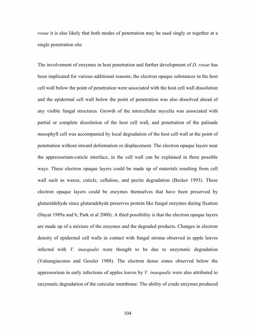
rosae it is also likely that both modes of penetration may be used singly or together at a
single penetration site.
The involvement of enzymes in host penetration and further development of D. rosae has
been implicated for various additional reasons; the electron opaque substances in the host
cell wall below the point of penetration were associated with the host cell wall dissolution
and the epidermal cell wall below the point of penetration was also dissolved ahead of
any visible fungal structures. Growth of the intercellular mycelia was associated with
partial or complete dissolution of the host cell wall, and penetration of the palisade
mesophyll cell was accompanied by local degradation of the host cell wall at the point of
penetration without inward deformation or displacement. The electron opaque layers near
the appressorium-cuticle interface, in the cell wall can be explained in three possible
ways. These electron opaque layers could be made up of materials resulting from cell
wall such as waxes, cuticle, cellulose, and pectin degradation (Becker 1993). These
electron opaque layers could be enzymes themselves that have been preserved by
glutaraldehyde since glutaradehyde preserves protein like fungal enzymes during fixation
(Hayat 1989a and b, Park et al 2000). A third possibility is that the electron opaque layers
are made up of a mixture of the enzymes and the degraded products. Changes in electron
density of epidermal cell walls in contact with fungal stroma observed in apple leaves
infected with V. inaequalis were thought to be due to enzymatic degradation
(Valsangiacomo and Gessler 1988). The electron dense zones observed below the
appressorium in early infections of apples leaves by V. inaequalis were also attributed to
enzymatic degradation of the cuticular membrane. The ability of crude enzymes produced
104

by V. inaequalis to degrade apple leaf cell walls has been demonstrated (Valsangiacomo
et al 1992).
Upon penetration of the host cuticle by D. rosae an infection vesicle is formed in the host
cell wall. In previous studies on D. rosae there is no mention of the infection vesicles
formed upon penetration. This infection vesicle seems to serve as a holding centre for all
fungal cellular contents immediately after penetration. The initial subcuticular and
intercellular hyphae are formed from this vesicle. The subcuticular hyphae grow in bands
of at least 2 or more parallel hyphae. New subcuticular hyphal strands are formed at acute
angles. The acute branching angle can be observed in Frick’s (1944) drawings although
she did not comment on its size. Palmer et al (1978) reported that the subcuticular hyphal
strands arise at right angles; but they could have been interpreting a meeting point of two
hyphal strands as a branching point or the formation of intramural hyphae, which are
formed at variable angles. Dodge (1931) observed that the two new hyphal branches
formed upon division of a parent cell at first widely diverged before converging to
become tightly pressed together, this initial diverging and converging of new branches
was not observed by the writer.
The growth of the subcuticular hyphae of D. rosae can be compared to Venturia
inaequalis and V. nashicola, the causal agents of scab on apples and Asian pears
respectively. These two pathogens grow between the epidermis and the cuticular
membrane without penetrating the cells or invading the intercellular spaces (Park et al
2000, Valsangiacomo et al 1989). The mycelia of V. nashicola only grow subcuticularly
within the pectin layers of the periclinal walls of epidermal cell without invading the
105

epidermal cells themselves (Park et al 2000). V. nashicola is suspected to produce
enzymes, cutinase and pectinase to enable it to inhabit these pectin layers. In apple leaves
infected with V inaequalis, translucent zones observed in host cell walls were associated
with local degradation of the host cell wall (Valsangiacomo et al 1989). In rose leaves
infected with D. rosae, the host wall surrounding the subcuticular or intramural hyphae
stained lighter with toluidine blue and was wider in diameter. This indicated a probable
change in the composition of the wall due to extracellular fungal products such as
enzymes. The intramural hyphae probably used enzymes to grow through the host cell
wall. Aronescu (1934) attributed the swellings in the host cell wall to enzymatic action.
As D. rosae continues to develop in the host tissue it leads to the development of brown
or black spots on the leaf surface. According to Dodge (1931), the brown or black spots
of discolouration were confined to the upper side of the leaf for some time and
occasionally he found sori on the lower side of the leaf directly below the brown or black
discolouration. He presumed that the infection sometimes occurred naturally on the lower
side. Aronescu (1934) clearly stated that spots on the lower surface were formed by the
fungus infection on the upper side growing through the leaf. In this study however,
observations made on naturally and artificially infected leaves revealed that the fungal
mycelia growing from the upper side of the leaf penetrated up to the palisade mesophyll
cells only. Infections established on the upper side of the leaf did not lead to symptom
development on the lower side of the leaf and vice versa. Infections on the lower side
were rare under natural conditions.
106

The initial browning of the leaf surface, during symptom development, was due to
browning of the palisade mesophyll layer, followed by the browning of the subcuticular
hyphae and the acervuli. The writer assumed that the browning of the mesophyll cells
was due to formation of phenolic compounds, while browning of the subcuticular hyphae
and the acervuli was due to pigmentation. Phenolic compounds are produced as a host
defence mechanism to the invasion by pathogens as summarized by Agrios (1997). The
pigmentation in the subcuticular hyphae and acervuli was assumed to be due to melanin
formation. Melanin production by fungal pathogens has been well researched as reported
by Butler and Day (1998), and Henson et al (1999) in their reviews. Although some fungi
produce melanin constitutively, others have been observed to undergo melanogenesis in
response to environmental stresses, such as exposure to toxic metals, desiccation,
hyperosmotic conditions, temperature extremes, limited nutrients and UV radiation to
mention but a few. In general, melanin is important for the survival and longevity of
fungal propagules (Bell and Wheeler 1986). Wolf (1912) thought that the black
discoloration was due to disorganization of the cells of the leaf and not due to the mycelia
of the fungus, which are colourless. Dodge (1931) was however convinced that the
browning was due to the collapse of the infected cells, while Aronescu (1934) attributed
the browning to the colour of the older mycelia. Frick (1944) thought the browning was
due to the changes in the contents of the infected epidermal cells but the aged thick
walled parallel hyphae contributed to the typical black spot symptoms.
The scope of the tools that previous researchers on the life cycle of D. rosae used was
limited and could have led to erroneous conclusions. Palmer et al (1978) observed that
few intercellular hyphae were formed and that the most of them were often between the
107

palisade cells, but in this study extensive intercellular growth was evident and almost all
of the intercellular growth took place in the epidermal layer. Wolf (1912) reported that
the acervuli stromata were connected to the internal mycelia below by hyphae growing
between or in the epidermal cells. In this study the stromata of the initial acervuli were
connected only to the subcuticular mycelia from which they were formed. As opposed to
the findings of several authors, the writer observed extensive plant tissue disorganization
in fallen or detached infected leaves that were disintegrating. Wolf (1912) observed
extensive disorganization and break down of mesophyll tissue directly below the
acervulus stroma during the acervulus development but in Dodge’s (1931) report this
disorganization and break down was said to be limited. According to Palmer et al (1978),
at the disease developmental stage when the visible symptoms were present, there was
extensive destruction of the host tissue such that the upper epidermis, upper portions of
the palisade and xylem parenchyma could not be histologically distinguished. This is not
surprising because they used detached leaves for their studies. Frick (1944) however saw
no disorganization. In this study there was minimal destruction of host tissue during the
biotrophic phase. However, in old infections sites with advanced necrotrophy there was
massive destruction of the epidermal cells, with the whole epidermal cell being filled
with intracellular hyphae and the cell wall disintegrating. Since Wolf (1912) does not
state how old the infection sites he observed were, it is possible that he observed very old
infection sites with abundant secondary hyphae, hence the erroneous conclusion.
In this study, the intracellular mycelia were observed from about the 7-8th dpi on leaves
still attached to the plant and about the 4th to the 5th dpi on detached leaves, which was
just before the formation of the acervuli in both cases. The number of intracellular
108
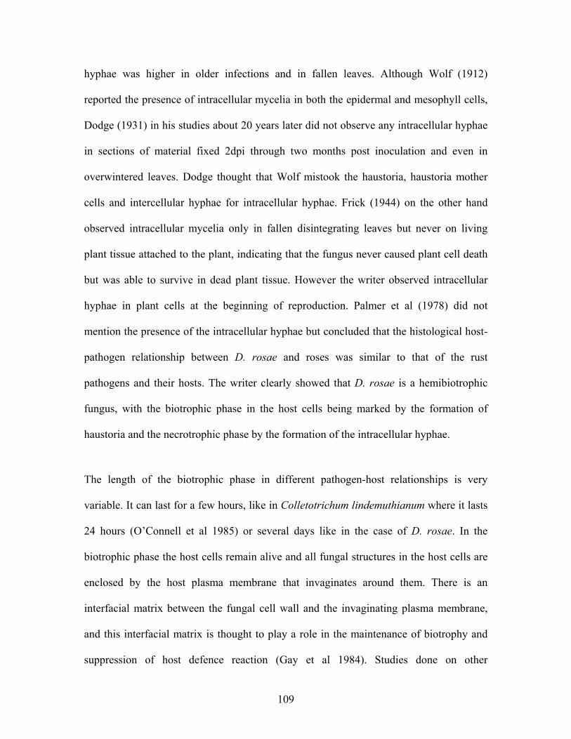
hyphae was higher in older infections and in fallen leaves. Although Wolf (1912)
reported the presence of intracellular mycelia in both the epidermal and mesophyll cells,
Dodge (1931) in his studies about 20 years later did not observe any intracellular hyphae
in sections of material fixed 2dpi through two months post inoculation and even in
overwintered leaves. Dodge thought that Wolf mistook the haustoria, haustoria mother
cells and intercellular hyphae for intracellular hyphae. Frick (1944) on the other hand
observed intracellular mycelia only in fallen disintegrating leaves but never on living
plant tissue attached to the plant, indicating that the fungus never caused plant cell death
but was able to survive in dead plant tissue. However the writer observed intracellular
hyphae in plant cells at the beginning of reproduction. Palmer et al (1978) did not
mention the presence of the intracellular hyphae but concluded that the histological host-
pathogen relationship between D. rosae and roses was similar to that of the rust
pathogens and their hosts. The writer clearly showed that D. rosae is a hemibiotrophic
fungus, with the biotrophic phase in the host cells being marked by the formation of
haustoria and the necrotrophic phase by the formation of the intracellular hyphae.
The length of the biotrophic phase in different pathogen-host relationships is very
variable. It can last for a few hours, like in Colletotrichum lindemuthianum where it lasts
24 hours (O’Connell et al 1985) or several days like in the case of D. rosae. In the
biotrophic phase the host cells remain alive and all fungal structures in the host cells are
enclosed by the host plasma membrane that invaginates around them. There is an
interfacial matrix between the fungal cell wall and the invaginating plasma membrane,
and this interfacial matrix is thought to play a role in the maintenance of biotrophy and
suppression of host defence reaction (Gay et al 1984). Studies done on other
109

hemibiotrophic pathogens have shown that the intracellular hyphae mark the onset of the
necrotrophic phase (Perfect et al 2001). During the biotrophic phase of C.
lindemuthianum the host and fungal plasma membranes are separated by an interfacial
matrix (Perfect and Green 2001), which is not formed in the necrotrophic phase.
Formation of secondary hyphae by C. lindemuthianum is associated with secretion of
large amounts of cell-wall degrading enzymes (Centis et al 1997). In D. rosae, the
haustoria and the large hyphae that formed on the inner side of the host cell wall were
surrounded by part of the host plasma membrane and are therefore part of the biotrophic
phase. The intracellular hyphae were not enclosed by part of the host plasma membrane
but were surrounded by a moderately electron opaque layer. The intracellular hyphae are
also associated with extensive host cell wall dissolution suggesting secretion of cell wall-
degrading enzymes. The intracellular hyphae therefore marked the onset of the necrotic
phase. The penetration of neighbouring cells by the intercellular and intracellular hyphae
probably involves use of cell wall degrading enzymes, because the cell wall thickness is
reduced at the point of penetration compared to the surrounding wall and the wall
microfibrils are cleanly severed.
The initial haustoria were formed very early in the disease development, Aronescu (1934)
observed them as early as 15 dpi. All haustoria were formed inside the host cell wall but
exterior to the plasma membrane a fact that the reports of early writers like Palmer et al
(1978) disputed. In this study the haustoria formed were of varying sizes and shapes,
from pyriform for the very young ones through ventricose to oval and clavate for the
older ones, but Palmer et al (1978) had observed all haustoria to be only belled shaped.
The haustoria of D. rosae grow in size after their formation. Haustoria are specialized
110

hyphae for nutrient absorption and metabolism and are recognized as one of the
distinguishing characteristics of a biotrophic fungi (Mendgen and Hahn 2002) or the
biotrophic phase of a hemibiotroph (Perfect and Green 2001). The size and shape of the
haustoria in fungi like powdery mildew (Bushnell 1972) and possibly in D. rosae have
been thought to play a role increasing the surface area of the haustoria–host plasma
membrane interface, hence the surface for nutrient absorption.
Haustoria of the rust fungi have been well researched on and much can be gleaned from
these studies. The extrahaustorial matrix has been reported to consist mainly of
carbohydrates and proteins that are primarily of plant but partly of fungal origin (Harder
and Chong 1991) and serves as a point of transfer of nutrients and signals between the
plant and the pathogen (Murdoch 1998, Stark-Urnau and Mendgen 1995, Hu and
Rijkenberg 1998, Heath and Skalamera 1997). Studies by Mendgen and Hahn (2002)
revealed that the bulk of the sugars and amino acids are taken up by haustoria. The
haustoria are therefore the feeding organ of the fungus in the biotrophic phase. Voegele
and Mendgen (2003) convincingly argued that since upon switching of the
hemibiotrophic fungus from the biotrophic to the necrotrophic phase the plasma
membrane surrounding the hyphae disintegrates, it seems like the interfacial matrix plays
a role in maintaining the biotrophy. An overall view of the growth pattern of the D. rosae
reveals that it forms very many haustoria as it continues to grow away from the point of
penetration. This leads to the assumption that the above arguments concerning the role of
the haustoria as a feeding organ associated with biotrophy hold true also in D. rosae, and
the haustoria therefore provide the food need for the subcuticular hyphae to reach parts
far removed from the infection point and for the fungus to ramify the host tissue
111

intramurally. Intramural mycelia grow in the spaces between the hyphal fascicles thus
helping the fungus to absorb nutrients from the epidermal cells between the hyphal
fascicles. In the end haustoria are found in very many of the host cells and more than one
haustorium can be found in a single cell. Heath (1990) showed that haustoria formation is
under signals from the host that either induce or complete the differentiation of haustoria.
This explains D. rosae does form haustoria in artificial grow media.
The end of an asexual cycle of D. rosae is signified by the formation of bicelled conidia
in subcuticular acervuli. In this study two-celled conidia and microconidia (spermatia)
were formed in the same subcuticular acervuli just as already described by Dodge in
1931. He observed that in many of the wintered leaves either wholly spermatial pustules
or in mixed ones with normal two cell conidia. However in this study no subcuticular
acervuli consisting of only microconida were observed. The role of the microconidia in
the life cycle of D. rosae has not been established and further investigations need to be
done to establish their role in propagation of the pathogen.
From the growth pattern observed the following conclusions can be drawn: D. rosae
penetrates the cuticle either by mechanical force or enzymatic degradation of the cell wall
or both. After that it establishes by an initial subcuticular and intramural necrotrophy that
is associated with swelling and dissolution of the host cell wall. The fungus spreads and
ramifies the host tissue subcuticularly and intercellularly. Parallel to this the fungus
invades the host cell to form haustoria that are enclosed by part of the host plasma
membrane. Large hyphae are formed on the inner side of the cell wall but exterior to the
plasma membrane thereby establishing a biotrophic phase in the host cells. There is an
112

increase in intramural ramification, which is associated with haustoria formation in the
epidermal cells and it is followed by intracellular necrotrophy. Therefore the necrotic and
biotrophic phases occur at the same infection site in different parts of the host tissue. Host
defence reaction is induced by the invasion of the cells to form haustoria, but the
formation of callose as part of the host cell defence does not stop the formation of the
haustorial body. The callose material is therefore formed as a collar around the haustorial
neck and the proximal end of the haustorial body, but never around the whole of the
haustorial body. In infections caused by E. graminis the formation of the callose is
stopped soon after the emergence of the young haustorium (Bushnell 1972). Renewed
growth or expansion of haustoria in host cells also leads to formation of callose where the
new growth takes place. The callose is a host cell wall wound reaction (Agrios 1997).
When the biotrophic phase of D. rosae is compared to biotrophic life cycle of a biotroph
like the bean rust fungus, Uromyces appendiculatus (Harding et al 1999) where minimal
collar material is formed and the host defence reaction is suppressed during the
development of the haustoria, then one can conclude that D. rosae is a hemibiotrophic
fungus whose biotrophic phase has not fully evolved, since the host defence reaction is
not fully suppressed.
For the survival of the fungal population, the fungus has to have a mechanism to survive
the unfavourable weather conditions. The findings in this study confirmed earlier reports
that D. rosae overwinters in fallen leaves and on the cane of rose plants. Some diseased
leaves remained attached to the plants during winter and can also serve as source of
inoculum in spring. Lyle (1943) reported on the ability of this fungus to overwinter on
canes, while Cook (1981) reported on its ability to overwinter on canes and in bud scales.
113

Frick (1944) demonstrated that the saprophytic mycelia found in overwintered leaves
formed conidia when placed in a damp chamber at room temperature. These conidia
germinated and caused blackspot lesions when inoculated on rose leaves.
Wolf (1912) described the ascospores of D. rosae for the first time. Aronescu (1934)
observed them also in the USA some years later and they have since been reported in
Great Britain (Knight and Wheeler 1977, Cook 1981) and in Russia (Dudin 1972) The
formation of these ascospores is rare and their role in genetic recombination is not fully
elucidated (Walker et al 1995). According to Wolf (1926) mycelium invades the deeper
layers of a fallen leaf to form stroma between the epidermis and the palisade parenchyma.
If the weather is favourable for the formation of ascocarps, then apothecia with asci are
expected in spring, but if the conditions for the formation of the asci are unfavourable,
the deep-seated fruiting bodies are filled with apothecial conidia (Dodge 1931). In this
study, heavily ornamented structures were observed in overwintering leaves from
December. By April these structures had an opening through one could see small oval
shaped cells. It is however unclear what these structures are and the writer assumed that
they were fruiting structures in which apothecial conidia (the small oval shaped
structures) were found. The deep-seated fruiting bodies with a shape similar to that of the
apothecia described by Wolf (1912) were also observed in this study but no asci had
developed. The writer assumed that the weather in Bonn during the winter of 2003 was
not favourable for the complete development of the ascocarps and the conidia formed
from the overwintered saprophytic mycelia on cane or leaves was the main source of
primary inoculum in spring. In the experiments carried out by Frick (1944) with infected
rose leaves collected from various parts of Switzerland and overwintered in Zurich, she
114

was not able to find any apothecia. With respect to overwintering, D. rosae can be poorly
compared with V. inaequalis, another ascomycete that grows subcuticularly and forms
ascospores as in overwintered plant material. In D. rosae the conidia formed from the
overwintered saprophytic mycelia are the main source of primary inoculum in spring
(Frick 1944), but in the case of V. inaequalis the ascospores formed in the overwintered
leaves are the main source of primary inoculum (MacHardy 1996). How D. rosae
survives the dry spells in the tropical countries is yet to be studied.
D. rosae grew in detached leaves. Although the fungal structures that developed in the
detached leaves were similar to those that developed in leaves of the intact plants, the
fungal structures were formed earlier in detached leaves than in leaves on intact plants.
The biotrophic phase of the fungus was shorter in detached leaves than in leaves on intact
plants. This is thought to be because of the degeneration of the detached leaves after
some time therefore causing the fungus to switch to a saprophytic way of feeding. The
ability of the fungus to form many haustoria in the detached leaves within such a short
time was attributed to the fact the plant defence reaction on the detached leaves is
minimal, therefore fungus is able to grow unhindered.
The saprophytic competence of the fungus demonstrated by its ability to live on dead or
senescent plant tissue is further confirmed by its ability to grow on artificial media. The
fungus grew and reproduced on all artificial growth media tested: water agar, PDA, MEA
and biomalt agar. However the highest numbers of conidia were formed on biomalt-based
media followed by malt extract agar based medium and the least formed on PDA. Both
biomalt agar and malt extract agar contain malt extract. Malt extract agar also contains
115

peptone from soja-bean meal, while biomalt may be enriched with other additives such
iron, calcium, vitamins etc which may have been responsible for the added advantage in
biomalt in comparison to MEA. The fungus sporulates better on malt extract based
products than on potato dextrose agar, an observation that had also been made by
Shirikawa (1955) and Drewes-Alvarez (1992). Although no measurements of the biomass
produced were carried out, D. rosae produced more mycelia on PDA than on MEA. The
differences in the amount of biomass formed on PDA and biomalt agar were not evident
and need to be measured. However, fungal cultures of D. rosae maintained on artificial
media have been reported to lose pathogenicity within a period of three, five, and six to
nine months by Frick (1944), Drewes-Alvarez (1992), and Stewart and Semeniuk (1965)
respectively.
Accurate knowledge of the lifecycle of a pathogen provides the opportunity for better
control in the sense that one knows when to the pathogen is most likely to be sensitive to
the given control methods. In the case of fungicide application this knowledge leads not
only to timely applications but also to more economical fungicide use. Timely
applications lead to fewer fungicide applications in a season (Bowen and Roark 2001).
The disease control aspect of this study focused on the effectiveness of the new class of
fungicides, the strobilurins, in the control of D. rosae in comparison to those fungicides
that are already in the market. In general, pre-inoculation applications of the strobilurins
led to 100 % conidia germination inhibition but post-inoculation applications did control
the disease. None of them was able to stop the development of disease symptoms at all
post-inoculation intervals tested. Despite the fact that an early application of
116

pyraclostrobin significantly reduced the disease severity compared to the untreated
control, it did not stop the development of the disease symptoms. The application of the
strobilurins when the fungus was fully established in the host led to minimal or no
reduction in the disease severity. This can be explained by the fact although the
strobilurins led to the destruction of the subcuticular mycelia and haustoria within four
days after application, new subcuticular hyphae and haustoria were formed with time
presumably from the deep seated mycelia, hence the further development of the fungus
leading to disease symptom expression. The growth of the fungus in the treated plants
was still so robust that the disease symptoms were formed on the same day in both the
fungicide treated and the untreated plants. Pyraclostrobin was more effective than
trifloxystrobin in all the post inoculation applications, which maybe due to the higher
sytemic activity of pyraclostrobin in comparison to trifloxystrobin (Bartlett et al 2001).
The DMI fungicides on the other hand allowed some germination of the D. rosae conidia
in all their pre-inoculation applications, but none of the germinated conidia were able to
lead to disease development. Their application early in the pathogen development led to
an almost complete inhibition of symptom development and there was no yellowing or
premature defoliation. However, their application just at the onset of symptom
development arrested symptom development but in the case of myclobutanil there was
yellowing and premature defoliation. This indicates that the DMI fungicides destroyed
the fungal structures in the host tissues in such a way that the fungus could not recover.
This can be explained by the fact the remnant fungal structures observed after treatment
of the plants with the DMI fungicides were scattered with no connection to each other
and therefore could not develop further. In addition in plants treated with tebuconazole,
117

the fungal structures were destroyed beyond recognition, Noegel (1998) had reported on
the ability of tebuconazole to deform fungal structures due to its interference with the
fungal cell membranes. Tebuconazole was therefore more effective than myclobutanil.
In all post inoculation applications the DMIs were significantly more effective in
inhibiting mycelial and haustoria development and in reducing disease severity than the
strobilurins. This is maybe because DMIs are more systemic (Kuck et al 1996, Newmann
and Jacob 1996, Bartlett et al 2001). Trifloxystrobin and pyraclostrobin have very low
movement into the leaf, low translaminar movement and are not xylem or phloem mobile
(Bartlett et al 2001), while the DMIs can either be translocated in the apoplast or
symplast (Newmann and Jacob 1996). In the post-inoculation treatments all the
fungicides were more effective in destroying the subcuticular hyphal network than the
haustoria. This is most likely due to the accessibility of the subcuticular hyphae in
comparison to the haustoria.
The strobilurins interfere with energy production by blocking ATP synthesis
(Ammermann et al 1992, Becker et al 1982, Zheng et al 1997). The inhibition of spore
germination by strobilurins is related to the spore’s dependency on mitochodrial
respiration during the germination process (Gold et al 1996). The two fungicide active
ingredients, trifloxystrobin and pyraclostrobin, were however not able to completely stop
the development of the D. rosae once it was inside the plant. This may be due to the
weaker dependency of the mycelia on respiration for vegetative growth and/ or the
possibility that an alternate pathway for electron transport may be inducible in the
mycelia (Gold et al 1996, Mizutani et al 1995). Despite the fact that the DMIs
118

(myclobutanil and tebuconazole) allowed germination and germ tube formation, they
strongly inhibited the development of and destroyed subcuticular mycelia. The effect of
the DMIs can be explained by the fact that during the initial phase of infection, fungi
sensitive to sterol inhibitors contain enough ergosterol in the germinating spores to
produce germ tubes and infection structures and to penetrate into the host tissue (Noegel
1998). When the fungus has depleted its ergosterol and has absorbed the fungicides a.i.
from the plant tissue the effects the chemical become obvious. In DMI sensitive fungi,
the take up of the chemical inhibits the production of ergosterol (vital in stabilizing the
cell membranes in the fungi) by the fungus and the fungus is killed by the depletion of
the sterol building blocks necessary for the cell membrane.
Trifloxystrobin has in previous experiments showed excellent protective as well as
curative activities against apple scab (Knauf-Beiter et al 2000). Protective application of
6 mg a.i./l inhibited spore germination by 80% and reduced penetration frequency by
85%, and no visible macroscopic symptoms developed. Absence of macroscopic
symptoms led to the assumption that the development of all subcuticular stromata formed
at the successful penetration site were completely hindered. At the rate of 20 mg a.i/l
spore germination reduced by 85 %, penetration frequency by 98 % and the development
of visible disease symptoms completely inhibited. In post inoculation treatments, the
application 6mg a.i./l 24 hours post inoculation almost completely inhibited penetration,
application 3 dpi led to such great reductions in the stroma development that the disease
development was suppressed. Eradicative treatment led to collapse of the already
developed fungal structures such as runner hyphae, conidiophores and conidia. The apple
scab fungus, Venturia inaequalis, develops only subcuticular stromata and their
119

destruction inhibited further development of the fungus. A single application of
trifloxystrobin led to destruction of the subcuticular mycelia of D. rosae as well, but did
not limit the fungus from further development because the deep seat mycelia formed new
subcuticular stromata. This can also be explained in terms of the low systemic properties
of trifloxystrobin, therefore the active ingredients is not accessible to the haustoria in the
host cells. The fungus therefore continues to absorb nutrients that supports further growth
and replacement of the destroyed structures.
In field trials with trifloxystrobin in combination with DMI fungicides, it was found to
control major ear and leaf diseases on cereals at the rate of 125-187.5 g a.i./ ha and it
provided long lasting protection (Margot et al 1998). On grapes, trifloxystrobin has been
found to be effective in controlling the oomycete fungal pathogen Plasmopara viticola,
which causes downy mildew of grapes and the ascomycete Uncinula necator, which
causes powdery mildew of grapes (Reuveni 2001). Trifloxystrobin was very effective
against conidial germination of U. necator in vitro and it inhibited mycelia growth and
sporulation of both fungi in-vivo. Trifloxystrobin had a partial effect on zoospore
discharge and suppressed zoospore motility and formation of germ tubes by P. viticola.
The fungicide a.i. strongly inhibited germination of U. necator at low concentrations and
had excellent prophylactic activity against both diseases of grapes. It suppressed
sporulation and further development of U. necator on grape leaves bearing sporulating
colonies or on mildewed clusters in the field. Trifloxystrobin controlled apple diseases,
scab and powdery mildew, at relatively low rates; scab at 3.75-5 g a.i /hl in protective or
curative schedules and powdery mildew at 5-7.5 g/hl. It controlled the foliar disease of
peanut (early and late leaf spots, and rust) at 70-105 g a.i./hl or in combination with
120

propiconazole at 62.5 + 62.5 g a.i./ hl. Control of black sigatoka of banana was achieved
with 70-90 g a.i./hl.
In this study the pre-inoculation application of the strobilurins completely inhibited
conidia germination and germ tube formation. A single application of 100 ppm of
trifloxystrobin or pyraclostrobin led to limited destruction of the subcuticular mycelia.
Studies were carried out by Wojdyla and Orlikowski (1999) to determine the
effectiveness of two other strobulurins, kresoxim-methyl and azoxystrobin, on
germination of the conidia of D. rosae revealed that when the two fungicides were
applied 14 d pre-inoculation the inhibition was 86% and less than 40% respectively.
Kresoxim-methyl inhibited the germination of D. rosae conidia by 86%, while with
azoxystrobin the germination inhibition varied from 9-38%. However weekly sprays of
the two fungicides on plants with black spot symptoms reduced the disease very
significantly compared to the untreated control plants (Wojdyla and Orlikowski 1999). In
their study they showed that the two fungicides completely protected the rose shrubs
against D. rosae. These weekly applications are similar to the protective applications,
since the a.i. residues are effective on every new generation of conidia formed.
Pyraclostrobin is relatively new in the market. It has been found to control several plant
pathogens; it provided excellent control of white rust of spinach caused by Albugo
occidentalis, on overwintered spinach (Damicone and Trent 2001), and is incorporated
into control programmes of several cereal diseases, fruit crops like grapes and apples, and
vegetables like beans and potatoes (BASF 2003).
121

The findings in this study compare very well with the findings of Gold et al (1996) on the
effectiveness of kresoxim-methyl (referred to as BAS 490 F in their report), another
strobilurin. Their observations revealed that pre-infection treatments 1 d before
inoculation with kresoxim-methyl inhibited spore germination and germ tube growth and
penetration of the host plant by all tested fungi. Post infection treatment with kresoxim-
methyl strongly inhibited mycelial growth and sporulation of Erysiphe graminis tritici
and Uncinula necator, whose mycelium and spores develop on the leaf surface, but had
less pronounced effects on the development of the mycelia of V. inaequalis and
Uromyces appendiculatus, whose mycelia develop in the plant. Since the mycelia of most
powdery mildew fungi develop exclusively on the leaf surface, Ypema and Gold (1999)
argued that this growth characteristic of the powdery mildew fungal mycelia makes all
development stages accessible to the kresoxim-methyl. Gold et al (1996) argued that
limited effect on the endoparasites may have been due to the limited up take of the a.i., its
metabolic breakdown in the leaves and also due to the relative differences in sensitivity
between spore germination and mycelial growth. A single eradicative treatment at 6 dpi,
a time when the V. inaequalis had developed extensive subcuticular mycelium and the
first conidia were forming, of apple plants with kresoxim-methyl at its recommended rate
of 67 mg AI l-1 completely inhibited macroscopically visible apple scab symptoms and
almost completely blocked sporulation. These findings agree with those of Knauf-Beiter
et al (2000) in which trifloxystrobin was found to greatly reduce the development of V.
inaequalis such that the disease development was suppressed. The apple scab fungus, V.
inaequalis, forms only subcuticular mycelia and therefore the inhibition of the
macroscopically visible symptoms can be taken to signify the destruction of these
122

subcuticular mycelia. There was little inhibition of mycelia growth in U. appendiculatus
whose mycelium develops in the mesophyll layer. The mycelia of D. rosae are formed
both in the subcuticular and mesophyll layers of the plant tissue, therefore the
effectiveness of kresoxim-methyl on the development of V. inaequalis whose mycelia
develop in the subcuticular layers and of U. appendiculatus in the mesophyll layers of
their respective hosts, can be compared to that of D. rosae. In the case of D. rosae the
destruction of the subcuticular mycelia by the strobilurins did not stop the fungus from
developing further. This is assumed to be because the mycelia in the epidermal and
mesophyll layers formed new subcuticular mycelia, leading to further growth and the
development of disease symptoms.
In this study a single application of 100 ppm tebuconazole or myclobutanil as soon as the
symptoms had developed gave very good control of the disease in that the disease spots
did not develop further. However in treatments with myclobutanil the leaves still turned
yellow and defoliation followed soon after that. In previous studies Hagan et al (1991)
had shown that weekly sprays of tebuconazole at the rates of 20, 40 and 80 ppm gave
only some modest reductions in the disease and that the weekly applications of the
fungicide at all these rates were equally effective in controlling the blackspot disease.
Although the application of tebuconazole at the above mentioned rates gave a season
long reduction is disease ratings compared to the untreated control, heavy leaf spotting
and some leaf shed was observed on plants treated annually with this fungicide (Hagan et
al 1991). In their studies on plants treated with weekly applications of 48 ppm
myclobutanil only isolated spots developed. Based on the results obtained in this study
tebuconazole (100 ppm) gave better results than myclobutanil (100 ppm), but Hagan et al
123

(1991) reported myclobutanil (24 ppm) to be more efficacious than tebuconazole (40
ppm).
In summary, the strobilurins provided excellent protective control and only a minimal
curative effect in the control of D. rosae. This was attributed to the growth pattern of the
fungus and the systemic properties of the strobilurins. In the pre-inoculation applications
of the DMI fungicides they did not completely inhibit the germination of the conidia but
the germinated conidia did not lead to disease development. Their post-inoculation was
very effective. Their late application however still led to disease symptom expression but
gave significant reductions in disease severity.
Since the strobilurins inhibited conidia germination but were not able to completely stop
the fungus once it was inside the leaves, they should be used protectively. The
prophylactic treatments should advisably be made as soon as the weather provides the
environmental conditions favourable for the development of D. rosae. Prophylactic
treatments should be on weekly basis where the temperature is above 15 oC and the
leaves are wet. At 10 oC and below no fungicide applications are required because the
fungus does not lead to disease development at these temperatures. Weekly applications
on plants already showing the disease symptoms may be effective at inhibiting the
germination of each generation of conidia formed and in the long run reduce disease
severity. However where the disease symptoms can already be observed it is advisable to
use the DMI fungicides because they are more effective at this stage. In advance disease
state mixture of a strobilurin and DMI may give good results. Repeated applications of
the strobilurins in a single season should be avoided, to reduce the chance of resistance
124

build up to these single site inhibitors. The heavy chemical residues associated with the
applications of myclobutanil are not desirable and therefore the fungicides should not be
applied just before harvest especially in the cut roses.
Recommendations of the control of this pathogen should consider the prevailing weather
conditions, the disease pressure, and the mode of action of the fungicides. Bowen and
Roark (2001) recommended the winter application of fungicides when the temperature
roses above -2.2 oC for 30 days. According to their field studies winter application of the
fungicides led to a delay of disease onset in the following spring, to reduced disease
severity and defoliation, increased plant vigour and increased flower production in the
following season compared to the untreated plants. In this study the fungus did not lead to
disease development at 10 oC even after 20 days of incubation, therefore based on these
findings it is not necessary to apply fungicides to control D. rosae at temperatures below
10 oC. However studies should be done to find out if there are isolates of D. rosae that
infect rose plants at these low temperatures. A fungicide application in late autumn to
destroy the inoculum already present on the plants may reduce the overwintering
potential of the fungus. Applications in early spring should be determined by the
prevailing weather conditions.
Disease management that combines cultural practices and minimal fungicide is more
desirable. Therefore cultural practices such as pruning of spent stems, removal of blind
shoots to allow good air circulation within the roses bushes and collection of fallen leaves
may reduce the disease pressure. Effective non-fungicidal solutions such as sodium
biocarbonate (Bowen et al 1995, Horst et al 1992) where disease pressure is low may
125

serve as alternative to fungicidal sprays. Planting of resistant cultivars where applicable is
recommendable. In the control of D. rosae fungicides should be used only when
necessary. Repeated application of these two classes of fungicides per season should not
exceed the manufacturer recommendations because of the risk of resistance development.
Based on the findings of this study, the early treatment of the infected plants has to be
emphasized: in late application some fungicides arrested symptom development but
defoliation still occurred. This showed that the damage caused by the disease is greater
than just the development of the lesions on the infected leaves. Prophylactic treatments
need to be applied before the visible symptoms because the pathogen has a latent period
of at least 7 days and not all fungicides are able to arrest the development of symptoms
when applied after visible symptoms are already present. Much data has been generated
from the various studies done on the interaction of D. rosae and its host, and the various
methods of control. However a disease-forecasting model is still needed for a more
economical and optimised use of all the available methods of control. A disease-
forecasting model will also optimise the timings of fungicide applications.
126

5 References
Aegerter BJ, Nuñez JJ and Davis RM 2003. Environmental factors affecting rose
downy mildew and development of a forecasting model for nursery production systems.
Plant Dis. 87: 732-738.
Agrios GN 1997. Plant pathology. Fourth edition, Academic press. San Diego,London,
Boston, New York, Sydney, Tokyo, Toronto pp 103.
Alexopoulos CJ, Mims CW and Blackwell M. 1996. Introductory Mycology. Fourth
edition, John Wiley and Sons Inc, New York.
Ali A, Hall AM, Aquino deMuro M and Cannon P 2000. The investigation of the
biology of the rose blackspot fungus, Diplocarpon rosae leading to improved methods of
control. Brighton Crop Prot. Conf., www.herts.ac.uk.
Ammermann E, Lorenz G, Schelberger K, Wenderoth B, Sauter H and Rentzea C
1992. BAS 490F: A broad spectrum-fungicide with a new mode of action. Brighton Crop
Prot. Conf., pp 403-410.
Aronescu A 1934. Diplocarpon rosae from spore germination to haustorium formation.
Bull. Theory Bot. Club 61: 291-329.
Arora A, Sairam RK and Srivastava GC 2002. Oxidative stress and antioxidative
system in plants. Current Science 82: 1227-1238.
Baker JC, Harmon GL, Glazener AJ and Orlandi EW 1995. A non-invasive
technique for monitoring peroxidative and H2O2 scavenging activities during interactions
between bacterial plant pathogens and suspension cells. Plant Physiol. 108: 353-359.
Baker KF 1948. The history, distribution and nomenclature of the Rose black spot
fungus. Plant Dis. Rep. 32: 260-274,
127

Bartlett DW, Clough JM, Godfrey CRA, Godwin JR, Hall AA, Heaney SP and
Maund SJ 2001. Understanding the strobilurin fungicides. Pesticide Outlook 4: 143-148.
BASF 2003. F500 the premium fungicide. www.basf.de.
Beales P 1997. Classic roses: An illustrated encyclopedia and grower’s manual of old
roses, shrub roses and climbers. Harvill Press. London.
Becker CM 1993. The cytology and histology of apple scab. In: Handbook of Cytology,
Histology, and Histochemistry of Fruit Tree Diseases. Biggs AR (ed), CPR Press, Boca
Raton, FL, pp 35-90
Behe B.K, Williams JD, Cobb P, Hagan AK and Stritikus G 1993. Growing roses.
Ala. Coop. Ext. Serv. Circ. ANR-157.
Bell AA and Wheeler MH 1986. Biosynthesis and functions of fungal melanins. Annu.
Rev. Phytopathol. 24: 411-51.
Black WA, Byrne DH and Pemberton HB 1994. Field study of black spot resistance in
rose. HortScience 29: 525.
Bolton AT and Svejda FJ 1979. A new race of Diplocarpon rosae capable of causing
severe black spot on Rosa rugosa hybrids. Can. Plant Dis. Surv. 59: 38-40.
Bowen KL and Roark RS 2001. Management of black spot of rose with winter
fungicide treatment. Plant Dis. 85: 393-398.
Bowen KL, Young B and Behe BK 1995. Management of blackspot of rose in the
landscape in Alabama. Plant Dis. 79: 250-253.
Brandes W and Kaspers H 1989. Tebuconazol - ein neues Botrytis fungizid für den
Weinbau. Pflanzenschutz-Nachrichten Bayer 42: 149-161.
128

Braskaran R, Purushothaman D and Ranganathan K 1974. Physiological changes in
rose leaves infected by Diplocarpon rosae. Phytopathol. Z. 79: 231-236.
Breese WA, Shattock RC, Williamson B and Hackett C 1994. In vitro spore
germination and infection of cultivars of Rubus and Rosa by downy mildews from both
hosts. Ann. Appl. Biol. 125: 73-85.
Bucheli P, Doares SH, Albersheim P and Darvill A 1990. Host-pathogen interactions
XXXVI. Partial purification and characterization of heat-labile molecules secreted by rice
blast pathogen that solubilize plant cell wall fragments that kill plant cells. Physiol. Mol.
Plant Pathol. 36: 159-173.
Butler MJ and Day AW 1998. Fungal melanins: A review. Can. J. Microbiol. 44: 1115-
1136.
Byrne DH, Black W and Ma Y 1995. The use amphiploidy in the development of the
black spot resistant rose germplasm. Acta Hort. 424: 269-272.
Carlson-Nilsson BU and Davidson CG 1995. Research on black spot pathogen
Diplocarpon rosae (Marssonina rosae) and resistance in Rosa. Acta Hort. 508: 141-147.
Castledine P, Grout BWW and Roberts AV 1981. Cuticular resistance to Diplocarpon
rosae. Trans. Br. Mycol. Soc. 77: 665-666.
Centis S, Guillas I, Séjalon N, Esquerré-Tugayé M-T and Dumas B 1997.
Endopolygalacturonase genes from Collectrotrichum lindemuthianum: Cloning of
CLPG2 and comparison of its expression to that of CLPG1 during saprophytic and
parasitic growth of the fungus. Mol. Plant-Microbe Int. 10: 769-775.
Chumley FG and Valent B 1990. Genetic analysis of melanin-deficient non-pathogenic
mutants of Magnaporthe grisea. Mol. Plant-Microbe Int. 3: 135-43.
129

Clergeot P-H, Gourgues M, Cots J, Laurans F, Latorse M-P and Pepin R, et al 2001.
PLS1, a gene encoding a tetraspanin-like protein, is required for penetration of rice leaf
by the fungal pathogen Magnaporthe grsea. Proc Natl Acad Sci USA 98: 6963-6968.
Coddington A and Gould DS 1992. Use of RFPLs to identify races of fungal pathogens.
In: Techniques for the rapid detection of plant pathogens. Duncan JM and Torrance L
(eds), Blackwell Scientific Publications, Oxford, London, Edinburgh, British Society for
plant pathology, pp 162-176.
Cook RTA 1981. Overwintering of Diplocarpon rosae at Winsley. Trans. Br.
Mycol.soc.77: 549-556
Couch HB 1995. Diseases of turf grasses. 3rd edition, Krieger Publishing, Malabar, Fla.
Dalton AJ 1955. A chrome-osmium fixative for electron microscopy. Anat. Rec. 121:
281.
Damicone JP and Trent MA 2001. Evaluation of spray programmes for the control of
spinach white rust, 2000. Fung. Nemat. Tests 56: V86.
Dean RA 1997. Signal pathways and appressorium morphogenesis. Annu. Rev.
Phytopathol. 35: 211-34.
Debener T 1996. Charakterisierung und Evaluierung genetischer Ressourcen in Gattung
Rosa. Jahresbericht der Bundesanstalt fuer Zuechtungsforshung an Kulturpflanzen (BAZ)
Germany, pp 23.
Debener T 2003. Genetics/Inheritance of characteristics. In: Encyclopaedia of rose
science. Roberts AV, Debener T and Gudin S (eds), Elsevier Academic Press
Amsterdam, pp 288-292.
130

Debener T and Mattiesch L 1995. Genetic analysis of the molecular markers in crosses
between diploid roses. Acta Hort. 424: 249-251.
Debener T, Drewes-Alvarez R and Rockstroh K 1998. Identification of five
physiological races of black spot, Diplocarpon rosae Wolf, on roses. Plant Breed. 117:
267-270.
Debener T, Kaufmann H, Mattiesh L and Genseleiter L 1996. Charakterisierung und
Isolierung von wirtschaftlich wichtigen Genen aus Rosa spec. Jahresbericht der
Bundesanstalt fuer Zuechtungsforshung an Kulturpflanzen (BAZ) Germany, pp 21-23.
Deising H, Heiler S, Rauscher M, Xu H, Mendgen K 1996. Cellular aspects of rust
infection structure differentiation. In: Histology, Ultrastructure and Molecular Cytology
of Plant-Micro-organisms Interactions. Nicole M and Gianinazzi-Pearson V (eds),
Kluwer, Amsterdam, pp 135-156.
Dodge BO 1931. A further study of the morphology and life of the rose black spot
fungus. Mycologia 23: 446-62.
Dohm A, Frehe K, Ludwig C and Debener T 1996. Resistenz- Induktion von
Resistenzgegenüber pilzlichen Schaderregern in Rosen durch Transformation.
Jahresbericht der Bundesanstalt für Züchtungsforschung an Kulturpflanzen, pp 36-37
Doke N 1983. Involvement of superoxide anion generation in the hypersensitive response
of potato tuber tissues to infection with an incompatible race of Phytophthora infestans
and to the hyphal wall components. Physiol. Plant Pathol. 23: 345-357.
Doke N 1992. A further study on the role of hypersensitivity in resistance in potato
cultivars to infection by an incompatible race of Phytophthora infestans. Physiol. Plant
Pathol. 21: 85-95.
131

Drewes-Alvarez R 1992. Untersuchungen am Pathosystem Sternrusstau (Marssonina
rosae (Lib.) Died)-Rose (Rosa L.) zur Klaerung der Frage nach pilzlichen Rassen und zur
Uebertragung der Resistenz aus diploiden Rosa multiflora Thunb. auf tetraploide
Gartenrosen. PhD Thesis, University of Hamburg, Germany.
Drewes-Alvarez R 2003. Disease/Black spot. In: Encyclopaedia of rose science. Roberts
AV, Debener T and Gudin S (eds), Elsevier Academic Press Amsterdam, pp 148-153.
Dubin VS 1972. Sumchatoe sporonoshenie kak istochnik vesennego vozobnovleniya
chernoi pyatnistosti na Roze. -(Ascus sporulation as a source of spring renewal of black
spot of rose).- Rev. Plant Pathol. 51: 450.
Ebeling M, Heimann K-G, Schoefer S and Sonder K 2003. The human and
environmental safety aspects of trifloxystrobin. In: Pflanzenschutz-Nachrichten Bayer 56:
231-245.
Elstner EF 1997. Metabolism of activated oxygen species. In: The Biochemistry of
Plants. Davies DD (ed.), vol 11, Academic Press, San Diego, CA, pp 253-315.
Emmett RW, and Parbery DG 1975. Appressoria. Annu. Rev. Phytopathol. 13: 147-
167.
Englehand AW 1969. Fungicidal efficacy of three bezimidazole compounds and
Dithane-45 against Diplocarpon rosae on two rose cultivars. Phytopathol. 59: 113.
Erincik O, Madden LV, Ferree DC, and Ellis MA 2003. Temperature and wetness-
duration requirements for grape leaf and cane infection by Phomopsis viticola. Plant Dis.
87: 832-840.
Eurostat 2000. EU imports of flowers and ornamental plants.
www.ids.ac.uk/ids/global/pdfs/et&Gender_kenyaReport3.pdf.
132

Fäber R B K, Chin KM and Leadbeiter N 2002. Sensitivity of Venturia inaequalis to
trifloxystrobin. Pest Manag. Sci. 58: 261-267.
Fernando TH, Jayasinge CK and Wijesundera RL 2001. Cell wall degrading enzyme
secretion by Colletotrichim acutatum, the causative fungus of secondary leaf fall of
Hevea brasiliensis. Mycol. Res. 105: 195-201.
Francis SA, Dewey FM and Gurr SJ 1996. The role of cutinase in germling
development and infection by Erysiphe graminis f. sp. hordei. Physiol. Mol. Plant Pathol.
49: 201-11.
Frick L 1944. Untersuchungen über die Biologie und Pathogenität von Diplocarpon
rosae (Lib.) Wolf. Phytopathol. Zeitschrift. 14: 525-591.
Gay JL 1984. Mechanism of biotrophy in fungal pathogens. In: Plant Diseases:
Infection, Damage, and Loss. Wood RKS and Jellis J (eds), Blackwell Scientific
Publications, Oxford, pp 49-59
Geyer G 1973. Ultrahistochemie. Gustav Fisher Verlag, Stuttgart.
Gold RE and Leinhos GM 1995. Fungicidal effects of BAS 490F on the development
and fine structure of plant pathogenic fungi. Pestic. Sci. 43: 250-253.
Gold RE, Ammerman E, Koehle H, Leinhos GM and Sauter H 1996. The synthetic
strobilurin BAS 490F: profile on the modern fungicide. In: Modern Fungicides and
Antifungal Compounds. Lyr H, Russell PE and Sisler HD (eds), Intercept, Andover,
Hants, UK, pp 79-92.
Gus-Mayer S, Naton B, Hahlbrock K and Schmelzer E 1998. Local mechanical
stimulation induces components of pathogen defence responses in parsley. Proc. Natl.
Acad. Sci. USA 95: 8398-8403.
133

Hagan AK, Gilliam CH, Fare DC and Bowen K 1991. Application rates and spray
schedules of ergosterol-biosynthesis inhibitor fungicides for control of blackspot of rose.
Plant Dis. 75: 1143-1146.
Hagan AK, Gillian CH, and Fare D 1987. Fungicides for black spot control.
Phytopathol. 77: 1728
Hagan AK, Gillian CH, and Fare DC 1988. Evaluation of new fungicides for the
control of rose black spot. J. Environ. Hort. 6: 67-69.
Harder DE and Chong J 1991. Rust haustoria. In: Electron microscopy of plant
pathogens. Mendgen K and Lesemann D-E (eds), Springer, Berlin, pp 235-250.
Harding MW, Stutz JC and Roberson RW 1999. Host-parasite relationships in bean
cultivars of varying susceptibility to bean rust. Can. J. Bot. 77: 1551-1559.
Hayat M A 1989. Chemical fixation. In: Principles and Techniques of Electron
Microscopy, 3rd ed. MacMillan Press, London, pp 1-78.
Hayat MA 1989. Positive staining. In: Principles and Techniques of Electron
Microscopy, 3rd ed. MacMillan Press London, pp 208-327.
Heath MC 1990. Influence of carbohydrates on the induction of haustoria of cowpea rust
fungus in vitro. Exp. Mycol. 14: 84-88.
Heath MC and Skalamera D 1997. Cellular interactions between plants and biotrophic
fungal parasites. Adv. Bot. Res. 24: 195-225.
Heatherington PJ and Meredith RH 1988. Disease control with HWG 1608 on cereals
and rape. Proc 1988 Brighton Crop Prot. Conf., pp 953-958.
Henson JM, Butler MJ and Day AW 1999. The Dark side of mycelium: melanins of
phytopathogenic fungi. Annu. Rev. Phytopathol. 37: 447-71.
134

Hoch HC and Staples RC 1991. Signalling for infection structure formation in fungi. In
The Fungal Spore and Disease Initiation in Plants and Animals. Cole GT and Hoch HC
(eds), Plenum, New York, pp 25-46.
Hood ME and Shew HD 1996. Applications of KOH-analine blue fluorescence in study
of plant-fungal interactions. Phytopathol. 86: 704-708.
Horst RK 1995. Compendium of Roses Diseases. APS Press, St. Paul MN, pp 7-10.
Horst RK 1983. Black spot. In: Compendium of Rose Diseases. APS Press. American
Phytopathological Society, St. Paul, MN, pp 7-11
Horst RK, Kawamoto SO and Porter LL 1992. Effect of sodium bicarbonate and oils
on the control of powdery mildew and black spot of roses. Plant Dis. 76: 247-251.
Howard RJ and Ferrari MA 1989. Role of melanin in appressorium function. Exp.
Mycol. 13: 403-418.
Howard RJ, Ferrari MA, Roach MA and Money NP. 1991. Penetration of hard
substrates by employing enormous turgor pressures. Proc. Natl. Acad. Sci. USA 88:
11281-11284.
Hu GG and Rijkenberg FHJ 1998. Ultrastructural studies of the intercellular hyphae
and haustorium of Puccinia recondita f. sp. tritici. J. Phytopathol. 146: 39-50.
Jellito TC, Page HA and Read ND 1994. Role of external signals in regulating the
prepenetration phase of infection by the rice blast fungus, Magnaporthe grisea. Planta
194: 471-77.
Jenkins WR 1995. Variability of pathogenicity and physiology of Diplocarpon rosae
Wolf, the rose blackspot fungus. Am. Rose Annu. 40: 92-97.
135

Jordan CM, Endo RM and Jordan LS 1988. Penetration and colonization of resistant
and susceptible Apium graveolens by Fusarium oxysporium f. sp. apii race 2: Callose as
structural response. Can. J. Bot. 66: 2385- 2391.
Joyaux F 2003. History of roses in cultivation/European (pre-1800). In: Encyclopedia of
Rose Science. Roberts AV, Debener T and Gudin S (eds), Elsevier Academic Press,
Amsterdam, pp 395-402.
Karnovsky MJ 1965. A formaldehyde-glutaraldehydehyde fixative of high osmolarity
for use in electron microscopy. J. Cell Biol. 27: 137-138.
Keen NT 1992. The molecular biology of disease resistance. Plant Mol. Biol. 19: 109-
122.
Kimura A, Takano Y, Furusawa I and Okuno T 2001. Peroxisomal metabolic
function is required for appressorium-mediated plant pathogenic infection by
Colletotrichum lagenarium. Plant Cell 13: 1945-1957.
Knauf-Beiter G, Küng-Färber R, Gugenheim R and Laird D 2000. Die Wirkung von
Trifloxystrobin gegenüber Apfelschorf. Deutsche Pflanzenschutztagung 52: 470-471.
Knight C and Wheeler BEJ 1977. Penetration of Diplocarpon rosae on rose leaves.
Trans. Br. Mycol. Soc. 69: 385-389.
Knight C and Wheeler BEJ 1978a. Evaluating the resistance of roses to blackspot.
Phytopathol. Z. 91: 218-229.
Knight C and Wheeler BEJ 1978b. The germination of Diplocarpon rosae on different
rose cultivars. Phytopathol.Z. 91: 346-354.
136

Kovtun Y, Chiu W-L, Tena G and Sheen J 2000. Functional analysis of oxidative
stress-activated mitogen-activated protein kinase cascade in plants. Proc. Natl. Acad. Sci.
USA 97: 2940-2945.
Kubo Y and Furusawa I 1991. Melanin biosynthesis: Prerequisite for successful
invasion of the plant host by appressoria of Colletotrichum and Pyricularia. In: The
Fungal Spore and Disease Initiation in Plants and Animals. Cole GT and Hoch HC (eds),
New York, Plenum, pp 205-18.
Kuck KH, Scheinpflug H and Pontzen R 1996. DMI fungicides. In: Modern Selective
fungicides; properties applications, mechanisms of action. Horst Lyr (ed), 2nd revised and
enlarged edition. Gustav Fisher Verlag. Jena, Stuttgart, New York, pp 205-258.
Leadbitter N, Amrein J, Knauf-Beiter G, Huggenberger F and Margot P 2000.
Trifloxystrobin: das neue Strobilurinfungizid von Novartis. Deutsche
Pflanzenschutztagung 52: 94.
Lee Y-H and Dean RA 1993. cAMP regulates infection structure formation in plant
pathogenic fungus Magnaporthe grisea. Plant Cell 5: 693-700.
Legendre L, Rueter S, Heinstein PF and Low PS 1993. Characterization of the
oligogalacturonide-induced oxidative burst in cultured soybean (Glycine max) cells. Plant
Physiol. 102: 233-240.
Lyle EW 1943. Black spot on rose canes. Am. Rose Annu. 155-156.
MacHardy WE 1996. Apple scab biology, epidemiology and management. APS Press,
Minnesota, pp 190-195.
137

Malek B and Debener T 1996. Resistenz- Erschliessung neuer Resistenzquellen
gegenueber dem Erreger des Sternrusstaus an Rosen. Jahresbericht der Bundesanstalt fuer
Züchtungsforschung an Kulturpflanzen, pp 37-38.
Malek BV, Rockstroh K, and Debener T 1996. Genetische und molekularbiologische
Charakterisierung der Sternrusstaureistenz aus Rosa multiflora. Jahresbericht der
Bundesanstalt fuer Zuechtungsforshung an Kulturpflanzen (BAZ) Germany, pp 20-21.
Margot P, Huggenberger F, Amrein J and Weiss B 1998. CGA 279202: A new broad-
spectrum strobilurin fungicide. Brighton Crop Prot. Conf. Pest Dis. 2: 375-382.
Mehdy CM, Sharma YK, Sathasivan K and Bays NW 1996. The role of activated
oxygen species in plant disease resistance. Plant Physiol. 98: 365-374.
Mellersh DG and Heath MC 2001. Plasma membrane-cell wall adhesion is required for
expression of plant defence responses during fungal penetration. Plant Cell 13: 413-424.
Mendgen K and Hahn M 2002. Plant infection and the establishment of fungal
biotrophy. Trends Plant Sci. 7: 352-356.
Mendgen K, Hahn M and Deising H 1996. Morphogenesis and mechanisms of
penetration by plant pathogenic fungi. Annu. Rev. Phytopathol. 34: 367-86.
Milosevic N and Slusarenko AJ 1996. Active oxygen metabolism and lignification in
the hypersensitive response in bean. Physiol. Mol. Plant Pathol. 49: 143-158.
Mims CW 1991. Using electron microscope to study plant pathogenic fungi. Mycologia
83: 1-19.
Mims CW, Rodriguez-Lother C and Richardson EA 2001. Ultrastructure of the host-
parasite interaction in leaves of Duchesnea indica infected by the rust fungus Frommeёla
mexicana var indicae as revealed by high pressure freezing. Can. J. Bot. 79: 49-57.
138

Mizutani A, Yukioka H, Tamura H, Miki N, Masuko M and Takeda 1995.
Respiratory characteristics in Pyricularia oryzae exposed to a novel
alkoxyiminoacetamide fungicide. Phytopathol. 85: 306-311.
Morrison LS and Russel CC 1976. Timing of fungicide-adjuvant mixtures for control
of rose black spot. Plant Dis.Rep. 60: 634-636.
Murdoch LJ, Kobayashi I and Hardham AR 1998. Production and characterization of
monoclonal antibodies to cell wall components of the flax rust fungus. Eur. J. Plant
Pathol. 104: 331-346.
Nair KRS and Ellingboe AH 1965. Germination of conidia of Erysiphe graminis f. sp.
tritici. Phytopathol. 55: 365-68.
Newnann S and Jacob F 1996. Principle of uptake and systemic transport of fungicides
within the plant. In: Modern Selective Fungicides; properties applications, mechanisms of
action. Horst Lyr (ed), 2nd revised and enlarged edition. Gustav Fisher Verlag Jena
Stuttgart, New York, pp 53-73.
Nicholas RO, Williams DW and Hunter PA 1994. Investigation of the value of β-
glucan-specific fluorochromes for predicting the β-glucan content of the cell walls of
zoopathogenic fungi. Mycol. Res. 98: 694-698.
Niks RE 1990. Effect of germ tube length on the fate of sporelings of Puccinia hordei in
susceptible and resistant barley. Phytopathol. 80: 57-60.
Noegel KA 1998. Folicur fungicide. www.wheatimprovement.org/Forum/1/Noegel.htm.
O’Connell RJ, Bailey JA and Richmond DV 1985. Cytology and physiology of
infection of Phaseolus vulgaris by Colletotrichum lindemuthianum. Physiol. Plant Pathol.
27: 75-98.
139

Orpin C, Bauer A, Bieri R, Faugeron JM and Sidddi G 1986. Myclobutanil, a broad
spectrum systemic fungicide for use in fruit, vines, and a wide range of other crops. Proc.
1986 Brit. Crop Protect. Conf., pp 55-62.
Ortega F, Steiner U and Dehne H-W 1998. Induced resistance to apple scab:
microscopic studies on the infection cycle of Venturia inequalis (Cke.) Wint. J.
Phytopathol 146: 1-7.
Palmer JG and Semeniuk P 1960. Relative susceptibilities to black spot of some rose
varieties in excised leaflet assay during 1959. Phytopathol. 50: 572.
Palmer JG and Semeniuk P 1961. Comparable susceptibilities of 50 species and hybrid
roses inoculated with black spot fungus from plants field-grown in Maryland in 1959.
Am. Rose Annu. 46:125-133.
Palmer JG and Semeniuk P 1962. Temperature effects on host pre-disposition and
development of black spot. Phytopathol. 52: 1220.
Palmer JG, Sachs IB, and Semeniuk P 1978. The leafspot caused by Marssonina rosae
observed in scanning electron and light microscopy. Scanning Electron Microscopy 2:
1019-1026.
Palmer JG, Semeniuk P and Stewart R N 1966. Roses and blackspot. I. Pathogenicity
to excised leaflets of Diplocarpon rosae from seven geographical locations. Phytopathol.
56: 1277-1282.
Palmer JG, Semeniuk P and Stewart RN 1966. Roses and blackspot. II. Seasonal
variation in host susceptibility and decline of virulence in culture of conidia from
Diplocarpon rosae. Phytoplathol. 56: 1283-1286.
140

Park P, Ishii H, Adachi Y, Kanematsu S, Ieki H and Umemoto S 2000. Infection
behaviour of Venturia nashicola, the cause of scab on Asian pears. Phytopathol. 90:
1209-1216.
Perfect SE and Green JR 2001. Infection structures of biotrophic and hemibiotrophic
fungal plant pathogens. Mol. Plant Pathol. 2: 101-108.
Plattner H 1981. Chemische fixierung. In: Methodensammlung EM 2.2.1, Schimmel G,
Vogell W (eds), Wiss. Verlag, Stuttgart.
Podila GK, Rogers LM and Kolattukudy PE 1993. Chemical signals from avocado
surface wax trigger germination and appressorium formation in Collectotrichum. Plant
Physiol. 103: 267-72.
Pring RJ, Nash C, Zakaria M and Bailey JA 1995. Infection process and host range of
Colletotrichum capsici. Physiol. Mol. Plant Pathol. 46: 137-152.
Pryce-Jones E, Carver T and Gurr SJ 1999. The role of cellulase enzymes and
mechanical force in host penetration by Erysiphe graminis f. sp. hordei. Physiol. Mol.
Plant Pathol. 55: 175-82.
Quinn JA, Fujimoto TT, Egan AR and Shabers SH 1986. The proprieties of RH3866,
a new triazole fungicide. Pestic. Sci. 17: 37-362.
Reddy S, Spencer JA and Newman SE 1992. Leaflet surfaces of blackspot-resistant and
susceptible roses and their reactions to fungal invasion. HortScience 27: 133-135
Reinecke P, Kaspers H, Scheinpflug H and Holmwood G 1986. BAY HWG 1608, a
new fungicide for foliar spray and seed treatment use against a wide spectrum of fungal
pathogens. Proc. Brit. Crop Protect. Conf., pp 41-46.
141

Reuveni M 2001. Activity of trifloxystrobin against powdery and downy mildew
diseases of grapevines. Can. J. Pathol. 23: 52-59.
Reynolds ES 1963. The use of lead citrate at high pH as an electron opaque stain in
electron microscopy. J. Cell Biol. 17: 208-212.
Sauter H, Ammermann E, Benoit R, Brand S, Gold RE, Grammenos W, Koehle H,
Lorenz G, Muller B, Rohl F, Schirmer U, Speakman JB, Wenderoth B and Wingert
H 1995. Mitochondrial respiration as target for antifungals: Lessons learned from
research on strobilurins. In: Antifungal Agents-Discovery and Mode of Action. Dixon
GK, Copping LG, and Hollomon DW (eds), BIOS Scientific Publishers, Oxford UK, pp
173-191.
Shirakawa HS 1955. The nutrition of Diplocarpon rosae. Amer. J. Bot. 42: 379-384.
Shumack R, Cobb P and Hagan AK 1988. Growing roses. Ala.Coop. Exten. Serv. Circ.
ANR-157.
Sitte H 1982. Ultramikrotomie-Häufige Probleme und Fehler. Supplement
Microscopy/Electronmikroscopie. GIT Lab. Med., pp 9-32.
Sitte H 1985. Ultramikrotomie. Mta-J. Extra. 10: 9-25.
Smith IM, Dunez J, Lelliott RA, Phillips DH and Archer SA 1988. European
Handbook of Plant Diseases. Blackwell Scientific Publications, Oxford.
Smith JE, Korsten L and Aveling TAS 1999. Infection process of Colletrotrichum
dematium on cowpea stems. Mycol. Res. 103: 230-234.
Smith MM and McCully ME 1978. A critical evaluation of the specificity of aniline
blue induced fluorescence. Protoplasma 95: 229-254.
142

Spurr AR 1969. A low viscosity epoxy resin embedding medium for electron
microscopy. J. Ultrastruct. Res. 26: 31-43.
Stanghellini ME, Rasmussen SL and Vandemark GJ 1993. Relationship of callose
deposition to resistance of letture to Plasmopara lactucae-radicis. Phytopathol. 83: 1498-
1501.
Stark-Urnau M and Mendgen K 1995. Sequential deposition of plant glycoproteins and
carbohydrates into the host-parasite interface of Uromyces vignae and Vigna sinensis.
Evidence for endocytosis and secretion. Protoplasma 186: 1-11.
Stark-Urnau M, Gold R, Guggenheim R and Dueggelin M 1997. Sensitivity of
different mildew and rust fungi against kresoxim-methyl. In: Proc. Eur. Mediterr. Cereal
Rusts Powdery Mildews Conf. 9th, Lunteren, The Netherlands, pp 268-271.
Steiner U and Oerke E-C 2001. The role of melanin in the infection process of Venturia
inaequalis. Phytopathol. 91 (suppl.): S85.
Stewart RN and Semeniuk P 1965. Report on rose research in the U.S. Department of
agriculture. Am. Rose Annu. 50: 99-105.
Svejva FJ and Bolton AT 1980. Resistance of rose hybrids to three races of
Diplocarpon rosae. Can. J. Plant Pathol. 2: 23-25.
Sweigard JA, Chumley FG and Valent B 1992a. Cloning and analysis of CUT1, a
cutinase gene from Magnaporthe grisea. Mol. Gen. Genet. 232:174-82.
Sweigard JA, Chumley FG and Valent B 1992b. Disruption of a Magnaporthe grisea
cutinase gene. Mol. Gen. Genet. 232: 183-90.
143

Terhune BT, Bojko RJ and Hoch HC 1993. Deformation of stomatal guard cell lips
and microfabricated artificial topographies during appressorium formation by Uromyces.
Exp. Mycol. 17: 70-78.
Thines E, Weber RWS and Talbot NJ 2000. MAP kinase and protein kinase A-
dependent mobilization of triacylgycerol and glycogen during appressorium turgor
generation by Magnaporthe grisea. Plant Cell 12: 1703-18.
Tucker SL and Talbot NJ 2001. Surface attachment and pre-penetration stage
development by plant pathogenic fungi. Annu. Rev. Phytopathol. 39: 38-417.
Valsangiacomo C and Gessler C 1988. Role of cuticular membrane in ontogenic and
Vf-resistance of apple leaves against Venturia inaequalis. Phytopathol. 78: 1066-1069
Valsangiacomo C, Ruckstuhl M and Gessler C 1992. In vitro degradation of cell walls
of apple leaves by pectinolytic enzymes of the scab fungus, Venturia inaequalis, and by
commercial pectinolytic and cellulolytic enzyme preparations. J. Phytopathol. 135: 20-
27.
Valsangiacomo C, Wagner K, Stadler B, Ruckstuhl M, Manini-Gessler P, Michel M
and Gessler C 1989. Aspects of host resistance and pathogenensis in the reaction
between Venturia inaequalis and apple leaves. In: Integrated control of pome fruit
diseases. Gessler C, Butt DJ and Koller B (eds), IOBC Bulletin Vol. II, pp 346.
Van Sengbush P, Hechler J and Muller U 1983. Molecular architecture of fungal cell
walls. An approach by use of fluorescent markers. Eur. J. Cell. Biol. 30: 305-312.
Voegele RT and Mendgen K 2003. Rust haustoria: nutrient uptake and beyond. New
Phytologist 159: 93-100.
144

von Tiedemann A 1997. Evidence for a primary role of active oxygen species in
induction of host cell death during infection of bean leaves with Botrytis cinerea. Physiol.
Mol. Plant Pathol. 50: 151-166
Walker S, Mandegaran Z and Roberts AM 1995. Screening roses for resistance to
Diplocarpon rosae. Acta Hort. 424: 209-213.
Ward E, Unwin M and Stoessel A 1975. Sesquiterpenoid phytoalexins from fruits of
egg plants. Phytopathol. 65: 859-863.
Wenefida I and Spencer JA 1993. Marssonina rosae variants in Mississippi and their
virulence on selected rose cultivars. Plant Dis. 77: 246-248.
Wharton PS, Julian AM and O’Connell RJ 2001. Ultrastructure of the infection of
Sorghum bicolour by Colletotrichum sublineolum. Phytopathol. 91: 149-158.
Wheeler MH and Bell AA 1988. Melanins and their importance in pathogenic fungi.
Curr. Top. Med. Mycol. 2: 338-387.
Widholm JM 1972. The use of fluorescein diacetate and phenosafranine for determining
viability of cultured plant cells. Stain Technol. 47: 185-194.
Wiggers RJ, West JG and Taylor J 1997. Conidial germination and infection by
Diplocarpon rosae on susceptible and resistant rose species. Mycologia 89: 103-108.
Wohlfahrt-Bottermann KE 1957. Die kontrastierung tierischer Zellen und Gewebe im
Rahmen ihrer elektronen-mikroskropischen Untersuchung an ultradünnen Schnitten.
Naturwiss. 44: 287-288.
Wojdyla AD and Orlikowski LB 1999. Strobilurin compounds in the control of rust,
powdery mildew and blackspot on some ornamental plants. Inter. Sym. Crop Prot. (Gent)
64: 539-545.
145

Wolf FA 1912. The perfect stage of Actinonema Rosae. Bot. Gaz. 54: 218-234.
Wolf FA 1926. Leaf scorch disease of strawberries. N.C. Agr. Exp. Sta. Tech. Bull. 28:
1-16.
Woloshuk CP and Sisler HD 1982. Tricyclazole, pyroquilon, tetraclorophthalide,
PCBA, coumarine and related compounds inhibit melanization and epidermal penetration
by Pyricularia oryzae. J. Pestic. Sci. 7: 161-66.
Wybou AP 1989. Field experiments with Folicur for the control of black sigatoka
(Mycosphaerella fijiensis) in banana growing at the atlantic coast of Costa Rica. Pflanzen
schutz-Nachrichten Bayer 42: 162-180.
Xiao JZ, Ohshima A, WatanabeT, Kamakura T and Yamaguchi I 1994. Studies on
cellular differentiation of Magnaporthe grisea. Physico-chemical aspects of substratium
surfaces in relation to appressorium formation. Physiol. Mol. Plant pathol. 44: 227-236.
Xue AG and Davidson CG 1998. Components of partial resistance to black spot disease
(Diplocarpon rosae Wolf) in garden roses. Hort. Sci. 33: 96-99.
Ypema HL and Gold RE 1999. Kresoxim-methyl: Modification of a naturally occurring
compound to produce a new fungicide. Plant Dis. 83: 4-19.
Zheng D and Köller W 1997. Characterization of the mitochondrial cytochrome b gene
encoding the target site of strobilurin fungicides from Venturia inaequalis. Phytopathol.
87 (suppl.): S109.
Ziegler H, Benet-Buchholz J, Etzel W and Gayer H 2003. Trifloxystrobin: a new
strobilurin fungicide with an outstanding biological activity. Pflanzenshutz-Nachrichten
Bayer 56: 213-214.
146

Acknowledgements
I would like to express my gratitude to Prof. Dr. H.-W. Dehne for giving me the
opportunity to carry out my research in his laboratory. I thank him for his guidance and
constructive criticism during my study.
I am grateful to Prof. Dr. W. Schumacher for accepting to read my work as the second
examiner.
My sincere gratitude goes to PD Dr. U. Steiner for supervising this work. I am thankful
for her guidance, patience and for her time during my research work and the thesis write-
up. I owe much to PD Dr. J. Hamacher for his time and advice especially during electron
microscopy. My gratitude goes to Mrs Neukirchen for all her technical assistance. I
would like to thank Dr. H. Hindorf for all his help in various ways. I also express my
gratitude to Prof. Dr. G. Siboe, Evans Njambere and Anthony Gichohi for their assistance
during the sample collection in Kenya.
I am very grateful to Dr. Simeon Kotchoni for his love and support during my stay in
Germany. My heartfelt gratitude goes to my parents Mr and Mrs Gachomo for their
encouragement and support. I wish to sincerely thank my son Bryan Gachomo for his
patience during my long periods of absence from home. I express my deepest
appreciation to all the big family members Karwega, Wanjeri, Wanjiru, Kamau, Gitonga,
Mwangi and my late sister Nyambura. You have all been of very helpful to Gachomo and
me.
Finally, I thank the German Academic Exchange Service (DAAD) for awarding me a
scholarship again. This second scholarship enabled me to do my Ph D in Germany.
147