student : rattaphong pokkaew ( 李平 ) student id : 0970456 department of food science
DESCRIPTION
Physical and mechanical properties of cardboard panels made from used beverage carton with veneer overlay. Ayrilmis, N . ; Candan , Z . ; Hiziroglu , S . 2008. 29, 1897–1903. Student : Rattaphong Pokkaew ( 李平 ) Student ID : 0970456 Department of Food Science. Outline. - PowerPoint PPT PresentationTRANSCRIPT

Student : Rattaphong Pokkaew (李平 ) Student ID : 0970456
Department of Food Science
Ayrilmis, N. ; Candan, Z. ; Hiziroglu, S. 2008. 29, 1897–1903
Physical and mechanical properties of cardboard panels made from used beverage carton with veneer overlay

OutlineOutline1. 1. IntroductionIntroduction2. 2. The objectiveThe objective3. Methods3. Methods4. Results4. Results5. Conclusion5. Conclusion

IntroductionIntroduction

Other recycled
Beverage carton,
313000, 3%

The peanut (Arachis hypogaea L.) The peanut is called as the “king” of oil seeds.
Peanuts are also known as : • Ground nuts, earthnuts, goobers, goober peas, pindas, jack nuts, pinders, manila nuts and monkey balls. • In the UK these are sold as monkey nuts.
Peanut

Peanuts are a good source of tocopherols, phytosterol and phospholipid
The tocopherol content of peanuts varies with variety and production location
Peanut oil mainly contains -tocopherol (50–373 ppm) and γ-tocopherols (90–390 ppm)
Tocopherols in peanut
(Firestone, 1999)
Sturm et al. (1966) determined tocopherol content of peanut oil from 17 varieties. Runner varieties had higher levels of -, γ-and -tocopherols than the Spanish varieties
Hashim et al. (1993) reported that there were significant differences in tocopherol content among Runner- and Virginia-type peanut cultivars
Tocopherols in peanut

Phospholipids (PL) contribute to the smoothness, texture, and mouthfeel of foods
Phospholipids improve the stability of the product because of their inherent antioxidant properties
Phospholipid in peanut

Breast cancerBreast cancer Breast cancer is the erratic growth of cells that originate in the breast
tissue
A group of rapidly dividing cells may form a lump or mass of extra
tissue. These masses are called tumors
The World Health Organization reported more than 1.2 million people worldwide will be diagnosed with breast cancer this year

The occurrence of breast cancer varies widely among women from different countries and cultures
Higher incidences in European and North American women Lower in women in less developed countries and countries relying more heavily on vegetarian diets (American Cancer Society, 2003)
Breast cancerBreast cancer
Dietary factors, specifically the proportion of animal versus plant fats, may play a role in the development of breast cancer (Messina and Barnes, 1991; Cho et al., 2003)

Plant foodstuffs contain specific phytochemicals which may offer protection from breast cancer.
An attractive hypothesis which may account for the cancer protective action of phytosterols is that phytosterols induce apoptosis or programmed cell death in highly proliferative tumor cells.
Breast cancer & PhytosterolBreast cancer & Phytosterol
Plant foodstuffs contain specific phytochemicals which may offer protection from breast cancer.

Source : Moreau et al., 2002; Berger et al., 2004; Kritchevsky and Chen, 2005
Fig 1. Structure of some representative phytosterols

Apoptosis, or programmed cell death, is associated with several fundamental biological processes, including cell development, differentiation and response to injury.
AApoptosispoptosis
Apoptosis is defined as a set of events that once initiated induce lethal changes that include membrane blebbing, mitochondrial break down and DNA fragmentation
Apoptosis occurs via two main pathways• The intrinsic or mitochondrial pathway• The extrinsic or death receptor-mediated pathway

Fig 2. TFig 2. The intrinsic or mitochondrial pathwayhe intrinsic or mitochondrial pathway
Apoptoticprotease-activating factor-1 (Apaf
-1)
Cytochrome c
Pro-apoptotic
factors

Fig 3. TFig 3. The he exextrinsic or trinsic or death receptor-mediateddeath receptor-mediated pathway pathway
FADD
Fas ligand
Fas receptor
Fas-associateddeath domain
TNF receptorassociated
death domain

The objectiveThe objective
The objective of this study was to assess the effect of cellular supplementation with either -sitosterol or cholesterol on the extrinsic caspase-8 pathway in the two breast cancer cell lines, MCF-7 and MDA-MB-231. Hypothesized of -sitosterol, that may potentiate Fas-death domain signaling, leading to caspase-8 activation and ultimately to apoptosis.
To test the hypothesis, the breast cancer cell lines were treated with -sitosterol or with cholesterol as a control and effects on cell growth, sterol incorporation in cell membranes, expression of Fas-death domain signaling proteins, and caspase-8 activity were determined.

MethodsMethods

Cell cultureCell culture
MCF-7 and MDA-MB-231 cells
5% CO2 / 95% air as monolayers using RPMI 1640 growth medium
2 g/l sodium bicarbonate
5% fetal bovine serum
1% antibiotic–antimycotic
Cultured at 37 C

Preparation of sterol supplemented media andPreparation of sterol supplemented media andmeasurement of cell growthmeasurement of cell growth
2-hydroxypropyl beta-cyclodextrin; CD complexes(RPMI 1640 media supplemented with cholesterol or b-sitosterol)
Growth medium(8–16 mMsterols: 5mM CD)
Control groups-Cell were treated
with 5mM CD
Studies groups

Measurement of sterol content of cell membraneMeasurement of sterol content of cell membranes by GLCs by GLC
Cells were seeded into 6-well plates
harvested by scraping
frozen 80 C in 350 ml 10mM Tris and 20mM mannitol buffer (pH 7.4)
Samples were thawedand briefly sonicated on ice

Samples were thensaponified at 80 C in 95% ethanolic KOH
Saponified samples
The upper organic phase
Dried under nitrogen
2 ml of water2ml of hexane
The lower phase
GLC

Determination of caspase-8 activityDetermination of caspase-8 activity
Cells were seeded in T-75 flasks; 24 h(10,000 cells/cm2)
Cells were scraped
Washed in PBS (pH 7.4)
The media were replaced with sterol-supplem
entedor control medi
a
3 day treatment

Lysed by suspension on ice of 107 cells/200 ml in buffer
The lysates were aspirated
Centrifuged at 10,000g ; 4 C ; 30 min
After 30 min

The supernatants (cell lysates) were analyzed by
the Bio-Rad DC protein assay
Cell lysates (100 mgprotein) were assayed
caspase-8 activity

ResultsResults

ResultsResults
Fig. 4. Effect of sterol supplementation on growth of MCF-7 cells. Cells were grown in RPMI-1640 growth medium supplemented with different concentrations of sterols

ResultsResults
Fig. 5. Effect of sterol supplementation on growth of MDAMB-231 cells. Cells were grown in RPMI-1640 growth medium supplemented with different concentrations of sterols

Supplementation Cholesterol -Sitosterol Total sterol -Sitosterol (mg/mg protein) (mg/mg protein) (mg/mg protein) (% total sterol)
CD vehicle 43 2a 0.0 0a 43 2a 0a
Cholesterol 49 2b 0.0 0a 49 2a 0a
-Sitosterol 40 1a 50 5b 90 5b 56b
Table 1. Effect of 2 d-sterol supplementation on sterol content of MCF-7 cell membranes*
*MCF-7 cells were treated for 2 d with 16 mM sterol or vehicle and sterol contents of membranes determined by gas–liquid chromatography as described. Data are means SEM (n = 3) and letters (a, b) of values in each column are si
gnificantly different (p<0.05).

Supplementation Cholesterol -Sitosterol Total sterol -Sitosterol (mg/mg protein) (mg/mg protein) (mg/mg protein) (% total sterol)
CD vehicle 49 7a 0.0 0a 49 7a 0a
Cholesterol 73 4b 0.0 0a 73 4b 0a
-Sitosterol 33 2a 51 6b 84 8b 61b
Table 2. Effect of 2 d-sterol supplementation on sterol content of MDA-MB-231 cell membranes*
*MDA-MB-213 cells were treated for 2 d with 16 mM sterol or vehicle and sterol contents of membranes determined by gas–liquid chromatography as described. Data are means SEM (n = 3) and letters (a, b) of values in each colum
n are significantly different (p<0.05)

Supplementation Caspase-8 activity of Caspase-8 activity of MCF-7 lysates MDA-MB-231 lysates
CD vehicle 6000 800a 4600 200a
Cholesterol 5000 200a 4600 200a
-Sitosterol 9800 400b 8100 800b
Table 3. Effect of 3 d-sterol supplementation on caspase-8 activity of MCF-7 and MDA-MB-231 cell lysates*
*Cells were treated for 3 d with 16 mM sterol or vehicle and caspase-8 activities of cell lysates determined. Data are RFU/100 mg lysate protein and represent means SEM (n = 3). Letters (a, b) of values in each column are significantl
y different (p<0.05).

Fas
FasL
FADD
p-FADD
Caspase-8
Actin
SITCHOLCONT
Fig. 6. Effect of sterol supplementation on expression levels of Fas-related signaling proteins in MCF-7 cells. MCF-7 cells were treated with sterols
for 24 h as described. Expression of Fas-related signaling pathway proteins was quantified by immunoblot.
Fas
FasL
FADD
p-FADD
Caspase-8
Actin
SITCHOLCONT
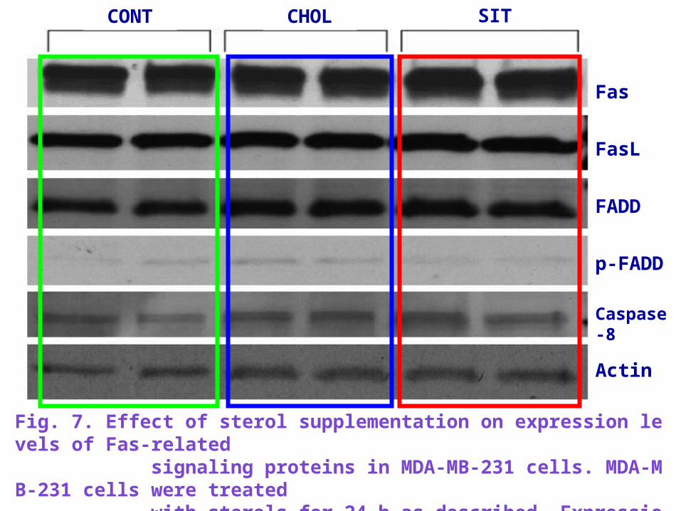
Fig. 7. Effect of sterol supplementation on expression levels of Fas-related signaling proteins in MDA-MB-231 cells. MDA-MB-231 cells were treated with sterols for 24 h as described. Expression of Fas-related signaling proteins was quantified by immunoblot.

ConclusionConclusion

ConclusionConclusion -sitosterol can be affect the amounts and activity of components of the extrinsic apoptotic pathway in human breast adenocarcinoma cells
-sitosterol induces a reduction in membrane sphingomyelin and an increase the ceramide levels in some tumor cells
The effect of -sitosterol treatment to increase caspase-8 activity and apoptosis in these cells may be mediated, at least in part, by changes in membrane sterol content and effects on the Fas apoptotic pathway

THANK YOUTHANK YOU FOR YOUR ATTENTION ! FOR YOUR ATTENTION !


Arachis hypogaea L
Source : http://upload.wikimedia.org/wikipedia/commons/1/1f/Koeh-163.jpg http://en.wikipedia.org/wiki/Image:Peanuts.jpg

Preparation of sterol supplemented media andPreparation of sterol supplemented media andmeasurement of cell growthmeasurement of cell growth
Studies groups
cells wereseeded at 5000 cells/cm2 into 24-well plates
incubated for 24 h
media were replaced with that containing -sitosterol or cholesterol in graded concentrations or CD vehicle

Preparation of sterol supplemented media andPreparation of sterol supplemented media andmeasurement of cell growthmeasurement of cell growth
Cells were trypsinized and counted on days 2, 4 and 6 by Coulter Counter using the electrical
sensing zone method
Growth curves were generated from the Coulter Counter data
Media were similarly changed on days 3 and 5
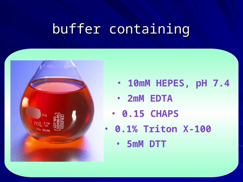
buffer containingbuffer containing
• 10mM HEPES, pH 7.4• 2mM EDTA• 0.15 CHAPS• 0.1% Triton X-100• 5mM DTT

Source : Moreau et al., 2002; Berger et al., 2004; Kritchevsky and Chen, 2005
Fig 1. Structure of some representative phytosterols